Back to Journals » Neuropsychiatric Disease and Treatment » Volume 13
Sexual dimorphism in Parkinson’s disease: differences in clinical manifestations, quality of life and psychosocial functioning between males and females
Authors Farhadi F, Vosoughi K, Shahidi GA, Delbari A ![]() , Lökk J
, Lökk J ![]() , Fereshtehnejad SM
, Fereshtehnejad SM ![]()
Received 18 October 2016
Accepted for publication 9 December 2016
Published 1 February 2017 Volume 2017:13 Pages 329—338
DOI https://doi.org/10.2147/NDT.S124984
Checked for plagiarism Yes
Review by Single anonymous peer review
Peer reviewer comments 2
Editor who approved publication: Dr Roger Pinder
Farzaneh Farhadi,1 Kia Vosoughi,1 Gholam Ali Shahidi,2 Ahmad Delbari,3,4 Johan Lökk,3,5 Seyed-Mohammad Fereshtehnejad3,6,7
1Medical Student Research Committee, 2Movement Disorders Clinic, Department of Neurology, Faculty of Medicine, Iran University of Medical Sciences, Tehran, Iran; 3Division of Clinical Geriatrics, Department of Neurobiology, Care Sciences, and Society (NVS), Karolinska Institutet, Stockholm, Sweden; 4Iranian Research Center on Aging, University of Social Welfare and Rehabilitation, Tehran, Iran; 5Department of Geriatric Medicine, Karolinska University Hospital, Stockholm, Sweden; 6Department of Neurology and Neurosurgery, McGill University, Montreal, QC, Canada; 7Firoozgar Clinical Research Development Center, Firoozgar Hospital, Iran University of Medical Sciences, Tehran, Iran
Introduction: Sex-related differences in clinical manifestations and consequences of Parkinson’s disease (PD) have been poorly explored. Better understanding of sexual dimorphism in neurologic diseases such as PD has been announced as a research priority. The aim of our study was to determine independent sex differences in clinical manifestations and subtypes, psychosocial functioning, quality of life (QoL) and its domains between male and female individuals with PD.
Patients and methods: A comprehensive list of demographics, motor symptoms and subtypes, nonmotor features, health-related quality of life (HRQoL), psychosocial functioning and general aspects of daily life was assessed in 157 individuals (108 males and 49 females) with idiopathic PD. In order to control for potential confounding variables, we applied Orthogonal Partial Least Squares – Discriminant Analysis (OPLS-DA) to explore the strength of each feature to discriminate male and female patients with PD.
Results: While no sex difference was found in the total Unified Parkinson’s Disease Rating Scale (UPDRS) score and cumulative daily dose of levodopa, females had significantly more severe anxiety (mean difference =2.2 [95% confidence interval, CI: 0.5–4.0], P=0.011), worse nutritional status (23.8 [standard deviation, SD =4.2] vs 25.8 [SD =2.6], P=0.003) and poorer QoL (28.3 [SD =15.7] vs 17.9 [SD =14.2], P<0.001). Based on multivariate discriminant analysis, emotional well-being, bodily discomfort, social support, mobility and communication domains of HRQoL, together with anxiety, depression and psychosocial functioning, were the strongest features with more severe/worse status in females after adjustment for potential statistical confounders.
Conclusion: Our study provides a comprehensive understanding of sexual dimorphism in PD. Anxiety, depression, specific domains of HRQoL (mobility, emotional well-being, social support and bodily discomfort) and psychosocial functioning were significantly worse in female individuals with PD. Sexual dimorphism in PD highlights the features that are more likely to be affected in each sex and should be specifically targeted when managing male and female individuals with PD.
Keywords: Parkinson’s disease, sexual dimorphism, male, female, quality of life, psychosocial functioning
Introduction
Parkinson disease (PD) is a progressive neurodegenerative disorder with widely heterogeneous manifestations, including motor, nonmotor and neuropsychiatric symptoms. It affects ~1% of the people older than 60 years.1–3 The major pathologic feature of PD is the loss of dopaminergic neurons in substantia nigra pars compacta and, consequently, basal ganglia.4,5
Sex differences in prevalence, clinical presentations and severity of PD have been reported in some previous studies.6–9 Although the exact sources of these differences in PD are unknown, sex hormones, genetic factors and variations in dopaminergic pathways have been proposed as possible underlying reasons.10–14 Identifying sexual variations influences prevention, therapeutic strategies and understanding of sex-related neurobiological differences.15,16 Exploration of sexual dimorphism in neurologic diseases such as PD has been recently announced as a research need.3 While studies are unanimous about the higher prevalence of PD among men, there are conflicting data regarding sex differences in clinical manifestations, progression and treatment outcome.17–23 Motor presentations (ie, tremor and dyskinesia), psychological manifestations (especially depression), sleep behavior disorders and cognitive impairment have been reported as potential aspects of sexual dimorphism in PD.17–23 However, data are still controversial, inconclusive and even conflicting in some aspects. As an example, a recent randomized clinical trial did not find any sex differences in daily activities and main motor features.24 In addition to lack of comprehensive data, these controversies can be partly due to interrelationship and confounding effects of clinical and demographic variables.3 Some clinical features that were previously reported to be sex related failed to show any difference between males and females with PD when controlled for other demographic and clinical variables. For instance, cognitive impairment was reported to be more common in men;9 however, the difference was much less pronounced when the effect of age, level of education and disease severity were taken into account.23 In another large study, univariate analysis showed that daily activity was more impaired in females, but multivariate analysis revealed no significant sex difference.24
Having collected comprehensive data on different PD-related and general features, we aimed to further investigate sexual dimorphism in PD. Our objective was to determine independent differences in clinical manifestations and subtypes, psychosocial functioning, quality of life (QoL) and its domains between males and females with idiopathic Parkinson’s disease (IPD) using a powerful statistical approach. Our a priori research hypothesis was that females with PD experienced more severe nonmotor manifestations, which would also influence their QoL and psychosocial functioning more prominently than males.
Patients and methods
Study setting
This study was conducted on 157 consecutive patients with IPD from an outpatient referral movement disorders clinic in Tehran, Iran, between October 2011 and December 2012. This was a collaborative project between Iran University of Medical Sciences (Tehran, Iran) and Karolinska Institute (Stockholm, Sweden).
Ethics
The study protocol was approved by the ethics committee of the Neurology Department at Firoozgar Clinical Research Development Center (FCRDC; affiliated to Iran University of Medical Sciences). Data were stored and treated according to the ethical guidelines of biomedical research. Prior to the launch of the study, all patients were informed about the aims and procedures. All participants provided their verbal informed consent to participate in this study. Since the project was designed as an observational research, verbal form of consent was approved by the aforementioned ethics committee. Participation in this study was voluntary, and the patients were free to withdraw from the project whenever they decided. Furthermore, the identity of research participants was protected, since the data files were anonymous and all names were omitted.
Patients’ requirement
Recruited patients fulfilled the following inclusion criteria: diagnosis of IPD based on the UK brain bank criteria,25 which was assessed by the same neurologist who was specialized in movement disorders for all participants; current age of 30 years or older and motor disability in the mild-to-severe range but not in the advanced stages that needs wheelchair or hospitalization according to the Hoehn and Yahr (H&Y) criteria (stage <5).26 Patients with moderate to severe dementia were excluded from the study, as were those with atypical Parkinsonism, including multiple system atrophy (MSA), progressive supranuclear palsy (PSP) and vascular or drug-induced Parkinsonism.
Assessments
Data collection was performed through face-to-face interviews with eligible patients by a trained group of medical interns by means of validated questionnaires and checklists. Patients were also examined for clinical assessments and diagnosis by one neurologist specialized in movement disorders. The demographic checklist consisted of baseline variables (age and sex), educational status, history of smoking, comorbidities, duration of PD (time passed from diagnosis) and history of levodopa administration. Total comorbidity burden was calculated by summing up the number of chronic comorbid conditions in each participant, including depression, hypertension, interstitial heart disease (IHD), diabetes, stroke/transient ischemic attack (TIA) and osteoporosis. Data were collected based on participants’ self-reports and their medical records at the referral center. Clinical characteristics of PD were assessed using the following scales and/or definitions:
- Motor severity: Unified Parkinson’s Disease Rating Scale (UPDRS) subscales I–IV, dyskinesia score (sum of UPDRS – Part IV items 32–34), fluctuation score (sum of UPDRS – Part IV items 36–39), H&Y staging and Schwab and England activities of daily living (ADL);
- Motor subtypes: postural instability–gait difficulty (PIGD) score (sum of UPDRS – Part III items concerning rise, gait and postural instability) and freezing–speech–swallowing (FOSS) score (sum of UPDRS – Part II items on freezing, speech and swallowing);
- Predominance of core manifestations: proportion of UPDRS – Part III on motor scores accounted for tremor (items 20–21), rigidity (item 22), bradykinesia (items 23–26 and 31) and gait (items 27–30) in percentage;
- Asymmetry Index: absolute differences in UPDRS between sides divided by the total UPDRS – Part III items 20–26;
- Axial/limb ratio: sum of UPDRS – Part III items 18, 19, 22 and 27–30 divided by the sum of UPDRS – Part III items 20–26;
- Other motor symptoms: presence of falls and freezing and
- Nonmotor manifestations: the Hospital Anxiety and Depression Scale (HADS) questionnaire to measure anxiety and depression,27 Fatigue Severity Scale (FSS) to investigate fatigue,28 the Mini-Nutritional Assessment (MNA) questionnaire together with anthropometric measurements for nutritional status,29 Persian-translated and validated version of the Scales for Outcomes in Parkinson’s Disease – Psychosocial (SCOPA-PS) questionnaire30 to assess psychosocial functioning and validated Persian version of the 39-item PD Questionnaire (PDQ-39)31 to evaluate health-related quality of life (HRQoL). All clinical assessments were done when the patients were in the “on” state.
Statistical analysis
Statistical descriptions and univariate and multivariate regression analyses were performed using the IBM SPSS Statistics for Windows, version 23 (IBM Corporation, Armonk, NY, USA). Mean (standard deviation [SD]) and frequency percentages were used to describe numerical and categorical variables, respectively. Kolmogorov–Smirnov test was applied to check the normality assumption for continuous variables. Since the normality of distribution was met, we used parametric tests for between-group comparisons. Univariate comparisons between males and females were performed using either independent samples t-test, chi-square test or Fisher’s exact test where appropriate. We used multivariate linear regression model to adjust the between-group differences in some numeric variables of interest for the baseline differences in disease duration and level of education. A two-tailed P-value of <0.05 was considered as the threshold to show statistical significant differences.
In order to evaluate the strength of each variable to discriminate male and female patients with PD, we applied Orthogonal Partial Least Squares – Discriminant Analysis (OPLS-DA) method using SIMCA software, version 14.1 (MKS Umetrics AB, Umeå, Sweden). Unit variance (UV) scaling was used to transform crude data prior to the OPLS-DA modeling. The OPLS-DA method divides the systematic variation in the X-block consisting of a comprehensive list of demographic and PD-related features to separate males and females with PD into two model parts. One part models the co-variation between X and Y, and another part expresses the X-variation that is not related to Y and is shown by the orthogonal component(s). This method results in a better class resolution for a discriminant problem such as the case in our study. Furthermore, the OPLS-DA method made it possible to compare the strength of different variables with various measurement units and scales to discriminate males and females with PD by means of values of standardized loading, their standard error (SE) and 95% confidence interval (CI). We visualized findings from the OPLS-DA method by three different plots with the following parameters:
- Score scatter plot: to show the performance of the entire OPLS-DA model to separate males and females where t1 refers to the score of each participant from the main discriminant component in the X-block (horizontal axis), while t2 shows the score of each participant from the Y-orthogonal component (vertical axis);
- Loadings bars: this is a one-dimensional plot representing loading value (p1) of each feature to discriminate males and females based on the main discriminant component of the OPLS-DA model;
- Loadings scatter plot: in this plot, p1 refers to the loading values of each feature from the main discriminant component to discriminate males and females (horizontal axis) and p2 represents the loadings of each variable from the Y-orthogonal component for the differences between features that are not related to sex (vertical axis).
Results
Overall, 157 individuals consisting of 108 males and 49 females with PD were recruited in our study. Table 1 summarizes the results for univariate comparison of the demography, motor severity and medications, motor subtypes, nonmotor features and QoL indicators between the two subgroups.
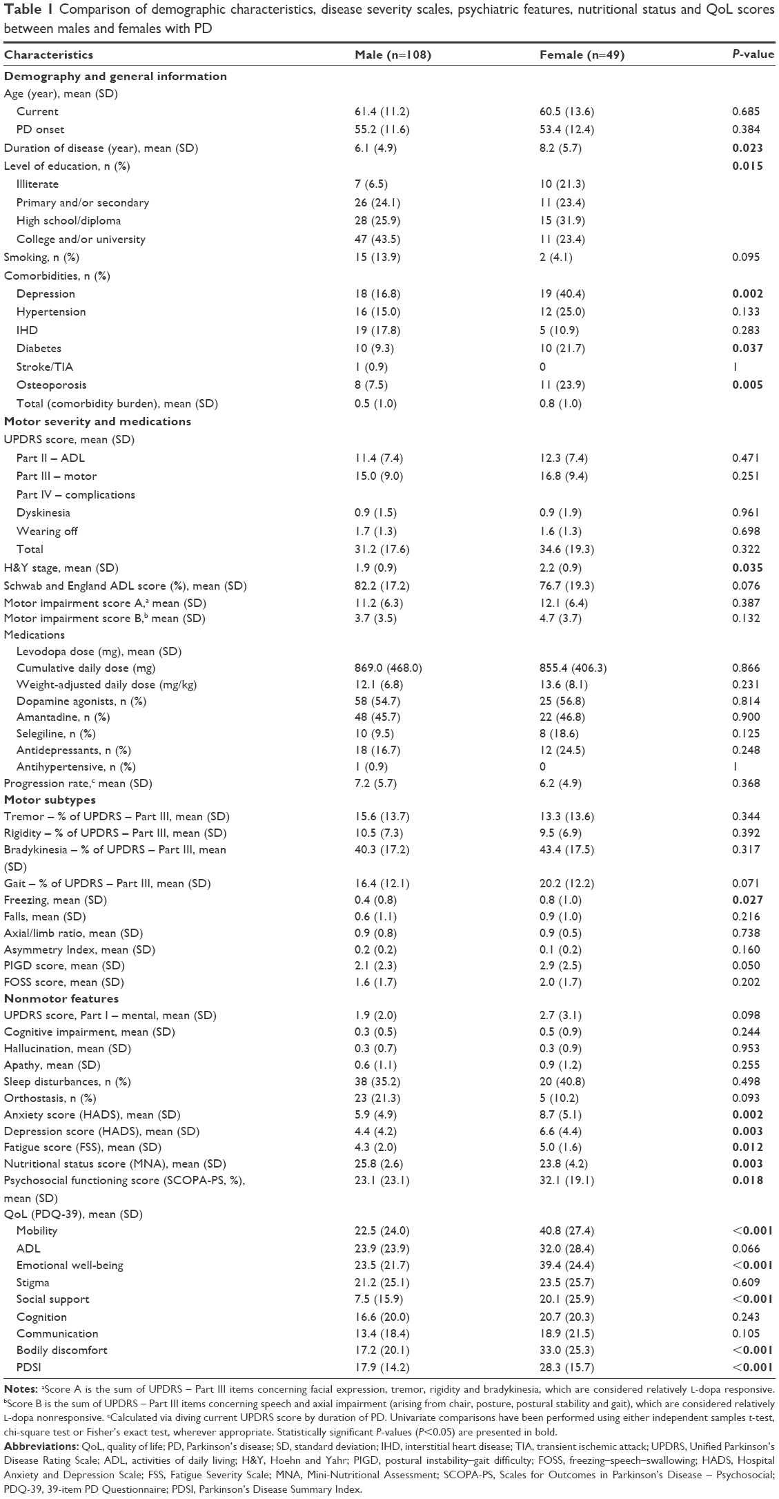 | Table 1 Comparison of demographic characteristics, disease severity scales, psychiatric features, nutritional status and QoL scores between males and females with PD |
Univariate and multivariate comparisons
The age at PD onset did not significantly differ between males and females (55.2 [SD =11.6] year vs 53.4 [SD =12.4] year, P=0.384), however, disease duration was longer in females at the time of recruitment (6.1 [SD =4.9] year vs 8.2 [SD =5.7] year, P=0.023). PD-related medication profile did not differ between males and females. Univariate differences in motor features such as total UPDRS and freezing score disappeared after adjustment for baseline difference. Among nonmotor features, anxiety, depression, fatigue and psychosocial functioning were all significantly more severe in female PD patients (Table 1). After statistical adjustment for the baseline differences in disease duration and level of education, female individuals still showed significantly higher score in anxiety (mean difference =2.2 [95% CI: 0.5–4.0], P=0.011) and borderline higher score in depression (mean difference =1.3 [95% CI: −0.2–2.7], P=0.079). Furthermore, female PD patients had significantly worse nutritional status and higher Parkinson’s Disease Summary Index (PDSI), both of which remained statistically significant after multivariate adjustment (mean difference for MNA score =−1.5 [95% CI: −2.5 to −0.4], P=0.009; mean difference for PDSI score =6.9 [95% CI: 1.9–11.8], P=0.006).
Among different domains of QoL, females were significantly worse in mobility (40.8 [SD =27.4] vs 22.5 [SD =24.0], P<0.001), emotional well-being (39.4 [SD =24.4] vs 23.5 [SD =21.7], P<0.001), social support (20.1 [SD =25.9] vs 7.5 [SD =15.9], P<0.001) and bodily discomfort (33.0 [SD =25.3] vs 17.2 [SD =20.1], P<0.001). All of these differences were still significant even after multivariate adjustment for the baseline differences in disease duration and level of education between males and females, resulting in the mean difference of 11.9 (95% CI: 3.5–20.3, P=0.006) in mobility, 12.5 (95% CI: 4.7–20.3, P=0.002) in emotional well-being and 11.0 (95% CI: 0.9–21.5, P=0.001) and 14.2 (95% CI: 6.4–21.9, P<0.001) in bodily discomfort domain of QoL.
Discriminant analysis
The OPLS-DA model that fitted the best to our dataset had the overall cross-validated predictive R2 value of 0.33. The score scatter plot in Figure 1 shows the performance of this model to discriminate males and females with PD. The corresponding loading value of each variable in the OPLS-DA model and its 95% CI are shown in Figure 2, and the loadings scatter plot is illustrated in Figure 3. Based on the absolute loading value for each variable in the OPLS-DA model, emotional well-being (mean =−0.22, SE =0.14), bodily discomfort (mean =−0.18, SE =0.13), social support (mean =−0.17, SE =0.19), mobility (mean =−0.17, SE =0.04) and communication (mean =−0.12, SE =0.15) domains of QoL, together with anxiety (mean =−0.18, SE =0.10), depression (mean =−0.17, SE =0.08) and psychosocial functioning (mean =−0.16, SE =0.11), were the strongest features with higher values in favor of females to discriminate the two sexes in PD population (Figures 2 and 3). Nutritional status (mean =0.13, SE =0.07), Schwab and England ADL score (mean =0.06, SE =0.13), and orthostasis (mean =0.06, SE =0.05) were also found to be strong discriminators in the model, nevertheless, with higher values in favor of the male PD patients.
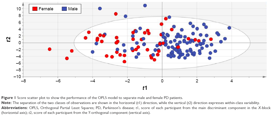 | Figure 1 Score scatter plot to show the performance of the OPLS model to separate male and female PD patients. |
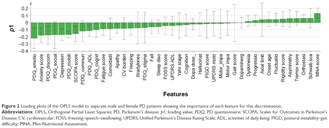 | Figure 2 Loading plots of the OPLS model to separate male and female PD patients showing the importance of each feature for this discrimination. |
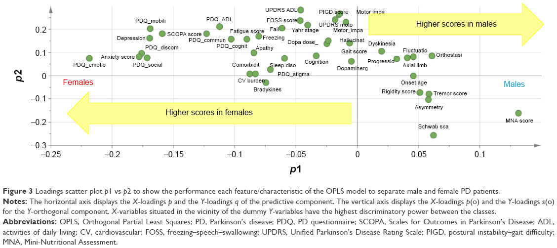 | Figure 3 Loadings scatter plot p1 vs p2 to show the performance each feature/characteristic of the OPLS model to separate male and female PD patients. |
Discussion
Our study is one of the few attempts to comprehensively investigate sexual dimorphism in PD. We applied powerful multivariate statistical methods to explore independent sex differences in clinical manifestations, HRQoL and psychosocial functioning of people with PD. In general, females with PD were significantly worse in psychological features such as anxiety and depression, nutritional status and specific domains of QoL, namely, mobility, emotional well-being, social support and bodily discomfort. Neuropsychiatric symptoms are among the main manifestations of sexual dimorphism in PD. Studies have suggested that emotional symptoms of PD, especially depression, are more common and more severe in females than in males.32–34 However, these findings were not approved by a large prospective study, which found no difference in the prevalence or severity of depression evaluated by Beck Depression Inventory (BDI) between females and males.24 In our study, females with PD tended to be more affected from psychological symptoms. We found that the scores for anxiety, psychosocial functioning and emotional well-being were all higher among females demonstrating a worse status. Conflicting results can be, in part, explained by ethnic, cultural and environmental differences as well as the use of different depression scales. In some cultures and environments, women are more prone to be stigmatized by disabling conditions,35,36 which make them more vulnerable to emotional and psychosocial adverse effects of PD.
Social and physical well-being, measured separately or as parts of QoL questionnaires, are important contributors of patient’s QoL.37,38 In our study, physical discomfort, including mobility, fatigue and bodily discomfort, was more severe among female participants. This finding is confirmed by previous studies of PD populations,17,34,39 as well as those of general population showing more severe physical discomfort in healthy older women compared to males of the same age group.40–42 Our results showed that despite the lack of statistically significant difference in social stigma, females displayed worse communication and psychosocial functioning. These sex-related differences can be the consequences of more severe emotional disturbance, physical discomfort, and poorer social supports in females compared to males with PD.
In our study, self-reported cognitive impairment was higher in female participants based on the cognition dimension of PDQ-39. In contrast, some studies have suggested that cognitive decline in PD is more severe among males.18,23 Nonetheless, as different dimensions of cognitive performance have been shown to be sex related in healthy elderly adults (for instance, males have better visuospatial ability, while females perform better on face and verbal recognition and semantic fluency tests43–45), different dimensions of cognitive performance must be investigated in order to clarify sexual dimorphism of cognition in PD.
Studies have reported several sex differences in motor symptoms of PD. Tremor and dyskinesia are reported to occur more frequently in females,8,18,33 whereas gait disturbance is more common in males.19 However, in line to one prospective study that had also adjusted the differences for the confounding effects of demographics and clinical covariates,24 we also could not find sex-related differences in motor symptoms.
Sleep quality is the other symptom of PD, which is believed to be sex related, but still remained a controversial issue. Rapid eye movement (REM) sleep behavior disorder (RBD) was reported as a male-related symptom in PD;33,46–48 some other sleep disturbances were found to be more severe among females.34,49 We did not find any difference in sleep disturbance between males and females using the nonmotor items of the UPDRS. A more thorough assessment with a valid and specific tool such as polysomnography is required to investigate sexual dimorphism in sleep disorders in people with PD.
The comprehensive list of PD-related features and general aspects of daily life is the most important strength of our study. Previous studies have investigated sexual dimorphism in manifestations of PD with rather small sample size not being controlled for the potential confounders.3,50 In our study, we evaluated a large list of variables (some of which were considered for sexual dimorphism in PD for the first time) and applied a strong multivariate statistical approach, OPLS-DA, to take into account all potential confounding interactions. Nevertheless, we also acknowledge our limitations. This study was designed as a cross-sectional research that restricts any causal inferences and going beyond associations. There are other aspects of sexual dimorphism in PD that need to be addressed in future research, namely, course of progression and mortality. There is a potential risk for selection bias since all recruited patients were from an outpatient neurology clinic where the majority of patients had mild-to-moderate IPD. As a result, our findings may not be generalized to all people with PD, particularly those with end-stage disease. Considering time restriction for data collection spent in each participant, we ought to rely on the items from UPDRS for some variables, which might not be sensitive enough to show between-group differences on this summary scale. Therefore, more sensitive gold-standard tools might potentially show different results for such features. Our rather small sample size in comparison with few large previous studies should also be noted. Augustine et al24 reported that studies with smaller sample sizes were more likely to report sex differences in different clinical features. We should also add the uneven number of males and females in our study as another limitation; however, it could be expected for PD that affects men more frequently.51 Yet the comprehensive list of variables and appropriate multivariate statistical methods of the present study overcame these issues and strengthened our findings.
Conclusion
This study provides a comprehensive understanding of sexual dimorphism in different motor and nonmotor features of PD and various domains of daily life by using sophisticated statistical analysis to evaluate the independent differences between males and females. After controlling for potential confounders, anxiety, depression, mobility, emotional well-being, social support, bodily discomfort and psychosocial functioning were significantly worse in female individuals with PD. On the other hand, male PD patients had better nutritional status (though with rather small effect size for difference) and ADL but also more severe orthostasis. Conclusively, there are considerable differences in psychiatric nonmotor manifestations. Concordance of depression, anxiety and overall psychosocial burden in being more severe in women is in favor of the presence of a considerable sex dimorphism in neuropsychiatric symptoms, which in turn suggest its clinical importance. QoL and psychosocial consequences of PD between males and females should be addressed for developing a more personalized caring approach. These aspects of sexual dimorphism in PD also enlighten the features that are more likely to be affected in each sex and should be specifically targeted when managing male and female individuals with PD. As for clinical implication, clinicians should consider sex differences in the evaluation and management of patients with PD. For example, they should be aware of the higher risk of psychological manifestations in women and therefore should screen them accurately in a timely manner and, if necessary, refer them to a psychologist in the early stages of these manifestations. Sex-specific symptoms highlight the importance of sex-specific treatments; hence, future studies should investigate the effects of specific interventions for females. Moreover, educational interventions are also needed in order to improve social support for women with PD and reduce their extra psychosocial burden.
Acknowledgments
The authors are grateful to the colleagues from the Movement Disorder Clinic who contributed in data collection: Hasti Hadizadeh, Mahdiyeh Shafieesabet, Nader Naderi, Dena Khaefpanah, and Ms Mahmoudi. The authors also thank all patients and their caregivers for their collaboration to collect the data for this project.
Disclosure
The authors report no conflicts of interest in this work.
References
Langston JW. The Parkinson’s complex: parkinsonism is just the tip of the iceberg. Ann Neurol. 2006;59(4):591–596. | ||
Elbaz A, Bower JH, Maraganore DM, et al. Risk tables for parkinsonism and Parkinson’s disease. J Clin Epidemiol. 2002;55(1):25–31. | ||
Pavon JM, Whitson HE, Okun MS. Parkinson’s disease in women: a call for improved clinical studies and for comparative effectiveness research. Maturitas. 2010;65(4):352–358. | ||
Stuendl A, Kunadt M, Kruse N, et al. Induction of alpha-synuclein aggregate formation by CSF exosomes from patients with Parkinson’s disease and dementia with lewy bodies. Brain. 2016;139(pt 2):481–494. | ||
Kalia LV, Kalia SK. alpha-synuclein and lewy pathology in Parkinson’s disease. Curr Opin Neurol. 2015;28(4):375–381. | ||
Kowal SL, Dall TM, Chakrabarti R, Storm MV, Jain A. The current and projected economic burden of Parkinson’s disease in the United States. Mov Disord. 2013;28(3):311–318. | ||
Amantea D, Russo R, Bagetta G, Corasaniti MT. From clinical evidence to molecular mechanisms underlying neuroprotection afforded by estrogens. Pharmacol Res. 2005;52(2):119–132. | ||
Haaxma CA, Bloem BR, Borm GF, et al. Gender differences in Parkinson’s disease. J Neurol Neurosurg Psychiatry. 2007;78(8):819–824. | ||
Miller IN, Cronin-Golomb A. Gender differences in Parkinson’s disease: clinical characteristics and cognition. Mov Disord. 2010;25(16):2695–2703. | ||
Smith KM, Dahodwala N. Sex differences in Parkinson’s disease and other movement disorders. Exp Neurol. 2014;259:44–56. | ||
Czech DP, Lee J, Sim H, Parish CL, Vilain E, Harley VR. The human testis-determining factor SRY localizes in midbrain dopamine neurons and regulates multiple components of catecholamine synthesis and metabolism. J Neurochem. 2012;122(2):260–271. | ||
Dewing P, Chiang CW, Sinchak K, et al. Direct regulation of adult brain function by the male-specific factor SRY. Curr Biol. 2006;16(4):415–420. | ||
Lavalaye J, Booij J, Reneman L, Habraken JB, van Royen EA. Effect of age and gender on dopamine transporter imaging with [123I]FP-CIT SPET in healthy volunteers. Eur J Nucl Med. 2000;27(7):867–869. | ||
Simunovic F, Yi M, Wang Y, Stephens R, Sonntag KC. Evidence for gender-specific transcriptional profiles of nigral dopamine neurons in Parkinson disease. PLoS One. 2010;5(1):e8856. | ||
Pancholy SB, Sharma PS, Pancholy DS, Patel TM, Callans DJ, Marchlinski FE. Meta-analysis of gender differences in residual stroke risk and major bleeding in patients with nonvalvular atrial fibrillation treated with oral anticoagulants. Am J Cardiol. 2014;113(3):485–490. | ||
Anderson GD. Gender differences in pharmacological response. Int Rev Neurobiol. 2008;83:1–10. | ||
Shulman LM. Gender differences in Parkinson’s disease. Gend Med. 2007;4(1):8–18. | ||
Lyons KE, Hubble JP, Troster AI, Pahwa R, Koller WC. Gender differences in Parkinson’s disease. Clin Neuropharmacol. 1998;21(2):118–121. | ||
Scott B, Borgman A, Engler H, Johnels B, Aquilonius SM. Gender differences in Parkinson’s disease symptom profile. Acta Neurol Scand. 2000;102(1):37–43. | ||
Kompoliti K, Comella CL, Jaglin JA, Leurgans S, Raman R, Goetz CG. Menstrual-related changes in motoric function in women with Parkinson’s disease. Neurology. 2000;55(10):1572–1575. | ||
Mozley LH, Gur RC, Mozley PD, Gur RE. Striatal dopamine transporters and cognitive functioning in healthy men and women. Am J Psychiatry. 2001;158(9):1492–1499. | ||
Elbers R, van Wegen EE, Rochester L, et al. Is impact of fatigue an independent factor associated with physical activity in patients with idiopathic Parkinson’s disease? Mov Disord. 2009;24(10):1512–1518. | ||
Nazem S, Siderowf AD, Duda JE, et al. Montreal cognitive assessment performance in patients with Parkinson’s disease with “normal” global cognition according to mini-mental state examination score. J Am Geriatr Soc. 2009;57(2):304–308. | ||
Augustine EF, Perez A, Dhall R, et al. Sex differences in clinical features of early, treated Parkinson’s disease. PLoS One. 2015;10(7):e0133002. | ||
Hughes AJ, Daniel SE, Kilford L, Lees AJ. Accuracy of clinical diagnosis of idiopathic Parkinson’s disease: a clinico-pathological study of 100 cases. J Neurol Neurosurg Psychiatry. 1992;55(3):181–184. | ||
Hoehn MM, Yahr MD. Parkinsonism: onset, progression, and mortality. Neurology. 1967;17(5):427–442. | ||
Montazeri A, Vahdaninia M, Ebrahimi M, Jarvandi S. The hospital anxiety and depression scale (HADS): translation and validation study of the Iranian version. Health Qual Life Outcomes. 2003;1:14. | ||
Fereshtehnejad SM, Hadizadeh H, Farhadi F, Shahidi GA, Delbari A, Lokk J. Reliability and validity of the Persian version of the fatigue severity scale in idiopathic Parkinson’s disease patients. Parkinsons Dis. 2013;2013:935429. | ||
Ghazi L, Fereshtehnejad SM, Abbasi Fard S, Sadeghi M, Shahidi GA, Lokk J. Mini nutritional assessment (MNA) is rather a reliable and valid instrument to assess nutritional status in Iranian Healthy Adults and Elderly with a chronic disease. Ecol Food Nutr. 2015;54(4):342–357. | ||
Fereshtehnejad SM, Farhadi F, Hadizadeh H, Shahidi GA, Delbari A, Lökk J. Cross-cultural validity, reliability, and psychometric properties of the Persian version of the scales for outcomes in Parkinson’s disease-psychosocial questionnaire. Neurol Res Int. 2014;2014:260684. | ||
Nojomi M, Mostafavian Z, Shahidi GA, Jenkinson C. Quality of life in patients with Parkinson’s disease: translation and psychometric evaluation of the Iranian version of PDQ-39. J Res Med Sci. 2010;15(2):63–69. | ||
Fernandez HH, Lapane KL, Ott BR, Friedman JH. Gender differences in the frequency and treatment of behavior problems in Parkinson’s disease. SAGE Study Group. Systematic assessment and geriatric drug use via epidemiology. Mov Disord. 2000;15(3):490–496. | ||
Martinez-Martin P, Falup Pecurariu C, Odin P, et al. Gender-related differences in the burden of non-motor symptoms in Parkinson’s disease. J Neurol. 2012;259(8):1639–1647. | ||
Kovacs M, Makkos A, Aschermann Z, et al. Impact of sex on the nonmotor symptoms and the health-related quality of life in Parkinson’s disease. Parkinsons Dis. 2016;2016:7951840. | ||
Khan N, Kausar R, Khalid A, Farooq A. Gender differences among discrimination & stigma experienced by depressive patients in Pakistan. Pak J Med Sci. 2015;31(6):1432–1436. | ||
Asiedu GB, Myers-Bowman KS. Gender differences in the experiences of HIV/AIDS-related stigma: a qualitative study in Ghana. Health Care Women Int. 2014;35(7–9):703–727. | ||
Kalfoss M, Halvorsrud L. Important issues to quality of life among Norwegian older adults: an exploratory study. Open Nurs J. 2009;3:45–55. | ||
Molzahn AE, Kalfoss M, Schick Makaroff K, Skevington SM. Comparing the importance of different aspects of quality of life to older adults across diverse cultures. Age Ageing. 2011;40(2):192–199. | ||
Baba Y, Putzke JD, Whaley NR, Wszolek ZK, Uitti RJ. Gender and the Parkinson’s disease phenotype. J Neurol. 2005;252(10):1201–1205. | ||
Manton KG. A longitudinal study of functional change and mortality in the United States. J Gerontol. 1988;43(5):S153–S161. | ||
Kelly-Hayes M, Jette AM, Wolf PA, D’Agostino RB, Odell PM. Functional limitations and disability among elders in the Framingham Study. Am J Public Health. 1992;82(6):841–845. | ||
Guerra RO, Alvarado BE, Zunzunegui MV. Life course, gender and ethnic inequalities in functional disability in a Brazilian urban elderly population. Aging Clin Exp Res. 2008;20(1):53–61. | ||
Ruitenberg A, Ott A, van Swieten JC, Hofman A, Breteler MM. Incidence of dementia: does gender make a difference? Neurobiol Aging. 2001;22(4):575–580. | ||
Letenneur L, Gilleron V, Commenges D, Helmer C, Orgogozo JM, Dartigues JF. Are sex and educational level independent predictors of dementia and Alzheimer’s disease? Incidence data from the PAQUID project. J Neurol Neurosurg Psychiatry. 1999;66(2):177–183. | ||
Shin JY, Pohlig RT, Habermann B. Self-reported symptoms of Parkinson’s disease by sex and disease duration. West J Nurs Res. Epub 2016 Sep 23. | ||
Scaglione C, Vignatelli L, Plazzi G, et al. REM sleep behaviour disorder in Parkinson’s disease: a questionnaire-based study. Neurol Sci. 2005;25(6):316–321. | ||
Ozekmekci S, Apaydin H, Kilic E. Clinical features of 35 patients with Parkinson’s disease displaying REM behavior disorder. Clin Neurol Neurosurg. 2005;107(4):306–309. | ||
Yoritaka A, Ohizumi H, Tanaka S, Hattori N. Parkinson’s disease with and without REM sleep behaviour disorder: are there any clinical differences? Eur Neurol. 2009;61(3):164–170. | ||
Pandey S, Bajaj BK, Wadhwa A, Anand KS. Impact of sleep quality on the quality of life of patients with Parkinson’s disease: a questionnaire based study. Clin Neurol Neurosurg. 2016;148:29–34. | ||
Van Den Eeden SK, Tanner CM, Bernstein AL, et al. Incidence of Parkinson’s disease: variation by age, gender, and race/ethnicity. Am J Epidemiol. 2003;157(11):1015–1022. | ||
Kalia LV, Lang AE. Parkinson’s disease. Lancet. 2015;386(9996):896–912. |
 © 2017 The Author(s). This work is published and licensed by Dove Medical Press Limited. The
full terms of this license are available at https://www.dovepress.com/terms
and incorporate the Creative Commons Attribution
- Non Commercial (unported, 3.0) License.
By accessing the work you hereby accept the Terms. Non-commercial uses of the work are permitted
without any further permission from Dove Medical Press Limited, provided the work is properly
attributed. For permission for commercial use of this work, please see paragraphs 4.2 and 5 of our Terms.
© 2017 The Author(s). This work is published and licensed by Dove Medical Press Limited. The
full terms of this license are available at https://www.dovepress.com/terms
and incorporate the Creative Commons Attribution
- Non Commercial (unported, 3.0) License.
By accessing the work you hereby accept the Terms. Non-commercial uses of the work are permitted
without any further permission from Dove Medical Press Limited, provided the work is properly
attributed. For permission for commercial use of this work, please see paragraphs 4.2 and 5 of our Terms.