Back to Journals » International Journal of Nanomedicine » Volume 21
SERS for Infectious Disease Diagnostics: An Advanced Platform for Pathogen Detection and Antimicrobial Resistance Analysis
Authors Liao Y, Wang X, Kang H ![]() , Tao Y, Chen H
, Tao Y, Chen H
Received 15 December 2025
Accepted for publication 16 February 2026
Published 5 March 2026 Volume 2026:21 589169
DOI https://doi.org/10.2147/IJN.S589169
Checked for plagiarism Yes
Review by Single anonymous peer review
Peer reviewer comments 3
Editor who approved publication: Prof. Dr. RDK Misra
Yiqun Liao,1 Xiaoling Wang,1 Huapei Kang,2 Yuanming Tao,2 Hui Chen3
1Department of Laboratory Medicine, First Affiliated Hospital of Gannan Medical University, Ganzhou, People’s Republic of China; 2The First School of Clinical Medicine, Gannan Medical University, Ganzhou, People’s Republic of China; 3Department of Healthcare-Associated Infection Management, First Affiliated Hospital of Gannan Medical University, Ganzhou, People’s Republic of China
Correspondence: Hui Chen, Department of Healthcare-associated Infection Management, First Affiliated Hospital of Gannan Medical University, NO. 23 Qingnian Road, Zhanggong District, Ganzhou, Jiangxi, 341000, People’s Republic of China, Email [email protected]
Abstract: Infectious diseases caused by pathogenic bacteria remain a major challenge for global public health. Rapid and accurate pathogen identification, as well as antimicrobial resistance (AMR) analysis, are crucial for the timely control and treatment of infectious diseases. Surface-enhanced Raman spectroscopy (SERS) is an analytical technique that combines Raman spectroscopy with the localized surface plasmon resonance (LSPR) effect of nanomaterials, featuring rapidity, non-destructiveness, high sensitivity, and specificity. This demonstrates significant potential for the diagnosis and treatment of infectious diseases. This article primarily expounds on the application of SERS in the detection of bacteria, viruses, fungi, and AMR; explores the use of multi-modal innovative technologies integrating SERS with nanotechnology, microfluidics, and deep learning in pathogen identification and AMR analysis; and discusses the challenges and prospects for clinical translation of SERS.
Keywords: SERS, infectious diseases, pathogen detection, antimicrobial resistance analysis, nanomaterials
Introduction
Infectious diseases are a category of diseases caused by pathogenic microorganisms, such as bacteria, viruses, and fungi, that invade the human body, multiply, and trigger immune responses that lead to tissue and organ infections or damage. These diseases pose a severe threat to human and global public health. The COVID-19 outbreak was initially classified as a public health emergency of international concern by the World Health Organization (WHO). As of February 25, 2022, over 431 million cases had been reported in 206 countries and regions, resulting in at least 5.9 million deaths worldwide.1 The WHO estimated that 13.7 million deaths were related to infections in all ages and in males and females across 204 countries and territories in 2019. Of these, 7.7 million deaths were associated with 33 bacterial pathogens across 11 infectious syndromes, accounting for 13.6% of global deaths and 56.2% of all sepsis-related deaths. Staphylococcus aureus (S. aureus), Escherichia coli (E. coli), Streptococcus pneumoniae (S. pneumoniae), Klebsiella pneumoniae (K. pneumoniae), and Pseudomonas aeruginosa (P. aeruginosa) were responsible for 54.9% of the deaths among the investigated bacteria.2 Therefore, rapid and accurate pathogen detection is crucial for diagnosis, treatment, prevention, and control of infectious diseases.
Microbial culture remains the gold standard for etiological diagnosis. However, it is time-consuming and has low positive rates,3 failing to meet the demands of rapid clinical diagnosis. With the rapid development of biotechnology, novel methods such as immunological testing, polymerase chain reaction (PCR), gene sequencing, and matrix-assisted laser desorption/ionization time-of-flight mass spectrometry (MALDI-TOF MS) have been applied for etiological diagnosis. Each of these methods has its advantages and limitations. For example, immunological methods have a narrow detection range and relatively low sensitivities. PCR involves complex operational procedures and requires stringent experimental conditions. Gene sequencing demands costly equipment and specialized expertise, and the target sequence often limits its results. The accuracy of the MALDI-TOF MS results is highly dependent on the comprehensiveness and quality of the reference database. In recent years, surface-enhanced Raman spectroscopy (SERS) has emerged as a novel technology for infectious disease diagnostics, owing to its advantages, such as rapidity, non-destructiveness, label-free nature, high sensitivity, and high specificity. This article primarily focuses on the application of SERS in pathogen identification and antimicrobial resistance (AMR) analysis and further explores the integrated application and development of SERS with cutting-edge technologies, as well as challenges for its clinical translation.
Raman Spectroscopy and SERS
In 1928, Indian physicist C.V. Raman first confirmed the Raman scattering effect by observing the inelastic scattering of light by benzene and toluene.4 When a substance is illuminated by incident light, the photons interact with its molecules, resulting in scattered light at various frequencies. Most of the scattered light retains the same frequency as the incident light, a phenomenon known as elastic scattering (Rayleigh scattering). However, a small fraction undergoes a frequency shift, termed inelastic scattering (Raman scattering). Raman spectroscopy is a vibrational spectroscopy technique based on this inelastic scattering. It measures the energy difference between incident and scattered photons, producing Raman shifts. The positions and intensities of these shifts provide insights into molecular bond vibrations, such as those in proteins, nucleic acids, and lipids, forming a unique spectral “fingerprint”.5 This fingerprint enables the analysis of molecular structure, chemical composition, and intermolecular interactions. Despite its utility, Raman spectroscopy faces a key limitation: the inherently weak Raman scattering signal, which makes detection challenging and restricts its practical applications.
In 1974, Fleischmann et al first reported potential-dependent Raman shifts and intensity variations of pyridine adsorbed on roughened silver electrodes.6 Later, in 1977, Van Duyne and Creighton’s team demonstrated that this Raman signal enhancement originated from the rough surfaces of metal nanoparticles (NPs),7,8 formally naming the phenomenon surface-enhanced Raman scattering. Figure 1 illustrates the workflow of conventional Raman spectroscopy and SERS.9 As a powerful analytical technique, SERS relies on the adsorption of target molecules onto nanostructured noble metal substrates. Under laser excitation, localized surface plasmon resonance (LSPR) generates intense electromagnetic “hot spots”, amplifying Raman signals by up to 104–1011-fold.10 The primary enhancement mechanisms involve electromagnetic enhancement and chemical enhancement, as illustrated in Figure 2. Electromagnetic enhancement arises from LSPR effects of metal NPs; Chemical enhancement involves molecule-substrate interactions (eg, charge transfer, chemisorption, and polarization). Owing to its rapidity, non-destructiveness, label-free operation, and exceptional sensitivity and specificity, SERS has been broadly applied in food science, biomedical research, and clinical diagnostics.11–13
 |
Figure 1 Schematic diagram of the workflow of conventional Raman spectroscopy and SERS. (A) Conventional Raman spectroscopy workflow. Molecular structural information was obtained through molecular vibrations for bacterial identification and antimicrobial resistance. (B) SERS workflow. Target molecules were adsorbed onto noble metal nanoparticle surfaces to enhance Raman signal intensity through SERS. Reproduced with permission.9 Copyright 2021, Frontiers in Microbiology. CC BY, https://creativecommons.org/licenses/. Abbreviations: CCD, charge-coupled device; NIR, near-infrared. |
 |
Figure 2 Schematic diagram of the electromagnetic and chemical enhancement mechanisms for SERS. (By FigDraw). Abbreviation: LSPR, localized surface plasmon resonance. |
Clinical Applications
Application of SERS in Bacterial Detection
Different bacterial species possess unique chemical compositions, which produce distinct Raman spectral fingerprints. By comparing the Raman spectra of unknown bacterial samples against a standardized clinical reference database, rapid and accurate classification and identification results can be obtained. This approach offers high specificity and sensitivity, requires no culturing, and enables swift identification at the species level. In bacterial detection, SERS currently relies on two primary methodologies: label-free and label-based approaches, illustrated in Figure 3.14
 |
Figure 3 Schematic diagram of label-based and label-free SERS methods for bacterial detection. (A) Label-based SERS method. (B) Label-free SERS method. Reproduced with permission.14 Copyright 2023, Journal of Advanced Research. CC BY-NC-ND 4.0, https://creativecommons.org/licenses/by-nc-nd/4.0/. |
In the label-free approach, target bacteria interact directly with plasmonic metal NPs exhibiting SERS enhancement; the generated Raman signal originates intrinsically from the bacteria themselves, enabling pathogen identification through characteristic spectral analysis. In contrast, the label-based method, commonly termed SERS tagging, employs functionalized plasmonic NPs conjugated with a distinct Raman reporter molecule (eg, 4-mercaptobenzoic acid or 4-aminothiophenol) and a biorecognition element (eg, antibody, aptamer, or peptide). Upon specific binding to the target pathogen, these engineered probes generate intense and signature Raman signals, thereby facilitating indirect bacterial detection.15 The comparison between label-free and label-based SERS methods for pathogen detection strategies is presented in Table 1. Liu et al successfully developed a multifunctional two-dimensional BP@MoS2 nanocomposite. The unique flower-like surface microstructure of this nanocomposite promoted target enrichment efficiency. While its hybridized band structure facilitated powerful charge transfer, thereby enabling label-free SERS for the detection of E. coli.16 Furthermore, the modified E. coli could be effectively eliminated by the versatile nanocomposites under the action of near-infrared laser, with a high sterilization rate of 99.66% within 8 minutes. Yuan et al used antimicrobial peptide (AMP)-functionalized magnetic NPs as “capture” probes for bacterial isolation. Employing gold-coated silver decorated graphene oxide (Au@Ag-GO) nanocomposites modified with 4-mercaptophenylboronic acid (4-MPBA) as SERS tags, they successfully isolated and identified E. coli, S. aureus, and P. aeruginosa from blood samples, with detection limits as low as 10 cfu/mL for all three pathogens.17 Furthermore, the AMP-modified Fe3O4 NPs exhibit high antibacterial activity, demonstrating the high performance of the multifunctional biosensor in simultaneously bacterial isolation, identification, and elimination.
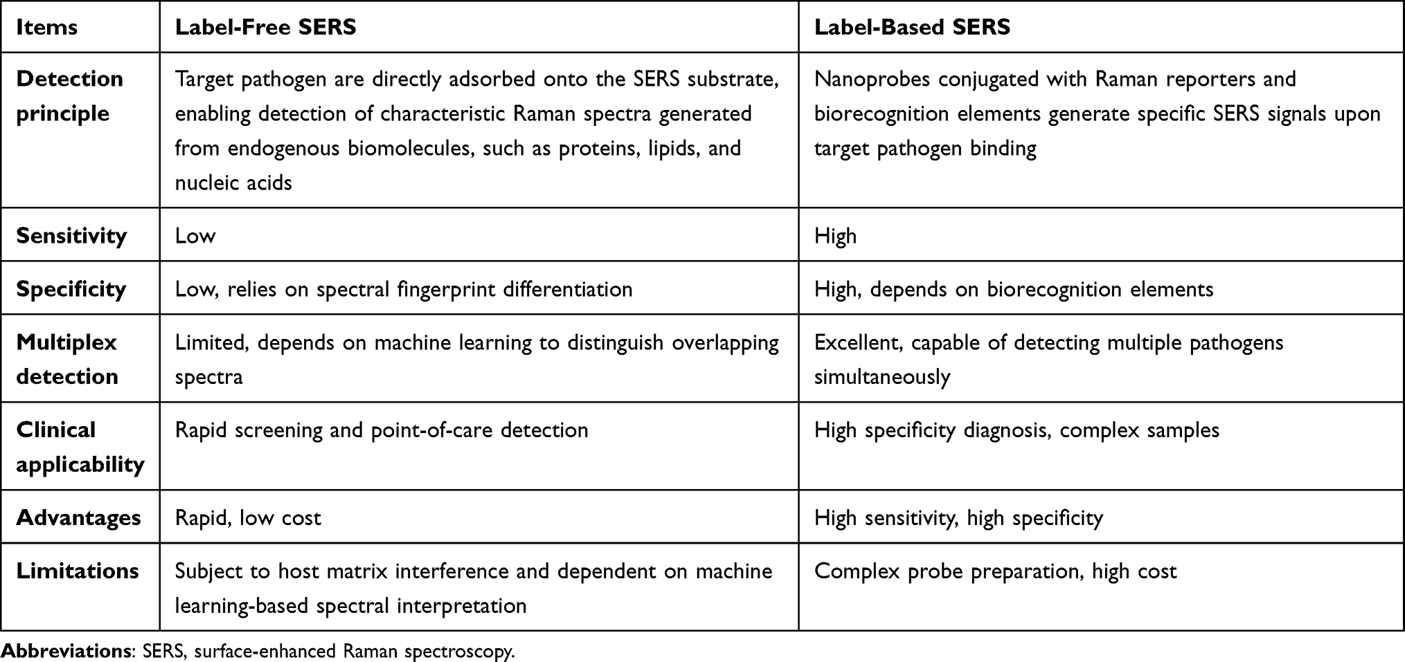 |
Table 1 The Comparison between Label-Free and Label-Based SERS Methods for Pathogen Detection Strategies |
However, Raman spectroscopy data are complex, and bacterial SERS fingerprint information is abundant. Some bacterial components and structures are similar, making it challenging to distinguish between pathogens merely through spectral comparisons. Therefore, algorithms are necessary for bacterial identification, which has led to the emergence of SERS analysis methods based on machine learning (ML) algorithms. Almeida et al developed an SERS protocol with silver nanostars for the discrimination of Acinetobacter baumannii (A. baumannii) and K. pneumoniae species, as well as their globally disseminated and clinically relevant antibiotic-resistant clones.18 They mixed bacterial colonies with silver nanostars and deposited them on a filter paper for SERS spectrum acquisition. Using principal component analysis (PCA) and partial least squares discriminant analysis (PLS-DA), they successfully distinguished A. baumannii and K. pneumoniae species along with their representative clones. Wang et al investigated 30 bacterial species belonging to nine different bacterial genera that were isolated from clinical samples via SERS.19 A total of 17,149 SERS spectra were harvested and further analyzed using ML approaches. The results indicated that the convolutional neural networks (CNNs) algorithm attained classification accuracies of 99.80% at the genus level and 98.37% at the species level, achieving its optimal performance in 97 seconds and 94 seconds, respectively.
The integration of SERS and nanotechnology has demonstrated significant advantages of rapidity and sensitivity for bacterial detection and therapy. It holds great potential in addressing the issues of long-term consumption and low positive rate of traditional culture methods. However, there are current challenges such as substrate reproducibility, biosafety, and clinical standardization, which significantly limit its clinical application.
Application of SERS in Viral Detection
Different viral species possess unique structural components, including protein capsids, lipid envelopes, and nucleic acids, which produce distinctive Raman spectral signatures. These characteristic vibrational fingerprints enable Raman spectroscopy to serve as an effective tool for both viral detection and typing. Furthermore, viral infection induces systemic alterations in serum composition, particularly in proteins, lipids, and metabolites, which can be captured through serum Raman spectroscopy and subsequently analyzed using ML algorithms to accurately determine infection status and identify viral subtypes. Yadav et al utilized a highly optimized silver nanorod array as the SERS substrate. Distinct signature peaks for varying concentrations (102–106 copies/mL) were identified in five HIV-1 subtypes (A, B, C, D, and CRF02_AG).20 The purified viruses were spiked in water and healthy plasma to capture pure HIV-1 characteristic peaks; the multivariate statistical analysis revealed that the SERS platform could effectively detect and differentiate HIV-1. Zhang et al developed a gold biconical nanoparticle-enhanced substrate that captured the SERS characteristic fingerprint spectra of SARS-CoV-2, adenovirus, influenza virus, S. aureus, and K. pneumoniae label-free.21 Additionally, this assay, combined with PCA, can be used to differentiate respiratory pathogens in serum and saliva within 2 minutes. Jiang et al developed a detection platform combining SERS technology with ML methods that utilize iodine ion incubation and calcium ion aggregation to prepare silver NPs(Ag@ICNPs) as SERS substrates. This platform is ultra-fast, highly sensitive, and label-free. It captures the characteristic fingerprint spectra of respiratory syncytial virus (RSV), influenza A virus (IFA), and human adenovirus (HAdV).22 By integrating multiple ML methods with SERS technology, the platform could rapidly identify these three viruses with a detection limit as low as 1.0×10² copies/mL within 3 minutes, and an accuracy rate of 100%. This study further established a robust linear correlation between viral concentration and the intensity of the characteristic SERS peaks, underscoring potential utility for clinical monitoring of antiviral treatment efficacy through quantitative assessment of viral load dynamics.
SERS has emerged as a transformative analytical platform for viral detection, offering advantages in detection speed, single-molecule sensitivity, and molecular specificity. The COVID-19 pandemic particularly highlighted the unique capability of SERS for rapid viral typing and point-of-care diagnosis, overcoming critical limitations of conventional nucleic acid amplification methods in terms of turnaround time and operational complexity. Nevertheless, clinical implementation necessitates addressing three critical challenges: ensuring reproducible fabrication of high-performance SERS substrates, achieving robust suppression of biological matrix interference, and conducting comprehensive multicenter clinical trials with demographically diverse cohorts.
Application of SERS in Fungal Detection
Fungi represent a diverse group of eukaryotic microorganisms characterized by their heterotrophic nature, chitinous cell walls, and morphological forms, including yeasts, molds, and dimorphic species. Their unique biochemical composition, featuring structural components (chitin and β-glucans), membrane sterols (ergosterol), and metabolites (mycotoxins and pigments), generates distinctive vibrational signatures detectable by SERS. This technique employs plasmonic nanostructures to amplify Raman signals by several orders of magnitude, obtaining characteristic spectral fingerprints of fungal cellular components. When integrated with multivariate analysis, SERS facilitates rapid species identification, label-free detection, and targeted molecular profiling of fungal pathogens. Wang et al carried out in situ growth of AgNPs on the surface of Candida albicans, Cryptococcus neoformans, and Aspergillus fumigatus to obtain the SERS spectra of these three species. Twenty Raman spectra were recorded for each fungus. PCA and orthogonal partial least squares discriminant analysis (OPLS-DA) were used to identify them.23 Dina et al synthesized AgNPs via chemical reduction; analyzed the SERS spectra of three fungi isolated from patient materials using PCA and linear discriminant analysis (LDA), achieving the accurate identification of Aspergillus fumigatus, cryptic A. fumigatus complex species, and Rhizomucor pusillus.24 Witkowska et al employed AgNPs as the SERS substrate, and by analyzing the characteristic Raman peaks of fungi combined with PCA, they achieved high-accuracy discrimination among Trichophyton, Microsporum, and Epidermophyton genera and successfully identified four common Trichophyton species.25
SERS has shown promise for rapid and culture-free detection of fungal pathogens, particularly in the early diagnosis of cutaneous mycoses and invasive fungal infections. However, relative to bacterial SERS reference databases, fungal taxonomy is considerably more diverse, and no standardized, clinically representative SERS spectral database exists for common pathogenic fungi currently. Furthermore, validation in real-world clinical settings remains scarce. Consequently, the identification capacity and diagnostic accuracy of SERS for fungi require rigorous, multicenter clinical experimental verification.
Application of SERS in Mycoplasma and Chlamydia Detection
Mycoplasma, the smallest prokaryotic organism lacking a cell wall, primarily includes Mycoplasma pneumoniae (M. pneumoniae) and Mycoplasma genitalium (M. genitalium) as human pathogens. The major surface protein P1 of M. pneumoniae requires complex formation with proteins P30 and P40/P90 to mediate receptor binding. Genetic variations in the P1 and P40/P90 proteins classify M. pneumoniae into two principal genotypes. Henderson et al developed a silver nanorod array SERS biosensing platform to detect M. pneumoniae in clinical throat swab samples with high sensitivity and specificity. The platform can distinguish between the reference strains of the two main M. pneumoniae.26 Berus et al developed an SERS biosensor for identifying Neisseria gonorrhoeae, Mycoplasma hominis, M. genitalium, Ureaplasma urealyticum, and Haemophilus ducreyi in male urethral swab samples.27 Multivariate methods were employed to differentiate the five sexually transmitted pathogens, with a prediction accuracy of 100% for PLS-DA.
Chlamydia are obligate intracellular prokaryotic pathogens characterized by a unique developmental cycle. The clinically significant species Chlamydia pneumoniae (C. pneumoniae) and Chlamydia trachomatis (C. trachomatis) are responsible for a range of diseases, including respiratory infections, ocular pathologies (trachoma and conjunctivitis), and genitourinary tract infections. The SERS spectrum of C. trachomatis is induced by the vibrational features of the cell surface proteins. Chen et al developed SiO2-coated Au and Ag nanoparticle substrates, as well as a multivariate analysis program. Leveraging SERS spectra excited at 785 nm on two different substrates, they successfully distinguished C. trachomatis from Neisseria gonorrhoeae within 1 hour via distinct spectral signatures induced by the nucleotide metabolites of Neisseria gonorrhoeae and the surface protein profiles of C. trachomatis.28
SERS has expanded its applicability beyond conventional bacterial pathogens to include fastidious microorganisms, with notable potential for the rapid detection of difficult-to-cultivate sexually transmitted pathogens. While current studies have successfully validated this approach under controlled laboratory conditions, several critical challenges, encompassing standardization of sample preparation protocols, establishment of robust spectral databases, and multicenter validation of analytical models, must be addressed before clinical implementation.
Application of SERS in AMR Analysis
Antimicrobial agents mediate bactericidal effects through targeted interactions with bacterial cellular components, including cell wall peptidoglycans, membrane phospholipids, and essential metabolic enzymes. These interactions induce distinct biochemical alterations that can be precisely monitored via SERS, achieving differentiation between drug-resistant and susceptible bacteria. Two principal SERS-based approaches have emerged for antimicrobial susceptibility testing (AST). One approach is detecting characteristic Raman peak modifications (intensity shifts, bandwidth changes, and area variations) to reflect drug-induced structural transformations in cellular components. The other method is isotope labeling coupled with SERS to track real-time metabolic changes during antibiotic exposure.29 Mushtaq et al employed SERS to characterize colistin-resistant and colistin-susceptible E. coli strains based on their distinct SERS spectral features. PCA was used to differentiate between colistin-susceptible and colistin-resistant E. coli strains owing to alterations in the biochemical composition of the bacterial cells. PLS-DA was utilized on the SERS spectral datasets to discriminate between resistant and susceptible E. coli strains with 100% specificity, 100% accuracy, and 99.8% sensitivity.30 Thrift et al used a CNN model integrated with SERS sensors to analyze bacterial metabolic profiles after antibiotic exposure. Remarkably, the platform demonstrated 99% discrimination accuracy in differentiating antibiotic-exposed E. coli and P. aeruginosa from untreated cells via SERS spectral analysis within just 10 minutes of treatment.31 Skvortsova et al utilized SERS combined ML for the sensitive detection of characteristic gene fragments responsible for the appearance and spread.32 They used bacterial plasmids, with or without the characteristic gene fragment encoding β-lactam antibiotic resistance, as the starting biological object. Following enzymatic digestion, the resulting DNA fragments were immobilized on functionalized SERS substrates for spectral acquisition. The acquired SERS spectral data were systematically compiled into a dedicated database to facilitate ML model training and cross-validation. The results demonstrated that SERS-ML technology enabled the detection of bacterial plasmids containing characteristic gene fragments, with a detection sensitivity of up to 10−7 times the initial plasmid concentration.
Therefore, SERS offers distinct advantages for the rapid detection of both bacterial resistance phenotypes and genotypes. The turnaround time is markedly shorter than that of conventional AST, which typically requires 24–48 hours. However, challenges including clinical sample heterogeneity, the complexity and diversity of resistance mechanisms, and the absence of standardized protocols limit SERS to applications in rapid screening and preliminary AMR assessment. The development of a globally coordinated and curated SERS reference database for AMR profiles holds significant promise for advancing SERS into a core analytical platform for real-time AMR surveillance and mitigation of the global AMR crisis.
Innovative Technologies
SERS and Microfluidics
Microfluidics is a technology that integrates multiple functional modules, including sample pretreatment, separation, and detection, into microscale devices such as chips. Manipulating fluids within microchannels enables high-throughput and intelligent detection of biological samples. This technology offers notable advantages, including a compact system footprint, minimal sample and reagent consumption, and a high degree of automation. It allows precise control of liquid samples at the microliter or even nanoliter level.33
The integration of microfluidics with SERS further enhances experimental precision and accuracy, creating a synergistic platform for advanced bioanalytical applications. Park et al developed an SERS-based microdroplet sensor to dramatically improve the detection limit and reproducibility of SARS-CoV-2. Compared to the SERS-based magnetic beads in a microtube, the microdroplet sensor platform significantly improved the detection limit of SARS-CoV-2 from 36 PFU/mL to 0.22 PFU/mL, with a coefficient of variation decreasing from 21.2% to 1.79%.34 Dogan et al constructed a capillary-driven microfluidic chip with specific antibody-modified magnetic NPs as bacterial capture probes and 4-aminothiophenol (4-ATP)-labeled gold nanorods as Raman probes in the capillary-driven microfluidic chip, thereby forming sandwich immunoassay structures (Figure 4). Based on SERS, 101–107 cfu/mL E. coli can be detected within 1 hour.35 Liu et al developed a microfluidic platform for rapid enrichment and ultrasensitive SERS detection of bacteria.36 The platform comprised ZnO nanoflower arrays with three-dimensional reticular columnar structures as the substrate, and AgNPs were evenly distributed on the substrate to enhance SERS efficiency and improve sensing reproducibility. The results demonstrated that the microfluidic sensor exhibited excellent bacterial capture and sensitive recognition capabilities, successfully identifying E. coli, S. aureus, E. faecalis, and Bacillus subtilis with a limit of detection as low as 102 cfu/mL. Liao et al designed an integrated array microcavity automated microfluidic chip system for AST.37 This system enabled on-chip reagent replacement, bacterial capture, and buffer exchange. In situ SERS-AST detection successfully distinguished between ampicillin-susceptible and ampicillin-resistant E. coli within 3.5 hours.
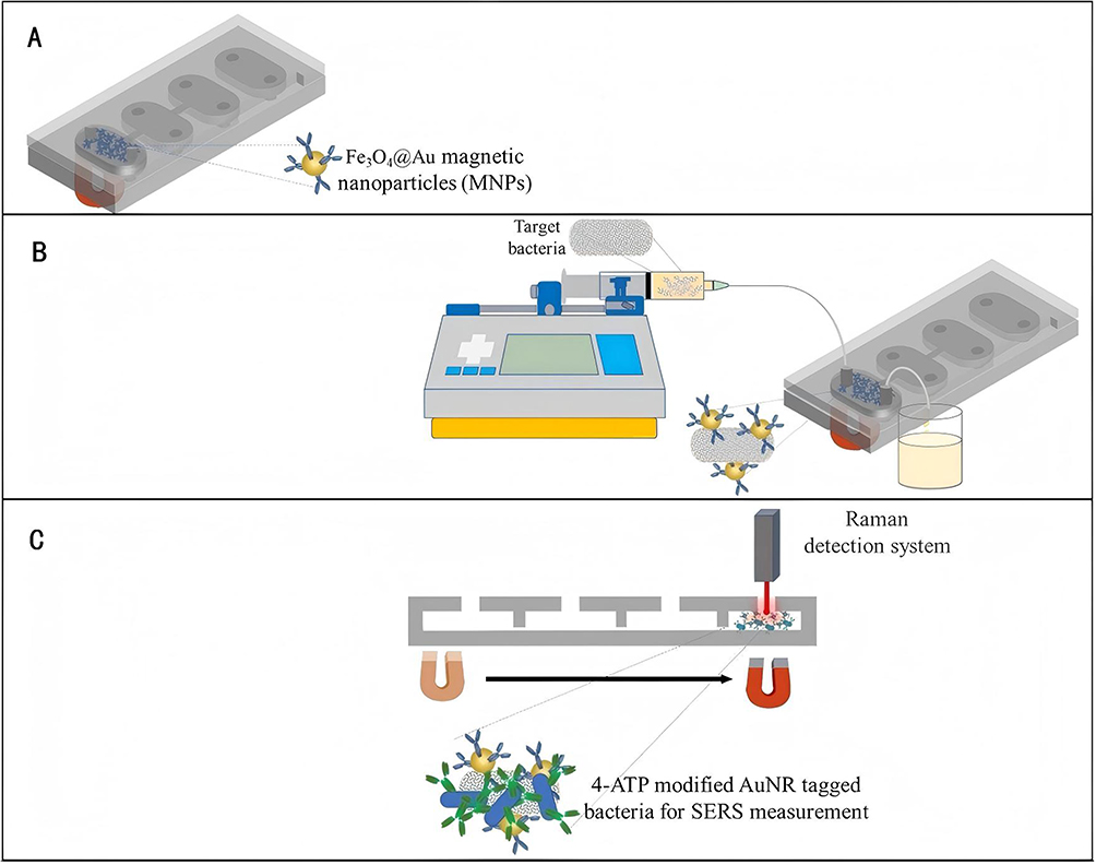 |
Figure 4 Schematic diagram of experimental strategy for SERS-based microfluidic chips. (A) Loading of antibody-conjugated Fe3O4@Au MNPs into the chip. (B) Capturing the target bacteria from the sample. (C) SERS measurement. Reproduced with permission.35 Copyright 2022, MDPI, Basel, Switzerland. CC BY 4.0, https://creativecommons.org/licenses/by/4.0/. |
The integration of microfluidics with SERS represents a transformative advancement in clinical microbial diagnostics. Once challenges such as scalable chip fabrication and cross-platform data standardization are resolved, this synergistic approach holds immense potential for revolutionizing rapid pathogen identification and AMR analysis.
SERS and Deep Learning
With the advent of the big data era, artificial intelligence (AI) has become increasingly integral to scientific research. As a subset of ML, deep learning (DL), which falls under representation learning, comprises a class of sophisticated ML algorithms. Raman spectroscopy can be effectively combined with DL architectures, such as CNNs, residual networks (ResNets), and recurrent neural networks (RNNs), to build high-accuracy classification systems for intelligent pathogen identification. This methodology involves acquiring large-scale single-cell Raman spectral datasets from diverse bacterial species, which are divided into training and testing subsets. A DL model is trained on the training set, and its discriminatory performance is systematically validated using the test set, thereby achieving reliable and precise bacterial classification. Ding et al measured 1,854 SERS spectra of Salmonella enteritidis, Salmonella typhimurium, and Salmonella paratyphi serotypes by combining SERS with a multiscale CNN. Multidimensional SERS spectral features were extracted using a multiscale CNN model, achieving a recognition accuracy of over 97%.38 Fu et al constructed an intelligent identification model for SERS spectra based on the DL technique, realizing the rapid, ultrasensitive, and non-labeled detection of pathogenic bacteria. They collected SERS spectra from 18 isolates belonging to six species of common urinary tract infection (UTI) bacteria, achieving accurate identification of bacterial species, antibiotic susceptibility, and multidrug resistance (MDR) via CNNs.39 This method significantly simplifies Raman data processing while achieving a classification accuracy above 96%. Ho et al created an extensive dataset of bacterial Raman spectra and applied the DL technique to accurately identify 30 common pathogens. The approach demonstrated 99.7% identification accuracy and 97.0 ± 0.3% accuracy of antibiotic treatment identification. It also exhibited 89 ± 0.1% differentiation accuracy between methicillin-resistant S. aureus (MRSA) and methicillin-susceptible S. aureus (MSSA).40
The integration of SERS and DL represents a paradigm shift in pathogen identification and AMR prediction. Nevertheless, clinical translation of this technology remains contingent upon resolving several critical challenges, including ensuring data quality and standardization across diverse clinical samples, improving model interpretability and generalizability to different pathogens, enhancing instrumentation robustness for point-of-care applications, and establishing appropriate regulatory frameworks. Successful bench-to-bedside implementation will require concerted interdisciplinary efforts to achieve technical standardization, demonstrate cost-effectiveness, and develop scalable solutions that meet clinical needs.
Single-Cell Raman Spectroscopy
Single-cell Raman Spectroscopy (SCRS) is an optical analysis technique that combines Raman scattering effects with precise single-cell manipulation. It can obtain vibrational spectral information on endogenous biomolecules at the level of a single living cell, thereby reflecting the chemical composition, metabolic state, and physiological phenotype of cells. The integration of diverse analytical methods facilitates the rapid and accurate identification of bacteria. Liu et al constructed an SCRS database encompassing 10 strains of S. aureus with diverse phenotypic traits and developed a CNN to predict these phenotypes from Raman spectra. The CNN model achieved 93.90%, 98.73%, and 98.66% accuracy in identifying enterotoxin-producing strains, MRSA, and growth stages, respectively.41 These demonstrate the significant potential of integrating SCRS and ML for rapid bacterial phenotypic identification.
Currently, AST based on SCRS primarily relies on the label-free identification of characteristic Raman spectral peaks and heavy water-labeled Raman techniques. Upon exposure to antibiotics, the susceptible bacterial strains undergo a series of biochemical changes. The efficacy of antibiotics can be assessed by monitoring alterations in specific Raman peak intensities or positions, thereby differentiating between susceptible and resistant strains and determining the minimum inhibitory concentration (MIC). Götz et al employed spectral changes arising from the interaction between antibiotics and E. coli to identify the characteristic Raman spectral peaks in bacterial strains exposed to antibiotics. These peaks were quantified using the normalized sum score (NSS), and a differentiation threshold between susceptible and resistant strains was established using a receiver operating characteristic (ROC) curve. Susceptibility testing of two E. coli laboratory strains and 12 clinical isolates to ciprofloxacin, cefotaxime, and piperacillin was completed within 3 hours.42 SCRS with heavy water labeling monitors bacterial metabolic activity via carbon-deuterium (C-D) bond formation, enabling antibiotic susceptibility assessment through C-D peak intensity analysis.43 Yi et al developed a fast Raman-assisted antibiotic susceptibility test (FRAST) that uses SCRS to detect single bacterial metabolic activity in the presence of antibiotics.44 After successfully detecting six clinical standard quality control strains in response to 38 antibiotics, they applied FRAST to real clinical analysis of nine urinary infectious samples and three sepsis samples. The turnaround time of “sample to report” could be reduced to 3 hours for urinary samples and 21 hours for sepsis samples, with the results consistent with MALDI-TOF identification and conventional AST. Wang et al completed the test of P. aeruginosa resistance to meropenem, ceftazidime, and cefepime within 4 hours using SCRS combined with a heavy water-labeled technique. The results demonstrated a high degree of consistency with those obtained using the broth microdilution method.45 These findings indicate that SCRS can offer rapid and accurate bacterial phenotypic identification and AMR profiling.
Challenges and Prospects
SERS technology has demonstrated significant potential for application in infectious disease diagnostics owing to its advantages, including rapid detection, non-destructive analysis, high sensitivity, and high specificity. However, the translation of SERS from laboratory research to widespread clinical implementation continues to present several challenges.
Firstly, the reproducibility and biosafety of SERS substrates are insufficient. Currently, most SERS substrates used for practical detection are metal NPs such as gold and silver. SERS signals are highly dependent on the distribution of nanostructure “hotspots”. However, most substrates exhibit problems such as significant batch-to-batch variations, high signal fluctuations, and poor long-term stability.46 These issues lead to difficulties in quantitative analysis and establishing standardized detection protocols, as well as limit multicenter clinical validation. Furthermore, the long-term toxicity profiles of NPs remain inadequately characterized. They have the potential to induce immune reactions, cause cytotoxic effects, or even alter gene expression patterns,47 raising significant concerns regarding their risks to human health. Consequently, further investigation is urgently needed to evaluate the biocompatibility and potential toxicity of metal-based nanomaterials, ensuring their long-term stability and safety in physiological environments. In addition, current biosafety assessment methodologies for such materials remain underdeveloped, lacking systematic and standardized evaluation criteria and protocols.48 This gap critically hinders the translation of metal nanomaterials into clinical applications.
Secondly, the fluorescence interference is severe. Complex clinical samples, such as blood, sputum, and urine, contain large amounts of proteins, lipids, and carbohydrates, which can generate strong fluorescence backgrounds or non-specific adsorption under laser excitation,14 completely masking the effective spectra of target pathogens.
Thirdly, the database and methodological standardization are insufficient. While Raman spectral “fingerprints” exhibit high specificity, the field currently lacks a unified, comprehensive microbial Raman spectroscopy reference database. Critical variations in experimental parameters, including laser wavelengths, substrate materials, and spectral preprocessing protocols, introduce substantial challenges for cross-study spectral comparison. Notably, research has demonstrated that SERS signal intensity directly correlates with the size and morphology of gold or silver NPs substrates,49 highlighting the necessity for standardized substrate fabrication protocols in clinical SERS applications.
Lastly, the technique faces practical implementation barriers due to its technical complexity. Current SERS analysis requires specialized software and trained personnel, significantly limiting its adoption in primary healthcare settings and point-of-care applications.
To address these challenges, future research should focus on the following aspects: (1) Develop highly reproducible and mass-producible SERS substrates to improve the reliability and consistency of quantitative detection; conduct in-depth research on the biosafety of metal nanomaterials, including their biocompatibility, toxicity, and degradability, to promote the development of sustainable and environmentally friendly nanomaterials; establish a comprehensive safety evaluation system for nanomaterials, and implement standardized assessments and regulatory oversight, to ensure the safety of their practical applications. (2) Integrate microfluidic systems with automated sample pretreatment modules to construct an integrated “sample-in, result-out” platform, thereby minimizing manual intervention and enhancing detection robustness. (3) Establish standardized operating protocols for SERS-based detection to promote standardization and facilitate clinical translation. (4) Build a multicenter pathogen SERS spectral database, encompassing diverse bacterial species and AMR phenotypes, and develop an AI-driven diagnostic system for automated pathogen identification and antimicrobial susceptibility prediction. (5) Develop intelligent and portable point-of-care testing (POCT) devices for deployment in resource-limited settings, such as primary healthcare facilities and epidemic-affected regions. The advantages, limitations, and potential solutions of SERS technology are summarized in Table 2.
 |
Table 2 The Advantages, Limitations, and Potential Solutions of SERS Technology |
Conclusion
SERS has emerged as a transformative platform for the rapid diagnostics of infectious diseases. Owing to its distinctive advantages, including rapid analysis, label-free and non-destructive detection, exceptional sensitivity, and high specificity, SERS enables precise identification of pathogenic bacteria and their AMR profiles. Notably, it exhibits superior performance in detecting low-abundance pathogens. Despite persistent challenges, including substrate reproducibility, biosafety concerns, complex sample preprocessing requirements, and the absence of standardized clinical protocols, ongoing advances in nanotechnology, synergistic integration of AI-driven data analysis, and strengthened regulatory frameworks for nanomaterial biosafety assessment are synergistically accelerating the maturation of SERS toward intelligent, high-throughput, and clinically deployable. With continued translational efforts, SERS is poised to transition successfully from laboratory studies to clinical applications and is expected to develop a cornerstone technology for next-generation infectious disease diagnostics, thereby advancing precision medicine and strengthening public health surveillance infrastructure.
Funding
This study was supported by the Science and Technology Project of the Health Commission of Jiangxi Province, China (no. 202610563).
Disclosure
The authors declare no conflicts of interest regarding the publication of this article.
References
1. Xiao M, Tian F, Liu X, et al. Virus detection: from state-of-the-art laboratories to smartphone-based point-of-care testing. Adv Sci. 2022;9(17):e2105904. doi:10.1002/advs.202105904
2. GBD 2019 Antimicrobial Resistance Collaborators. Global mortality associated with 33 bacterial pathogens in 2019: a systematic analysis for the Global Burden of Disease Study 2019. Lancet. 2022;400(10369):2221–13. doi:10.1016/S0140-6736(22)02185-7
3. Wang C, Zhang Z, Wang X, et al. Detection of respiratory pathogenic bacterial nucleic acid detection by loop-mediated isothermal amplification in patients with bacterial pulmonary infections. Pract Lab Med. 2023;37:e00344. doi:10.1016/j.plabm.2023.e00344
4. Raman CV, Krishnak KS. A new type of secondary radiation. Nature. 1928;121(3048):501–502.
5. Yu M, Wang JK, Lin KC, et al. Application progress on Raman spectroscopy in the diagnosis of pathogenic infections. J Microb Infect. 2022;17(2):102–109.
6. Fleischmaann M, Hendra PJ, Mcquillan AJ. Raman spectra of pyridine adsorbed at a silver electrode. Chem Phys Lett. 1974;26(2):163–166.
7. Jeanmaire DL, Van Duyne RP. Surface Raman spectroelectrochemistry: part I. heterocyclic, aromatic, and aliphatic amines adsorbed on the anodized silver electrode. J Electroanal Chem Interfacial Electrochem. 1977;84(1):1–20.
8. Albrecht MG, Creighton JA. Anomalously intense Raman spectra of pyridine at a silver electrode. J Am Chem Soc. 1977;99(15):5215–5217.
9. Wang L, Liu W, Tang JW, et al. Applications of Raman spectroscopy in bacterial infections: principles, advantages, and shortcomings. Front Microbiol. 2021;12:683580. doi:10.3389/fmicb.2021.683580
10. Zong C, Xu M, Xu LJ, et al. Surface-Enhanced Raman spectroscopy for bioanalysis: reliability and challenges. Chem Rev. 2018;118(10):4946–4980. doi:10.1021/acs.chemrev.7b00668
11. Li H, Gao R, Hu X, et al. Noble Metal-Based nanocomposites for surface-enhanced raman spectroscopy detection of food contaminants. Foods. 2025;14(17):3108. doi:10.3390/foods14173108
12. Chaloupková Z, Gajdošová K, Poláková K, et al. Surface-enhanced Raman spectroscopy-assisted lateral flow test for adenine and IgG analysis. Anal Chim Acta. 2025;1376:344595. doi:10.1016/j.aca.2025.344595
13. Jiang Y, Yanagida R, Saito M, et al. Sensitive, Low-Cost, early detection of periodontal disease using surface-enhanced raman spectroscopy with easy-to-use silver nanomesh-coated periopaper. Anal Chem. 2025;97(39):21550–21557. doi:10.1021/acs.analchem.5c03876
14. Usman M, Tang JW, Li F, et al. Recent advances in surface enhanced Raman spectroscopy for bacterial pathogen identifications. J Adv Res. 2023;51:91–107. doi:10.1016/j.jare.2022.11.010
15. Meng ZC, Ling DY, Yang LB. Research progress of surface-enhanced raman spectroscopy in bacterial detection. J Light Scatter. 2023;35(2):142–149.
16. Liu X, Dai Y, Yao W, et al. Development of multifunctional BP@MoS2 two-dimensional hybrid nanomaterials for label-free surface-enhanced Raman spectroscopy detection and synergistic light-conversion depredation of pathogenic bacteria. Talanta. 2026;297:128700. doi:10.1016/j.talanta.2025.128700
17. Yuan K, Mei Q, Guo X, et al. Antimicrobial peptide based magnetic recognition elements and Au@Ag-GO SERS tags with stable internal standards: a three in one biosensor for isolation, discrimination and killing of multiple bacteria in whole blood. Chem Sci. 2018;9(47):8781–8795. doi:10.1039/c8sc04637a
18. de Almeida MP, Rodrigues C, Novais Â, et al. Silver Nanostar-Based SERS for the discrimination of clinically relevant Acinetobacter baumannii and Klebsiella pneumoniae species and clones. Biosensors. 2023;13(2):149. doi:10.3390/bios13020149
19. Wang L, Tang JW, Li F, et al. Identification of bacterial pathogens at genus and species levels through combination of raman spectrometry and deep-learning algorithms. Microbiol Spectr. 2022;10(6):e0258022. doi:10.1128/spectrum.02580-22
20. Yadav S, Senapati S, Desai D, et al. Portable and sensitive ag nanorods based SERS platform for rapid HIV-1 detection and tropism determination. Colloids Surf B Biointerfaces. 2021;198:111477. doi:10.1016/j.colsurfb.2020.111477
21. Zhang Z, Zhang Y, Lv XM, et al. Detection of respiratory pathogens based on gold biconical nanoparticles with surface-enhanced raman spectroscopy. Spectrosc Spectral Anal. 2023;43(7):175–176.
22. Jiang H, Lv ZW, Li Y. A novel strategy for viral detection in acute respiratory infections: combining SERS with machine learning. Spectrosc Spectral Anal. 2025;45(5):1217–1224.
23. Wang MF, Huang JW, Liu CL, et al. Identification of invasive fungal disease related pathogens using surface-enhanced raman spectroscopy. J Tianjin Univer. 2023;56(09):927–934.
24. Dina NE, Gherman AMR, Chiş V, et al. Characterization of clinically relevant fungi via SERS fingerprinting assisted by novel chemometric models. Anal Chem. 2018;90(4):2484–2492. doi:10.1021/acs.analchem.7b03124
25. Witkowska E, Jagielski T, Kamińska A. Genus-and species-level identification of dermatophyte fungi by surface-enhanced Raman spectroscopy. Spectrochim Acta A Mol Biomol Spectrosc. 2018;192:285–290. doi:10.1016/j.saa.2017.11.008
26. Henderson KC, Benitez AJ, Ratliff AE, et al. Specificity and strain-typing capabilities of nanorod array-surface enhanced Raman spectroscopy for Mycoplasma pneumoniae detection. PLoS One. 2015;10(6):e0131831. doi:10.1371/journal.pone.0131831
27. Berus SM, Adamczyk-Popławska M, Młynarczyk-Bonikowska B, et al. SERS-based sensor for the detection of sexually transmitted pathogens in the male swab specimens: a new approach for clinical diagnosis. Biosens Bioelectron. 2021;189:113358. doi:10.1016/j.bios.2021.113358
28. Chen Y, Premasiri WR, Ziegler LD. Surface enhanced Raman spectroscopy of chlamydia trachomatis and neisseria gonorrhoeae for diagnostics, and extra-cellular metabolomics and biochemical monitoring. Sci Rep. 2018;8(1):5163. doi:10.1038/s41598-018-23562-5
29. Chen C, Hong W. Recent development of rapid antimicrobial susceptibility testing methods through metabolic profiling of bacteria. Antibiotics. 2021;10(3):311. doi:10.3390/antibiotics10030311
30. Mushtaq A, Nawaz H, Irfan Majeed M, et al. Surface-enhanced Raman spectroscopy (SERS) for monitoring colistin-resistant and susceptible E. coli strains. Spectrochim Acta A Mol Biomol Spectrosc. 2022;278:121315. doi:10.1016/j.saa.2022.121315
31. Thrift WJ, Ronaghi S, Samad M, et al. Deep learning analysis of vibrational spectra of bacterial lysate for rapid antimicrobial susceptibility testing. ACS Nano. 2020;14(11):15336–15348. doi:10.1021/acsnano.0c05693
32. Skvortsova A, Trelin A, Guselnikova O, et al. Surface enhanced Raman spectroscopy and machine learning for identification of beta-lactam antibiotics resistance gene fragment in bacterial plasmid. Anal Chim Acta. 2024;1329:343118. doi:10.1016/j.aca.2024.343118
33. Yue S, Fang J, Xu Z. Advances in droplet microfluidics for SERS and Raman analysis. Biosens Bioelectron. 2022;198:113822. doi:10.1016/j.bios.2021.113822
34. Park S, Su Jeon C, Choi N, et al. Sensitive and reproducible detection of SARS-CoV-2 using SERS-based microdroplet sensor. Chem Eng J. 2022;446:137085. doi:10.1016/j.cej.2022.137085
35. Dogan Ü, Sucularlı F, Yildirim E, et al. Escherichia coli enumeration in a capillary-driven microfluidic chip with SERS. Biosensors. 2022;12(9):765. doi:10.3390/bios12090765
36. Liu Y, Su G, Wang W, et al. A novel multifunctional SERS microfluidic sensor based on ZnO/Ag nanoflower arrays for label-free ultrasensitive detection of bacteria. Anal Methods. 2024;16(14):2085–2092. doi:10.1039/d4ay00018h
37. Liao CC, Chen YZ, Lin SJ, et al. A microfluidic microwell device operated by the automated microfluidic control system for surface-enhanced Raman scattering-based antimicrobial susceptibility testing. Biosens Bioelectron. 2021;191:113483. doi:10.1016/j.bios.2021.113483
38. Ding J, Lin Q, Zhang J, et al. Rapid identification of pathogens by using surface-enhanced Raman spectroscopy and multi-scale convolutional neural network. Anal Bioanal Chem. 2021;413(14):3801–3811. doi:10.1007/s00216-021-03332-5
39. Fu Q, Zhang Y, Wang P, et al. Rapid identification of the resistance of urinary tract pathogenic bacteria using deep learning-based spectroscopic analysis. Anal Bioanal Chem. 2021;413(30):7401–7410. doi:10.1007/s00216-021-03691-z
40. Ho CS, Jean N, Hogan CA, et al. Rapid identification of pathogenic bacteria using Raman spectroscopy and deep learning. Nat Commun. 2019;10(1):4927. doi:10.1038/s41467-019-12898-9
41. Liu L, Xue J, Song Y, et al. A pilot study on single-cell raman spectroscopy combined with machine learning for phenotypic characterization of Staphylococcus aureus. Microorganisms. 2025;13(6):1333. doi:10.3390/microorganisms13061333
42. Götz T, Dahms M, Kirchhoff J, et al. Automated and rapid identification of multidrug resistant Escherichia coli against the lead drugs of acylureidopenicillins, cephalosporins, and fluoroquinolones using specific Raman marker bands. J Biophotonics. 2020;13(8):e202000149. doi:10.1002/jbio.202000149
43. Tao Y, Wang Y, Huang S, et al. Metabolic-Activity-Based assessment of antimicrobial effects by D2O-Labeled single-cell raman microspectroscopy. Anal Chem. 2017;89(7):4108–4115. doi:10.1021/acs.analchem.6b05051
44. Yi X, Song Y, Xu X, et al. Development of a fast Raman-Assisted Antibiotic Susceptibility Test (FRAST) for the antibiotic resistance analysis of clinical urine and blood samples. Anal Chem. 2021;93(12):5098–5106. doi:10.1021/acs.analchem.0c04709
45. Wang FC, Niu L, Ye HY, et al. Study on rapid antimicrobial susceptibility test of pseudomonas aeruginosa by D2O-Labeled single-cell raman spectroscopy. Spectrosc Spectral Anal. 2025;45(08):2253–2258.
46. Ali A, Nettey-Oppong EE, Effah E, et al. Miniaturized Raman instruments for SERS-Based point-of-care testing on respiratory viruses. Biosensors. 2022;12(8):590. doi:10.3390/bios12080590
47. Kothari D, Kumar A. Metallic, carbon-based, and polymeric nanomaterials: transforming dairy farming practices for sustainability. Food Chem X. 2025;29:102640. doi:10.1016/j.fochx.2025.102640
48. Allan J, Belz S, Hoeveler A, et al. Regulatory landscape of nanotechnology and nanoplastics from a global perspective. Regul Toxicol Pharmacol. 2021;122:104885. doi:10.1016/j.yrtph.2021.104885
49. Ramirez-Perez JC, Reis TA, Olivera CLP, et al. Impact of silver nanoparticles size on SERS for detection and identification of filamentous fungi. Spectrochim Acta A Mol Biomol Spectrosc. 2022;272:120980. doi:10.1016/j.saa.2022.120980
 © 2026 The Author(s). This work is published and licensed by Dove Medical Press Limited. The
full terms of this license are available at https://www.dovepress.com/terms
and incorporate the Creative Commons Attribution
- Non Commercial (unported, 4.0) License.
By accessing the work you hereby accept the Terms. Non-commercial uses of the work are permitted
without any further permission from Dove Medical Press Limited, provided the work is properly
attributed. For permission for commercial use of this work, please see paragraphs 4.2 and 5 of our Terms.
© 2026 The Author(s). This work is published and licensed by Dove Medical Press Limited. The
full terms of this license are available at https://www.dovepress.com/terms
and incorporate the Creative Commons Attribution
- Non Commercial (unported, 4.0) License.
By accessing the work you hereby accept the Terms. Non-commercial uses of the work are permitted
without any further permission from Dove Medical Press Limited, provided the work is properly
attributed. For permission for commercial use of this work, please see paragraphs 4.2 and 5 of our Terms.