Back to Journals » Open Access Emergency Medicine » Volume 11
Septic shock in the ER: diagnostic and management challenges
Authors Worapratya P, Wuthisuthimethawee P ![]()
Received 13 November 2018
Accepted for publication 21 February 2019
Published 11 April 2019 Volume 2019:11 Pages 77—86
DOI https://doi.org/10.2147/OAEM.S166086
Checked for plagiarism Yes
Review by Single anonymous peer review
Peer reviewer comments 2
Editor who approved publication: Dr Hans-Christoph Pape
Panita Worapratya, Prasit Wuthisuthimethawee
Department of Emergency Medicine, Faculty of Medicine, Prince of Songkla University, Hat Yai, Songkhla 90110, Thailand
Abstract: Sepsis is a common presentation in the emergency department and a common cause of intensive care unit admissions and death. Accurate triage, rapid recognition, early resuscitation, early antibiotics, and eradication of the source of infection are the key components in delivering quality sepsis care. Evaluation of the patient’s volume status, optimal hemodynamic resuscitation, and evaluation of patient response is crucial for sepsis management in the emergency department.
Keywords: sepsis, Sepsis-3, resuscitation, lactate, fluid responsiveness
Introduction
Sepsis is a common presentation in the emergency department and common cause of intensive care unit admissions and death.1,2 Even though, there is a sepsis campaign guideline, the mortality from sepsis worldwide is still high at 34–46%.3,4 Rapid recognition, early resuscitation, early antibiotics, and eradication of the source of infection are the key components in delivering quality sepsis care.5–7 Since sepsis8 is defined as a dysregulated host response to infection and causes organ dysfunction which cannot be diagnosed by superficial assessment, triage and recognizing sepsis in the emergency department can be a challenge.
Early and optimal hemodynamic resuscitation is crucial. Either under or over fluid resuscitation can lead to an unfavorable patient outcome. Evaluation of the patient’s volume status and response is the cornerstone for sepsis management in the emergency department.
From this point of view, we discuss some practical points of sepsis triage, initial resuscitation, hemodynamic monitoring, and the target end point of resuscitation for sepsis patients in the emergency department based on the current evidence.
New and old definitions of sepsis: how does it matter for sepsis triage?
Systemic inflammatory response syndrome (SIRS) – body temperature >38°C or <36°C, heart rate >90 beats/min, respiratory rate >20 breaths/min, and white blood cell count >12,000/nm3 or >10% immature neutrophils – has been used as part of the definition of sepsis for decades.9,10 A recent study shows that the elderly and immunocompromised patients may have an absence of fever and present with leukopenia instead of leukocytosis, meanwhile tachycardia, increased respiratory rate, and high body temperature are not specific for infection. For these reasons, the SIRS criteria are inadequate and not specific to make a diagnosis of sepsis.11–13
The new definition of sepsis according to Sepsis-3 pays attention to organ dysfunction and hypoperfusion rather than an inflammation.14 Subsequently, the term “severe sepsis” was removed from the definition.8 Therefore, the sepsis task force proposed a new definition of sepsis as a life-threatening organ dysfunction, which is defined by a sequential organ failure assessment (SOFA) score ≥2 (Table 1). Septic shock was defined as the need for a vasopressor to maintain a mean arterial pressure of at least 65 mmHg and serum lactate level over 2 mmol/L (>18 mg/dL) in the absence of hypovolemia which can increase the mortality rate to 40%.15
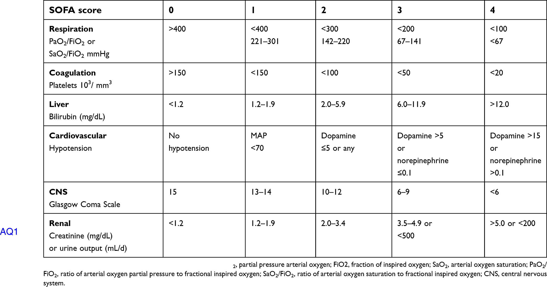 | Table 1 SOFA score |
However, the SOFA score needs several laboratory results which mostly are not available in the triage area of an emergency department. The time needed to obtain the test results can cause delayed detection of a septic patient.
The quick SOFA (qSOFA) score (Box 1) was introduced in Sepsis-314 and is a tool to predict risk of death and extended ICU stay, but it is not designed to stand alone as an early warning signal of sepsis or identify which patients should be transferred to the ICU.16,17 Two recent cohort studies found that the validity of the qSOFA score criteria, that includes altered mental status (Glasgow Coma Scale score <15), respiratory rate >22, systolic blood pressure <100 mmHg, and with and without serum lactate greater than >2 mmol/L, were good indicators to predict hospital mortality equally as well as the SOFA score.14,18 Unfortunately, outside the ICU sepsis group, 30% had no SIRS criteria and 41% had no SOFA points. A recent systematic review and data from a meta-analysis showed that the qSOFA outside the ICU had poor sensitivity (0.51) when used as a screening tool in the emergency department.19,20 Meanwhile, the National Early Warning Score (NEWS) (Table 2) and Modified Early Warning Score (MEWS) (Table 3), which are based on the clinical parameters of body temperature, heart rate, respiratory rate, oxygen saturation, systolic blood pressure, and level of consciousness, were shown to be more feasible for monitoring and early recognition of septic patients in both the emergency department and outside the ICU. Recent data seem to indicate that the sensitivity of the NEWS criteria is superior to the MEWS and qSOFA scores. The sensitivity of a NEWS ≥5 is 79%, which is similar to the SIRS criteria ≥2 (sensitivity 80%) and higher than qSOFA ≥2 (sensitivity 74%). NEWS had a similar AUROC (AUROC =0.65; 95% CI, 0.61 to 0.68) to qSOFA (AUROC =0.62; 95% CI, 0.59 to 0.66).21 When the sensitivity was compared for in-hospital mortality, the NEWS ≥5, MEWS ≥5, qSOFA ≥2, and SIRS ≥2 criteria had sensitivities of 95.1%, 71.4%, 68.7%, and 93.8%, respectively.22
 | Box 1 Quick sequential organ failure assessment |
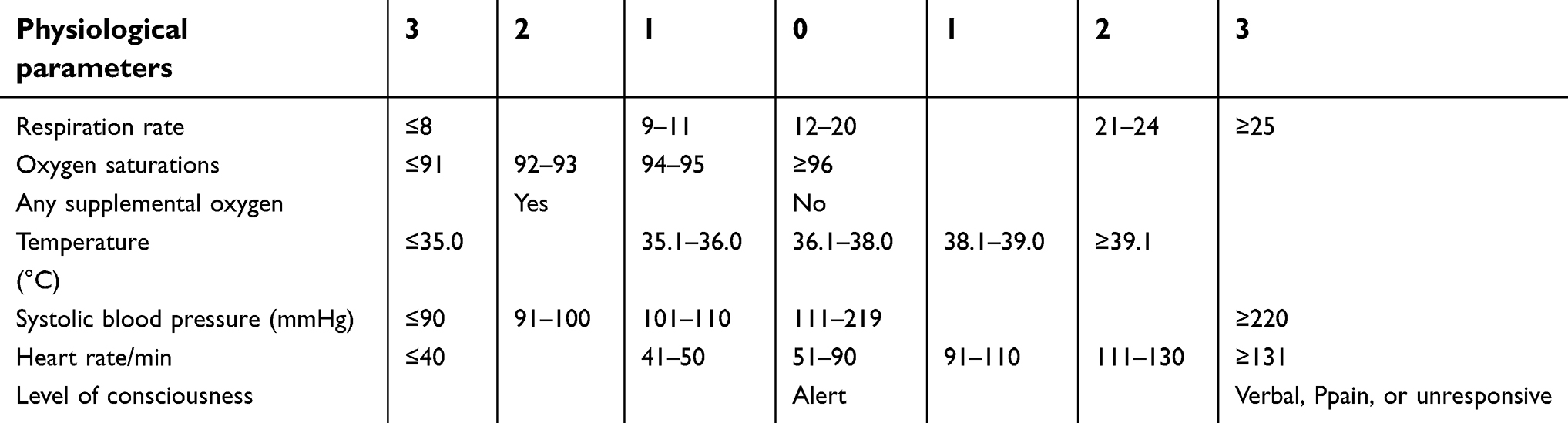 | Table 2 National Early Warning Score (NEWS) |
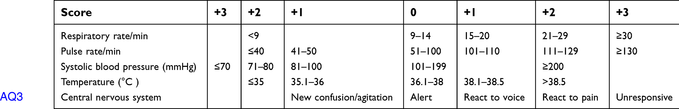 | Table 3 Modified Early Warning Score (MEWS) |
In summary, the new definition of sepsis focuses on organ dysfunction and hypoperfusion. SIRS was removed from the sepsis clinical syndrome and is not a part of the definition of sepsis and septic shock is now defined as a subset of sepsis.14 However, the new sepsis-3 definition is not without controversy. There is debate about whether the new definition, which relies on organ dysfunction, can fail in early recognition and delayed resuscitation of sepsis patients. The qSOFA score is not a part of the definition of sepsis and cannot be used as a sepsis screening tool. However, it should alert clinicians to patients who are in need of further assessment of organ dysfunction. NEWS is superior to MEWS as a screening tool for sepsis patients.18 However, the Surviving Sepsis Campaign (SSC) guideline does not recommend any specific tool for screening septic patients.14
Fluid resuscitation in sepsis: time, types, and dose
For decades, fluid resuscitation has been recommended as the first priority to treat septic shock.5,8,23 The physiology of septic shock is due to increased insensible fluid loss, alteration of venous capacitance, and vascular leakage that results in generating “relative hypovolemia”.24 Therefore, fluid management in sepsis may differ between the phases of sepsis and fluid choice and the volume of resuscitation affects the patient’s outcome.25
Understanding the 4 phases of septic shock
A recent conceptual model of circulatory shock was published, and it identifies the 4 phases of resuscitation as rescue, optimization, stabilization, and de-escalation.26
The rescue phase or life-threatening phase occurs within minutes to hours characterized by strong vasodilation and causes hypotension and impaired organ perfusion. During the first 3 to 6 hrs after initiation of therapy, fluid resuscitation is the aim for early and adequate fluid administration to prevent cardiovascular collapse and death. Early goal-directed therapy (EGDT) has been recommended for a decade as the best standard protocol for resuscitation of septic shock patients according to the SSC.9,23 Results from three international independent multicenter trials (ProCESS, ARISE, and ProMISe)8,27,28 showed no benefit of EGDT over standard care. Many systematic reviews and meta-analyses reported that EGDT does not decrease mortality compared with conventional care.29–33 Furthermore, one study showed a worse outcome in the EGDT group compared to conventional care.34 The Sepsis Campaign Guideline 201835 recommends immediate fluid administration at a dose of 30 mL/kg of intravenous crystalloid fluid in all septic patients who have hypotension or an elevated lactate level. However, the goal of fluid administration should be individualized. An evaluation of the need for fluid can be achieved by many methods, such as the fluid challenge test, the passive leg raise test, and the end-expiratory occlusion test.36
The optimization phase, which is also referred to as the ischemia and reperfusion phase, occurs within hours. During this time, careful assessment of the intravascular volume status and determination of the need for further fluid administration are crucial. Optimum fluid resuscitation in this phase is still being debated. Some studies showed a higher mortality rate in the fluid bolus group,37 while another study showed a relationship between increased fluid balance and increased mortality in septic patients.38 Fluid responsiveness is determined by a change in the stroke volume or cardiac output (CO) of approximately 12–15% after a bolus dose of fluid administration.39 The routine use of invasive CO monitoring devices, such as central venous catheter and pulmonary artery catheter, have been associated with risks without significant benefits.40 Current data show poor sensitivity and specificity of central venous pressure (CVP) to evaluate volume status and fluid response in shock patients.41,42 Some studies gave evidence that the passive leg raising test and the end-expiratory occlusion test have potential to predict volume responsiveness.43–46 The velocity time integral (VTI) is another parameter to predict volume response in septic patients which can be done by bedside echocardiography. Lamia et al47 studied patients with and without mechanical ventilation for ICU patients with shock and demonstrated that a 12.5% change in the VTI had a sensitivity and specificity of 77% and 100%, respectively, for a >15% increase in CO following fluid loading with an associated area under the curve of 0.96. The caval index can be calculated as the maximum diameter on expiration (IVC max) minus the minimum diameter on inspiration (IVC min) divided by IVC max. Some research demonstrated that the IVC diameter may predict CVP in intubated, mechanically ventilated patients and in spontaneously breathing patients.48–50 A meta-analysis from 8 studies reported that the pool sensitivity and specificity of the caval index to predict volume responsiveness in shock patients were 76% and 86%, respectively, and the caval index performance was better in patients on mechanical ventilation than in spontaneously breathing patients.51
Once adequate intravascular volume status has been achieved, rapid administration of vasopressor therapy in the setting of fluid-refractory shock is a time-critical intervention. Delayed initiation of vasopressor therapy can lead to excessive fluid resuscitation and increased morbidity and mortality. Mortality increases of 5.3% were estimated to occur for every 1 hr delay in vasopressor initiation.52 Initial vasopressor choice in septic shock is norepinephrine, starting at a dose of 0.5 mcg/kg/min.53
The stabilization phase usual occurs within a few days and after optimized fluid was given which is manifested by a stable hemodynamic state. The goal of this phase is to maintain intravascular volume, replace ongoing fluid loss, support organ dysfunction, and avoid iatrogenic harm with unnecessary intravenous fluid administration.54 This step assesses the adequacy of organ perfusion and microcirculatory resuscitation is crucial. Several studies demonstrated that a high CVP (≥8 mmHg) could be associated with prolonged mechanical ventilation, longer hospital length of stay, kidney injury, lower pO2/FiO2, and increased mortality.55,56 A ScvO2 between 70% and 89% would suggest an adequate VO2/DO2 balance, while a supranormal ScvO2 value ≥90% suggests poor oxygen utilization, tissue dysoxia, and it is associated with mortality.57 The use of lactate and lactate clearance (([initial lactate – 6 hrs lactate]/initial lactate) x 100)) seem to be the best options to date.58 Multiple studies showed a lower mortality by achieving the lactate clearance.58–60 Once the end point of resuscitation is achieved, daily fluid balance should be closely monitored and maintenance fluid should be given only to cover daily needs including insensible fluid loss and gastrointestinal loss.61
The de-escalation phase is characterized by organ recovery and weaning from mechanical ventilation and vasopressor support. Excessive fluid balance in this phase is significantly related to mortality.55,62 The goal of this phase is to achieve an overall negative fluid balance.63 Recently, more clinical studies have demonstrated an independent association between an increased positive fluid balance and increased mortality in patients with sepsis.38,64,65 One randomized study showed that aggressive fluid loading was associated with a significant increase in the risk of death.37
A moderate fluid management strategy encompasses the avoidance of fluid loading and getting rid of fluid overload is a key component for improved survival.36 Starting fluid removal should be done carefully without inducing hypotension and reducing CO.66 The risk in this phase is to be too aggressive in discontinuing fluids which may cause hemodynamic deterioration. To avoid this problem, testing preload responsiveness might be useful.36
Time is crucial!!
One of the most important concerns of fluid resuscitation is time. Many studies have shown that delayed fluid resuscitation is related to mortality.67–69 This was confirmed by a cohort study in 11,182 sepsis patients which demonstrated the mortality benefit of early fluid resuscitation within 30 mins after diagnosis. The mortality rate was lower in the <30-min group (17.8%) than in the >30-min group (24.5%).70 In a recent study that evaluated the effects of Ringer’s lactate solution or 4% albumin on the microvascular circulation, fluid administration improved microvascular perfusion in the early but not in the late phase of sepsis.71 This gave assurance that the type of fluid used is likely to be less important than timing.71,72
Type of fluid: colloid versus crystalloid
Fluid resuscitation plays a major role in septic shock patients in the acute phase. However, the best choice of intravenous fluid for sepsis patients has been debated. Ideal fluid resuscitation in sepsis should be physiologically balanced with improvement in the patient’s outcome in a cost-effective manner.
Normal saline solution (NSS) has been used for volume resuscitation as standard treatment in shock patients for decades.73,74 NSS is isotonic to extracellular fluid, but contains a higher chloride concentration than plasma. Hartmann’s solution, lactated Ringer’s solution (LRS), and Plasma-Lyte may be slightly hypotonic to extracellular fluid, but these solutions provide more physiologic ions and pH control.24 A randomized, double-blind crossover study demonstrated a reduction of renal cortical perfusion and renal blood flow by magnetic resonance imaging in healthy volunteers who received a 2-L bolus of NSS.75 Another retrospective analysis of 1,940 ICU patients found that hyperchloremia at 72 hrs after ICU admission was significantly associated with mortality and every 5 mEq/L increase of serum chloride concentration was associated with a further increase in mortality.76 Both experimental77,78 and clinical79,80 studies suggested that resuscitation with normal saline has detrimental effects on the kidneys, acid–base balance, and electrolyte homeostasis, and may affect tissue perfusion.81 A prospective pilot study in a single ICU, which implemented a chloride-restricted fluid policy, found a significant decrease in acute kidney injury and the reduced need for renal replacement therapy although mortality was unchanged.82
LRS, Hartmann’s solution, and Plasma-Lyte are commonly known as balanced crystalloids because they have lower concentrations of chloride ions compared to normal saline.24 LRS is considered to be superior to NSS in terms of acid balance and has shown to improve survival in septic patients.83,84 The lactate in LRS is metabolized in the liver to form bicarbonate which is the key buffer in preventing acidosis without increasing the circulating lactic acid concentration in patients who are in the state of hypoperfusion.85,86
Recently, a cohort study in SIRS patients87 and a prospective observational study in abdominal surgery patients88 reported the same results from a comparison between Plasma-Lyte and NSS. Plasma-Lyte was associated with lower rates of acute respiratory failure, coagulopathy, electrolyte abnormality, and renal failure, in addition to a shorter length of hospital stay and lower in-hospital mortality.
Albumin and hydroxyethyl starch (HES) are considered to be colloid solutions. Albumin is commonly used and recommended in septic patients due to the anti-inflammatory effect, fluid sparing, and may decrease mortality in septic shock patients.89,90 Sepsis guidelines (Grade 2C) currently suggest that albumin should be considered as a resuscitation fluid in patients with severe sepsis, particularly if those patients are not responding to crystalloid infusion.23,91 However, the benefits of using albumin for resuscitation in septic patients need to be clarified. A subgroup analysis among septic patients in the Saline versus Albumin Fluid Evaluation study (SAFE)89 showed significant mortality reduction in the albumin group with a mortality rate of 30.7% compared with 35.3% in the NSS group. The Albumin Italian Outcome Sepsis study (ALBIOS)92 randomized 1,818 severe sepsis and septic shock patients and showed no statistically significant difference in the 28-day mortality between the albumin and NSS groups. A third trial, Early Albumin Resuscitation During Septic Shock (EARSS), randomized septic shock patients who received vasopressin within 8 hrs were administered 100 mL of 20% albumin or 0.9% NSS 100 mL every 8 hrs for 3 days. Among 798 patients, vasopressor-free days were higher in the albumin group (24.1% vs 26.3%) without improvement of 28-day mortality.
HES is a semisynthetic colloid solution derived from chemically modified plant starch. An HES solution contains molecules of various different molecular weights that range from 70 to 670 kDa93 and has been associated with several problems, such as altered hemostasis, immunological consequence, and altered renal function.94 In 2004, the Efficacy of Volume Substitution and Insulin Therapy in Severe Sepsis (VISEP) trial95 had to stop early due to a trend of increased 90-day mortality (41.0% vs 33.9%) and increased acute kidney injury with HES 200/0.5 (34.9% vs 22.8%) compared with the LRS group. Later, the larger Scandinavian Starch for Severe Sepsis/Septic Shock (6S) trial compared the use of HES and LRS as resuscitation fluids in 804 septic patients and showed the same results as the VISEP trial. The author concluded that patients who received HES had a higher risk of death at 90 days and were more likely to need renal-replacement therapy compared to LRS.96 The harmful effects of HES were later confirmed further in the CRYSTMAS, CHEST, and CRISTAL studies.97–99 The latest systematic review and meta-analysis affirmed that HES was related to acute kidney injury and mortality.100
Conclusion
The new concept of sepsis is dysregulation of a host’s response to infection defined as an organ dysfunction score ≥2.8 Septic shock is defined as persistent hypotension, which needs a vasopressor to maintain hemodynamic stability, and hyperlactatemia (serum lactate ≥2 mmol/L).23 The concept of septic shock is believed to be due to an inflammatory process that causes changes in vessel permeability and cardiac dysfunction rather than volume depletion.101,102 The qSOFA, which is recommended as the standard sepsis screening tool in the Sepsis-3 guideline,23 had poor sensitivity when used in the ED.19,20 NEWS was shown to be superior to MEWS and qSOFA as screening tools for sepsis in the ED.22 The lower mortality benefit of early fluid resuscitation within 30 mins after diagnosis is superior to the type of fluid resuscitation.70,72 Optimized volume resuscitation is the second priority for sepsis shock resuscitation to decrease mortality.37,103 The type of fluid, whether crystalloid or colloid is still controversial92 while albumin resuscitation in sepsis has tended to be superior to a crystalloid solution without statistical significance.89 LRS is recommended in first-line treatment of sepsis resuscitation rather than NSS due to the benefit of lower mortality80 and renal replacement therapy.82 CVP is not a single good indicator to evaluate fluid responsiveness.104,105 However, if CVP monitoring is needed, the CVP should be kept lower than 8 mmHg.55 Lactate clearance can be used as a goal targeted therapy as well as ScvO2.106,107 Bedside echocardiography to evaluate VTI seems to be the best parameter to guide fluid resuscitation in the ED with high sensitivity and specificity.108,109 Early antibiotic treatment within 1 hr to eliminate the source of infection is still recommended.5–8 Physicians who care for sepsis patients should use various physiologic parameters to adjust fluid resuscitation rather than rely on a single parameter.
Disclosure
The authors report no conflicts of interest in this work.
References
1.
2. Brun-Buisson C, Meshaka P, Pinton P, Vallet B. EPISEPSIS study group. EPISEPSIS: a reappraisal of the epidemiology and outcome of severe sepsis in French intensive care unit. Intensive Care Med. 2004;30(4):580–588. doi:10.1007/s00134-003-2121-4
3. Martin GS. Sepsis, severe sepsis and septic shock: changes in incidence, pathogens and outcomes. Expert Rev Anti Infect Ther. 2012;10(6):701–706. doi:10.1586/eri.12.50
4. Fleischmann C, Scherag A, Adhikari NK, et al. Global burden of sepsis: a systematic review. Crit Care. 2015;19(suppl 1):P21. doi:10.1186/cc14101
5. Bozza FA, Carnevale R, Japiassú AM, Castro-Faria-Neto HC, Angus DC, Salluh JI. Early fluid resuscitation in sepsis: evidence and perspectives. Shock. 2010;34(Suppl 1):40–43. doi:10.1097/SHK.0b013e3181e7e668
6. Westphal GA, Koenig Á, CaldeiraFilho M, et al. Reduce mortality after the implementation of a protocol for early detection of sepsis. J Crit Care. 2011;26(1):76–81. doi:10.1016/j.jcrc.2010.08.001
7. Liu VX, Fieling-Singh V, Greene JD, et al. The timing of early antibiotics and hospital mortality in sepsis. Am J RespirCrit Care Med. 2017;196(7):856–863. doi:10.1164/rccm.201609-1848OC
8. Singer M, Deutshman CS, Seymour CW, et al. The third international consensus definition for sepsis and septic shock (Sepsis-3). JAMA. 2016;315:801–810. doi:10.1001/jama.2016.0287
9. Levy MM, Dellinger RP, Townsend SR, et al. The surviving sepsis Campaign: results of an international guideline-based performance improvement program targeting severe sepsis. Intensive Care Med. 2010;36(2):222–231. doi:10.1007/s00134-009-1738-3
10. Bone RC, Balk RA, Cerra FB, et al. Definitions for sepsis and organ failure and guidelines for the use of innovative therapies in sepsis. The ACCP/SCCM consensus conference committee. American college of chest physicians/society of critical care medicine. Chest. 1992;101(6):1644–1655.
11. Rumbus Z, Matics R, Hegyi P, et al. Fever is associated with reduced, hypothermia with increased mortality in septic patients: a meta-analysis of clinical trials. PLoS One. 2017;12(1):e0170152. doi:10.1371/journal.pone.0170152
12. Levy MM, Evans LE, Rhodes A. The surviving sepsis Campaign bundle: 2018 update. Intensive Care Med. 2018;44(6):925–928. doi:10.1007/s00134-018-5085-0
13. Churpek MM, Zadravecz FJ, Winslow C, Howell MD, Edelson DP. Incidence and prognostic value of the systemic inflammatory response syndrome and organ dysfunctions in ward patients. Am J RespirCrit Care Med. 2015;192(8):958–964. doi:10.1164/rccm.201502-0275OC
14. Seymour CW, Liu VX, Iwashyna TJ, et al. Assessment of clinical criteria for sepsis: for the third international consensus definitions for sepsis and septic shock (Sepsis-3). JAMA. 2016;315(8):762–774. doi:10.1001/jama.2016.0288
15. Kaukonen K-M, Bailey M, Pilcher D, Cooper DJ, Bellomo R. Systemetic inflammatory response syndrome criteria in defining severe sepsis. N Engl J Med. 2015;372:1629–1638. doi:10.1056/NEJMoa1415236
16. Donnelly JP, Safford MM, Shapiro NI, Baddley JW, Wang HE. Application of the third international consensus definitions for sepsis (Sepsis-3) classification: a retrospective population-based cohort study. Lancet Infect Dis. 2017;17(6):661–670. doi:10.1016/S1473-3099(17)30117-2
17. Seymour CW, Coopersmith CM, Deutschman CS, et al. Application of a framework to assess the usefulness of alternative sepsis criteria. Crit Care Med. 2016;44(3):e122–30. doi:10.1097/CCM.0000000000001724
18. Shankar-Hari M, Phillips GS, Levy ML, et al. Developing a new definition and assessing new clinical criteria for septic shock for the third international consensus definitions for sepsis and septic shock (Sepsis-3). JAMA. 2016;315(8):775–787. doi:10.1001/jama.2016.0289
19. Haydar S, Spanier M, Weems P, Wood S, Strout T. Comparison of qSOFa and SIRS criteria as a screening mechanisms for emergency department sepsis. Am J Emerg Med. 2017;35(11):1730–1733. doi:10.1016/j.ajem.2017.07.001
20. Song JU, Sin CK, Park HK, Shim SR, Lee J. Performance of the quick sequential (sepsis-related) organ failure assessment score as a prognostic tool in infected patients outside the intensive care unit; a systematic review and meta-analysis. Crit Care. 2018;22(1):28. doi:10.1186/s13054-017-1926-4
21. Maitra S, Som A, Bhattacharjee S. Accuracy of quick Sequential Organ Failure Assessment (qSOFA) score and systemic inflammatory response syndrome (SIRS) criteria for predicting mortality in hospitalized patients with suspected infection: a meta-analysis of observational studies. Clin Microbiol Infect. 2018;24(11):1123–1129. doi:10.1016/j.cmi.2018.03.032
22. Goulden R, Hoyle M-C, Monis J, et al. qSOFA, SIRS and NEWS for predicting inhospital mortality and ICU admission in emergency admissions treated as sepsis. Emerg Med J. 2018;35(6):345–349. doi:10.1136/emermed-2017-207120
23. Dellinger RP, Levy MM, Rhodes A, et al. Surviving sepsis campaign: international guidelines for management of severe sepsis and septic shock: 2012. Crit Care Med. 2013;41(2):580–637. doi:10.1097/CCM.0b013e31827e83af
24. Semler MW, Rice TW. Sepsis resuscitation: fluid choice and dose. Clin Chest Med. 2016;37(2):241–250. doi:10.1016/j.ccm.2016.01.007
25. Marik P, Bellomo R. A rational approach to fluid therapy in sepsis. Br J Anaesth. 2016;116(3):339–349. doi:10.1093/bja/aev349
26. Vincent J-L, De Backer D. Circulatory shock. N Engl J Med. 2013;369(18):1726–1734. doi:10.1056/NEJMra1208943
27.
28.
29. Angus DC, Barnato AE, Bell D, et al. A systematic review and meta-analysis of early goal-directed therapy for septic shock: the ARISE, ProCESS and ProMISe investigators. Intensive Care Med. 2015;41(9):1549–1560. doi:10.1007/s00134-015-3822-1
30. Lu J, Wang X, Chen Q, et al. The effect of early goal-directed therapy on mortality in patients with severe sepsis and septic shock: a meta-analysis. J Surg Res. 2016;202(2):389–397. doi:10.1016/j.jss.2015.12.048
31. Chen X, Zhu W, Tan J, et al. Early outcome of early-goal directed therapy for patients with sepsis and septic shock: a systematic review and meta-analysis of randomized control trials. Oncotarget. 2017;8(16):27510–27519. doi:10.18632/oncotarget.15550
32. Winters ME, Sherwin R, Vilke GM, Wardi G. Does early goal-directed therapy decrease mortality compared with standard care in patients with septic shock? J Emerg Med. 2017;52(3):379–384. doi:10.1016/j.jemermed.2016.10.028
33. Gottlieb M, Bailitz J. Comparison of Early goal-directed therapy with usual care for severe sepsis and septic shock. Ann Emerg Med. 2015;66(6):632–634. doi:10.1016/j.annemergmed.2015.05.025
34. Kalil AC, Johnson DW, Lisco SJ, Sun J. Early goal-directed therapy for sepsis: a novel solution for discordant survival outcomes in clinical trials. Crit Care Med. 2017;45(4):607–614. doi:10.1097/CCM.0000000000002235
35. Levy MM, Evans LE, Rhodes A. The surviving sepsis campaign bundle: 2018 update. Crit Care Med. 2018;46(6):997–1000. doi:10.1097/CCM.0000000000003119
36. Malbrain MLNG, Van Regenmortel N, Saugel B, et al. Principles of fluid management and stewardship in septic shock: it is time to consider the four D’s and the four phases of fluid therapy. Ann Intensive Care. 2018;8(1):66. doi:10.1186/s13613-018-0402-x
37. Maitland K, Kiguli S, Opoka RO, et al. Mortality after fluid bolus in African children with severe infection. N Engl J Med. 2011;364(26):2483–2495. doi:10.1056/NEJMoa1101549
38. Samoni S, Vigo V, Reséndiz LI, et al. Impact of hyperhydration on the mortality risk in critically ill patients admitted in intensive care units: comparison between bioelectrical impedance vector analysis and cumulative fluid balance recording. Crit Care. 2016;20:95. doi:10.1186/s13054-016-1362-x
39. McLean AS. Echocardiography in shock management. Crit Care. 2016;20:275. doi:10.1186/s13054-016-1362-x
40. Harvey S, Harrison DA, Singer M, et al. Assessment of the clinical effectiveness of pulmonary artery catheter in management of patients in intensive care (PAC-Man): a randomized controlled trial. Lancet. 2005;366(9484):472–477. doi:10.1016/S0140-6736(05)67061-4
41. Farcy D, Jain A, Dalley M, Scalea T. Pitfalls in using central venous pressure as a marker of fluid responsiveness. Emerg Med. 2016;48(1):18–28. doi:10.12788/emed.2016.0004
42. De Backer D, Vincent JL. Should we measure the central venous pressure to guide fluid management? Ten answers to 10 questions. Crit Care. 2018;22(1):43. doi:10.1186/s13054-017-1926-4
43. Wu J, Wang Z, Wang T, et al. Evaluation of the fluid responsiveness in patients with septic shock by ultrasound plus the passive leg raising test. J Surg Res. 2018;224:207–214. doi:10.1016/j.jss.2017.12.014
44. Georges D, de Courson H, Lanchon R, Sesay M, Nouette-Gaulain K, Biais M. End-expiratory occlusion maneuver to predict fluid responsiveness in the intensive care unit: an echocardiographic study. Crit Care. 2018;22(1):32. doi:10.1186/s13054-017-1926-4
45. Monnet X, Osman D, Ridel C, Lamia B, Richard C, Teboul J-L. Predicting volume responsiveness by using the end-expiratory occlusion in mechanically ventilated intensive care unit patients. Crit Care Med. 2009;37(3):951–956. doi:10.1097/CCM.0b013e3181968fe1
46. Biais M, Larghi M, Henriot J, de Courson H, Sesay M, Nouette-Gaulain K. End-expiratory occlusion test predicts fluid responsiveness in patients with protective ventilation in the operating room. AnesthAnalg. 2017;125(6):1889–1895.
47. Lamia B, Ochagavia A, Monnet X, Chemla D, Richard C, Teboul J-L. Echocardiographic prediction of volume responsiveness in critically ill patients with spontaneously breathing activity. Intensive Care Med. 2007;33(7):1125–1132. doi:10.1007/s00134-007-0646-7
48. Arthur ME, Landolfo C, Wade M, Castresana MR. Inferior vena cava diameter (IVCD) measured with transesophageal echocardiography (TEE) can be used to derive the central venous pressure (CVP) in anesthetized mechanically ventilated patients. Echocardiography. 2009;26(2):140–149. doi:10.1111/j.1540-8175.2008.00772.x
49. Jue J, Chung W, Schiller NB. Does inferior vena cava size predict right atrial pressures in patients receiving mechanical ventilation? J Am SocEchoca diogr. 1992;5(6):613–619. doi:10.1016/S0894-7317(14)80327-1
50. Airapetian N, Maizel J, Alyamani O, et al. Does inferior vena cava respiratory variability predict fluid responsiveness in spontaneously breathing patients? Crit Care. 2015;19:400. doi:10.1186/s13054-015-1100-9
51. Zhang Z, Xu X, Ye S, Xu L. Ultrasonographic measurement of the respiratory variation in the inferior vena cava diameter is predictive of fluid responsiveness in critically ill patients: systematic review and meta-analysis. Ultrasound Med Biol. 2014;40(5):845–853. doi:10.1016/j.ultrasmedbio.2013.12.010
52. Bai X, Yu W, Ji W, et al. Early versus delayed administration of norepinephrine in patients with septic shock. Crit Care. 2014;18(5):532. doi:10.1186/cc13712
53. De Backer D, Aldecoa C, Njimi H, Vincent J-L. Dopamine versus norepinephrine in the treatment of septic shock: a meta-analysis*. Crit Care Med. 2012;40(3):725–730. doi:10.1097/CCM.0b013e31823778ee
54. Greenwood JC, Orloski CJ. End points of sepsis resuscitation. Emerg Med Clin North Am. 2017;35(1):93–107. doi:10.1016/j.emc.2016.09.001
55. Boyd JH, Forbes J, Nakada T-A, Walley KR, Russell JA. Fluid resuscitation in septic shock: a positive fluid balance and elevated central venous pressure are associated with increased mortality. Crit Care Med. 2011;39(2):259–265. doi:10.1097/CCM.0b013e3181feeb15
56. Wang X-T, Yao B, Liu D-W, Zhang H-M. Central venous pressure dropped early is associated with organ function and prognosis in septic shock patients: a retrospective observational study. Shock. 2015;44(5):426–430. doi:10.1097/SHK.0000000000000445
57. Pope JV, Jones AE, Gaieski DF, Arnold RC, Trzeciak S, Shapiro NI. Multicenter study of central venous oxygen saturation (ScvO(2)) as a predictor of mortality in patients with sepsis. Ann Emerg Med. 2010;55(1):40–46. doi:10.1016/j.annemergmed.2009.08.014
58. Ryoo SM, Lee J, Lee Y-S, et al. Lactate level versus lactate clearance for predicting mortality in patients with septic shock defined by sepsis-3. Crit Care Med. 2018;46(6):
59. Bakker J, Coffernils M, Leon M, Gris P, Vincent JL. Blood lactate levels are superior to oxygen-derived variables in predicting outcome in human septic shock. Chest. 1991;99(4):956–962.
60. Ryoo SM, Kim WY. Clinical applications for lactate testing in patients with sepsis and septic shock. J Emerg Crit Care Med. 2018;2:14.
61. Monnet X, Bleibtreu A, Ferré A, et al. Passive leg-raising and end-expiratory occlusion tests perform better than pulse pressure variation in patients with low respiratory system compliance. Crit Care Med. 2012;40(1):152–157. doi:10.1097/CCM.0b013e31822f08d7
62. Sakr Y, RubattoBirri PN, Kotfis K, et al. Higher fluid balance increases the risk of death from sepsis: results from a large international audit. Crit Care Med. 2017;45(3):386–394. doi:10.1097/CCM.0000000000002189
63. Malbrain MLNG, Marik PE, Witters I, et al. Fluid overload, de-resuscitation, and outcomes in critically ill or injured patients: a systematic review with suggestions for clinical practice. Anaesthesiol Intensive Ther. 2014;46(5):361–380. doi:10.5603/AIT.2014.0060
64. Sadaka F, Juarez M, Naydenov S, O’Brien J. Fluid resuscitation in septic shock: the effect of increasing fluid balance on mortality. J Intensive Care Med. 2014;29(4):213–217. doi:10.1177/0885066613478899
65. Smith SH, Perner A. Higher vs. lower fluid volume for septic shock: clinical characteristics and outcome in unselected patients in a prospective, multicenter cohort. Crit Care. 2012;16(3):R76. doi:10.1186/cc11333
66. Bellamy MC. Wet, dry or something else? Br J Anaesth. 2006;97(6):755–757. doi:10.1093/bja/ael290
67. Leisman DE, Doerfler ME, Schneider SM, Masick KD, D’Amore JA, D’Angelo JK. Predictors, prevalence, and outcomes of early crystalloid responsiveness among initially hypotensive patients with sepsis and septic shock. Crit Care Med. 2018;46(2):189–198. doi:10.1097/CCM.0000000000002834
68. Leisman D, Wie B, Doerfler M, et al. Association of fluid resuscitation initiation within 30 mins of severe sepsis and septic shock recognition with reduced mortality and length of stay. Ann Emerg Med. 2016;68(3):298–311. doi:10.1016/j.annemergmed.2016.02.044
69. Morley PT. Early fluid management in sepsis: yes. Crit Care Med. 2018;46(2):327–328. doi:10.1097/CCM.0000000000002880
70. Leisman DE, Goldman C, Doerfler ME, et al. Patterns and outcomes associated with timeliness of initial crystalloid resuscitation in a prospective sepsis and septic shock cohort. Crit Care Med. 2017;45(10):1596–1606. doi:10.1097/CCM.0000000000002574
71. Ospina-Tascon G, Neves AP, Occhipinti G, et al. Effects of fluids on microvascular perfusion in patients with severe sepsis. Intensive Care Med. 2010;36(6):949–955. doi:10.1007/s00134-010-1843-3
72. Vincent J-L, Gerlach H. Fluid resuscitation in severe sepsis and septic shock: an evidence-based review. Crit Care Med. 2004;32(11 Suppl):S451–4.
73. Mullins RJ. Management of Shock. Philadelphia, Stamford, CT: Appleton & Lange; 1996.
74. Maier RV. Shock. In: Greenfield LJ, Mulholland MW, Oldham KT, Zelenock GB, Lillemoe KD, editors. Surgery: Scientific Principles and Practice.
75. Chowdhury AH, Cox EF, Francis ST, Lobo DN. A randomized, controlled, double-blind crossover study on the effects of 2-L infusions of 0.9% saline and plasma-lyte® 148 on renal blood flow velocity and renal cortical tissue perfusion in healthy volunteers. Ann Surg. 2012;256(1):18–24. doi:10.1097/SLA.0b013e318256be72
76. Neyra JA, Canepa-Escaro F, Li X, et al. Acute kidney injury in critical illness study group: association of hyperchloremia with hospital mortality in critically ill septic patients. Crit Care Med. 2015;43(9):1938–1944. doi:10.1097/CCM.0000000000001161
77. Zhou F, Peng ZY, Bishop JV, Cove ME, Singbartl K, Kellum JA. Effects of fluid resuscitation with 0.9% saline versus a balanced electrolyte solution on acute kidney injury in a rat model of sepsis*. Crit Care Med. 2014;42(4):e270–8. doi:10.1097/CCM.0000000000000145
78. Healey MA, Davis RE, Liu FC, Loomis WH, Hoyt DB. Lactated ringer’s is superior to normal saline in a model of massive hemorrhage and resuscitation. J Trauma. 1998;45(5):894–899.
79. Smith CA, Gosselin RC, Utter GH, et al. Does saline resuscitation affect mechanisms of coagulopathy in critically ill trauma patients? An exploratory analysis. Blood Coagul Fibrinolysis. 2015;26(3):250–254. doi:10.1097/MBC.0000000000000154
80. Hadimioglu N, Saadawy I, Saglam T, Ertug Z, Dinckan A. The effect of different crystalloid solutions on acid-base balance and early kidney function after kidney transplantation. Anesth Analg. 2008;107(1):264–269. doi:10.1213/ane.0b013e3181732d64
81. Gruartmoner G, Mesquida J, Ince C. Fluid therapy and the hypovolemic microcirculation. Curr Opin Crit Care. 2015;21(4):276–284. doi:10.1097/MCC.0000000000000220
82. Yunos NM, Bellomon R, Hegarty C, Strory D, Ho L, Bailey M. Association between a chloride-liberal vs chloride restrictive intravenous fluid administration strategy and kidney injury in critically ill adults. JAMA. 2012;308(15):1566–1572. doi:10.1001/jama.2012.13356
83. Raghunathan K, Shaw A, Nathanson B, et al. Association between the choice of IV crystalloid and in-hospital mortality among critically ill adults with sepsis*. Crit Care Med. 2014;42(7):1585–1591. doi:10.1097/CCM.0000000000000305
84. Rochwerg B, Alhazzani W, Sindi A, et al. Fluid in sepsis and septic shock group. Fluid resuscitation in sepsis: a systematic review and network meta-analysis. Ann Intern Med. 2014;161(5):347–355. doi:10.7326/M14-0178
85. Mane AS. Fluid resuscitation: ringer lactate versus normal saline - a clinical study. Ijcmr. 2017;4(11):2290–2293.
86. James PM
87. Shaw AD, Schermer CR, Lobo DN, et al. Impact of intravenous fluid composition on outcomes in patients with systemic inflammatory response syndrome. Crit Care. 2015;19:334. doi:10.1186/s13054-015-1045-z
88. Shaw AD, Bagshaw SM, Goldstein SL, et al. Major complications, mortality, and resource utilization after open abdominal surgery: 0.9% saline compared to plasma-lyte. Ann Surg. 2012;255(5):821–829. doi:10.1097/SLA.0b013e31825074f5
89. Study Investigators SAFE, Finfer S, McEvoy S, et al. Impact of albumin compared to saline on organ function and mortality of patients with severe sepsis. Intensive Care Med. 2011;37(1):86–96. doi:10.1007/s00134-010-2039-6
90. Delaney AP, Dan A, McCaffrey J, Finfer S. The role of albumin as a resuscitation fluid for patients with sepsis: a systematic review and meta-analysis. Crit Care Med. 2011;39(2):386–391. doi:10.1097/CCM.0b013e3181ffe217
91. Reinhart K, Perner A, Sprung CL, et al. Consensus statement of the ESICM task force on colloid volume therapy in critically ill patients. Intensive Care Med. 2012;38(3):368–383. doi:10.1007/s00134-012-2472-9
92. Caironi P, Tognoni G, Masson S, et al. Albumin replacement in patients with severe sepsis or septic shock. N Engl J Med. 2014;370(15):1412–1421. doi:10.1056/NEJMoa1305727
93. Petroni RC, Soriano FG. Fluid resuscitation in septic patients [Internet]; 2016 [
94. Vincent J-L, Gottin L. Type of fluid in severe sepsis and septic shock. Minerva Anestesiol. 2011;77(12):1190–1196.
95. Brunkhorst FM, Engel C, Bloos F, et al. Intensive insulin therapy and pentastarch resuscitation in severe sepsis. N Engl J Med. 2008;358(2):125–139. doi:10.1056/NEJMoa070716
96. Perner A, Haase N, Guttormsen AB, et al. Hydroxyethyl starch 130/0.42 versus Ringer’s acetate in severe sepsis. N Engl J Med. 2012;367(2):124–134. doi:10.1056/NEJMoa1204242
97. Guidet B, Martinet O, Boulain T, et al. Assessment of hemodynamic efficacy and safety of 6% hydroxyethylstarch 130/0.4 vs. 0.9% NaCl fluid replacement in patients with severe sepsis: the CRYSTMAS study. Crit Care. 2012;16(3):R94. doi:10.1186/cc11358
98. Myburgh JA, Finfer S, Bellomo R, et al. Hydroxyethyl starch or saline for fluid resuscitation in intensive care. N Engl J Med. 2012;367(20):1901–1911. doi:10.1056/NEJMoa1209759
99. Annane D, Siami S, Jaber S, et al. Effects of fluid resuscitation with colloids vs crystalloids on mortality in critically ill patients presenting with hypovolemic shock: the CRISTAL randomized trial. JAMA. 2013;310(17):1809–1817. doi:10.1001/jama.2013.280502
100. Zarychanski R, Abou-Setta AM, Turgeon AF, et al. Association of hydroxyethyl starch administration with mortality and acute kidney injury in critically ill patients requiring volume resuscitation: a systematic review and meta-analysis. JAMA. 2013;309(7):678–688. doi:10.1001/jama.2013.430
101. Sanfilippo F, Corredor C, Fletcher N, et al. Diastolic dysfunction and mortality in septic patients: a systematic review and meta-analysis. Intensive Care Med. 2015;41(6):1004–1013. doi:10.1007/s00134-015-3748-7
102. Brown SM, Pittman JE, Hirshberg EL, et al. Diastolic dysfunction and mortality in early severe sepsis and septic shock: a prospective, observational echocardiography study. Crit Ultrasound J. 2012;4(1):8. doi:10.1186/2036-7902-4-8
103. MongeGarcía MI, Guijo González P, Gracia Romero M, et al. Effects of fluid administration on arterial load in septic shock patients. Intensive Care Med. 2015;41(7):1247–1255. doi:10.1007/s00134-015-3898-7
104. Marik PE, Cavallazzi R. Does the central venous pressure predict fluid responsiveness? An updated meta-analysis and a plea for some common sense. Crit Care Med. 2013;41(7):1774–1781. doi:10.1097/CCM.0b013e31828a25fd
105. Gottlieb M, Hunter B. Utility of central venous pressure as a predictor of fluid responsiveness. Ann Emerg Med. 2016;68(1):114–116. doi:10.1016/j.annemergmed.2016.02.009
106. Arnold RC, Shapiro NI, Jones AE, et al. Multicenter study of early lactate clearance as a determinant of survival in patients with presumed sepsis. Shock. 2009;32(1):35–39.
107. Jansen TC, van Bommel J, Schoonderbeek FJ, et al. Early lactate-guided therapy in intensive care unit patients: a multicenter, open-label, randomized controlled trial. Am J Respir Crit Care Med. 2010;182(6):752–761. doi:10.1164/rccm.200912-1918OC
108. Marik PE, Levitov A, Young A, Andrews L. The use of bioreactance and carotid Doppler to determine volume responsiveness and blood flow redistribution following passive leg raising in hemodynamically unstable patients. Chest. 2013;143(2):364–370. doi:10.1378/chest.12-1274
109. Haydar SA, Moore ET, Higgins GL
 © 2019 The Author(s). This work is published and licensed by Dove Medical Press Limited. The
full terms of this license are available at https://www.dovepress.com/terms
and incorporate the Creative Commons Attribution
- Non Commercial (unported, 3.0) License.
By accessing the work you hereby accept the Terms. Non-commercial uses of the work are permitted
without any further permission from Dove Medical Press Limited, provided the work is properly
attributed. For permission for commercial use of this work, please see paragraphs 4.2 and 5 of our Terms.
© 2019 The Author(s). This work is published and licensed by Dove Medical Press Limited. The
full terms of this license are available at https://www.dovepress.com/terms
and incorporate the Creative Commons Attribution
- Non Commercial (unported, 3.0) License.
By accessing the work you hereby accept the Terms. Non-commercial uses of the work are permitted
without any further permission from Dove Medical Press Limited, provided the work is properly
attributed. For permission for commercial use of this work, please see paragraphs 4.2 and 5 of our Terms.