Back to Journals » International Journal of Nanomedicine » Volume 20
Selenium Nanoparticles: Biomedical Application Advances and New Stomatology Pathways
Authors Zhang Y ![]() , Qin X
, Qin X ![]() , Wan Z, Deng C
, Wan Z, Deng C ![]() , Deng T
, Deng T
Received 28 August 2025
Accepted for publication 17 December 2025
Published 31 December 2025 Volume 2025:20 Pages 16027—16053
DOI https://doi.org/10.2147/IJN.S563790
Checked for plagiarism Yes
Review by Single anonymous peer review
Peer reviewer comments 3
Editor who approved publication: Dr Sachin Mali
Yao Zhang,1,* Xiaopeng Qin,1,* Zhen Wan,2,* Chenxin Deng,3,4,* Tian Deng1
1The State Key Laboratory Breeding Base of Basic Science of Stomatology(Hubei MOST) & Key Laboratory of Oral Biomedicine Ministry of Education School & Hospital of Stomatology, Wuhan University, Wuhan, 430079, People’s Republic of China; 2Taikang Tongji (Wuhan) Hospital, Wuhan, Hubei, 430050, People’s Republic of China; 3Department of Geriatrics, Tongji Hospital, Tongji Medical College, Huazhong University of Science and Technology, Wuhan, 430030, People’s Republic of China; 4Key Laboratory of Vascular Aging, Ministry of Education, Tongji Hospital, Tongji Medical College, Huazhong University of Science and Technology, Wuhan, 430030, People’s Republic of China
*These authors contributed equally to this work
Correspondence: Tian Deng, The State Key Laboratory Breeding Base of Basic Science of Stomatology(Hubei MOST) & Key Laboratory of Oral Biomedicine Ministry of Education School & Hospital of Stomatology, Wuhan University, Wuhan, 430079, People’s Republic of China, Email [email protected]
Abstract: Selenium nanomaterials, as current emerging materials, have garnered significant attention in the medical field due to their remarkable bio-compatibility, low toxicity, and environmental sustainability. Due to their facile synthetic accessibility and tunable physicochemical properties, they exhibit significant potential as novel adjuvant therapeutic strategies for treating inflammation, bacterial infections, and other pathological conditions. This review first systematically outlines three primary synthesis strategies for Selenium nanoparticles(SeNPs): physical, chemical, and biological approaches, highlighting their respective underlying mechanisms and unique advantages. Then it summarizes the properties of various SeNPs and the advantages and disadvantages of each method, assessing and providing a comprehensive comparison of the strengths and limitations associated with each synthesis method. Furthermore, this review introduces the molecular mechanisms underlying the anti-inflammatory, antioxidant, and antimicrobial activities of SeNPs, with a focus on the signaling pathways and enzymatic interactions through which SeNPs exert their therapeutic effects in vivo. Finally, this review summarizes recent advancements in the application of SeNPs in three critical areas: antimicrobial therapy, cancer treatment, and anti-inflammatory/antioxidant interventions, focusing on summarizing the current application of SeNPs and exploring the possibility of their application in the field of stomatology by elucidating their strengths and weaknesses, which provides a theoretical basis for SeNPs’ application in the field of stomatology.
Keywords: selenium nanoparticles, synthesis, antibacterial, anti-inflammatory and antioxidant, anti-cancer, stomatology
Introduction
Selenium (Se) is an essential trace element in human physiology, playing a vital role in maintaining health and supporting normal physiological functions. As a key constituent of many selenoproteins and selenium-containing cysteine enzymes,1–3 Se deficiency disrupts the synthesis of various selenoproteins, leading to significant physiological consequences.4 The glutathione peroxidase (GPx) family, the first identified group of selenoproteins,5 is found in a wide range of organisms.6 It functions as a core enzyme responsible for scavenging reactive oxygen species (ROS) and alleviating oxidative stress.7 Through the catalytic activity of selenocysteine residues, GPx facilitates the decomposition of H2O2 to remove oxides, thereby conferring anti-inflammatory and antioxidant effects.8–10 Moreover, Se contributes to cancer prevention by serving as a catalytic center for enzymes such as GPx and as a precursor for anticancer metabolites, including hydrogen selenide (H2Se),11–14 methylselenide,11,14 and selenocysteine.15–17 These diverse functionalities distinguish selenium-based materials among anti-inflammatory and antioxidant agents.
Among various selenium-containing materials, selenium nanoparticles (SeNPs) have attracted extensive interest in agriculture, food science, and medicine18–20 due to their distinctive physicochemical properties and multifunctional advantages. Numerous studies have demonstrated that SeNPs possess higher biocompatibility,21,22 along with remarkable anti-inflammatory, antioxidant, and antibacterial activities.23–25 Meanwhile, they are also less toxic and exhibit superior performance in biological systems compared to active selenoproteins or other selenium-containing compounds.26,27 For example, in vitro studies have indicated that chitosan-stabilized SeNPs reduce cytotoxicity by 20–30% relative to sodium selenite and show selective anti-proliferative activity against cancer cells.27 Currently, most reported SeNPs are stabilized with agents such as water-soluble Ganoderma lucidum polysaccharide derivatives,28 SiO2,29 or chitosan,30 which enhance their stability and bioactivity. This progress provides an important foundation for their potential applications in stomatology. The core mechanisms of current stabilizers lie in four pathways, including steric effects, potential regulation, chemical bonding and oxidation inhibition.31 For example, chitosan is a commonly used stabilizer for SeNPs. Hector Estevez et al have studied and shown that SeNPs modified by chitosan have a highly positive ζ- potential value of 62.5±5.2 mV that falls in the zone of colloidal stability. Furthermore, experiments have demonstrated that the steric effects of chitosan are also an important mechanism for stabilizing SeNPs. Therefore, chitosan mainly achieves the stability of SeNPs through three pathways: potential regulation, steric hindrance and oxidation inhibition.32 This indicates that a stabilizer achieves its stabilizing effect through multiple pathways rather than a single one.
Oral diseases, including periodontitis, dental caries, and oral mucosal infections, continue to pose significant public health challenges worldwide. Conventional treatments often face limitations such as antibiotic resistance, prolonged therapy duration, and risk of tissue damage.33,34 For instance, periodontitis, which is associated with pathogens like Porphyromonas gingivalis and Enterococcus faecalis,35 frequently results in chronic inflammation and alveolar bone resorption.36 Similarly, dental caries caused by Streptococcus mutans is complicated by biofilm resistance.37 The antibacterial, anti-inflammatory, and antioxidant properties of SeNPs align well with the therapeutic requirements of these oral diseases. Despite their demonstrated efficacy in models of diabetes,38,39 Parkinson’s disease,40,41 cancer,42,43 SeNPs are still in the stage of clinical development. There remains a scarcity of systematic research and application in the field of stomatology. Key unresolved issues include insufficient data on the stability of SeNPs in the oral microenvironment, unclear optimal dosing for oral conditions, and a lack of targeted studies supporting clinical translation for oral diseases. This review systematically summarizes the principal synthesis methods for SeNPs, including physical, chemical, and biological approaches, and evaluates their respective advantages and limitations. It further elaborates on the molecular mechanisms behind the antibacterial, anti-inflammatory, and antioxidant activities of SeNPs, with a particular emphasis on their potential to address oral health challenges. By integrating existing research progress and identifying current bottlenecks, this review aims to provide a theoretical foundation for the development and clinical translation of SeNPs in the field of stomatology.
The Synthesis of SeNPs
At present, there are three main methods of SeNPs synthesis: physical synthesis, chemical synthesis and biosynthesis.1 Different methods lead to different characteristics of SeNPs.44 Therefore, experimental personnel often select the corresponding synthesis method by evaluating the relevant characteristics of the required SeNPs (Figure 1).
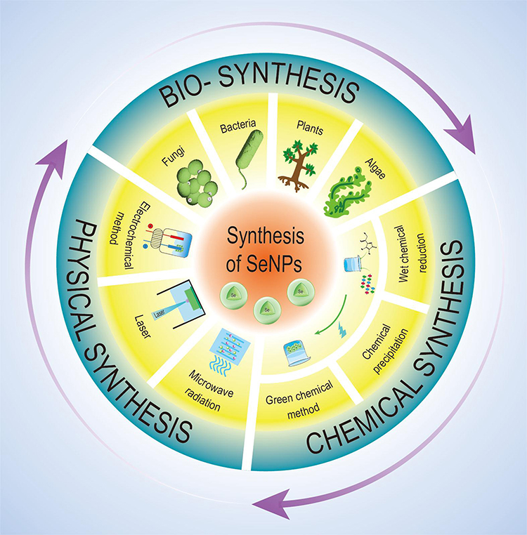 |
Figure 1 Synthesis methods of selenium nanoparticles. |
Physical Synthesis
Physical synthesis utilizes various energy forms to produce elemental selenium (Se0), which is directly converted into SeNPs.45 Currently, the main physical synthesis methods include electrochemical synthesis, microwave radiation, laser ablation, and laser irradiation.
Microwave radiation is one of the most commonly used physical synthesis methods. Compared to traditional heating methods, microwave radiation enables faster heating and cooling rates through more uniform particle size distribution, making it easier for materials to absorb energy. This process generates high thermal efficiency, resulting in green, energy-saving, and enhanced reaction selectivity.46–48 Kosar Sheikhlou et al mixed walnut extract with Se solution and subjected the mixture to microwave radiation with a fixed power of 800 W for 4 minutes, successfully obtaining SeNPs. Then the experimental results demonstrated the stability and excellent antibacterial properties of SeNPs.49
In addition, laser ablation is also a commonly used green synthesis method at present. The nanoparticles(NPs), produced by pulsed laser ablation (PLAL), have the characteristics of uniformity, stability and ease of collection, which meet the requirements of modern medicine for residue-free material surfaces.50 At the same time, the PLAL method is environmentally, friendly and green, which can prevent adhesion during the generation of NPs, and has significantly promoted the application of SeNPs in the medical field.51 Grégory Guisbiers et al were the first to report the preparation of SeNPs using the PLAL method. Under conditions of 800nm infrared light, pulse duration less than 100 fs, and repetition frequency of 80±1 MHz, they obtained SeNPs. Then SEM and TEM observations revealed that the surface of the SeNPs was smooth with no residue. Bacterial experiments also demonstrated the excellent antibacterial properties of the material.52
While physical synthesis methods offer such core advantages as high product purity and environmental friendliness in SeNP synthesis. Notably (Table 1), the particle size and properties of SeNPs vary with different synthesis methods, and significant differences in SeNP properties are also observed when the same method is applied under different power conditions. In addition, compared with other synthesis approaches, physical synthesis methods are associated with the drawbacks of high energy consumption and excessive reliance on equipment.50 For instance, pulsed laser ablation can efficiently produce SeNPs, but it remains challenging to achieve precise control over their properties. Meanwhile, pulsed laser ablation is overly reliant on laser generation equipment and constrained by economic considerations and scalability.53,54 Furthermore, in microwave-assisted synthesis, the failure to achieve uniform microwave irradiation may result in the thermal decomposition of some SeNPs due to overheating, whereas others lack sufficient energy for conversion.55 Therefore, future efforts should focus on optimizing processes, upgrading energy-efficient equipment, and improving pollution control and resource recovery systems to overcome the existing technical bottlenecks of physical synthesis methods, while achieving the low-carbon, clean, and sustainable production of SeNPs.
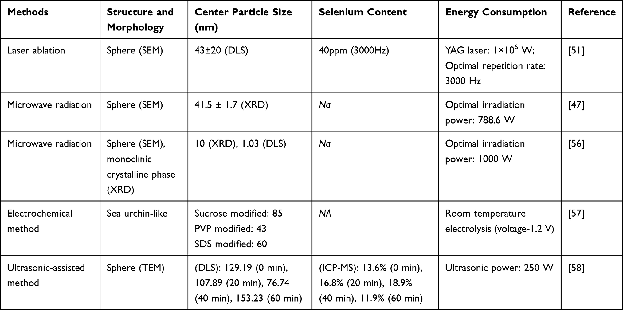 |
Table 1 A Summary of Physical Synthesis Methods of SeNPs. The Table Mainly Summarizes Some of the New Common Methods |
Chemical Synthesis
As the most widely adopted approach for SeNPs synthesis to date, chemical synthesis enables the formation of SeNPs through the reduction of Se ions in sodium selenite solution, facilitated by reducing agents and stabilizers.59 Commonly used reducing agents include ascorbic acid, glutathione and sulfur dioxide.60,61 Besides, polysaccharides, glycoproteins, polyphenols, etc., are commonly used to modify SeNPs to improve their stability (Table 2).
 |
Table 2 A Summary of a Variety of New Chemical Synthesis Methods |
It is noteworthy that different reaction parameters in the chemical synthesis system exert distinct effects on the properties of SeNPs. pH is a critical parameter requiring regulation during SeNPs synthesis. Daniela ˇ Stefankov´ et al found that a decrease in pH will significantly accelerate the reaction rate. And under acidic conditions (pH=3.16 or 2.5), the reaction is almost instantaneous. But the particles will connect with each other to form a dense network structure, leading to severe agglomeration.69 Besides, Meanwhile, product characteristics vary with the type of organic acid employed, primarily attributed to the stabilizing effect of organic acid anions, which modulate both the reducing capacity and the surface charge of the particles.70,71 Additionally, temperature exerts a notable impact on the properties of SeNPs. Several studies have demonstrated that low temperatures (25–40 °C) result in low Se atom mobility, typically leading to the formation of amorphous structures. In contrast, moderate to high temperatures (60–100 °C) enhance Se atom mobility and facilitate accelerated crystal growth, often yielding hexagonal or trigonal crystal systems with higher crystallinity.72,73 Thus, the selection and optimization of reaction parameters based on specific requirements represent a pivotal step in the chemical synthesis of SeNPs.
Although chemical synthesis is simple and economical, it often results in residues of chemical substances during the process, which may lead to the failure of treatments.73 Meanwhile, variations in materials such as stabilizers also affect the chemical properties of SeNPs. Polysaccharides, for instance, may induce the aggregation of SeNPs in acidic environments, which in turn reduces the bioavailability of the material. This places stringent requirements on its purity, toxicity and performance in medical applications.74 Furthermore, the selection or minor alterations of reaction parameters may induce uneven reactions of the material during synthesis, thereby leading to batch-to-batch variations. This may serve as a limitation to the widespread application of the chemical synthesis method.75 But, some researches indicate that ascorbic acid, when used as a reducing agent in the synthesis of SeNPs, can significantly reduce surface chemical residues, thereby decreasing external toxicity while further enhancing the purity of SeNPs.76,77 Boroumand et al utilized ascorbic acid as the reducing agent and chitosan and polyvinyl alcohol as stabilizers to prepare SeNPs. Through SEM and TEM observations, along with absorbance measurements, they found that the SeNPs had a smooth surface and minimal chemical residues. Additionally, in ROS detection and bacterial experiments, these particles demonstrated excellent antioxidant and antibacterial properties.76 Thus, the rational selection of reduction systems, stabilizers, and reaction parameters constitutes an urgent issue to be addressed for the efficient chemical synthesis of SeNPs.
Biosynthesis
Biosynthesis utilizes bionanotechnology, taking various biological systems as nanofactories to obtain corresponding extracts. These biological systems mainly include bacteria, fungi, plants, etc. The principle of this method is that Se salts, selenic acid, and Se dioxide are introduced into the so-called “bioreactor” as precursors for synthesizing SeNPs, and the production of SeNPs relies on the abundant precursors and reducing agents in biological resources. In these biological resources, due to the presence of enzymes, proteins, lipids, carbohydrates, etc., the biological systems exhibit strong reducing capabilities towards Se ions.78 The synthesis principle primarily involves the reduction of Se(IV) or Se(VI) to obtain SeNPs. The differences in the mechanisms of SeNPs synthesis among different biological systems mainly lie in the variations of reducing agents and enzymes involved in the process. According to current research findings, within bacterial systems, apart from non-enzymatic reduction pathways involving glutathione, sulfides, and siderophores, a variety of reductases including sulfite reductase, nitrite reductase, and glutathione reductase are also actively engaged in the reduction process.79 Compared to bacteria, fungi are easier to cultivate and can survive and reproduce at higher concentrations. Meanwhile, fungi can not only release reducing proteins and enzymes into the extracellular medium, allowing these biomolecules to reduce Se ions and then precipitate to form SeNPs, but also perform reduction intracellularly.80 For plant synthesis, SeNPs are mainly generated through proteins, amino acids, organic acids, vitamins, and some metabolites that act as reducing agents and stabilizers synthesized by plants. However, the products generated by plants are influenced by the plant parts and their functions.81 Additionally, other biological systems such as algae have also been confirmed to promote Se reduction and stabilization through various bioactive substances82 (Table 3).
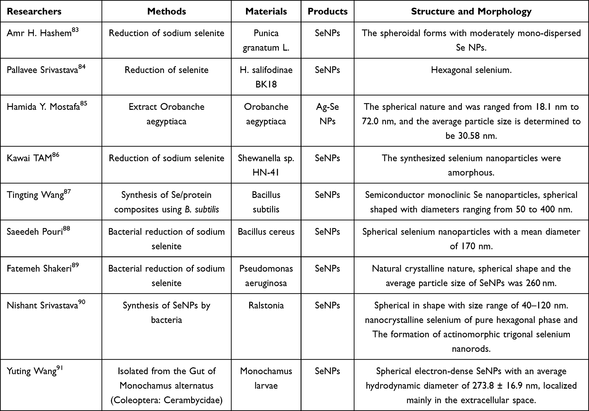 |
Table 3 Biomediated Synthesis of Selenium Nanoparticles |
Compared with physically and chemically synthesized SeNPs, biologically synthesized SeNPs have been reported to possess better bioactivity and lower toxicity.92 Moreover, due to the diversity of biosynthetic methods and relatively low synthesis costs, the biosynthesis of SeNPs shows promising prospects, yet there are also corresponding limitations tied to it. Firstly, the size of biologically synthesized SeNPs is not necessarily controllable. For instance, SeNPs synthesized by Bacillus subtilis have an average size of 536 nm with a wide particle size distribution range from 280 to 630 nm. Minor changes in selenate concentration and culture time can significantly affect particle morphology, resulting in poor batch-to-batch reproducibility.3 Furthermore, in large-scale production Helga Fernández-Llamosas et al reported that although Vibrio natriegens can rapidly synthesize SeNPs the yield is limited by selenate concentration up to 15 mM and relies on complex medium optimization, studies have indicated that when the selenate concentration exceeds 10 mM cell viability significantly decreases leading to a reduction in the release of nanoparticles, moreover the size of SeNPs produced by this method changes with time and concentration.93 Additionally, although Aspergillus oryzae can efficiently accumulate Se at 8462 mg/kg dry weight, the Se reduction pathway involves both assimilatory and dissimilatory mechanisms involving the coordinated regulation of more than 20 genes. This complexity makes it difficult to conduct targeted optimization of the SeNPs synthesis process due to the unclear mechanism and metabolic complexity.94 Moreover, SeNPs synthesized using plant extracts require multiple purification steps such as ultrafiltration and centrifugation. However, residual mixtures are difficult to completely remove. Studies have pointed out that the purification and separation process of this method is challenging and time-consuming, necessitating further process improvements.95 In conclusion, although product stability and process complexity still pose significant challenges to its production, biosynthesis has certain advantages in terms of toxicity, biocompatibility, and cost.
Mechanism and Application of SeNPs’ Activity
Infection
Antibacterial Activity of SeNPs in Biomedicine and Stomatology
The widespread misuse of antibiotics has led to the emergence of antimicrobial resistance in pathogenic microorganisms, posing a significant threat to global public health. Consequently, the development of novel antibacterial agents has become a critical focus in contemporary biomedical research. SeNPs exhibit considerable potential as antibacterial agents owing to their distinctive mechanochemical properties, biological activity, and intrinsic antimicrobial efficacy96 (Table 4).
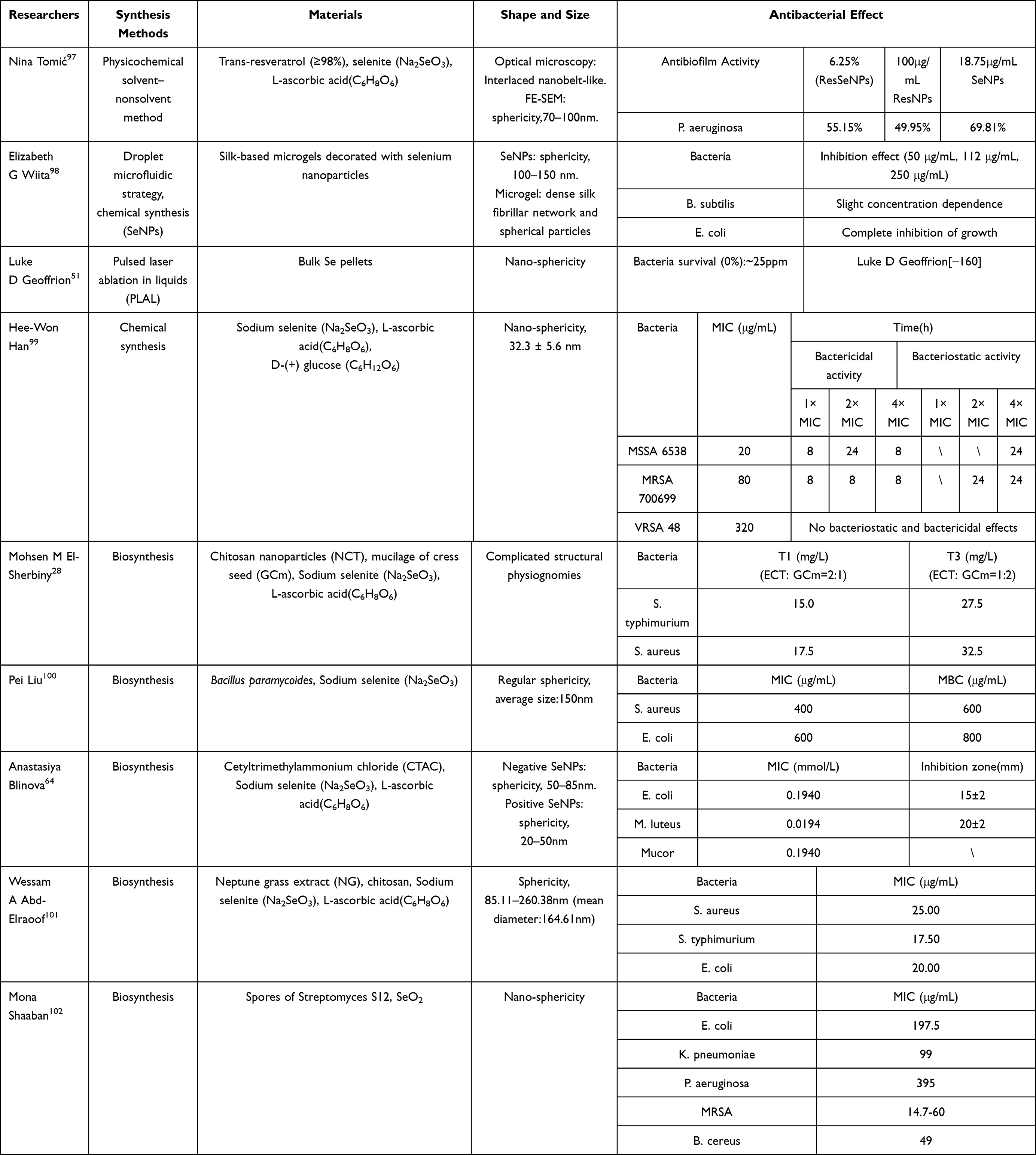 |
Table 4 The Application of Anti-Bacteria. This Table Mainly Summarizes the Bacteria Used in Antimicrobial Experiments and the Corresponding Antibacterial Results |
SeNPs demonstrate potent antimicrobial activity, with their mechanisms of action categorized into general and specific pathways. The general antibacterial mechanism of SeNPs involves multiple synergistic modes of action: electrostatic adsorption-mediated disruption of cellular membrane integrity and permeability; induction of reactive oxygen species (ROS) accumulation, resulting in oxidative stress; interference with energy metabolism through inhibition of ATP synthase activity; and suppression of essential biomolecule synthesis.103 The surface negative charge characteristics of SeNPs result in significantly differential binding affinities to bacterial cells depending on their membrane charge properties.104 The nanoscale dimensions and spherical architecture of SeNPs facilitate enhanced penetration through biofilm matrices.105
In the specific antibacterial mechanism of bacteria, for Gram-positive bacteria (such as Staphylococcus aureus) and Gram-negative bacteria (such as Porphyromonas gingivalis), SeNPs disrupt ATPase activity by releasing Se2⁺, consequently impairing cellular energy metabolism.106,107 Against drug-resistant strains (eg, MRSA, VRSA), SeNPs demonstrate three distinct mechanisms: (1) downregulation of antibiotic resistance-associated gene expression, (2) disruption of the thiol-based antioxidant defense system, and (3) potentiation of conventional antibiotic susceptibility.53,108–112 In the specific antibacterial mechanism of fungi, SeNPs can synergistically enhance nystatin to suppress the formation of fungal biofilm by selective interference with the RAS/cAMP/PKA signaling pathway against Candida albicans and other fungi.113 In addition, mechanism investigations showed that SeNPs disrupt the ergosterol biosynthesis pathway via modulation of the expression of key antifungal resistance genes ERG3, ERG11 and FKS1.114
Clinical studies in dental medicine have demonstrated that SeNPs exhibit broad-spectrum antimicrobial activity against major oral pathogens, including Staphylococcus aureus (S. aureus), Streptococcus mutans(S. mutans), Porphyromonas gingivalis(P. gingivalis), and Enterococcus faecalis (E.faecalis).115,116 In oral pathologies, these pathogens serve as primary etiological agents for oral mucosal infections, dental caries, periodontitis, and apical periodontitis. Their propensity for antimicrobial resistance development frequently complicates therapeutic interventions.117,118 Notably, methicillin-resistant Staphylococcus aureus (MRSA) and other multidrug-resistant (MDR) strains exhibit remarkable antibiotic resistance profiles. Importantly, clinical studies have demonstrated that SeNPs exert potent antibacterial activity against MRSA, highlighting their therapeutic potential for combating antimicrobial resistance. S. mutants, a primary etiological agent of human dental caries, colonizes biofilms on tooth surfaces. The antimicrobial mechanism of SeNPs involves membrane disruption and permeability alteration, resulting in biomacromolecule leakage and bacterial cell death - properties that suggest their therapeutic potential for caries prevention. Emerging evidence supports this application, as demonstrated by Dhanraj et al, who fabricated SeNPs and evaluated their antibacterial efficacy against oral pathogens, including S. mutans and S. aureus. Their findings revealed potent growth inhibition of S. mutans, indicating potential applications in controlling cariogenic biofilms or incorporation into dental sealants for secondary caries prevention.119 In refractory apical periodontitis, Enterococcus faecalis (E. faecalis) establishes persistent biofilms within root canal systems, penetrates deep into dentin tubules, and induces sustained release of pro-inflammatory mediators, resulting in progressive destruction of periapical tissues. This pathogenic behavior constitutes a key determinant of treatment failure in refractory cases.35 The distinctive physicochemical characteristics and inherent antimicrobial activity of SeNPs may offer novel therapeutic strategies for managing and preventing this pathological condition.
In addition, SeNPs exhibit broad-spectrum antimicrobial efficacy across diverse applications, underscoring their translational potential in dental medicine. They hold substantial promise in combination therapies, as adjuvants in combination therapies, and as functional additives in dental materials or nanocarriers or platform components for targeted delivery systems. A representative study by Samane Shahmoradi et al evaluated the synergistic effects of SeNPs with photodynamic therapy (PDT) against 24-hour-old planktonic cultures and biofilms of Streptococcus viridans. Through controlled experiments, they observed that SeNP-modified PDT achieved a significant reduction in colony-forming units (CFUs) of S. mutans biofilms. Notably, this combinatorial approach exhibited potent biofilm-disrupting activity against S. viridans.120 In dental restorative applications, the inherent antimicrobial properties of SeNPs address critical requirements for oral restorative materials. As demonstrated by Iqra Saleem et al, SeNPs potentiate the antimicrobial efficacy of resin-based dental composites through the incorporation of Se-doped zinc oxide nanoparticles (Se/ZnO NPs) as antibacterial nanofillers. Comparative analysis revealed that composites containing 1% Se/ZnO NPs exhibited enhanced antibacterial activity, improved biocompatibility, and superior mechanical strength relative to those with 1% ZnO NPs alone, establishing Se/ZnO NPs as high-performance antibacterial additives effective at reduced concentrations.121 Therefore, SeNPs demonstrate significant clinical potential not only for managing prevalent oral diseases (dental caries and periodontitis) but also exhibit distinctive advantages in dental restoration and preventive applications.
Application and Mechanism of SeNPs Against Virus
SeNPs exert broad-spectrum antiviral activity through multiple synergistic mechanisms. As an essential trace element, Se participates in the biosynthesis of pivotal immunoregulatory enzymes, thereby potentiating host antiviral immunity. Current evidence demonstrates that Se enhances immune cell proliferation, promotes T-cell maturation and differentiation, augments cytotoxic activity, induces apoptosis of virus-infected cells, and stimulates interferon (IFN) and cytokine production.122 Additionally, SeNPs exhibit antiviral activity through modulation of host cell signaling pathways. Specifically, ribavirin-modified SeNPs (Se@RBV) demonstrate inhibitory effects against H1N1 influenza virus by targeting the caspase-3-dependent apoptotic pathway. Treatment with Se@RBV significantly downregulates expression of caspase-3-associated proteins, including cleaved poly-ADP-ribose polymerase (PARP), caspase-8, and Bax, while concurrently suppressing phosphorylation of p38, JNK and p53.123 Furthermore, SeNPs mediate antiviral effects through dual mechanisms of ROS modulation: (1) inducing localized ROS generation to degrade viral nucleic acids and structural proteins, while (2) simultaneously suppressing excessive ROS accumulation to protect host cells from oxidative damage. This balanced redox modulation enables concurrent viral inactivation and host cytoprotection.124 SeNPs exhibit potent antiviral activity through direct structural disruption of viral particles. Mechanistic studies demonstrate that SeNPs effectively inhibit influenza virus neuraminidase activity, thereby preventing viral progeny release.125 However, the precise mechanisms underlying SeNP-virus structural interactions remain incompletely characterized. Drawing parallels from established nanoparticle-virus interaction paradigms, we hypothesize that SeNPs may similarly target viral envelope proteins and capsid components through analogous mechanisms.126,127 Furthermore, nanoparticles can block viral cellular entry by targeting surface antigens while simultaneously enhancing drug potency when utilized as antiviral nanocarriers.128 Collectively, these findings establish a conceptual framework and mechanistic directions for advancing SeNP-based antiviral research.
Herpes simplex virus type 1 (HSV-1) serves as the principal etiological agent for oral pathologies, including herpetic stomatitis and labial herpes, with established associations to nasopharyngeal carcinoma. Recent studies demonstrate that surface-modified SeNPs potently inhibit HSV-1 replication by targeting critical viral genes and structural proteins across multiple stages of the viral life cycle, effectively suppressing viral proliferation and assembly. These findings significantly advance understanding of SeNP-mediated antiviral mechanisms and their therapeutic potential for oral HSV-1 infections.129 Despite their promise as antiviral agents, SeNPs remain underexplored in clinical applications. Their development could offer novel therapeutic strategies for viral infections in oral medicine.
Anti-Inflammatory and Antitoxidant of SeNPs
Anti-Inflammatory Activity and Application of SeNPs
As a key component in histopathology, inflammation significantly influences pathological processes such as neurodegeneration, carcinogenesis, and immune system dysregulation.130–133 In the field of stomatology, periodontitis and gingivitis, as prevalent oral disorders, exhibit inflammatory processes that critically drive disease progression in oral ulcers and oral carcinomas.134–136 Therefore, timely treatment of inflammation is crucial for improving patients’ quality of life and maintaining health. At present, antibiotic-based therapies present limitations including prolonged treatment courses, side effects, and oxidative stress-induced tissue damage from sustained activities of anti-inflammatory agents.33,34,137,138 Notably, SeNPs demonstrate remarkable anti-inflammatory activity with potential for alleviating and treating various oral diseases, including periodontitis and gingivitis.
The anti-inflammatory mechanisms of SeNPs primarily involve blocking inflammatory signaling pathways and suppressing pro-inflammatory factor expression. Current evidence indicates SeNPs mitigate inflammatory responses through inhibition of NF-κB and MAPK pathways (Figure 2). Specifically, SeNPs can inhibit the NF-κB pathway and subsequently suppress the expression of pro-inflammatory cytokine target genes, leading to reduced activity of inflammatory responses. Wang et al mixed the complex SPS-SeNPs with Raw 264.7 cells and found that SeNPs inhibited the activation of the NF-κB pathway, thereby suppressing the production of NO and other anti-inflammatory factors,30 which demonstrates the significant role of the NF-κB signaling pathway in pro-inflammatory activation. In the classic NF-κB pathway, SeNPs block the degradation of Iκ-B to inhibit the activation of NF-κB, thereby affecting the translocation of NF-κB to the nucleus. Since NF-κB is an activator of pro-inflammatory cytokines, it further inhibits the process of LPS-induced pro-inflammatory molecule iNOS and achieves the suppression of inflammatory responses.30 In the alternative pathway of NF-κB, the use of SeNPs also inhibits the phosphorylation of p65 protein induced by LPS, thus inhibiting the activation of the NF-κB pathway.139 In addition, SeNPs also interfere with MAPK signaling pathways by inhibiting the phosphorylation of JNK and p38 pathways, thereby inhibiting LPS-induced iNOS expression and subsequent NO and TNF-α production.30 In this way, the expression of inflammatory cytokines such as IL-6, TNF-α, and IL-1β is reduced, and the secretion of anti-inflammatory cytokines such as IL-12 is increased, so as to inhibit the inflammatory response.139
 |
Figure 2 Anti-inflammatory and antioxidant mechanism of selenium nanoparticles. Notes: Created in BioRender. Q, X (2025) https://BioRender.com/g6ufr07. |
Periodontitis patients frequently exhibit oral epithelial cell damage and gingival collagen degradation due to chronic inflammation.140 This is also a key factor affecting supragingival scaling efficacy and prognosis. Therefore, how to more conveniently alleviate oral inflammation and reduce mucosal damage has become an urgent issue to address. Tritean et al prepared a kombucha dietary beverage rich in SeNPs and mixed it with gingival fibroblasts at different concentrations for culture. The detection of ROS generation rates showed that when the SeNPs concentration was 0.1μg/mL, the ROS generation rate decreased to 26.6%, and the cells exhibited good growth and proliferation status. Thus, the material demonstrated excellent anti-inflammatory capabilities and showed potential for promoting cell proliferation and repair.141 This makes it possible for patients to reduce periodontal inflammation and tissue damage by drinking dietary drinks on a daily basis. In addition, severe periodontitis can lead to inflammatory bone loss (alveolar bone resorption), which can lead to gum recession, tooth loosening and even tooth loss.36 This is also the reason why the implant process is suspended due to periodontal inflammation caused by the restoration. At present, most of the treatment methods use bone powders and other materials to promote bone regeneration.142,143 However, the presence of periodontitis may lead to an imbalance between bone resorption and bone regeneration. The mechanisms of bone resorption mainly involve two aspects: on one hand, immune cells such as macrophages and T cells are stimulated, secreting large amounts of pro-inflammatory factors which activate osteoclasts and inhibit osteoblasts through the RANKL-RANK-OPG axis; on the other hand, precursor cells of macrophages can differentiate into osteoclasts under the influence of relevant factors, further intensifying bone resorption.144,145 Therefore, Huang et al adopted a macrophage polarization-targeting strategy to address bone resorption, employing G3@SeHANs to scavenge circulating cell-free DNA (cfDNA). This approach concurrently promoted M2 macrophage polarization and suppressed production of pro-inflammatory mediators (TNF-β, IL-6).146 In summary, compared with other materials, SeNPs provide new solutions for the two major challenges of inflammation suppression and tissue repair in the treatment of periodontal disease and gingivitis.147 (Figure 3) At the same time, the advantages of simple application, rapid treatment and high efficiency of SeNPs can also reduce the pain in the treatment process of periodontitis patients and further improve the living standards of periodontitis patients for long-term treatment.
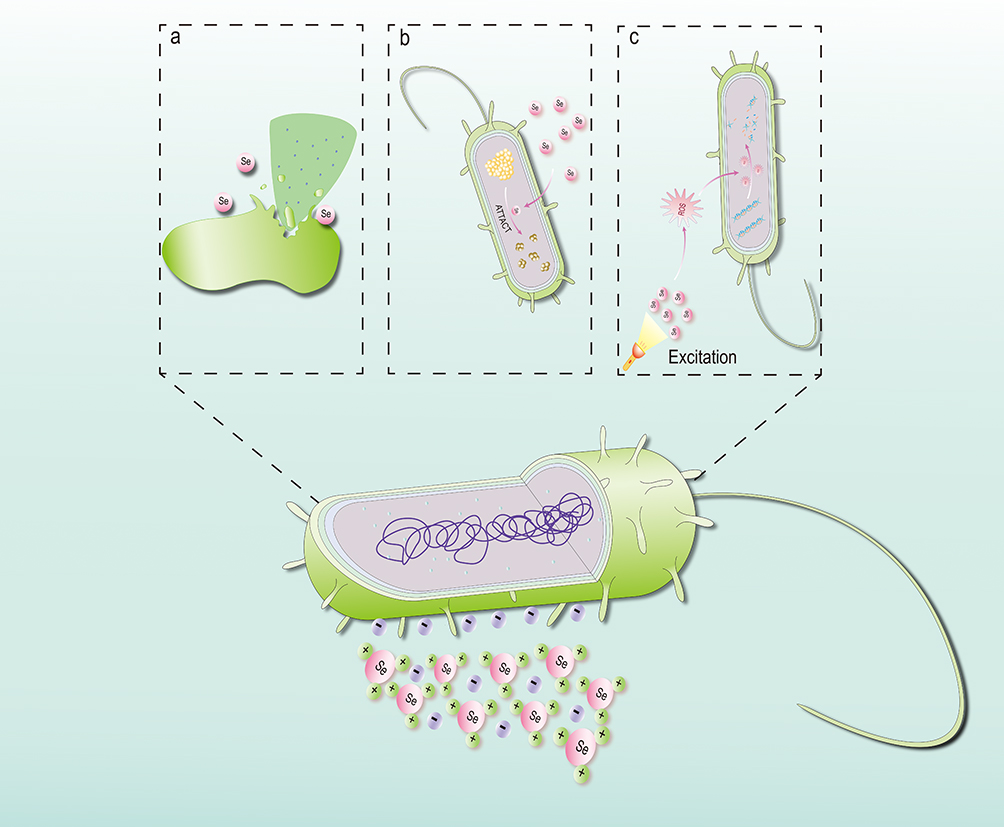 |
Figure 3 Antibacterial mechanism of selenium nanoparticles. (a) The bacterial cell membrane is broken and the contents are released, causing the bacteria to die. (b) SeNPs cleave and denature proteins in bacteria, killing the bacteria. (c) The SeNPs are excited by specific light and cleave the DNA which lead bacteria to die. |
Antioxidant Activity and Application of SeNPs
Oxidative injury serves as a fundamental mechanism underlying numerous diseases, including inflammation and cancer. It mainly causes lipid peroxidation, protein denaturation and DNA damage, following cell death and tissue damage through the production of ROS in the body.148 In a healthy body, oxidative damage and oxidative repair are in balanced states. However, when the body tissues are in pathological states (eg, cancer) or exposed to certain external influences (eg, radiation), the balance of the body will be disrupted due to oxidative stress, leading to excessive ROS accumulation and extensive tissue damage.149,150 This pathological process can further cause other diseases, such as diabetes, periodontitis, radiation jawbone osteomyelitis and so on. According to relevant studies, SeNPs have better antioxidant ability, which can significantly reduce the production and release of ROS, thereby offering a promising strategy for mitigating and repairing oxidative stress-induced damage.151
It is well known that selenoenzymes are the basis of redox homeostasis, and antioxidant enzymes such as glutathione peroxidase (GPx) and selenoprotein P are important enzymes in selenoproteins.152 SeNPs exert their antioxidative effects by leveraging the protective functions of selenoproteins, especially GPx.153 Se increases the level of GPx protein, thereby enhancing GPx enzyme activity and increasing the oxidative process of GSH-GSSG, reducing the production of methyl-induced ROS within cells. Moreover, SeNPs can prevent lipid oxidation through GPx, thus protecting cell membranes. Additionally, GPx protects biological systems by neutralizing peroxynitrite (ONOO−)-mediated oxidative damage.153 Intracellular superoxide dismutase (SOD) is a specific oxygen radical scavenger, which can be enhanced by SeNPs. SOD catalyzes the disproportionation of superoxide anions into H2O2, which can subsequently be reduced by GPx to exert antioxidant function.154
Diabetes mellitus (DM) represents a well-established risk factor for periodontitis.155 Hyperglycemia in diabetic patients can induce oxidative stress and immune dysregulation, which will promote the process of periodontitis.156 Conversely, periodontitis is also recognized to impact systemic inflammation, insulin resistance, lipid and glucose metabolism, thereby further influencing the development and progression of diabetes mellitus. Thus, diabetes and periodontitis exhibit a bidirectional relationship, with the core of their interaction lying in the exacerbation and dysregulation of inflammatory responses.155 Current clinical interventions for periodontitis primarily involve mechanical debridement (eg, scaling and root planing) and antibiotic therapy to disrupt biofilms and inhibit bacterial growth.157 However, because the incidence of this kind of periodontitis is multifactorial, the traditional treatment is always ineffective.158 So, SeNPs have become a potential treatment option for alleviating diabetic periodontitis due to their powerful antioxidant properties. At present, several experiments have proved that SeNPs have strong antioxidant capacity.159,160 In the study by Biju Thomas et al, 150 subjects were divided into three groups, and their serum levels of glutathione, catalase, and Se were measured respectively. The results showed that compared with subjects with periodontitis but otherwise healthy and those without periodontitis, patients with type 2 diabetes and periodontitis had significantly reduced serum levels of glutathione, catalase, and Se. This indicates that in the inflammatory response induced by diabetes and periodontitis, the body can employ glutathione peroxidase and catalase to scavenge ROS for self-protection. Se, as a cofactor of glutathione peroxidase, plays a crucial role in this antioxidant defense process.161 In a separate study, they found that compared with periodontitis patients without other systemic diseases, those with concurrent diabetes had significantly lower serum Se levels. This highlights that Se may enhance the body’s antioxidant defense by increasing glutathione peroxidase activity. Given the mechanisms underlying diabetes and periodontitis, this underscores the potential benefits of interventions targeting both conditions.162 And due to the similarity of the whole-body oxidation mechanism, SeNPs can achieve the reduction of the periodontal tissue damage caused by oxidative stress activated by diabetes, which especially improves the traditional therapy effect of periodontitis patients. Hence, SeNPs are expected to be used in the auxiliary treatment of diabetic patients with periodontitis.163,164 However, in current studies investigating the effects of Se supplementation on diabetes, relevant research indicates that Se supplementation may increase the prevalence of diabetes. When exploring the treatment of diabetic periodontitis, further evaluation of its potential implications for diabetes is thus required.165 However, according to relevant literature analysis, a non-linear dose-response relationship exists between serum Se levels and type 2 diabetes mellitus (T2DM). The results indicate that in populations with relatively low serum Se levels (<97.5 μg/l) and those with relatively high serum Se levels (>132.5 μg/l), serum Se levels are positively correlated with T2DM. This suggests a potential U-shaped non-linear dose-response relationship between serum Se and T2DM.166 Therefore, when considering the therapeutic application of SeNPs in diabetic periodontitis, a thorough evaluation of their safety profile is necessary. Although in vitro and animal studies have highlighted the potential of SeNPs, human trials are still insufficient. Meanwhile, it’s still unknown whether the bioavailability and long-term safety of SeNPs are stable, which is an urgent problem to be solved in order to achieve clinical application.
With the development of medical technology, radiotherapy (RT) has become the primary treatment for head and neck cancer. However, prolonged RT exposure frequently leads to osteoradionecrosis of the jaws (ORN). As reported by Annu Singh et al, ORN typically manifests within two years of RT treatment, particularly in patients receiving doses exceeding 60 Gy, with a predilection for the mandible.167 Despite technological advancements in RT, ORN remains the most severe complication within the treatment of head and neck cancer, reducing treatment efficacy and survival rates of patients.168 The pathogenesis of ORN unfolds in three stages: endothelial injury, osteoclast apoptosis and sequestrum formation.167 Prolonged RT induces oxidative stress of jawbone marrow, subsequently leading to accumulation of mass inflammatory factors and ROS, which culminates in marrow ischemia, osteocyte death, and bone necrosis.169 Therefore, reducing the degree of oxidative stress in the body during RT and inhibiting the production of inflammatory factors and ROS provides a new direction for alleviating or even treating ORN. In an experiment evaluating the efficacy of Se replacement in preventing radiation-related toxicity, J. BÜNTZEL et al found that patients who received sodium selenite exhibited a significant reduction in dysphagia and taste loss compared with those who underwent irradiation without any Se replacement. This indicates that Se replacement has a certain role in preventing radiation-related side effects. However, its effects on mucositis and xerostomia were not significantly affected, with limited efficacy and marginal effects. They attributed this to the fact that auxiliary detoxification is only one of the many pathways of excretion. Therefore, they suggested that in future studies investigating Se replacement for reducing radiation-related toxicity, it should be combined with other scavengers to further evaluate its efficacy.170 In a randomized controlled trial involving post-operative patients with differentiated thyroid cancer, Tong, Huimin et al found that Se supplementation exerted a protective effect on the salivary glands during high-dose155 I therapy. Furthermore, the combined administration of Se and vitamin C yielded superior outcomes. Specifically, antioxidant Se supplementation demonstrated significant protective effects on the excretory function of the parotid glands and the uptake and excretory functions of the submandibular glands.171 These experiments demonstrate that Se exhibits significant efficacy in scavenging ROS following radiotherapy. Furthermore, SeNPs can inhibit inflammation through multiple pathways and promote the production of GPx and SOD to clear ROS. Therefore, SeNPs may be an option for the treatment of ORN. However, due to the difficulty in simulating the osteomyelitis environment in vitro and the lack of clinical studies, further research is needed to determine whether SeNPs can achieve therapeutic effects on ORN (Figure 4).
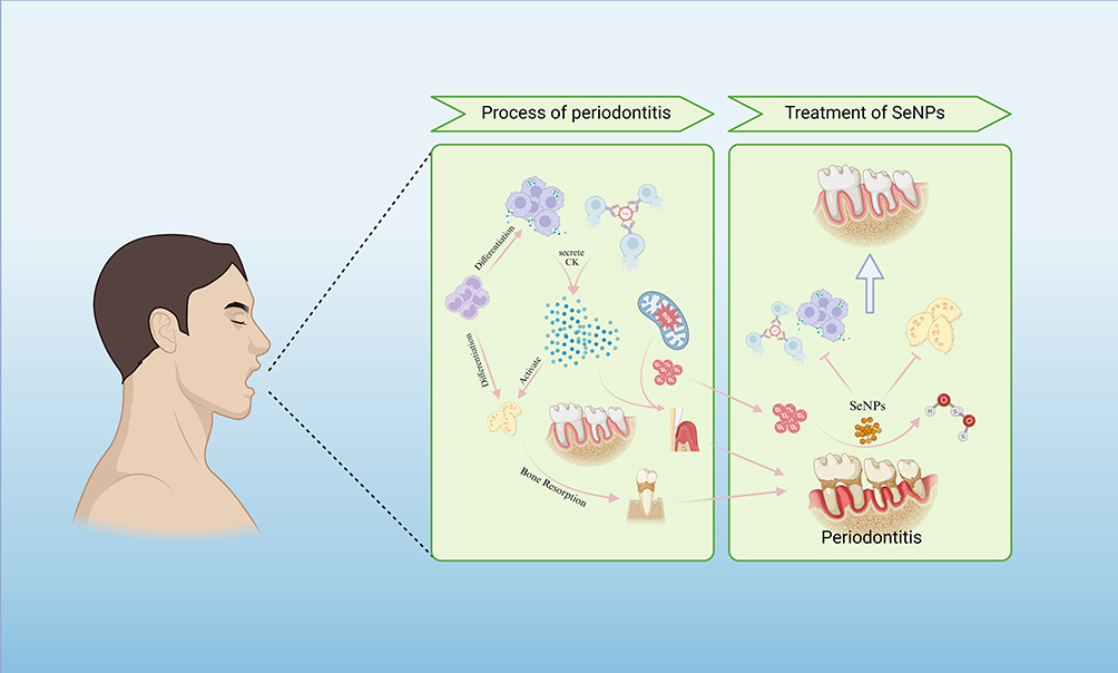 |
Figure 4 Pathogenesis of periodontitis and treatment of SeNPs particles. Notes: Created in BioRender. Q, X (2025) https://BioRender.com/f9sgtje. |
Application of SeNPs in Cancer
Oral cancer is one of the most prevalent types of cancer. According to relevant data, there were 15,730 new cases and 9090 deaths from oral cancer in 2025, with the incidence rate increasing year by year,172 which poses a certain threat to people’s life and health. Traditional treatment modalities involve surgical resection of tumors followed by chemotherapy and radiotherapy, which have side effects such as drug resistance and healthy cells damage.173,174 In recent years, emerging therapies including immunotherapy, gene therapy, and tumor treating fields (TTFields) have demonstrated promising advancements.174–176 Among them, nano-therapy has gained significant attention due to its high biocompatibility and low toxicity in cancer therapy, offering marvelous potential in anticancer applications177 (Table 5).
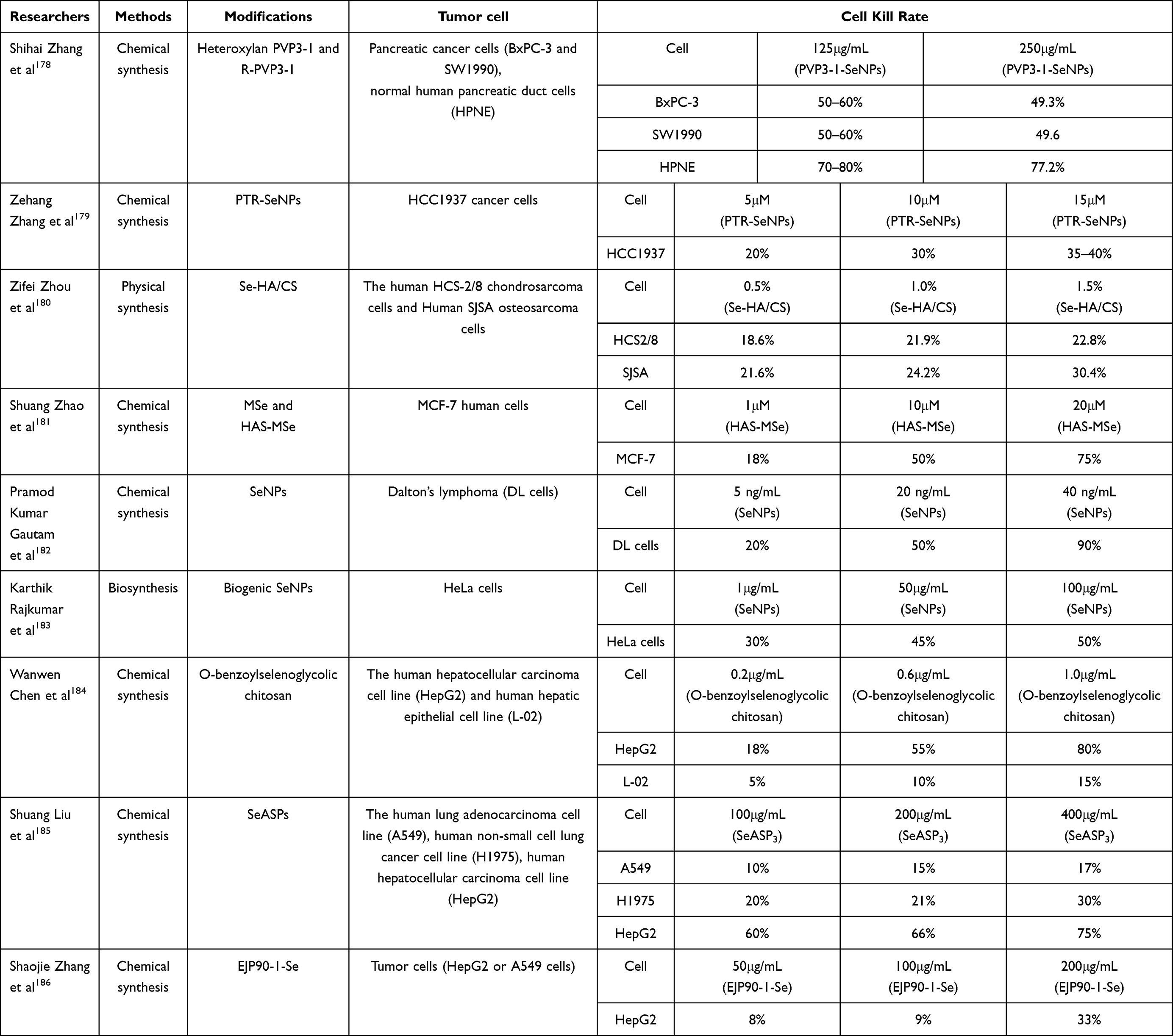 |
Table 5 The Application of Anti-Tumor. The Table Mainly Summarizes the Types of Tumor Cells Used in the Experiment and the Effect of the SeNPs |
As an essential trace element in humans, Se is intimately associated with tumorigenesis. Hughes et al conducted a prospective cohort study to investigate the relationship between Se supplement intake and prostate cancer risk. Their results demonstrated an inverse association between Se intake and prostate cancer risk, with the highest intake group exhibiting a 32% lower risk (RR=0.68, 95% CI =0.54–0.85).187 Furthermore, baseline Se concentration is closely correlated with cancer incidence. Fritz et al evaluated the association between baseline Se levels and cancer development via the Nutritional Prevention of Cancer (NPC) trial. They found that individuals with low baseline Se levels (serum<106ng/mL) who received Se supplementation experienced a 49% reduction in overall cancer risk and a hazard ratio (HR) of 0.42 for lung cancer (95% CI=0.18–0.96). Conversely, those with high baseline Se levels (serum≥121.6 ng/mL) showed an increased lung cancer risk (HR=1.25) subsequent to Se supplementation, alongside an elevated risk of diabetes (HR=2.70).188 These observations provide empirical data and a theoretical framework for the application of SeNPs in cancer therapy.
SeNPs not only have anti-inflammatory and antioxidant effects, but can also inhibit the occurrence and metastasis of tumor cells through pathway activation, stimulation of immune cell proliferation and inhibition of inflammatory processes.189,190 The relevant studies indicate that high concentrations of SeNPs have weakened anti-inflammatory and antioxidant ability, which is mostly manifested as inhibition and killing of tumor cells.152,191 At the same time, the excessive accumulation of SeNPs in tumor cells can effectively kill tumor cells, and the ROS production rate is significantly higher than that of sodium selenite and other substances.192 Therefore, it is feasible for SeNPs to be used in the treatment of oral squamous cell carcinoma (OSCC) and head and neck tumors.193 Chen et al developed a SeNP-indocyanine green (ICG) nanocomposite for combinatorial photothermal therapy. Using orthotopic oral squamous cell carcinoma (OSCC) murine models, they demonstrated 40% tumor growth inhibition via synergistic chemo-photothermal effects.194 In addition, SeNPs can also be validated as targeted drug delivery carriers, which can increase the enrichment of drugs in tumor cells and improve the killing rate of tumors, as reported by Jia et al,195 Xie et al.196 Furthermore, multiple studies have demonstrated that adequate Se supplementation can enhance the efficacy of conventional radiotherapy (RT) and chemotherapy (CT). Zou et al divided PC3 prostate tumor-bearing mouse models into four groups: the experimental groups received SeNPs alone, RT alone, and combination therapy. The final results showed that the tumor volumes in the three monotherapy and control groups were 1.36 cm3, 1.11 cm3, and 0.97 cm3, while the tumor volume in the combination therapy group was significantly reduced to 0.32 cm3. Meanwhile, the body weights of the mice remained stable throughout the study, confirming the efficacy and safety of SeNPs combined with RT.197 Besides, in a randomized, double-blind Phase II clinical trial for cervical cancer (NCT04815565), patients were randomly assigned to the control group (placebo plus chemoradiotherapy) and the experimental group (Se plus CRT). The results indicated that the incidence of grade 3 thrombocytopenia was significantly lower in the experimental group, accompanied by a more significant reduction in tumor volume. These findings further highlight the considerable potential of SeNPs in the field of cancer therapy.
Beyond their therapeutic potential, SeNPs also have significant advantages in cancer diagnosis. Traditional nanoimaging agents for cancer diagnosis and treatment often require coupling nanocarriers with photosensitizers or imaging agents.166–168 This hinders the release and transformation of materials, affecting the killing rate of cancer cells.169,170 Therefore, the label-free strategy has become a new direction for cancer diagnosis and treatment. Liu et al innovatively combined lentinan (LNT) and SeNPs, and used chemical exchange saturation transfer (CEST) MRI technology to detect and image a large number of free hydroxyl groups that exist in biomaterials within living organisms. Through fluorescence imaging, it was observed that LNT-SeNPs were extensively taken in by tumor cells with the assistance of TNF-α, achieving precise localization in CEST MRI images. Furthermore, continued cultivation of tumor cells demonstrated that LNT-SeNPs have excellent anticancer properties, with significant effects persisting even two weeks post-injection.
However, SeNPs do not exert exclusively positive therapeutic effects in cancer patients. Firstly, there is an overlap between their therapeutic and toxic doses, based on LD50 and NOAEL experiments in mice. Zhang et al categorized SeNPs doses into low (<200 μg Se/kg/day), medium (200–750 μg Se/kg/day), and high (>750 μg Se/kg/day) groups. The results indicated that although the therapeutic effect of high doses is obvious, there is obvious toxicity. This suggests that the use of SeNPs should be considered when using them in cancer treatment.179 Secondly, different tumor types exhibit varying sensitivity to SeNPs. Elena V. Karpukhina et al compared their cytotoxic effects across four human cancer cell lines (glioblastoma, colorectal cancer, breast cancer, and prostate cancer) and found that 0.5 μg/mL SeNPs significantly reduce glioblastoma cell viability, whereas the other three cell lines require 5 μg/mL or higher to achieve comparable effects.198 Furthermore, the intracellular redox environment is another key factor limiting the therapeutic efficacy of SeNPs. Xiong et al demonstrated that in tumor cells with a highly oxidative microenvironment, SeNPs are primarily converted into cytotoxic selenite (SeO32−), triggering reactive oxygen species (ROS) bursts and apoptosis. But in normal cells or subsets of tumor cells in a low redox state, SeNPs are preferentially converted into non-toxic selenium-containing amino acids for selenoprotein synthesis, which may be an important factor in promoting tumor proliferation.199 Additionally, due to the paucity of robust clinical research, translating SeNPs into clinical cancer therapy requires further research efforts, and continuous in-depth investigations are therefore warranted to design patient-specific SeNPs tailored to different tumor types and their distinct redox profiles, thereby facilitating the successful application of SeNPs in cancer diagnosis and treatment.
Summary and Outlook
Clinical Potential of SeNPs
With multiple advantages in antibacterial, anti-inflammatory and antioxidant, and tumor-targeted diagnosis and treatment, SeNPs have shown great potential to replace traditional therapies, especially for the early diagnosis of oral infections, inflammation, and tumors. (Figure 5) In terms of antibacterial, SeNPs can not only be used as an auxiliary sterilization method for periodontitis and other diseases, but also may achieve rapid sterilization of oral surgical instruments, which can avoid the disadvantages of traditional sterilization methods that are time-consuming and cumbersome to operate. In terms of anti-inflammatory and antioxidant properties, SeNPs may be used as adjunctive drugs to inhibit inflammation and promote tissue repair. In addition, in the field of cancer, SeNPs are expected to become an important means of cancer diagnosis and treatment. However, most of the existing studies focus on in vitro experiments and short-term effect verification. And its insufficient stability, unclear pharmacokinetic laws, and lack of data support for long-term biocompatibility have become key obstacles restricting its use from basic research to clinical application.
 |
Figure 5 SeNPs have a potential application perspective in the field of antibacteria, antitumor and anti-inflammation, which may provide new directions of disease treatment. |
Limitations on the Stability of SeNPs
At present, existing studies have obvious limitations in the stability verification of SeNPs. Most studies focus on a single environment in vitro and lack targeted consideration for practical clinical application scenarios. Due to the complex characteristics of the oral microenvironment, such as temperature fluctuations, dynamic changes in pH value, and salivary enzyme production, existing studies have not systematically verified the structural integrity and activity retention ability of SeNPs in this environment. Therefore, the complexity of the oral environment may become an important factor in the failure of SeNPs in the future. In addition, while numerous studies have employed surface modification strategies, including stabilizer coatings and inorganic encapsulation, to enhance nanoparticle stability, the majority of these investigations have focused solely on short-term efficacy, with insufficient evidence regarding long-term performance.200 Therefore, in practical applications, stabilizers may fail due to environmental variability, which will eventually lead to particle aggregation and settlement or even decomposition. Therefore, how to effectively improve the stability of SeNPs so that they can be better used for clinical treatment has become an urgent problem to be solved.
Limitations of SeNPs Metabolism and Biocompatibility
Drug metabolism and long-term compatibility are fundamental to the successful clinical application of therapeutics. Huang et al adopted a macrophage polarization-targeting strategy to address bone resorption studies indicate that after absorption via multiple routes, SeNPs primarily accumulate in the liver, kidneys, and spleen. Subsequently, SeNPs undergo partial conversion into Se compounds and are excreted in urine through renal metabolism. However, current pharmacokinetic research on SeNPs has predominantly focused on healthy individuals, with limited exploration in patients with oral diseases or other conditions.201 Meanwhile, regarding concentration-dependent effects, existing studies have only confirmed the occurrence of phenomena without conducting in-depth analysis of regulatory mechanisms, action targets, or signaling pathways at different concentrations. Furthermore, biocompatibility assessments of SeNPs are mostly confined to short-term experiments, creating a significant gap in long-term safety data. Studies have demonstrated that prolonged use of SeNPs may lead to chronic inflammation and organ damage in patients, which remains the greatest obstacle to translating SeNPs from basic research to clinical practice.202
Future Research and Directions for SeNPs
Therefore, researchers must continue to advance pharmacokinetic studies on SeNPs, particularly focusing on critical clinical parameters such as optimal dosing regimens and dosing intervals, to establish a robust foundation for their clinical application in disease treatment. Additionally, the stability of SeNPs remains a pivotal factor for successful clinical translation. Researchers should deepen theoretical investigations into stabilizer mechanisms, identify the most suitable stabilizers for diverse application scenarios, and enhance the anti-degradation performance of SeNPs in complex biological environments. Concurrently, establishing a multi-scenario stability testing system is essential to systematically evaluate the long-term stability and biocompatibility of SeNPs across different conditions. Ultimately, through targeted research efforts, we aim to facilitate the transition of SeNPs from basic research to standardized clinical practice, offering safer and more effective innovative solutions for diagnosing and treating oral infections, inflammatory diseases, and tumors—thereby improving human health outcomes.
Abbreviations
SeNPs, selenium nanoparticles; NPs, nanoparticles; GPx, Glutathione peroxidase; ROS, reactive oxygen species; SeCys, selenocysteine; H2Se, hydrogen selenide; Se0, elemental selenium; PLAL, pulsed laser ablation; TEM, transmission electron microscopy; SEM, scanning electron microscopy; DLS, dynamic light scattering; GSH, oxidation of glutathione; O2-, superoxide anions; H2O2, hydrogen peroxide, MSSA, methicillin-sensitive Staphylococcus aureus; MRSA, methicillin-resistant Staphylococcus aureus; VRSA, vancomycin-resistant Staphylococcus aureus; PDT, photodynamic therapy; CFUs, colony-forming units.
Funding
This work was supported by the Youth Fund of National Natural Science Foundation of China(82101021); Scientific and Technological Planning Project of Hubei Province(JCZRYB202500850, 2025AFB773).
Disclosure
The authors declare that they have no competing interests in this work.
References
1. Zambonino MC, Quizhpe EM, Mouheb L, Rahman A, Agathos SN, Dahoumane SA. Biogenic selenium nanoparticles in biomedical sciences: properties, current trends, novel opportunities and emerging challenges in theranostic nanomedicine. Nanomaterials. 2023;13(3):424. doi:10.3390/nano13030424
2. Chen N, Yao P, Zhang W, et al. Selenium nanoparticles: enhanced nutrition and beyond. Crit Rev Food Sci Nutr. 2023;63(33):12360–12371. doi:10.1080/10408398.2022.2101093
3. Ullah A, Yin X, Wang F, et al. Biosynthesis of selenium nanoparticles (via Bacillus subtilis BSN313), and their isolation, characterization, and bioactivities. Molecules. 2021;26(18):5559. doi:10.3390/molecules26185559
4. Holben DH, Smith AM. The diverse role of selenium within selenoproteins: a review. J Am Diet Assoc. 1999;99(7):836–843. doi:10.1016/S0002-8223(99)00198-4
5. Reeves MA, Hoffmann PR. The human selenoproteome: recent insights into functions and regulation. Cell Mol Life Sci. 2009;66(15):2457–2478. doi:10.1007/s00018-009-0032-4
6. Toppo S, Vanin S, Bosello V, Tosatto SCE. Evolutionary and structural insights into the multifaceted glutathione peroxidase (Gpx) superfamily. Antioxid Redox Signal. 2008;10(9):1501–1514. doi:10.1089/ars.2008.2057
7. Herbette S, Roeckel-Drevet P, Drevet JR. Seleno-independent glutathione peroxidases. FEBS J. 2007;274(9):2163–2180. doi:10.1111/j.1742-4658.2007.05774.x
8. Pei J, Pan X, Wei G, Hua Y. Research progress of glutathione peroxidase family (GPX) in redoxidation. Front Pharmacol. 2023;14:1147414. doi:10.3389/fphar.2023.1147414
9. Jomova K, Raptova R, Alomar SY, et al. Reactive oxygen species, toxicity, oxidative stress, and antioxidants: chronic diseases and aging. Arch Toxicol. 2023;97(10):2499–2574. doi:10.1007/s00204-023-03562-9
10. Fernández-Sánchez A, Madrigal-Santillán E, Bautista M, et al. Inflammation, oxidative stress, and obesity. Int J Mol Sci. 2011;12(5):3117–3132. doi:10.3390/ijms12053117
11. Morán-Serradilla C, Angulo-Elizari E, Henriquez-Figuereo A, Sanmartín C, Sharma AK, Plano D. Seleno-metabolites and their precursors: a new dawn for several illnesses? Metabolites. 2022;12(9):874. doi:10.3390/metabo12090874
12. Krakowiak A, Pietrasik S. New insights into oxidative and reductive stress responses and their relation to the anticancer activity of selenium-containing compounds as hydrogen selenide donors. Biology. 2023;12(6):875. doi:10.3390/biology12060875
13. Pan X, Song X, Wang C, Cheng T, Luan D, Xu K. H2Se induces reductive stress in hepg2 cells and activates cell autophagy by regulating the redox of HMGB1 protein under hypoxia. Theranostics. 2019;9(6):1794.
14. Kim SJ, Choi MC, Park JM, Chung AS. Antitumor effects of selenium. Int J Mol Sci. 2021;22(21):11844. doi:10.3390/ijms222111844
15. Hatfield DL, Tsuji PA, Carlson BA, Gladyshev VN. Selenium and selenocysteine: roles in cancer, health and development. Trends Biochem Sci. 2014;39(3):112–120. doi:10.1016/j.tibs.2013.12.007
16. DeAngelo SL, Győrffy B, Koutmos M, Shah YM. Selenoproteins and tRNA-Sec: regulators of cancer redox homeostasis. Trends Cancer. 2023;9(12):1006–1018. doi:10.1016/j.trecan.2023.08.003
17. Shimada BK, Swanson S, Toh P, Seale LA. Metabolism of selenium, selenocysteine, and selenoproteins in ferroptosis in solid tumor cancers. Biomolecules. 2022;12(11):1581. doi:10.3390/biom12111581
18. Song J, Yu S, Yang R, Xiao J, Liu J. Opportunities for the use of selenium nanoparticles in agriculture. NanoImpact. 2023;31:100478. doi:10.1016/j.impact.2023.100478
19. Dawood MAO, Basuini MFE, Yilmaz S, et al. Selenium nanoparticles as a natural antioxidant and metabolic regulator in aquaculture: a review. Antioxidants. 2021;10(9):1364. doi:10.3390/antiox10091364
20. Abdelsalam A, El-Sayed H, Hamama HM, Morad MY, Aloufi AS, Abd El-Hameed RM. Biogenic selenium nanoparticles: anticancer, antimicrobial, insecticidal properties and their impact on soybean (Glycine max L.) seed germination and seedling growth. Biology. 2023;12(11):1361. doi:10.3390/biology12111361
21. Skalickova S, Milosavljevic V, Cihalova K, Horky P, Richtera L, Adam V. Selenium nanoparticles as a nutritional supplement. Nutrition. 2017;33:83–90. doi:10.1016/j.nut.2016.05.001
22. Pandiyan I, Sri SD, Indiran MA, Rathinavelu PK, Prabakar J, Rajeshkumar S. Antioxidant, anti-inflammatory activity of Thymus vulgaris-mediated selenium nanoparticles: an in vitro study. J Conserv Dent. 2022;25(3):241–245. doi:10.4103/JCD.JCD_369_21
23. Hosnedlova B, Kepinska M, Skalickova S, et al. Nano-selenium and its nanomedicine applications: a critical review. Int J Nanomed. 2018;13:2107–2128. doi:10.2147/IJN.S157541
24. Sun Y, Liang L, Yi Y, et al. Synthesis, characterization and anti-inflammatory activity of selenium nanoparticles stabilized by aminated yeast glucan. Int J Biol Macromol. 2023;245:125187. doi:10.1016/j.ijbiomac.2023.125187
25. Ikram M, Javed B, Raja NI, Mashwani ZUR. Biomedical potential of plant-based selenium nanoparticles: a comprehensive review on therapeutic and mechanistic aspects. Int J Nanomed. 2021;16:249–268. doi:10.2147/IJN.S295053
26. Dhanjal S, Cameotra SS. Aerobic biogenesis of selenium nanospheres by Bacillus cereus isolated from coalmine soil. Microb Cell Fact. 2010;9:52. doi:10.1186/1475-2859-9-52
27. Estevez H, Garcia-Lidon JC, Luque-Garcia JL, Camara C. Effects of chitosan-stabilized selenium nanoparticles on cell proliferation, apoptosis and cell cycle pattern in HepG2 cells: comparison with other selenospecies. Colloids Surf B Biointerfaces. 2014;122:184–193. doi:10.1016/j.colsurfb.2014.06.062
28. El-Sherbiny MM, Orif MI, El-Hefnawy ME, et al. Fabrication of bioactive nanocomposites from chitosan, cress mucilage, and selenium nanoparticles with powerful antibacterial and anticancerous actions. Front Microbiol. 2023;14:1210780. doi:10.3389/fmicb.2023.1210780
29. Zheng Z, Deng G, Qi C, et al. Porous Se@SiO2 nanospheres attenuate ischemia/reperfusion (I/R)-induced acute kidney injury (AKI) and inflammation by antioxidative stress. Int J Nanomed. 2019;14:215–229. doi:10.2147/IJN.S184804
30. Wang J, Zhang Y, Yuan Y, Yue T. Immunomodulatory of selenium nano-particles decorated by sulfated Ganoderma lucidum polysaccharides. Food Chem Toxicol. 2014;68:183–189. doi:10.1016/j.fct.2014.03.003
31. Chen W, Cheng H, Xia W. Progress in the surface functionalization of selenium nanoparticles and their potential application in cancer therapy. Antioxidants. 2022;11(10):1965. doi:10.3390/antiox11101965
32. Estevez H, Garcia-Calvo E, Rivera-Torres J, Vallet-Regí M, González B, Luque-Garcia JL. Transcriptome analysis identifies novel mechanisms associated with the antitumor effect of chitosan-stabilized selenium nanoparticles. Pharmaceutics. 2021;13(3):356. doi:10.3390/pharmaceutics13030356
33. Hariharan S, Dharmaraj S. Selenium and selenoproteins: its role in regulation of inflammation. Inflammopharmacology. 2020;28(3):667–695. doi:10.1007/s10787-020-00690-x
34. Wang S, Xu M, Huang K, et al. Biocompatible metal-free organic phosphorescent nanoparticles for efficiently multidrug-resistant bacteria eradication. Sci China Mater. 2020;63(2):316–324. doi:10.1007/s40843-019-1191-9
35. Deng Z, Lin B, Liu F, Zhao W. Role of Enterococcus faecalis in refractory apical periodontitis: from pathogenicity to host cell response. J Oral Microbiol. 2023;15(1):2184924. doi:10.1080/20002297.2023.2184924
36. Usui M, Onizuka S, Sato T, Kokabu S, Ariyoshi W, Nakashima K. Mechanism of alveolar bone destruction in periodontitis — periodontal bacteria and inflammation. Japan Dent Sci Rev. 2021;57:201–208. doi:10.1016/j.jdsr.2021.09.005
37. Lemos JA, Palmer SR, Zeng L, et al. The biology of Streptococcus mutans. Microbiol Spectr. 2019;7(1). doi:10.1128/microbiolspec.GPP3-0051-2018
38. Gutiérrez RMP, Gómez JT, Urby RB, Soto JGC, Parra HR. Evaluation of diabetes effects of selenium nanoparticles synthesized from a mixture of luteolin and diosmin on streptozotocin-induced type 2 diabetes in mice. Molecules. 2022;27(17):5642. doi:10.3390/molecules27175642
39. Zeng S, Ke Y, Liu Y, et al. Synthesis and antidiabetic properties of chitosan-stabilized selenium nanoparticles. Colloids Surf B Biointerfaces. 2018;170:115–121. doi:10.1016/j.colsurfb.2018.06.003
40. Xu K, Huang P, Wu Y, et al. Engineered selenium/human serum albumin nanoparticles for efficient targeted treatment of parkinson’s disease via oral gavage. ACS Nano. 2023;17(20):19961–19980. doi:10.1021/acsnano.3c05011
41. Zhao H, Song J, Wang T, Fan X. Selenium nanoparticles decorated with polysaccharides from Sargassum fusiforme protects against 6-OHDA-induced neurotoxicity in PC12 cells and rat model of Parkinson’s disease. Nanomedicine. 2024;59:102755. doi:10.1016/j.nano.2024.102755
42. Yanhua W, Hao H, Li Y, Zhang S. Selenium-substituted hydroxyapatite nanoparticles and their in vivo antitumor effect on hepatocellular carcinoma. Colloids Surf B Biointerfaces. 2016;140:297–306. doi:10.1016/j.colsurfb.2015.12.056
43. Varlamova EG. Molecular mechanisms of the therapeutic effect of selenium nanoparticles in hepatocellular carcinoma. Cells. 2024;13(13):1102. doi:10.3390/cells13131102
44. Varlamova EG, Turovsky EA, Blinova EV. Therapeutic potential and main methods of obtaining selenium nanoparticles. Int J Mol Sci. 2021;22(19):10808. doi:10.3390/ijms221910808
45. Manjunatha C, Preran Rao P, Bhardwaj P, Raju H, Ranganath D. New insight into the synthesis, morphological architectures and biomedical applications of elemental selenium nanostructures. Biomed Mater. 2021;16(2):022010. doi:10.1088/1748-605X/abc026
46. Panahi-Kalamuei M, Salavati-Niasari M, Hosseinpour-Mashkani SM. Facile microwave synthesis, characterization, and solar cell application of selenium nanoparticles. J Alloy Compd. 2014;617:627–632. doi:10.1016/j.jallcom.2014.07.174
47. Mellinas C, Jiménez A, Del Garrigós MC. Microwave-assisted green synthesis and antioxidant activity of selenium nanoparticles using theobroma cacao L. bean shell extract. Molecules. 2019;24(22):4048. doi:10.3390/molecules24224048
48. Borowska M, Jiménez-Lamana J, Bierla K, Jankowski K, Szpunar J. A green and fast microwave-assisted synthesis of selenium nanoparticles and their characterization under gastrointestinal conditions using mass spectrometry. Food Chem. 2023;417:135864. doi:10.1016/j.foodchem.2023.135864
49. Sheikhlou K, Allahyari S, Sabouri S, Najian Y, Jafarizadeh-Malmiri H. Walnut leaf extract-based green synthesis of selenium nanoparticles via microwave irradiation and their characteristics assessment. Open Agriculture. 2020;5(1):227–235. doi:10.1515/opag-2020-0024
50. Sampath S, Sunderam V, Manjusha M, Dlamini Z, Lawrance AV. Selenium nanoparticles: a comprehensive examination of synthesis techniques and their diverse applications in medical research and toxicology studies. Molecules. 2024;29(4):801. doi:10.3390/molecules29040801
51. Geoffrion LD, Hesabizadeh T, Medina-Cruz D, et al. Naked selenium nanoparticles for antibacterial and anticancer treatments. ACS Omega. 2020;5(6):2660–2669. doi:10.1021/acsomega.9b03172
52. Guisbiers G, Lara HH, Mendoza-Cruz R, et al. Inhibition of Candida albicans biofilm by pure selenium nanoparticles synthesized by pulsed laser ablation in liquids. Nanomedicine. 2017;13(3):1095–1103. doi:10.1016/j.nano.2016.10.011
53. Guisbiers G, Wang Q, Khachatryan E, et al. Inhibition of E. coli and S. aureus with selenium nanoparticles synthesized by pulsed laser ablation in deionized water. Int J Nanomed. 2016;11:3731–3736. doi:10.2147/IJN.S106289
54. Geng L, Li L, Sun X, Cheng S, He J. Recent advances towards selenium nanoparticles: synthetic methods, functional mechanisms, and biological applications. Foods. 2025;14(21):3640. doi:10.3390/foods14213640
55. Varma RS. Microwave technology—applications in chemical synthesis. In: Kirk-Othmer Encyclopedia of Chemical Technology. John Wiley & Sons, Ltd; 2013:1–68. doi:10.1002/0471238961.1309031822011813.a01.pub2
56. Ranjitha VR, Ravishankar Rai V. Actinomycetes mediated microwave-assisted synthesis of nanoselenium and its biological activities. Part Sci Technol. 2023;41(6):904–914. doi:10.1080/02726351.2022.2159899
57. Ye X, Chen L, Liu L, Bai Y. Electrochemical synthesis of selenium nanoparticles and formation of sea urchin-like selenium nanoparticles by electrostatic assembly. Materials Letters. 2017;196:381–384. doi:10.1016/j.matlet.2017.03.072
58. Wang J, Wang X, Xiu W, et al. Ultrasound-assisted preparation of sweet corn cob polysaccharide selenium nanoparticles alleviates symptoms of chronic fatigue syndrome. Food Funct. 2025;16(1):133–146. doi:10.1039/D4FO04195J
59. Zhang T, Qi M, Wu Q, Xiang P, Tang D, Li Q. Recent research progress on the synthesis and biological effects of selenium nanoparticles. Front Nutr. 2023;10:1183487. doi:10.3389/fnut.2023.1183487
60. Wadhwani SA, Shedbalkar UU, Singh R, Chopade BA. Biogenic selenium nanoparticles: current status and future prospects. Appl Microbiol Biotechnol. 2016;100(6):2555–2566. doi:10.1007/s00253-016-7300-7
61. Sun F, Wang J, Wu X, Yang CS, Zhang J. Selenium nanoparticles act as an intestinal p53 inhibitor mitigating chemotherapy-induced diarrhea in mice. Pharmacol Res. 2019;149:104475. doi:10.1016/j.phrs.2019.104475
62. Shinde V, Desai K. Selenium-methionine-folic acid nanoparticles (SeMetFa NPs) and its in vivo efficacy against rheumatoid arthritis. Biol Trace Elem Res. 2024;202(5):2184–2198. doi:10.1007/s12011-023-03840-8
63. Blinov AV, Nagdalian AA, Siddiqui SA, et al. Synthesis and characterization of selenium nanoparticles stabilized with cocamidopropyl betaine. Scientific Reports. 2022;12:21975. doi:10.1038/s41598-022-25884-x
64. Blinova A, Blinov A, Kravtsov A, et al. Synthesis, characterization and potential antimicrobial activity of selenium nanoparticles stabilized with cetyltrimethylammonium chloride. Nanomaterials. 2023;13(24):3128. doi:10.3390/nano13243128
65. Guo L, Huang K, Liu H. Biocompatibility selenium nanoparticles with an intrinsic oxidase-like activity. J Nanopart Res. 2016;18(3):74. doi:10.1007/s11051-016-3357-6
66. Nie T, Wu H, Wong KH, Chen T. Facile synthesis of highly uniform selenium nanoparticles using glucose as the reductant and surface decorator to induce cancer cell apoptosis. J Mater Chem B. 2016;4(13):2351–2358. doi:10.1039/C5TB02710A
67. Huang Q, Lin W, Yang XQ, et al. Development, characterization and in vitro bile salts binding capacity of selenium nanoparticles stabilized by soybean polypeptides. Food Chem. 2022;391:133286. doi:10.1016/j.foodchem.2022.133286
68. Sentkowska A, Konarska J, Szmytke J, Grudniak A. Herbal polyphenols as selenium reducers in the green synthesis of selenium nanoparticles: antibacterial and antioxidant capabilities of the obtained SeNPs. Molecules. 2024;29(8):1686. doi:10.3390/molecules29081686
69. Štefanková D, Skrbek K, Pižl M, Bartůněk V. Nano and mesosized selenium and its synthesis using the ascorbic acid route. J Non-Cryst Solids. 2023;616:122462. doi:10.1016/j.jnoncrysol.2023.122462
70. Shah CP, Kumar M, Bajaj PN. Acid-induced synthesis of polyvinyl alcohol-stabilized selenium nanoparticles. Nanotechnology. 2007;18(38):385607. doi:10.1088/0957-4484/18/38/385607
71. Dwivedi C, Shah CP, Singh K, Kumar M, Bajaj PN. An organic acid-induced synthesis and characterization of selenium nanoparticles. J Nanotechnol. 2011;2011(1):651971. doi:10.1155/2011/651971
72. Wang H, Li C, Li Y, Gao J, Leng X, Huang D. Preparation, physicochemical characterization and functional properties of selenium nanoparticles stabilized by polysaccharides from the seeds of Plantago asiatica L. J Sci Food Agric. 2025;105(7):3582–3592. doi:10.1002/jsfa.14127
73. Hesabizadeh T, Subedi R, Webster TJ, et al. Selenium nanoparticles: effect of autoclave treatment on size, shape, phase and antimicrobial properties. Chalcogenide Lett. 2024;21(11):847–854. doi:10.15251/CL.2024.2111.847
74. Al-Bassam L, Naiyer MM, Morris CJ, Brocchini S, Williams GR. Selenium nanoparticles: synthesis, stability and in vitro evaluation in human lens epithelial cells. Pharmaceutics. 2025;17(9):1157. doi:10.3390/pharmaceutics17091157
75. Akçay FA, Avcı A. Effects of process conditions and yeast extract on the synthesis of selenium nanoparticles by a novel indigenous isolate Bacillus sp. EKT1 and characterization of nanoparticles. Arch Microbiol. 2020;202(8):2233–2243. doi:10.1007/s00203-020-01942-8
76. Boroumand S, Safari M, Shaabani E, Shirzad M, Faridi-Majidi R. Selenium nanoparticles: synthesis, characterization and study of their cytotoxicity, antioxidant and antibacterial activity. Mater Res Express. 2019;6(8):0850d8. doi:10.1088/2053-1591/ab2558
77. Filipović N, Ušjak D, Milenković MT, et al. Comparative study of the antimicrobial activity of selenium nanoparticles with different surface chemistry and structure. Front Bioeng Biotechnol. 2021;8:624621. doi:10.3389/fbioe.2020.624621
78. Hu D, Yu S, Yu D, et al. Biogenic trichoderma harzianum-derived selenium nanoparticles with control functionalities originating from diverse recognition metabolites against phytopathogens and mycotoxins. Food Control. 2019;106:106748. doi:10.1016/j.foodcont.2019.106748
79. Tan Y, Wang Y, Wang Y, et al. Novel mechanisms of selenate and selenite reduction in the obligate aerobic bacterium comamonas testosteroni S44. J Hazard Mater. 2018;359:129–138. doi:10.1016/j.jhazmat.2018.07.014
80. Fouda A, Hassan SED, Eid AM, Abdel‐Rahman MA, Hamza MF. Light enhanced the antimicrobial, anticancer, and catalytic activities of selenium nanoparticles fabricated by endophytic fungal strain, penicillium crustosum EP-1. Sci Rep. 2022;12(1):11834. doi:10.1038/s41598-022-15903-2
81. Zambonino MC, Quizhpe EM, Jaramillo FE, et al. Green synthesis of selenium and tellurium nanoparticles: current trends, biological properties and biomedical applications. Int J Mol Sci. 2021;22(3):989. doi:10.3390/ijms22030989
82. Muthusamy G, Thangasamy S, Raja M, Chinnappan S, Kandasamy S. Biosynthesis of silver nanoparticles from spirulina microalgae and its antibacterial activity. Environ Sci Pollut Res. 2017;24(23):19459–19464. doi:10.1007/s11356-017-9772-0
83. Hashem AH, Saied E, Ali OM, et al. Pomegranate peel extract stabilized selenium nanoparticles synthesis: promising antimicrobial potential, antioxidant activity, biocompatibility, and hemocompatibility. Appl Biochem Biotechnol. 2023;195(10):5753–5776. doi:10.1007/s12010-023-04326-y
84. Srivastava P, Braganca JM, Kowshik M. In vivo synthesis of selenium nanoparticles by Halococcus salifodinae BK18 and their anti-proliferative properties against HeLa cell line. Biotechnol Prog. 2014;30(6):1480–1487. doi:10.1002/btpr.1992
85. Mostafa HY, El-Sayyad GS, Nada HG, Ellethy RA, Zaki EG. Promising antimicrobial and antibiofilm activities of Orobanche aegyptiaca extract-mediated bimetallic silver-selenium nanoparticles synthesis: effect of UV-exposure, bacterial membrane leakage reaction mechanism, and kinetic study. Arch Biochem Biophys. 2023;736:109539. doi:10.1016/j.abb.2023.109539
86. Tam K, Ho CT, Lee JH, et al. Growth mechanism of amorphous selenium nanoparticles synthesized by shewanella sp. HN-41. Biosci Biotechnol Biochem. 2010;74(4):696–700. doi:10.1271/bbb.90454
87. Wang T, Yang L, Zhang B, Liu J. Extracellular biosynthesis and transformation of selenium nanoparticles and application in H2O2 biosensor. Colloids Surf B. 2010;80(1):94–102. doi:10.1016/j.colsurfb.2010.05.041
88. Pouri S, Motamedi H, Honary S, Kazeminezhad I. Biological synthesis of selenium nanoparticles and evaluation of their bioavailability. Braz Arch Biol Technol. 2018;60. doi:10.1590/1678-4324-2017160452
89. Shakeri F, Zaboli F, Fattahi E, Babavalian H. Biosynthesis of selenium nanoparticles and evaluation of its antibacterial activity against pseudomonas aeruginosa. Adv Mater Sci Eng. 2022;2022(1):4118048. doi:10.1155/2022/4118048
90. Srivastava N, Mukhopadhyay M. Green synthesis and structural characterization of selenium nanoparticles and assessment of their antimicrobial property. Bioprocess Biosyst Eng. 2015;38(9):1723–1730. doi:10.1007/s00449-015-1413-8
91. Wang Y, Shu X, Zhou Q, et al. Selenite reduction and the biogenesis of selenium nanoparticles by alcaligenes faecalis Se03 isolated from the gut of monochamus alternatus (coleoptera: cerambycidae). Int J Mol Sci. 2018;19(9):2799. doi:10.3390/ijms19092799
92. Ndwandwe BK, Malinga SP, Kayitesi E, Dlamini BC. Advances in green synthesis of selenium nanoparticles and their application in food packaging. Int J Food Sci Technol. 2021;56(6):2640–2650. doi:10.1111/ijfs.14916
93. Fernández-Llamosas H, Castro L, Blázquez ML, Díaz E, Carmona M. Speeding up bioproduction of selenium nanoparticles by using vibrio natriegens as microbial factory. Sci Rep. 2017;7(1):16046. doi:10.1038/s41598-017-16252-1
94. Gao L, Li X, Li Y, et al. Biochemical characterization, biosynthesis mechanism, and functional evaluation of selenium-enriched aspergillus oryzae A02. Int J Biol Macromol. 2024;275(Pt 2):133714. doi:10.1016/j.ijbiomac.2024.133714
95. Sentkowska A, Pyrzynska K. Does the type matter? Verification of different tea types’ potential in the synthesis of SeNPs. Antioxid. 2022;11(12):2489. doi:10.3390/antiox11122489
96. Yin IX, Zhang J, Zhao IS, Mei ML, Li Q, Chu CH. The antibacterial mechanism of silver nanoparticles and its application in dentistry. Int J Nanomed. 2020;15:2555–2562. doi:10.2147/IJN.S246764
97. Tomić N, Stevanović MM, Filipović N, et al. Resveratrol/selenium nanocomposite with antioxidative and antibacterial properties. Nanomaterials. 2024;14(4):368. doi:10.3390/nano14040368
98. Wiita EG, Toprakcioglu Z, Jayaram AK, Knowles TPJ. Selenium-silk microgels as antifungal and antibacterial agents. Nanoscale Horiz. 2024;9(4):609–619. doi:10.1039/d3nh00385j
99. Hw H, Kd P, Jh K, et al. Selenium nanoparticles as candidates for antibacterial substitutes and supplements against multidrug-resistant bacteria. Biomolecules. 2021;11(7). doi:10.3390/biom11071028
100. Liu P, Long H, Cheng H, et al. Highly-efficient synthesis of biogenic selenium nanoparticles by Bacillus paramycoides and their antibacterial and antioxidant activities. Front Bioeng Biotechnol. 2023;11:1227619. doi:10.3389/fbioe.2023.1227619
101. Abd-Elraoof WA, Tayel AA, El-Far SW, et al. Characterization and antimicrobial activity of a chitosan-selenium nanocomposite biosynthesized using Posidonia oceanica. RSC Adv. 2023;13(37):26001–26014. doi:10.1039/d3ra04288j
102. Shaaban M, El-Mahdy AM. Biosynthesis of Ag, Se, and ZnO nanoparticles with antimicrobial activities against resistant pathogens using waste isolate streptomyces enissocaesilis. IET Nanobiotechnol. 2018;12(6):741–747. doi:10.1049/iet-nbt.2017.0213
103. Hemeg HA. Nanomaterials for alternative antibacterial therapy. IJN. 2017;12:8211–8225. doi:10.2147/IJN.S132163
104. Tran PA, O’Brien-Simpson N, Reynolds EC, Pantarat N, Biswas DP, O’Connor AJ. Low cytotoxic trace element selenium nanoparticles and their differential antimicrobial properties against S. aureus and E. coli. Nanotechnology. 2015;27(4):045101. doi:10.1088/0957-4484/27/4/045101
105. Hariharan H, Al-Harbi NA, Karuppiah P, Rajaram SK. Microbial synthesis of selinium nanocomposite using Saccharomyces cerevisiae and its antimicrobial activity against pathogens causing nosocomial infection. Chalcogenide Lett. 2012;9(12):509–515.
106. Serov DA, Khabatova VV, Vodeneev V, Li R, Gudkov SV. A review of the antibacterial, fungicidal and antiviral properties of selenium nanoparticles. Materials. 2023;16(15):5363. doi:10.3390/ma16155363
107. Selenium nanoparticles and omega-3 fatty acid enhanced thermal tolerance in fish against arsenic and high temperature - ScienceDirect. Available from: https://www.sciencedirect.com/science/article/pii/S153204562200182X?via%3Dihub.
108. Lin W, Zhang J, Xu JF, Pi J. The advancing of selenium nanoparticles against infectious diseases. Front Pharmacol. 2021;12. doi:10.3389/fphar.2021.682284
109. Wang Q, Larese-Casanova P, Webster TJ. Inhibition of various gram-positive and gram-negative bacteria growth on selenium nanoparticle coated paper towels. IJN. 2015;10(1):2885–2894. doi:10.2147/IJN.S78466
110. Liu W, Golshan NH, Deng X, et al. Selenium nanoparticles incorporated into titania nanotubes inhibit bacterial growth and macrophage proliferation. Nanoscale. 2016;8(34):15783–15794. doi:10.1039/c6nr04461a
111. Medina Cruz D, Mi G, Webster TJ. Synthesis and characterization of biogenic selenium nanoparticles with antimicrobial properties made by Staphylococcus aureus, methicillin-resistant Staphylococcus aureus (MRSA), Escherichia coli, and Pseudomonas aeruginosa. J Biomed Mater Res A. 2018;106(5):1400–1412. doi:10.1002/jbm.a.36347
112. Yazhiniprabha M, Vaseeharan B. In vitro and in vivo toxicity assessment of selenium nanoparticles with significant larvicidal and bacteriostatic properties. Mater Sci Eng. 2019;103:109763. doi:10.1016/j.msec.2019.109763
113. Nile SH, Thombre D, Shelar A, et al. Antifungal properties of biogenic selenium nanoparticles functionalized with nystatin for the inhibition of candida albicans biofilm formation. Molecules. 2023;28(4):1836. doi:10.3390/molecules28041836
114. Hosseini Bafghi M, Zarrinfar H, Darroudi M, Zargar M, Nazari R. Green synthesis of selenium nanoparticles and evaluate their effect on the expression of ERG3, ERG11 and FKS1 antifungal resistance genes in Candida albicans and Candida glabrata. Lett Appl Microbiol. 2022;74(5):809–819. doi:10.1111/lam.13667
115. Truong LB, Medina-Cruz D, Mostafavi E, Rabiee N. Selenium nanomaterials to combat antimicrobial resistance. Molecules. 2021;26(12):3611. doi:10.3390/molecules26123611
116. Al-Saggaf MS, Tayel AA, Ghobashy MOI, Alotaibi MA, Alghuthaymi MA, Moussa SH. Phytosynthesis of selenium nanoparticles using the costus extract for bactericidal application against foodborne pathogens. Green Proc Synth. 2020;9(1):477–487. doi:10.1515/gps-2020-0038
117. Cuesta AI, Jewtuchowicz VM, Brusca MI, Mujica MT, Rosa AC. Antibiotic susceptibility of Staphylococcus aureus isolates in oral mucosa and pockets of patients with gingivitis-periodontitis. Acta Odontol Latinoam. 2011;24(1):35–40.
118. Taymour N, Ali MAM, Taher ES, et al. Functionalized nanodiamonds in dentistry: multifunctional frontiers for oral and maxillofacial regeneration. J Drug Delivery Sci Technol. 2025;114:107448. doi:10.1016/j.jddst.2025.107448
119. Dhanraj G, Rajeshkumar S. Anticariogenic effect of selenium nanoparticles synthesized using Brassica oleracea. J Nanomater. 2021;2021:e8115585. doi:10.1155/2021/8115585
120. Shahmoradi S, Shariati A, Amini SM, Zargar N, Yadegari Z, Darban-Sarokhalil D. The application of selenium nanoparticles for enhancing the efficacy of photodynamic inactivation of planktonic communities and the biofilm of Streptococcus mutans. BMC Res Notes. 2022;15(1):84. doi:10.1186/s13104-022-05973-w
121. Saleem I, Rana NF, Tanweer T, et al. Effectiveness of Se/ZnO NPs in enhancing the antibacterial activity of resin-based dental composites. Materials. 2022;15(21):7827. doi:10.3390/ma15217827
122. Steinbrenner H, Al-Quraishy S, Dkhil MA, Wunderlich F, Sies H. Dietary selenium in adjuvant therapy of viral and bacterial infections12. Adv Nutr. 2015;6(1):73–82. doi:10.3945/an.114.007575
123. Lin Z, Li Y, Gong G, et al. Restriction of H1N1 influenza virus infection by selenium nanoparticles loaded with ribavirin via resisting caspase-3 apoptotic pathway. IJN. 2018;13:5787–5797. doi:10.2147/IJN.S177658
124. Liu X, Chen D, Su J, et al. Selenium nanoparticles inhibited H1N1 influenza virus-induced apoptosis by ROS-mediated signaling pathways. RSC Adv. 2022;12(7):3862–3870. doi:10.1039/d1ra08658h
125. Li Y, Lin Z, Guo M, et al. Inhibition of H1N1 influenza virus-induced apoptosis by functionalized selenium nanoparticles with amantadine through ROS-mediated AKT signaling pathways. Int J Nanomed. 2018;13:2005–2016. doi:10.2147/IJN.S155994
126. Ramya S, Shanmugasundaram T, Balagurunathan R. Biomedical potential of actinobacterially synthesized selenium nanoparticles with special reference to anti-biofilm, anti-oxidant, wound healing, cytotoxic and anti-viral activities. J Trace Elem Med Biol. 2015;32:30–39. doi:10.1016/j.jtemb.2015.05.005
127. Kabeerdass N, Thangaswamy S, Mohanasrinivasan V, et al. Green synthesis–mediated nanoparticles and their curative character against post COVID-19 skin diseases. Curr Pharmacol Rep. 2022;8(6):409–417. doi:10.1007/s40495-022-00303-x
128. Qiao Q, Liu X, Yang T, et al. Nanomedicine for acute respiratory distress syndrome: the latest application, targeting strategy, and rational design. Acta Pharmaceutica Sinica B. 2021;11(10):3060–3091. doi:10.1016/j.apsb.2021.04.023
129. Chen X, Yue J, Xu X, et al. Surface different charge ligands for modulating selenium nanoparticles formation and activating the interaction with proteins for effective anti-Herpes simplex virus l infection. Nanotechnology. 2024;36(6):065101. doi:10.1088/1361-6528/ad902b
130. Kwon T, Lamster IB, Levin L. Current concepts in the management of periodontitis. Int Dent J. 2021;71(6):462–476. doi:10.1111/idj.12630
131. Leńska-Mieciek M, Madetko-Alster N, Alster P, Królicki L, Fiszer U, Koziorowski D. Inflammation in multiple system atrophy. Front Immunol. 2023;14:1214677. doi:10.3389/fimmu.2023.1214677
132. Iqbal AJ, Fisher EA, Greaves DR. Inflammation-a critical appreciation of the role of myeloid cells. Microbiol Spectr. 2016;4(5). doi:10.1128/microbiolspec.MCHD-0027-2016
133. Li R, Hou M, Yu L, Luo W, Liu R, Wang H. Association between periodontal disease and oral squamous cell carcinoma: a systematic review and meta-analysis. Br J Oral Maxillofac Surg. 2023;61(6):394–402. doi:10.1016/j.bjoms.2023.05.004
134. Komlós G, Csurgay K, Horváth F, Pelyhe L, Németh Z. Periodontitis as a risk for oral cancer: a case-control study. BMC Oral Health. 2021;21(1):640. doi:10.1186/s12903-021-01998-y
135. Sahingur SE, Yeudall WA. Chemokine function in periodontal disease and oral cavity cancer. Front Immunol. 2015;6:214. doi:10.3389/fimmu.2015.00214
136. Dhingra K. Is periodontal disease a risk factor for oral cancer? Evid Based Dent. 2022;23(1):20–21. doi:10.1038/s41432-022-0245-z
137. Pihlstrom BL, Michalowicz BS, Johnson NW. Periodontal diseases. Lancet. 2005;366(9499):1809–1820. doi:10.1016/S0140-6736(05)67728-8
138. Bjørklund G, Shanaida M, Lysiuk R, et al. Selenium: an antioxidant with a critical role in anti-aging. Molecules. 2022;27(19):6613. doi:10.3390/molecules27196613
139. Xiao J, Li N, Xiao S, Wu Y, Liu H. Comparison of selenium nanoparticles and sodium selenite on the alleviation of early atherosclerosis by inhibiting endothelial dysfunction and inflammation in apolipoprotein E-deficient mice. International J Mol Sci. 2021;22(21):11612. doi:10.3390/ijms222111612
140. Zhao T, Chu Z, Chu CH, et al. Macrophages induce gingival destruction via Piezo1-mediated MMPs-degrading collagens in periodontitis. Front Immunol. 2023;14:1194662. doi:10.3389/fimmu.2023.1194662
141. Tritean N, Dima Ștefan O, Trică B, et al. Selenium-fortified kombucha-pollen beverage by in situ biosynthesized selenium nanoparticles with high biocompatibility and antioxidant activity. Antioxidants. 2023;12(9):1711. doi:10.3390/antiox12091711
142. Monje A, Pons R, Nart J, Miron RJ, Schwarz F, Sculean A. Selecting biomaterials in the reconstructive therapy of peri-implantitis. Periodontol 2000. 2024;94(1):192–212. doi:10.1111/prd.12523
143. Nasser Atia GA, Mohamed SZ, Taymour N, et al. Carbon dots as promising carbon nanomaterials for diagnostics, therapeutics, and regenerative orofacial applications. J Drug Delivery Sci Technol. 2025:106808. doi:10.1016/j.jddst.2025.106808
144. Cochran DL. Inflammation and bone loss in periodontal disease. J Periodontol. 2008;79(8 Suppl):1569–1576. doi:10.1902/jop.2008.080233
145. Hienz SA, Paliwal S, Ivanovski S. Mechanisms of bone resorption in periodontitis. J Immunol Res. 2015;2015:615486. doi:10.1155/2015/615486
146. Huang H, Pan W, Wang Y, et al. Nanoparticulate cell-free DNA scavenger for treating inflammatory bone loss in periodontitis. Nat Commun. 2022;13(1):5925. doi:10.1038/s41467-022-33492-6
147. Atia GA, Abdal Dayem A, Taher ES, et al. Urine-derived stem cells: a sustainable resource for advancing personalized medicine and dental regeneration. Front Bioeng Biotechnol. 2025;13:1571066. doi:10.3389/fbioe.2025.1571066
148. Caliri AW, Tommasi S, Besaratinia A. Relationships among smoking, oxidative stress, inflammation, macromolecular damage, and cancer. Mutat Res, Rev Mutat Res. 2021;787:108365. doi:10.1016/j.mrrev.2021.108365
149. Preci DP, Almeida A, Weiler AL, Mukai Franciosi ML, Cardoso AM. Oxidative damage and antioxidants in cervical cancer. Int J Gynecol Cancer. 2021;31(2):265–271. doi:10.1136/ijgc-2020-001587
150. Valko M, Rhodes CJ, Moncol J, Izakovic M, Mazur M. Free radicals, metals and antioxidants in oxidative stress-induced cancer. Chem-Biol Interact. 2006;160(1):1–40. doi:10.1016/j.cbi.2005.12.009
151. Lv Z, Zou L, Wei H, Liu S, Huang W, Zhao Q. Phosphorescent starburst Pt(II) porphyrins as bifunctional therapeutic agents for tumor hypoxia imaging and photodynamic therapy. ACS Appl Mater Interfaces. 2018;10(23):19523–19533. doi:10.1021/acsami.8b05944
152. Kiełczykowska M, Kocot J, Paździor M, Musik I. Selenium - a fascinating antioxidant of protective properties. Adv Clin Exp Med. 2018;27(2):245–255. doi:10.17219/acem/67222
153. Imam SZ, Ali SF. Selenium, an antioxidant, attenuates methamphetamine-induced dopaminergic toxicity and peroxynitrite generation. Brain Research. 2000;855(1):186–191. doi:10.1016/S0006-8993(99)02249-0
154. Soumya RS, Vineetha VP, Raj PS, Raghu KG. Beneficial properties of selenium incorporated guar gum nanoparticles against ischemia/reperfusion in cardiomyoblasts (H9c2). Metallomics. 2014;6(11):2134–2147. doi:10.1039/C4MT00241E
155. Preshaw PM, Alba AL, Herrera D, et al. Periodontitis and diabetes: a two-way relationship. Diabetologia. 2012;55(1):21–31. doi:10.1007/s00125-011-2342-y
156. Lalla E, Papapanou PN. Diabetes mellitus and periodontitis: a tale of two common interrelated diseases. Nat Rev, Endocrinol. 2011;7(12):738–748. doi:10.1038/nrendo.2011.106
157. Mathew JE, Jacob JJ, Kalra S. Periodontitis management in diabetes care. JPMA J Pak Med Assoc. 2021;71(8):2097–2099.
158. Salvi GE, Carollo-Bittel B, Lang NP. Effects of diabetes mellitus on periodontal and peri-implant conditions: update on associations and risks. J Clin Periodontol. 2008;35(8 Suppl):398–409. doi:10.1111/j.1600-051X.2008.01282.x
159. Dkhil MA, Zrieq R, Al-Quraishy S, Abdel Moneim AE. Selenium nanoparticles attenuate oxidative stress and testicular damage in streptozotocin-induced diabetic rats. Mol. 2016;21(11):1517. doi:10.3390/molecules21111517
160. Fan D, Li L, Li Z, et al. Biosynthesis of selenium nanoparticles and their protective, antioxidative effects in streptozotocin induced diabetic rats. Sci Technol Adv Mater. 2020;21(1):505–514. doi:10.1080/14686996.2020.1788907
161. Thomas B, Ramesh A, Suresh S, Prasad BR. A comparative evaluation of antioxidant enzymes and selenium in the serum of periodontitis patients with diabetes mellitus type 2. Contemp Clin Dent. 2013;4(2):176–180. doi:10.4103/0976-237X.114867
162. Thomas B, Prasad BR, Kumari NS, Radhakrishna V, Ramesh A. A comparative evaluation of the micronutrient profile in the serum of diabetes mellitus type II patients and healthy individuals with periodontitis. J Indian Soc Periodontol. 2019;23(1):12–20. doi:10.4103/jisp.jisp_398_18
163. Gerber PA, Rutter GA. The role of oxidative stress and hypoxia in pancreatic beta-cell dysfunction in diabetes mellitus. Antioxid Redox Signal. 2017;26(10):501–518. doi:10.1089/ars.2016.6755
164. Liguori I, Russo G, Curcio F, et al. Oxidative stress, aging, and diseases. Clin Interv Aging. 2018;13:757–772. doi:10.2147/CIA.S158513
165. Stranges S, Marshall JR, Natarajan R, et al. Effects of long-term selenium supplementation on the incidence of type 2 diabetes: a randomized trial. Ann Intern Med. 2007;147(4):217–223. doi:10.7326/0003-4819-147-4-200708210-00175
166. Wang XL, Yang TB, Wei J, Lei GH, Zeng C. Association between serum selenium level and type 2 diabetes mellitus: a non-linear dose-response meta-analysis of observational studies. Nutr J. 2016;15(1):48. doi:10.1186/s12937-016-0169-6
167. Singh A, Huryn JM, Kronstadt KL, Yom SK, Randazzo JR, Estilo CL. Osteoradionecrosis of the jaw: a mini review. Front Oral Health. 2022;3:980786. doi:10.3389/froh.2022.980786
168. Jereczek-Fossa BA, Orecchia R. Radiotherapy-induced mandibular bone complications. Cancer Treat Rev. 2002;28(1):65–74. doi:10.1053/ctrv.2002.0254
169. Spijkervet FKL, Brennan MT, Peterson DE, Witjes MJH, Vissink A. Research frontiers in oral toxicities of cancer therapies: osteoradionecrosis of the jaws. J Natl Cancer Inst Monogr. 2019;2019(53):lgz006. doi:10.1093/jncimonographs/lgz006
170. Büntzel J, Riesenbeck D, Glatzel M, et al. Limited effects of selenium substitution in the prevention of radiation-associated toxicities. results of a randomized study in head and neck cancer patients. Anticancer Res. 2010;30(5):1829–1832.
171. Tong H, Yue R, Fang J, et al. Effects of postoperative antioxidants on the salivary glands in patients with thyroid cancer undergoing radioactive iodine-131 treatment. Nucl Med Commun. 2024;45(4):312–320. doi:10.1097/MNM.0000000000001817
172. Siegel RL, Kratzer TB, Giaquinto AN, Sung H, Jemal A. Cancer statistics, 2025. CA Cancer J Clin. 2025;75(1):10–45. doi:10.3322/caac.21871
173. Arruebo M, Vilaboa N, Sáez-Gutierrez B, et al. Assessment of the evolution of cancer treatment therapies. Cancers. 2011;3(3):3279–3330. doi:10.3390/cancers3033279
174. Helmy KY, Patel SA, Nahas GR, Rameshwar P. Cancer immunotherapy: accomplishments to date and future promise. Ther Deliv. 2013;4(10):1307–1320. doi:10.4155/tde.13.88
175. Cross D, Burmester JK. Gene therapy for cancer treatment: past, present and future. Clin Med Res. 2006;4(3):218–227. doi:10.3121/cmr.4.3.218
176. Mun EJ, Babiker HM, Weinberg U, Kirson ED, Von Hoff DD. Tumor-treating fields: a fourth modality in cancer treatment. Clin Cancer Res. 2018;24(2):266–275. doi:10.1158/1078-0432.CCR-17-1117
177. Zaimy MA, Saffarzadeh N, Mohammadi A, et al. New methods in the diagnosis of cancer and gene therapy of cancer based on nanoparticles. Cancer Gene Ther. 2017;24(6):233–243. doi:10.1038/cgt.2017.16
178. Zhang S. Construction and anti-pancreatic cancer activity of selenium nanoparticles stabilized by Prunella vulgaris polysaccharide. Int J Biol Macromol. 2024;278:134924.
179. Zhang Z, Du Y, Liu T, Wong KH, Chen T. Systematic acute and subchronic toxicity evaluation of polysaccharide–protein complex-functionalized selenium nanoparticles with anticancer potency. Biomater Sci. 2019;7(12):5112–5123. doi:10.1039/C9BM01104H
180. Zhou ZF, Sun TW, Qin YH, et al. Selenium-doped hydroxyapatite biopapers with an anti-bone tumor effect by inducing apoptosis. Biomater Sci. 2019;7(12):5044–5053. doi:10.1039/C9BM00953A
181. Zhao S, Yu Q, Pan J, et al. Redox-responsive mesoporous selenium delivery of doxorubicin targets MCF-7 cells and synergistically enhances its anti-tumor activity. Acta Biomaterialia. 2017;54:294–306. doi:10.1016/j.actbio.2017.02.042
182. Gautam PK, Kumar S, Tomar MS, et al. Selenium nanoparticles induce suppressed function of tumor associated macrophages and inhibit Dalton’s lymphoma proliferation. Biochem Biophys Rep. 2017;12:172–184. doi:10.1016/j.bbrep.2017.09.005
183. Rajkumar K, MVS S, Koganti S, Burgula S. Selenium nanoparticles synthesized using pseudomonas stutzeri (MH191156) show antiproliferative and anti-angiogenic activity against cervical cancer cells. Int J Nanomed. 2020;15:4523–4540. doi:10.2147/IJN.S247426
184. Chen W, Cheng H, Chen L, Zhan X, Xia W. Synthesis, characterization, and anti-tumor properties of O-benzoylselenoglycolic chitosan. Int J Biol Macromol. 2021;193:491–499. doi:10.1016/j.ijbiomac.2021.10.086
185. Liu S, Hu J, Li M, et al. The role of Se content in improving anti-tumor activities and its potential mechanism for selenized Artemisia sphaerocephala polysaccharides. Food Funct. 2021;12(5):2058–2074. doi:10.1039/D0FO03013A
186. Zhang S, Zhang H, Shi L, et al. Structure features, selenylation modification, and improved anti-tumor activity of a polysaccharide from Eriobotrya japonica. Carbohydrate Polymers. 2021;273:118496. doi:10.1016/j.carbpol.2021.118496
187. Hughes DJ, Fedirko V, Jenab M, et al. Selenium status is associated with colorectal cancer risk in the european prospective investigation of cancer and nutrition cohort. Int J Cancer. 2015;136(5):1149–1161. doi:10.1002/ijc.29071
188. Fritz H, Kennedy D, Fergusson D, et al. Selenium and lung cancer: a systematic review and meta analysis. PLoS One. 2011;6(11):e26259. doi:10.1371/journal.pone.0026259
189. Ullah A, Mu J, Wang F, et al. Biogenic selenium nanoparticles and their anticancer effects pertaining to probiotic bacteria-a review. Antioxidants. 2022;11(10):1916. doi:10.3390/antiox11101916
190. Brozmanová J, Mániková D, Vlčková V, Chovanec M. Selenium: a double-edged sword for defense and offence in cancer. Arch Toxicol. 2010;84(12):919–938. doi:10.1007/s00204-010-0595-8
191. Khurana A, Tekula S, Saifi MA, Venkatesh P, Godugu C. Therapeutic applications of selenium nanoparticles. Biomed Pharmacother. 2019;111:802–812. doi:10.1016/j.biopha.2018.12.146
192. Zhao G, Wu X, Chen P, Zhang L, Yang CS, Zhang J. Selenium nanoparticles are more efficient than sodium selenite in producing reactive oxygen species and hyper-accumulation of selenium nanoparticles in cancer cells generates potent therapeutic effects. Free Radic Biol Med. 2018;126:55–66. doi:10.1016/j.freeradbiomed.2018.07.017
193. Hassan CE, Webster TJ. The effect of red-allotrope selenium nanoparticles on head and neck squamous cell viability and growth. Int J Nanomed. 2016;11:3641–3654. doi:10.2147/IJN.S105173
194. Chen Y, Stoll S, Sun H, Liu X, Liu W, Leng X. Stability and surface properties of selenium nanoparticles coated with chitosan and sodium carboxymethyl cellulose. Carbohydr Polym. 2022;278:118859. doi:10.1016/j.carbpol.2021.118859
195. Jia B, Yang W, Li H, et al. Ophiopogonis radix fructan-selenium nanoparticles for dual amelioration of ulcerative colitis and anti-colon cancer. Int J Biol Macromol. 2025;307(Pt 4):142327. doi:10.1016/j.ijbiomac.2025.142327
196. Xie F, Liu N, Liu X, et al. Insights into folic acid functionalization of self-assembled octenyl succinic anhydride starch micelles towards targeted delivery of selenium nanoparticles. Int J Biol Macromol. 2025;308:142352. doi:10.1016/j.ijbiomac.2025.142352
197. Zou Y, Xu H, Wu X, Liu X, Zhao J. Enhancing radiotherapy sensitivity in prostate cancer with lentinan-functionalized selenium nanoparticles: mechanistic insights and therapeutic potential. Pharmaceutics. 2024;16(9):1230. doi:10.3390/pharmaceutics16091230
198. Varlamova EG, Goltyaev MV, Mal’tseva VN, et al. Mechanisms of the cytotoxic effect of selenium nanoparticles in different human cancer cell lines. Int J Mol Sci. 2021;22(15):7798. doi:10.3390/ijms22157798
199. Xiong Z, He L, Pi F, Yu Y, Xiao Z, Chen T. Intracellular redox environment determines cancer-normal cell selectivity of selenium nanoclusters. Angew Chem. 2025;64(4):e202416006. doi:10.1002/anie.202416006
200. Sans-Serramitjana E, Obreque M, Muñoz F, et al. Antimicrobial activity of selenium nanoparticles (SeNPs) against potentially pathogenic oral microorganisms: a scoping review. Pharmaceutics. 2023;15(9):2253. doi:10.3390/pharmaceutics15092253
201. Ferro C, Florindo HF, Santos HA. Selenium nanoparticles for biomedical applications: from development and characterization to therapeutics. Adv Healthcare Mater. 2021;10(16):2100598. doi:10.1002/adhm.202100598
202. Xue SJ, Zhang XT, Li XC, et al. Multi-pathways-mediated mechanisms of selenite reduction and elemental selenium nanoparticles biogenesis in the yeast-like fungus Aureobasidium melanogenum I15. J Hazard Mater. 2024;470:134204. doi:10.1016/j.jhazmat.2024.134204
 © 2025 The Author(s). This work is published and licensed by Dove Medical Press Limited. The
full terms of this license are available at https://www.dovepress.com/terms
and incorporate the Creative Commons Attribution
- Non Commercial (unported, 4.0) License.
By accessing the work you hereby accept the Terms. Non-commercial uses of the work are permitted
without any further permission from Dove Medical Press Limited, provided the work is properly
attributed. For permission for commercial use of this work, please see paragraphs 4.2 and 5 of our Terms.
© 2025 The Author(s). This work is published and licensed by Dove Medical Press Limited. The
full terms of this license are available at https://www.dovepress.com/terms
and incorporate the Creative Commons Attribution
- Non Commercial (unported, 4.0) License.
By accessing the work you hereby accept the Terms. Non-commercial uses of the work are permitted
without any further permission from Dove Medical Press Limited, provided the work is properly
attributed. For permission for commercial use of this work, please see paragraphs 4.2 and 5 of our Terms.