Back to Journals » Clinical Ophthalmology » Volume 14
Scleral Fixation of Single-Piece Foldable IOL Using Double-Flanged Technique
Authors Samir A , ElHag YG, Elsayed AMA , Gamal TG , Lotfy A
Received 9 August 2020
Accepted for publication 4 September 2020
Published 8 October 2020 Volume 2020:14 Pages 3131—3136
DOI https://doi.org/10.2147/OPTH.S276226
Checked for plagiarism Yes
Review by Single anonymous peer review
Peer reviewer comments 3
Editor who approved publication: Dr Scott Fraser
Supplementary video of "Transconjunctival intrascleral fixation of foldable single piece IOLs" [ID 276226].
Views: 1165
Ahmed Samir,1 Yasser G ElHag,2 Ayman M Abdelrahman Elsayed,1 Tamer Gamal Elsayed,1 Ayman Lotfy1
1Assistant Professor of Ophthalmology, Ophthalmology Department, Zagazig University, Zagazig, Egypt; 2Ophthalmology Consultant, AlBasar International Foundation, Kano, Nigeria
Correspondence: Ahmed Samir
Zagazig University, 9 Lebanon Street, Zagazig, Egypt
Tel +966594988779
Email [email protected]
Purpose: To describe the efficiency and visual results of a new method of transconjunctival intrascleral fixation of single-piece foldable IOL using double-flanged 6/0 prolene suture.
Materials and Methods: Seventeen aphakic eyes of 17 patients without adequate capsular support were involved in this study. Lens was prepared by passing the 6/0 prolene suture in a track in the haptic of single-piece foldable IOL created by 31 g needle. The 6/0 prolene suture was retrieved through a 30 g needle transconjunctivally to outside the globe; then, IOL was implanted and a terminal bulb was created at the outer end of the prolene suture.
Results: All cases were aphakic after complicated phacoemulsification. In 10 cases hydrophilic IOLs were used and in 7 cases hydrophobic IOLs were used. There is clear statistically significant difference between pre-UCVA and post- UCVA. Complications included suture slippage in 2 cases and prolene bulb exposure in one case. There was no significant difference in endothelial cell count and IOP before and after 3 months.
Conclusion: Transconjunctival intrascleral fixation of foldable single-piece IOLs is a safe efficient method for correcting aphakia.
Keywords: scleral fixation, aphakia, foldable IOLs, prolene suture, double flanged
Synopsis
The described technique provides surgeons with a simple way for transconjunctival intrascleral fixation of single-piece foldable IOLs using double-flanged 6/0 prolene suture.
Introduction
Tran-scleral suturing of IOLs is an effective and well-established technique for IOL implantations in aphakic eyes with insufficient capsular support. Different IOLs were used from the PMMA (with or without eyelet in the haptics) to the foldable single or multiple piece IOLs which minimize incision size. The number of points of fixation to the sclera can vary between 2 and 4 points to minimize IOL tilt and maximize stability and centration. Many suture types are used as polypropylene and Gore-Tex sutures (Off label).1,2
A number of techniques were described to reduce the risk of suture erosion as burying the knot within a scleral groove, placing the sutures under scleral flaps or pockets. Two recent techniques started to replace IOL suturing to the sclera, the glued IOL and trans-conjunctival fixation by creating bulb at the haptic ends with cautery to be fixed within the sclera. In these techniques, three pieces of foldable IOL with prolene or PMMA haptics were used.3–6 In this work, we describe a new method of trans-conjunctival intra-scleral fixation of single-piece foldable IOL using double-flanged 6/0 prolene suture.
Materials and Methods
This technique was performed in 17 aphakic eyes in which the capsular support was not enough to implant posterior chamber IOL. All cases were operated during the period from July 2019 up to February 2020. This work was done in compliance with the Declaration of Helsinki and was approved by the institutional research ethics committee of Alpha vision center, Zagazig, Egypt. The procedure and the required follow up were disclosed to all patients and informed written consent was obtained.
Cases with glaucoma, posterior segment pathology or corneal opacity were excluded. Uncorrected and best-corrected visual acuity, tonometry, anterior segment slit-lamp biomicroscopy and fundus examination were recorded. IOL power calculation was done using IOL Master 700 apparatus (Carl Zeiss Meditec, USA) by SRK-T formula. Specular microscopy was done using Nidek (San Jose, CA, USA). All patients were operated under peri-bulbar block anesthesia. The limbus was marked at 180 degrees apart in the planned axis of fixation.
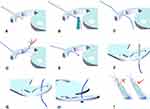
The lens was prepared first (Figure 1). The point of fixing the 6/0 prolene suture to the haptic was determined according to the lens design (the widest haptic span). A 31gauge needle was passed through the haptic, the 6/0 prolene suture was passed through the lumen of the needle. Then, the needle was withdrawn so that the 6/0 prolene suture now passed via the needle track through the haptic. A hand held ophthalmic cautery (Accu-Temp Cautery; Beaver Visitec, Waltham, MA) was approximated very closely to apply radiation heat to the free end of the prolene suture (located on the inner or inferior side of the haptic), and a terminal bulb was created (the flange) to prevent prolene suture slippage. The same was repeated in the other haptic. The two prolene sutures attached to the haptics were passed through the nozzle of the injector and the lens was loaded in the injector. Extreme care should be taken to keep the suture attached to haptics cleared and easily distinguishable from each other.
 |
Figure 1 (A–I) Lens preparation by passing the prolene suture in the track created by the 31-gauge needle. |
A 2.4 mm clear cornea incision was done. Anterior vitrectomy was done to clear any vitreous in the AC if present and to clear the visual axis from any capsular remnants. Any adhesion between the iris and the capsular remnants was severed to allow easy placement of the IOL. The anterior chamber was filled with OVD (Na Hyaluronate 1.4%). A 30 gauge was passed trans-conjunctiva through the sclera 2 mm behind the limbus to show through the pupil. The injector holding the prepared IOL was kept nearby the incision by the assistant. A micro-grasper or McPherson forceps was used to pass the prolene suture attached to the leading haptic into the lumen of the needle. The needle was withdrawn to retrieve the prolene suture outside the eye at the desired site of fixation. Then, the same was repeated on the other side (Figure 2).
 |
Figure 2 (A–C) The prolene suture is passed transconjunctivally through the 30-gauge needle. (D–F) The IOL is implanted and centered. |
After retrieving both prolene sutures through the scleral, the IOL was injected into the eye then centered under the iris with gentle equal traction on both prolene sutures. The prolene suture was pulled fully and was held proximally flushing with the sclera with a tying forceps. The hand-held cautery was used to create another terminal bulb (flange) at the outer end of the prolene suture. The flange was embedded at the entrance of the scleral track and was covered by the conjunctiva (Figure 3). The OVD was washed and the corneal wound was closed by intra-stromal hydration (Supplementary Video 1).
 |
Figure 3 (A–D) The outer bulb is created and kept in the scleral track under the conjunctiva. |
Patients were treated with topical broad spectrum antibiotic drops (moxifloxacin 0.5%) four times per day, topical steroid drops (prednisolone acetate 1%) four times per day and topical NSAIDs drop (nepafenac 0.1%) three times a day. The topical antibiotic and NSAIDs were stopped after two weeks and the topical steroid was tapered over four weeks. The cases were followed up for at least three months for the uncorrected visual acuity, best-corrected visual acuity, IOP, IOL centration, specular microscopy, lens stability and presence of complications.
Results
This technique was performed in 17 aphakic eyes after complicated phacoemulsification.
In 10 cases we used hydrophilic IOLs and in 7 cases we used hydrophobic IOLs. Clinical characteristics, preoperative and postoperative data are summarized in (Table 1).
 |
Table 1 Preoperative and Postoperative Characteristic Data |
There is clear statistically significant difference between Pre-UCVA and Post-UCVA. Also, there is no statistically significant difference between pre-BCVA and post-UCVA. All cases achieved well-centered stable IOL except one case which showed IOL tilt with capture of upper part of the optic that was treated by IOL repositioning after one week.
Slippage of the prolene filament from the haptic happened in 2 cases after implantation. The slippage in one case was due to a small flange size; it was managed by exteriorizing the haptic and replacement of the prolene with a bigger flange. The other case showed a suture slippage from the haptic while implanting the IOL as the trailing haptic got stuck with the plunger of the injector. The IOL was cut, explanted and another IOL was used. One case showed rolling of the prolene over each other inside the injector to create a conflict between the filaments that prevents placement of the IOL after implantation. It was managed by repeating the scleral track after retrieving one filament to clear the conflict.
One case showed exposure of the prolene bulb through the conjunctiva which was managed by burring the bulb in the scleral track and suturing the conjunctiva over it. All corneas were clear from the 1st postoperative day except two cases with mild corneal edema that resolved with frequent topical steroid eye drops. There was no significant difference in endothelial cell count before and after 3 months. There was no significant difference in regard to the IOP before and after 3 months. No cases of cystoid macular edema, endophthalmitis, vitreous hemorrhage or retinal detachment were recorded.
Discussion
The use of the intra-scleral fixated IOLs to manage cases of aphakia with inadequate capsular support is a widely used technique where the IOL is placed close to the anatomically desired position. Gabor and Agarwal et al described the technique of glue IOLs in which the exteriorized haptic is secured under a partial thickness scleral flaps using fibrin glue. In this technique, there is no need for conjunctival dissection, flap creation and fibrin glue use. Yamane et al described scleral fixated flanged IOL with suture-less prolene haptic by approximating a cautery to the end of the haptic to make a flange and pushing the haptics back in the scleral tunnel. The Yamane double-needle technique shows the difficulty of second haptic grasping, the risk of first haptic slippage, risk of haptic deformity or breakage, risk of IOL dislocation to the vitreous and long learning curve.1–3 In this technique, no risk of haptic slippage or IOL dislocation as the lens is secured all the time by the suture. This technique has a short learning curve where both anterior and posterior segment surgeon can do. Threading of 6/0 prolene suture can be done using McPherson forceps through the main incision without the need of end gripping forceps. Moreover, threading the prolene suture through the lumen of 30-gauge needle is much easier than threading of the haptic of the three-piece IOL so no need to use wider gauge needles as recommended by other techniques especially for the beginners.
Canabravo et al described the use of 5/0 prolene suture with flanges to fixate a nonfoldable PMMA IOL with eyelets in the haptics.7 The prolene suture was passed through the eyelet and retrieved through a 25 gauge needle. This technique required a larger incision while in our work we implanted the foldable IOL through a 2.4 sutureless corneal incisions.
The use of 30-gauge needle instead of wider needles will minimize the size of intra-scleral track so a smaller bulb is needed to prevent slippage.8 This technique has a short learning curve where both anterior and posterior segment surgeon can do. Threading of 6/0 prolene suture can be done using McPherson forceps through the main incision without the need of end gripping forceps. In this technique, we used a single-piece foldable IOL which are widely used worldwide. No need to use a special lens design or 3 piece lens.9 The 6/0 prolene is a non-absorbable suture that has a better longevity compared to the 10/0 prolene suture used in sutured scleral-fixated IOLs. Studies have been published using 6/0 prolene suture for intrascleral fixation of Ahmed segment and Cionni CTR.10,11 This is the first study using 6/0 prolene suture for single-piece foldable IOL fixation. Stem et al found that the most common postoperative complications of scleral fixated IOL are vitreous hemorrhage and cystoid macular edema.12 There were no cases of vitreous hemorrhage, cystoid macular edema or endophthalmitis.
Conclusions
Transconjunctival intrascleral fixation of foldable single-piece IOLs is a safe efficient method for correcting aphakia. It is a simple technique that avoids haptic manipulation, slippage and breakage.
Author Contributions
All authors contributed to data analysis, drafting or revising the article, have agreed on the journal to which the article will be submitted, gave final approval of the version to be published, and agree to be accountable for all aspects of the work.
Funding
There is no funding to report.
Disclosure
None of the authors have financial, consultant, institutional and other relationships that might lead to bias or a conflict of interest in the materials presented in this paper. The authors report no conflicts of interest for this work.
References
1. Yamane S, Sato S, Maruyama-Inoue M, et al. Flanged intrascleral intraocular lens fixation with double-needle technique. Ophthalmology. 2017;124(8):1136. doi:10.1016/j.ophtha.2017.03.036
2. Yamane S, Inoue M, Arakawa A, et al. Sutureless 27-gauge needle guided intrascleral intraocular lens implantation with lamellar scleral dissection. Ophthalmology. 2014;121(1):61–66. doi:10.1016/j.ophtha.2013.08.043
3. Almashad GY, Abdelrahman AM, Khattab HA, Samir A. Four-point scleral fixation of posterior chamber intraocular lenses without scleral flaps. Br J Ophthalmol. 2010;94(6):
4. Zhang Y, He F, Jiang J, et al. Modified technique for intrascleral fixation of posterior chamber intraocular lens without scleral flaps. J Cataract Refract Surg. 2017;43(2):162–166. doi:10.1016/j.jcrs.2016.10.029
5. Bonnell AC, Mantopoulos D, Wheatley HM, et al. Surgical technique for sutureless intrascleral fixation of a 3-piece intraocular lens using a 30-gauge needle. Retina. 2019;39 Suppl 1:S13–S15. doi:10.1097/IAE.0000000000001889
6. Chantarasorn Y, Techalertsuwan S, Siripanthong P, et al. Reinforced scleral fixation of foldable intraocular lens by double sutures: comparison with intra-scleral intraocular lens fixation. Jpn J Ophthalmol. 2018;62(3):365–372. doi:10.1007/s10384-018-0579-4
7. Canabrava S, Canêdo Domingos Lima AC, Ribeiro G. Four-flanged intrascleral intraocular lens fixation technique: no flaps, no knots, no glue. Cornea. 2020;39(4):527–528. doi:10.1097/ICO.0000000000002185
8. Kelkar AS, Fogla R, Kelkar J, Kothari AA, Mehta H, Amoaku W. Sutureless 27-gauge needle-assisted transconjunctival intrascleral intraocular lens fixation: initial experience. Indian J Ophthalmol. 2017;65(12):1450–1453. doi:10.4103/ijo.IJO_659_17
9. Veritti DMD, Grego LMD, Samassa FMD, Sarao VMD, Lanzetta PMD. Scleral fixation of a single-piece foldable acrylic IOL through a 1.80 mm corneal incision. J Cataract Refract Surg. 2020;46(5):662–666. doi:10.1097/j.jcrs.0000000000000138
10. Canabrava SMD, Lima CD, Ana Carolina MD. Novel double-flanged technique for managing Marfan syndrome and microspherophakia. J Cataract Refract Surg. 2020;46(3):333–339. doi:10.1097/j.jcrs.0000000000000116
11. Samir A, Abdelrahman Elsayed AM, Alyan A, Lotfy A. Double-flanged polypropylene suture for scleral fixation of cionni capsule tension ring. Clin Ophthalmol. 2020;14:1055–1058. doi:10.2147/OPTH.S244751
12. Stem MS, Wa CA, Todorich B, et al. 27-gauge suture less intra scleral fixation of intraocular lenses with haptic flanging: short-term clinical outcomes and a Disinsertion force study. Retina. 2019;39(11):2149–2154. doi:10.1097/IAE.0000000000002268
 © 2020 The Author(s). This work is published and licensed by Dove Medical Press Limited. The full terms of this license are available at https://www.dovepress.com/terms.php and incorporate the Creative Commons Attribution - Non Commercial (unported, v3.0) License.
By accessing the work you hereby accept the Terms. Non-commercial uses of the work are permitted without any further permission from Dove Medical Press Limited, provided the work is properly attributed. For permission for commercial use of this work, please see paragraphs 4.2 and 5 of our Terms.
