
Back to Journals » International Medical Case Reports Journal » Volume 18
SAPHO Syndrome Misdiagnosed as Spinal Infection: A Case Series
Authors Yang X  , Gu J, Zeng C, Pan D , Cao H, Qian Q, Chen L
Received 7 March 2025
Accepted for publication 9 October 2025
Published 16 October 2025 Volume 2025:18 Pages 1329—1341
DOI https://doi.org/10.2147/IMCRJ.S526910
Checked for plagiarism Yes
Review by Single anonymous peer review
Peer reviewer comments 2
Editor who approved publication: Dr Yu-Ping Ning
Xiaoyan Yang, Jina Gu, Chengjun Zeng, Danmei Pan, Hongchao Cao, Qinbin Qian, Lin Chen
Infectious Diseases Department, Ningbo No. 2 Hospital, Ningbo, Zhejiang, 315010, People’s Republic of China
Correspondence: Lin Chen, Infectious Diseases Department, Ningbo No. 2 Hospital, Ningbo, Zhejiang, CN 315010, Tel +8613566636155, Fax +86057487085742, Email [email protected]
Abstract: SAPHO (synovitis, acne, pustulosis, hyperostosis, and osteitis) syndrome is a rare autoinflammatory disorder characterized by osteoarticular symptoms and dermatological lesions. This case series aims to enhance clinical recognition and reduce misdiagnosis by presenting three patients initially misdiagnosed with spinal infection at external hospitals. All cases presented with worsening back pain and recurrent palmoplantar pustulosis (PPP), one of whom also had comorbid psoriasis. Imaging revealed multifocal purely osteolytic lesions in one patient and mixed osteolytic-osteosclerotic changes in the other two. After exclusion of infectious and neoplastic etiologies, all patients were diagnosed with SAPHO syndrome and showed significant improvement with symptomatic treatment. In conclusion, this case series demonstrated that SAPHO syndrome should be considered in patients with back pain and PPP after excluding infection and tumor.
Keywords: SAPHO syndrome, PPP, acne, bullhead sign, spinal infection
Introduction
SAPHO (synovitis, acne, pustulosis, hyperostosis, and osteitis) syndrome is a rare systemic inflammatory disorder first described by Chamot et al in 1987.1 With an estimated prevalence of less than 1 in 10,000,2,3 it can occur at any age but is uncommon in individuals over 603,4 and shows a predilection for middle-aged women.5,6 The diagnosis of SAPHO syndrome relies on a combination of clinical and imaging features.2,3,7–9 Typical clinical manifestations include musculoskeletal symptoms (eg, local pain, tenderness, swelling, or limited range of motion) and dermatological conditions (eg, palmoplantar pustulosis [PPP], or acne).3,7 Characteristic imaging findings comprise bony sclerosis, with bone scintigraphy often displaying the classic “bullhead sign”.10,11
Diagnosing SAPHO is straightforward when patients present with typical presentations and imaging findings. However, their clinical presentation is heterogeneous. The non-specific nature of the initial symptoms frequently leads to misdiagnosis or delayed diagnosis of this condition12.Without timely intervention, severe cases may progress to bone destruction and joint deformity. Early diagnosis and treatment are essential to halt structural damage and improve patients’ quality of life. While some reports describe sclerotic bone lesions in SAPHO,1,4,10 the co-occurrence of osteolytic and sclerotic lesions, or PPP and psoriasis, as observed in our patients, is exceedingly rare.
In this case series, we present three patients who were ultimately diagnosed with SAPHO syndrome. All initially exhibited atypical clinical features, leading to an initial misdiagnosis of spinal infection. The aim of this report is to emphasize that SAPHO syndrome should be considered as a differential diagnosis in patients presenting with osteoarticular and dermatological manifestations, particularly after infectious and neoplastic etiologies have been ruled out.
Case Reports
Case 1
A 68-year-old woman presented to our hospital with a two-month history of worsening pain in the neck, back, and left hip. During this period, she experienced recurrent fever. One month prior to admission, she developed palmoplantar pustulosis (PPP) affecting both hands and feet. Her previous medical history included heart valve replacement, methicillin-sensitive Staphylococcus aureus Prosthetic Valve Endocarditis, and thoracoscopic surgery for lung adenocarcinoma. Prior to admission, an 18F-fluorodeoxyglucose positron emission tomography-computed tomography (18FDG-PET/CT) performed at another hospital revealed multiple osteolytic lesions in the spine, right sternoclavicular joint, and left sacroiliac joint (Figure 1). In conjunction with an elevated serum C-reaction protein(CRP) level of 41.96mg/L, the doctors at the other hospital suspected spinal infection as the underlying etiology at that time.
 |
Figure 1 PET/CT of Case 1. Osteolytic bone destruction with increased FDG uptake was observed in the C6 vertebral body (A), right clavicle-sternum end (B), L5 vertebral body (C), and left sacroiliac joint and iliac surface (D). |
At admission, her physical examination revealed PPP (Figure 2) and restricted motion.Laboratory tests revealed a normal white blood cell count (WBC; 8.2*10^9/L), neutrophil ratio (N%; 64.9%), and lymphocyte ratio (L%,24.2%). Inflammatory markers were elevated, including C-reactive protein (CRP) (37.28 mg/L), interleukin (IL)-6 (7.60 pg/mL), and erythrocyte sedimentation rate (ESR; 73 mm/h). No infectious agents were identified through pus culture, blood cultures, or blood next-generation sequencing (NGS) (Detailed in Table 1).
 |
Table 1 Clinical Characteristics, Diagnostic Findings and Treatment of the Three Patients |
 |
Figure 2 Pustulosis of the hands (A and B) of Case 1. |
Contrast-enhanced magnetic resonance imaging (MRI) of her lumber and cervical spine showed bone marrow edema in the C5, C6, L5, and S1 vertebral bodies, as well as left sacroiliac joint (Figures 3 and 4). Histopathological examination of the spinal lesion revealed no evidence of malignancy, and tissue culture was negative. Spinal tissue NGS detected a low abundance of Cutibacterium acnes (sequence number: 17; relative abundance: 1.55%).
 |
Figure 3 Lumber MRI with contrast of Case 1. (A) T2-weighted and (B) T1-weighted images showed high signal intensity in the L5 vertebral body. (C) T1-weighted images revealed long T1 signal intensity in the left sacroiliac joint and S1 vertebral body, with enhancement. |
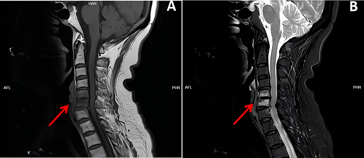 |
Figure 4 Cervical MRI of Case 1. (A) T1-weighted images exhibited low signal intensity in the C5 and C6 vertebral bodies. (B) T2-weighted images showed high signal intensity in the C5 and C6 vertebral bodies. |
A diagnosis of SAPHO syndrome was established. The patient was initially managed with non-steroidal anti-inflammatory drugs (NSAIDs) and alendronate for one month, which resulted in significant pain reduction but persistent skin rash. Her regimen was subsequently adjusted to include glucocorticoids, iguratimod, and alendronate. At the six-month follow-up, the patient showed significant symptomatic improvement.
Case 2
A 39-year-old woman presented to our hospital with a two-month history of progressively worsening soreness in the neck and bilateral shoulder. She had a history of recurrent rashes for four years, with pus-filled rashes in severe cases. One week before admission, she developed bilateral upper arm soreness and distention after excessive cervical spine activity.
Based on cervical computed tomography (CT) findings showing abnormal bone density in the C6 vertebral body, the doctors at an outside hospital had previously diagnosed her with a cervical spinal infection.
Physical examination revealed restricted cervical spine mobility, tenderness over the spinous processes and paraspinal regions, and multiple erythematous scaly patches of varying sizes on the right scalp, abdomen, waist, and lower limbs — findings consistent with a provisional diagnosis of psoriasis by the dermatologist.
Laboratory results were as follows: white blood cell count (WBC) 6.9×10⁹/L (normal), neutrophil ratio (N%) 72.9% (normal), lymphocyte ratio (L%) 22.1% (normal), C-reactive protein (CRP) 10.45 mg/L (elevated), erythrocyte sedimentation rate (ESR) 78 mm/h (elevated), and immunoglobulin A (IgA) 6.96 g/L (elevated). Antinuclear antibody indirect immunofluorescence assay was weakly positive (titer 1:80). Serum and urinary protein immunoelectrophoresis, bone marrow culture, and biopsy showed no evidence of hematologic disorders or infections (Detailed in Table 1).
Contrast-enhanced MRI of her cervical spine showed bone marrow edema and wedge deformation in the C6 vertebral body (Figure 5). PET/CT revealed osteolytic bone destruction with sclerosis in the right first rib and the manubriosternal junction, along with wedge deformation of C6. These lesions exhibited increased fluorodeoxyglucose (FDG) uptake (Figure 6). Therefore, the patient was diagnosed with SAPHO syndrome. She was initially treated with nonsteroidal anti-inflammatory drugs (NSAIDs) and alendronate. One month later, recombinant human tumor necrosis factor-alpha (TNF-α) inhibitor therapy was initiated. Significant symptomatic improvement was noted at the six-month follow-up.
 |
Figure 5 Cervical MRI with contrast of Case 2. (A) T1-weighted images displayed low signal intensity and wedge deformation in the C6 vertebral body. (B) T2-weighted images revealed mixed signal intensity and wedge deformation in the C6 vertebral body. |
 |
Figure 6 PET/CT images of Case 2. Osteolytic bone destruction with sclerosis in the right first rib (A and B) and the border of the manubrium and sternum body (C and D), and C6 vertebral body wedge deformation (E and F). |
Case 3
A 62-year-old woman presented to our hospital with a six-year history of progressive pain, in the neck, back, and left thigh and a three-year history of recurrent PPP affecting both feet. One year prior to admission, she had been hospitalized at another institution for severe back pain. At that time, laboratory parameters—including WBC, CRP, ESR, and procalcitonin—were within normal limits; however, MRI and PET/CT findings suggested vertebral inflammatory changes. She was diagnosed with a spinal infection and received anti-infective therapy. However, after two weeks, the patient discontinued the medication without medical advice due to perceived lack of symptomatic improvement and has not resumed treatment since. Within the two weeks preceding admission, she began experiencing new-onset soreness in the right thigh.
Physical examination revealed PPP (Figure 7) and limitation of motion.Laboratory tests: WBC 9.0×10⁹/L (normal), N% 53.9% (normal), CRP 45.78 mg/L (elevated), interleukin-6 (IL-6) 10.3 pg/mL (elevated), ESR 53 mm/h (elevated), and IgA 4.79 g/L (elevated). Antinuclear antibody testing was weakly positive (titer 1:80). Interferon-gamma release assay for tuberculosis was positive. The admission pus culture from the palm was negative. Two sets of blood cultures were obtained, one bottle of which grew Staphylococcus hominis (penicillin-resistant, oxacillin-sensitive), deemed a contaminant (Detailed in Table 1).
 |
Figure 7 Pustulosis of the hand (A) and foot (B) of Case 3. |
Thoracic spine MRI with contrast showed multiple abnormal signals in the vertebral bodies from T1 to T7, with bone destruction at the lower edge of T7 (Figure 8). PET/CT imaging revealed multiple osteolytic bone destruction with sclerosis in the costovertebral joints from C7 to T7, the left costovertebral joint of T12, the right sternoclavicular joints of T1 and T4, the pubic symphysis, and the right acetabulum (Figure 9).
 |
Figure 8 Thoracic MRI with contrast of Case 3. (A) T1-weighted images showed high signal intensity from T1 to T7 vertebral body. (B) T2-weighted images showed high signal intensity from T1 to T7 vertebral body. |
 |
Figure 9 PET/CT of Case 3. Osteolytic bone destruction or sclerosis in the costovertebral joints from C7 to T7 (A–H), the right sternoclavicular joints of T1 (C/D) and T4 (I), the left costovertebral joint of T12 (J), the right acetabulum (K), and the pubic symphysis (L) with increased FDG uptake. |
Biopsy and culture of a pubic bone lesion showed no evidence of infection or malignancy. Histopathological findings indicated fibro-osseous changes, medullary fibrous hyperplasia with hyaline degeneration, focal reactive bone and cartilage formation, and scattered inflammatory cell infiltration.
The diagnosis of SAPHO syndrome was made. She was initially managed with nonsteroidal anti-inflammatory drugs (NSAIDs) and alendronate. Due to suboptimal symptomatic response after two weeks, TNF-α inhibitor was introduced. Following one month of combination therapy, which yielded only modest improvement, TNF-α inhibitor was transitioned to JAK inhibitor. After 6 months of follow-up, the patient’s symptoms improved.
Discussion
Pathogenesis
SAPHO syndrome is a rare and highly heterogeneous disease of unknown etiology. Genetic, infectious, and immunological factors may contribute to its development.13–15 Studies have identified various single nucleotide polymorphisms that appear to be associated with SAPHO syndrome:3,7,13,15–17 T309G in the MDM2 proto-oncogene, G72C in the tumor protein p53 (TP53), (T>C) in CDK5 regulatory subunit associated protein 1 like 1 (CDKAL1), colony-stimulating factor 2 receptor subunit alpha (CSF2RA), nucleotide-binding oligomerization domain containing 2 (NOD2), multiple EGF like domains 6 (MEGF6), and ADAM metallopeptidase domain 5 (pseudogene) (ADAM5). Although 3.7%–30.0% of patients test positive for human leukocyte antigen (HLA)-B27, its pathogenic role remains unconfirmed.5–7,18 All three patients in our series were HLA-B27 negative.
Infections are considered significant environmental factors contributing to the development of SAPHO syndrome.2,7,13,18 Propionibacterium acnes antigen has been detected in 42% of bone lesion biopsies from SAPHO syndrome patients,19,20 as also seen in our first case. It is hypothesized that Propionibacterium acnes (P. acnes) may act as an initiator of the inflammatory process.18 When Propionibacterium acnes infects patients with immune system dysfunction, it cannot be effectively cleared, leading to dysregulation of transcriptional regulatory factors (nuclear factor kappa B [NF-κB]) and activation of pro-inflammatory cytokines (IL-1, IL-6, IL-17, IL-23, and TNF-α), ultimately resulting in systemic inflammation.2,3,7,8,19,21 However, this mechanism remains debated and requires further validation. Humoral immunity may also contribute to the development of SAPHO syndrome. Studies found that elevated serum IgG4 and monoclonal immunoglobulinG could be observed in patients with SAPHO syndrome.22–24Two patients in our study had elevated immunoglobulin A level. These findings suggest that persistent antigenic stimulation in SAPHO syndrome may drive polyclonal or monoclonal immunoglobulin production. However, further research is needed to validate this hypothesis.
Diagnosis
Several sets of diagnostic or classification criteria have been proposed, but none have been clinically validated.25–28 Diagnosing SAPHO syndrome still relies on the presence of clinical features and the exclusion of infections and tumors. Clinical symptoms are usually related to osteoarticular and dermatological manifestations. Common osteoarticular symptoms include localized pain, swelling, and limited mobility. The anterior chest wall is most frequently involved (65%–90%), followed by the spine (33%–79.2%), sacroiliac joints (32.4%–45.3%), peripheral joints (5.7%–35.3%), mandible (16.5%), and pelvis (13%–52%).2,5,6,29 In this study, all patients were diagnosed with SAPHO syndrome based on the Kahn criteria.28 Spinal lesions were observed in all cases, with two patients additionally demonstrating sternoclavicular joint involvement. Furthermore, one case presented with sacroiliitis, while another exhibited lesions in the pubic symphysis and right acetabulum. These imaging findings are consistent with previous researches.2,5,6,12,29
Clinical Manifestations
Studies have shown that imaging findings in SAPHO syndrome include synovitis, hyperostosis, osteitis, arthropathy, and enthesopathy.3,9 Bone sclerosis and “bullhead sign” are hallmark radiological findings.4,30 In our series, two patients had mixed osteolytic-sclerotic lesions, while one had rare purely osteolytic lesions. Studies have increasingly indicated that bone lesions present as osteolytic bone destruction in early-stage disease and osteosclerosis in late-stage disease.3,9,31 Kinoshita et al reported that bone lesions changed from purely osteolytic to osteosclerotic lesions during 15 months of follow-up.32 Our cases may represent the different osteoarticular phases of SAPHO syndrome.
Classic cutaneous manifestations include PPP and severe acne. PPP—characterized by sterile pustules, erythema, and hyperkeratosis on palms and soles—occurs in over 90% of patients; severe acne is seen in about 15%.5,6 In addition, other less common dermatological manifestations such as psoriasis, hidradenitis suppurativa, Sneddon–Wilkinson syndrome, necrotizing pustular purpura, and sweet syndrome may be observed.12,33,34
Some SAPHO syndrome patients present with multiple concurrent skin manifestations.35 In our case series, two patients had PPP alone, and one had both PPP and psoriasis. Notably, up to 15% of SAPHO patients lack skin lesions. Dermatological manifestations may occur before (54.2%), after (18.7%), and concurrently with (27.1%) osteoarticular symptoms.5 The skin lesions of two cases clearly developed after the osteoarticular lesions, but the absence of concurrent skin lesions does not rule out the possibility of SAPHO syndrome. In addition to the typical clinical manifestations of skin and joints, some patients may also present with a low fever.
Laboratory Tests
No specific laboratory test confirms SAPHO syndrome. In two studies, the ESR was elevated in 57.1%–62.5% and the CRP level was elevated in 70.8%–64.7% of patients with SAPHO syndrome.5,36 Serum levels of TNF-α, IL-6, and IL-17A were significantly elevated in patients with SAPHO syndrome,36 with IL-6 correlating with CRP and ESR.7 All our patients had elevated CRP and ESR, while two had elevated IL-6. One study found that 2.4% of patients with SAPHO syndrome were positive for HLA-B27 and 7.3% for antinuclear antibodies.5 Two of our patients had weakly positive ANA, possibly reflecting underlying autoimmune predisposition.
Imaging Findings
Radiographic examinations, including X-rays, CT, bone scintigraphy, MRI, and PET/CT, are used to assess patients with suspected SAPHO syndrome.37 Since X-rays cannot detect early lesions, CT has become the imaging modality of choice. CT can clearly visualize bone sclerosis and osteolytic lesions, but for early-stage changes such as synovitis and edema, MRI is better. Bone scintigraphy is a useful diagnostic modality that always reveals the “bullhead sign” (predominant high tracer uptake in the sternoclavicular region). The configuration of the bullhead sign is characteristic but not entirely sensitive. Only 22.9% of patients with SAPHO syndrome had the bullhead sign.38 As an emerging diagnostic modality, PET demonstrates high sensitivity. It is not only helpful for evaluating the involved sites in SAPHO syndrome but can also assist in differentiating tumors and infections.39
Comorbidities
An increased prevalence of autoimmune diseases (eg, hypothyroidism, diabetes, Sjögren syndrome, and antiphospholipid syndrome) and inflammatory bowel diseases (mainly Crohn’s disease) has been reported in patients with SAPHO syndrome.7,40,41 MGUS and amyloidosis are rare comorbidities.21,42 In recent years, emerging evidence implicates chronic antigen stimulation—resulting from autoimmunity, infection, or inflammation—in in the pathogenesis of MGUS.43 In patients with SAPHO syndrome complicated by MGUS, persistent antigenic challenge from Propionibacterium acnes may drive aberrant immunoglobulin production, potentially contributing to the emergence of MGUS. Further investigation is required to elucidate the clinical and mechanistic implications of this association.
Treatment
No standardized treatment guidelines exist due to the rarity of SAPHO syndrome. Current strategies target pathogenic mechanisms.
The first-line drugs for SAPHO syndrome are non-steroidal anti-inflammatory drugs (NSAIDs), which can reduce osteoarticular symptoms but do not affect skin changes.5,12,44 Patients with osteoarticular lesions that do not respond to NSAIDs could be treated with short-term corticosteroids.35
Bisphosphonates were administered to patients with vertebral lesions.45 These agents not only mitigate bone destruction by inhibiting osteoclast activity and bone resorption but also attenuate inflammation through suppression of pro-inflammatory cytokines such as IL-1, TNF-α, and IL-6.46
There are two forms of disease-modifying antirheumatic drugs(DMARDs): conventional synthetic disease-modifying anti-rheumatic drugs(csDMARDs); biological disease-modifying anti-rheumatic drugs(bDMARDs).2 The csDMARDs are effective in some patients and are usually used when symptoms persist despite NSAID treatment,5,13,44 whereas bDMARDs are used to treat refractory patients. Cytokine-blocking strategies could correct (at least partially) the cytokine imbalance associated with SAPHO syndrome.2,12,47,48 bDMARDs such as TNF-α inhibitors (infliximab, adalimumab, etanercept), IL-1 antagonists (anabolic leukocytoclasts), IL-6 inhibitors(tozolizumab), IL-17 inhibitors (Secukinumab), and IL-23 inhibitors (Ustekinumab, Tildrakizumab, and Risankizumab) have demonstrated good efficacy in SAPHO syndrome treatment. As emerging treatments, clinical application of JAK inhibitors (tofacitinib,baricitinib and upadacitinib) and apremilast requires further investigation to substantiate its efficacy and safety.
Due to the potential involvement of Propionibacterium acnes, antibiotics sometimes were used to treat patients with SAPHO syndrome, especially in patients with skin lesions.49–51 In our study, none of the patients received anti-Propionibacterium acnes therapy. While several treatment options exist for SAPHO syndrome, further studies are still urgently needed to improve the evidence base for its treatment.
Conclusion
SAPHO syndrome is a chronic inflammatory disorder characterized by recurrent systemic inflammation. It primarily manifests as recurrent cutaneous and osteoarticular manifestations. Radiological findings often include osteosclerosis and the “bull’s head” sign. When patients present with atypical osteoarticular and dermatological presentations, the possibility of SAPHO syndrome should be considered after excluding infection and malignancy.
Ethics Approval and Consent for Publication
This retrospective study was approved by the Research Ethics Committee of Ningbo No.2 Hospital (ref#2024-095-01,ref#2025-031-01). Institutional approval was not required to publish the case details. All patients provided written informed consent for publication of the clinical details and images.
Acknowledgments
The authors thank Junjie Fang from the Department of Radiology, Ningbo No. 2 Hospital, for his assistance.
Funding
This work was supported by Ningbo Clinical Research Center for Respiratory System Disease, Project No. 2022L004.
Disclosure
The authors report no conflicts of interest in this work.
References
1. Yousaf A, Muhammad S, Abdelazeem B, et al. Synovitis, Acne, Pustulosis, Hyperostosis, and Osteitis (SAPHO): a Case Report. Cureus. 2021;13(11):e19636. doi:10.7759/cureus.19636
2. Li SW, Roberts E, Hedrich CM. Treatment and monitoring of SAPHO syndrome: a systematic review. RMD Open. 2023;9(4):e003688. doi:10.1136/rmdopen-2023-003688
3. Demirci Yildirim T, Sari İ. SAPHO syndrome: current clinical, diagnostic and treatment approaches. Rheumatol Int. 2024;44;(11). doi:10.1007/s00296-023-05491-3
4. Duan N, Chen X, Liu Y, et al. Multimodal imaging findings of SAPHO syndrome with no skin lesions: a report of three cases and review of the literature. Exp Ther Med. 2016;12(4):2665–2670. doi:10.3892/etm.2016.3663
5. Li C, Zuo Y, Wu N, et al. Synovitis, acne, pustulosis, hyperostosis and osteitis syndrome: a single centre study of a cohort of 164 patients. Rheumatology. 2016;55(6):1023–1030. doi:10.1093/rheumatology/kew026
6. Cao Y, Li C, Xu W, et al. Spinal and sacroiliac involvement in SAPHO syndrome: a single center study of a cohort of 354 patients. Semin Arthritis Rheum. 2019;48(6):990–996. doi:10.1016/j.semarthrit.2018.10.017
7. Przepiera-Będzak H, Brzosko M. SAPHO syndrome: pathogenesis, clinical presentation, imaging, comorbidities and treatment: a review. Postepy Dermatol Alergol. 2021;38(6):937–942. doi:10.5114/ada.2021.112275
8. Cheng W, Li F, Tian J, et al. New insights in the treatment of SAPHO syndrome and medication recommendations. J Inflamm Res. 2022;15:2365–2380. doi:10.2147/JIR.S355247
9. Kakiuchi A, Yamamoto Y, Kanbe A, et al. SAPHO syndrome with a purely osteolytic form: a case report. Radiol Case Rep. 2023;18(10):3612–3614. doi:10.1016/j.radcr.2023.07.065
10. Liu SZ, Zhou X, Song A, et al. The SAPHO syndrome and the bullhead sign. QJM. 2020;113(2):129–130. doi:10.1093/qjmed/hcz263
11. Gupta N, Verma R, Belho ES. Bone Scan and SPECT/CT Scan in SAPHO Syndrome. Indian J Nucl Med. 2019;34(4):349–350. doi:10.4103/ijnm.IJNM_64_19
12. Ferraioli M, Levani J, De Luca R, et al. What is new and what is next for SAPHO syndrome management: a narrative review. J Clin Med. 2025;14(4):1366. doi:10.3390/jcm14041366
13. Liu S, Tang M, Cao Y, et al. Synovitis, acne, pustulosis, hyperostosis, and osteitis syndrome: review and update. Ther Adv Musculoskelet Dis. 2020;12:1759720X20912865. doi:10.1177/1759720X20912865
14. Hedrich CM, Morbach H, Reiser C, et al. New insights into adult and paediatric chronic non-bacterial osteomyelitis CNO. Curr Rheumatol Rep. 2020;22(9):52. doi:10.1007/s11926-020-00928-1
15. Assmann G, Wagner AD, Monika M, et al. Single-nucleotide polymorphisms p53 G72C and Mdm2 T309G in patients with psoriasis, psoriatic arthritis, and SAPHO syndrome. Rheumatol Int. 2010;30(10):1273–1276. doi:10.1007/s00296-009-1134-x
16. Hurtado-Nedelec M, Chollet-Martin S, Chapeton D, et al. Genetic susceptibility factors in a cohort of 38 patients with SAPHO syndrome: a study of PSTPIP2, NOD2, and LPIN2 Genes. J Rheumatol. 2010;37(2):401–409. doi:10.3899/jrheum.090661
17. Guo C, Tian X, Han F, et al. Copy Number Variation of Multiple Genes in SAPHO Syndrome. J Rheumatol. 2020;47(9):1323–1329. doi:10.3899/jrheum.190435
18. Berthelot JM, Corvec S, Hayem G. SAPHO, autophagy, IL-1, FoxO1, and Propionibacterium (Cutibacterium) acnes. Joint Bone Spine. 2018;85(2):171–176. doi:10.1016/j.jbspin.2017.04.010
19. Zimmermann P, Curtis N. The role of Cutibacterium acnes in auto-inflammatory bone disorders. Eur J Pediatr. 2019;178(1):89–95. doi:10.1007/s00431-018-3263-2
20. Rozin AP. SAPHO syndrome: is a range of pathogen-associated rheumatic diseases extended? Arthritis Res Ther. 2009;11(6):131. doi:10.1186/ar2810
21. Xu D, Liu X, Lu C, et al. Reduction of peripheral natural killer cells in patients with SAPHO syndrome. Clin Exp Rheumatol. 2019;37(1):12–18.
22. Li C, Xiang Y, Wu X, et al. Serum IgG4 elevation in SAPHO syndrome: does it unmask a disease activity marker? Clin Exp Rheumatol. 2020;38(1):35–41.
23. Horino T, Ogasawara M, Matsumoto T, et al. Monoclonal gammopathy of undetermined significance in a patient with synovitis, acne, pustulosis, hyperostosis and osteitis (SAPHO) syndrome: coincidence or association? J Clin Rheumatol. 2021;27(8S):S557–S558. doi:10.1097/RHU.0000000000001630
24. Liu C, Chen T, Wang Y, et al. SAPHO syndrome complicated by IgG4-related ophthalmic disease: a case report and literature review. Front Immunol. 2025;16:1563542. doi:10.3389/fimmu.2025.1563542
25. Witt M, Meier J, Hammitzsch A, et al. Disease burden, disease manifestations and current treatment regimen of the SAPHO syndrome in Germany: results from a nationwide patient survey. Semin Arthritis Rheum. 2014;43(6):745–750. doi:10.1016/j.semarthrit.2013.11.004
26. Benhamou CL, Chamot AM, Kahn MF. Synovitis-acne-pustulosis hyperostosis-osteomyelitis syndrome (SAPHO). A new syndrome among the spondyloarthropathies? Clin Exp Rheumatol. 1988;6(2):109–112.
27. Hayem G. SAPHO syndrome. Rev Prat. 2004;54(15):1635–1636.
28. Kahn MF. Proposed classification criteria of SAPHO syndrome. Arthritis Rheum. 2003;48(Suppl 9):S35–S750.
29. Earwaker JW, Cotten A. SAPHO: syndrome or concept? Imaging findings. Skeletal Radiol. 2003;32(6):311–327. doi:10.1007/s00256-003-0626-0
30. Jurik AG, Klicman RF, Simoni P, et al. SAPHO and CRMO: the Value of Imaging. Semin Musculoskelet Radiol. 2018;22(2):207–224. doi:10.1055/s-0038-1641571
31. Dong A, Bai Y, Cui Y, et al. FDG PET/CT in early and late stages of SAPHO syndrome: two case reports With MRI and bone scintigraphy correlation. Clin Nucl Med. 2016;41(4):e211–e215. doi:10.1097/RLU.0000000000001075
32. Kinoshita H, Ishii T, Kamoda H, et al. Synovitis, acne, pustulosis, hyperostosis, and osteitis syndrome with purely osteolytic, not osteosclerotic, lesions mimicking a malignant tumor. Case Rep Rheumatol. 2020;2020:6316921. doi:10.1155/2020/6316921
33. Rukavina I. SAPHO Syndrome: a Review. J Child Orthop. 2015;9(1):19–27. doi:10.1007/s11832-015-0637-0
34. Marzano AV, Borghi A, Meroni PL, et al. Pyoderma gangrenosum and its syndromic forms: evidence for a link with autoinflammation. Br J Dermatol. 2016;175(5):882–891. doi:10.1111/bjd.14691
35. Nguyen MT, Borchers A, Selmi C, et al. The SAPHO syndrome. Semin Arthritis Rheum. 2012;42(3):254–265. doi:10.1016/j.semarthrit.2012.05.006
36. Gao S, Deng X, Zhang L, et al. The comparison analysis of clinical and radiological features in SAPHO syndrome. Clin Rheumatol. 2021;40(1):349–357. doi:10.1007/s10067-020-05219-9
37. Heldmann F, Kiltz U, Baraliakos X, et al. SAPHO syndrome. Z Rheumatol. 2014;73(8):729–741. doi:10.1007/s00393-014-1415-y
38. Fu Z, Liu M, Li Z, et al. Is the bullhead sign on bone scintigraphy really common in the patient with SAPHO syndrome? A single-center study of a 16-year experience. Nucl Med Commun. 2016;37(4):387–392. doi:10.1097/MNM.0000000000000457
39. Xu T, Huang Y, Zhao Y, et al. 68Ga-DOTA-FAPI-04 PET/CT Imaging in a Case of SAPHO Syndrome. Clin Nucl Med. 2022;47(3):246–248. doi:10.1097/RLU.0000000000004023
40. Valkema PA, Luymes CH, Witteveen JE, et al. High prevalence of autoimmune disease in the rare inflammatory bone disorder sternocostoclavicular hyperostosis: survey of a Dutch cohort. Orphanet J Rare Dis. 2017;12(1):20. doi:10.1186/s13023-017-0580-x
41. Wang L, Yu Y, Zhang S, et al. Coexistence of Sjögren syndrome in patients with synovitis, acne, pustulosis, hyperostosis, and osteitis syndrome: a retrospective observational study. Medicine. 2021;100(12):e23940. doi:10.1097/MD.0000000000023940
42. Valentin R, Gürtler KF, Schaker A. Renal amyloidosis and renal failure--a novel complication of the SAPHO syndrome. Nephrol Dial Transplant. 1997;12(11):2420–2423. doi:10.1093/ndt/12.11.2420
43. Sigurbergsdóttir AÝ, Love TJ, Kristinsson SY. Autoimmunity, infections, and the risk of monoclonal gammopathy of undetermined significance. Front Immunol. 2022;13:876271. doi:10.3389/fimmu.2022.876271
44. Przepiera-Będzak H, Brzosko M. Clinical symptoms, imaging, and treatment of SAPHO syndrome: a single‑center study of 52 cases. Pol Arch Intern Med. 2018;128(6):396–399. doi:10.20452/pamw.4264
45. Taddio A, Ferrara G, Insalaco A, et al. Dealing with chronic non-bacterial osteomyelitis: a practical approach. Pediatr Rheumatol Online J. 2017;15(1):87. doi:10.1186/s12969-017-0216-7
46. Wang CR, Tsai YS, Whang-Peng J. Zoledronic acid monotherapy improves osteoarticular involvement in SAPHO syndrome. Scand J Rheumatol. 2020;49(5):419–421. doi:10.1080/03009742.2020.1744640
47. Wang Y, Gu M, Zheng Z, et al. Therapeutic approaches for SAPHO syndrome from the perspective of pathogenesis: a review of the literature. Front Immunol. 2025;16:1560398. doi:10.3389/fimmu.2025.1560398
48. Ferraioli M, Fiannacca L, Greco E, et al. Risankizumab efficacy in Synovitis, Acne, Pustulosis, Hyperostosis, and Osteitis (SAPHO) remission: a case report on rheumatologic and dermatologic disease manifestations with literature review. Case Rep Immunol. 2024;2024:9076852. doi:10.1155/2024/9076852
49. Assmann G, Kueck O, Kirchhoff T, et al. Efficacy of antibiotic therapy for SAPHO syndrome is lost after its discontinuation: an interventional study. Arthritis Res Ther. 2009;11(5):R140. doi:10.1186/ar2812
50. Govoni M, Colina M, Massara A, et al. SAPHO syndrome and infections. Autoimmun Rev. 2009;8(3):256–259. doi:10.1016/j.autrev.2008.08.003
51. Colina M, Trotta F. Antibiotics may be useful in the treatment of SAPHO syndrome. Mod Rheumatol. 2014;24(4):697–698. doi:10.3109/14397595.2014.885866
 © 2025 The Author(s). This work is published and licensed by Dove Medical Press Limited. The
full terms of this license are available at https://www.dovepress.com/terms
and incorporate the Creative Commons Attribution
- Non Commercial (unported, 4.0) License.
By accessing the work you hereby accept the Terms. Non-commercial uses of the work are permitted
without any further permission from Dove Medical Press Limited, provided the work is properly
attributed. For permission for commercial use of this work, please see paragraphs 4.2 and 5 of our Terms.
© 2025 The Author(s). This work is published and licensed by Dove Medical Press Limited. The
full terms of this license are available at https://www.dovepress.com/terms
and incorporate the Creative Commons Attribution
- Non Commercial (unported, 4.0) License.
By accessing the work you hereby accept the Terms. Non-commercial uses of the work are permitted
without any further permission from Dove Medical Press Limited, provided the work is properly
attributed. For permission for commercial use of this work, please see paragraphs 4.2 and 5 of our Terms.