
Back to Journals » Clinical Ophthalmology » Volume 19
Retinal Images with Catenary-Curved Contact Lenses [Letter]
Authors Westheimer G 
Received 4 November 2025
Accepted for publication 3 December 2025
Published 9 December 2025 Volume 2025:19 Pages 4543—4544
DOI https://doi.org/10.2147/OPTH.S579008
Checked for plagiarism Yes
Editor who approved publication: Dr Scott Fraser
Gerald Westheimer
Department of Neuroscience, University of California, Berkeley, CA, USA
Correspondence: Gerald Westheimer, Email [email protected]
View the original paper by Dr Tuan and colleagues
Dear editor
The recent publication of a clinical trial of lenses with catenary curved power profile by Tuan et al1 is an example of expanded-focus contact and intra-ocular lenses. The report is rare in that it includes the actual curvature specification, which allows independent evaluation of their diffraction-mandated resolution limit. Using the computational approach to the retinal image light distribution that had been applied to other non-traditional surface configurations,2 this problem is explored here.
After converting the curvatures to phase deviations of the wavefront converging on the retina, the diffraction image on the retina was calculated for monochromatic light, a round pupil of 4mm, and defocus in half-diopter steps from −1D to +4D, ie, target distances from 1m virtual to 25 cm real. To relate this information to the clinical reports, the computed point-spread functions were used to derive the light spread across the width of a 2 arcmin bright bar, the basic component of 20/40 (0.3 logMAR) letters on visual acuity charts. Because of its easy visualization of how defocus degrades the image through contrast reduction and light spread, this is a good measure for comparing patient data with the best performance allowed by physical theory.
Figure 1 is the computationally generated through-focus display in the −1D to +4 diopter target vergence range, side-by-side for three conditions: left, normal viewing (with a plane wavefront) and a 4mm pupil, middle, with the catenary-curve lens and a 4mm pupil, and, right, the catenary-curve and a 6mm pupil diameter. These data can now be compared with reports from the clinical trial of the visual acuity of patients wearing normal single vision as well as actual catenary-curved lenses. The smoothly augmented focus range (by about 1 Diopter) of the latter, Fig. 4 of Tuan et al,1 does not match the theoretical graph’s more widely extended if choppy focus extension nor are the clinical claims of barely attenuated maximum acuity paralleled by the severe contrast reduction in the model describing the performance of optical systems obeying physical systems.
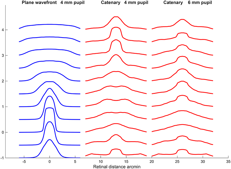 |
Figure 1 Through-focus depiction of computed light spread in the retinal image of a 2 arcmin-wide bright bar in aberration-free eye with monochromatic light, for (left) normal vision with plane wavefront and 4mm pupil diameter, (middle) wearing catenary -curved contact lens and 4mm pupil, and (right) catenary curved lens and 6 mm pupil. Target vergence from bottom up in ½ diopter steps from −1D (1m virtual) to 4D (25cm real). |
The objection that the computational approach is based on too simplified a model is partially met by the demonstration, in an equivalent connections, that it retains general validity when incorporating some defects found in real eyes: spherical and other low-order aberrations, chromatic aberrations, Stiles-Crawford effect and photoreceptor diversity.3 Although what finally counts is always the patient performance, nevertheless when it diverges significantly from the limits set by physical theory, reconciliation is called for.
Disclosure
The author reports no conflicts of interest in this communication.
References
1. Tuan K, Benoit D, O’Connor B. Evaluation of the functional visual range of a catenary curve-based, extended depth-of-focus contact lens for presbyopia. Clin Ophthalmol. 2024;18:2113–2123. doi:10.2147/OPTH.S468699
2. Westheimer G. Focused and defocused retinal images with Bessel and axicon pupil functions. J Opt Soc Am A. 2020;37(1):108–114. doi:10.1364/JOSAA.37.000108
3. Westheimer G. Multifocal contact lens myopia control: central and peripheral retinal image quality. Clin Exp Optom. 2023;106:538–543.
 © 2025 The Author(s). This work is published and licensed by Dove Medical Press Limited. The
full terms of this license are available at https://www.dovepress.com/terms
and incorporate the Creative Commons Attribution
- Non Commercial (unported, 4.0) License.
By accessing the work you hereby accept the Terms. Non-commercial uses of the work are permitted
without any further permission from Dove Medical Press Limited, provided the work is properly
attributed. For permission for commercial use of this work, please see paragraphs 4.2 and 5 of our Terms.
© 2025 The Author(s). This work is published and licensed by Dove Medical Press Limited. The
full terms of this license are available at https://www.dovepress.com/terms
and incorporate the Creative Commons Attribution
- Non Commercial (unported, 4.0) License.
By accessing the work you hereby accept the Terms. Non-commercial uses of the work are permitted
without any further permission from Dove Medical Press Limited, provided the work is properly
attributed. For permission for commercial use of this work, please see paragraphs 4.2 and 5 of our Terms.