Back to Journals » Drug Design, Development and Therapy » Volume 20
Resveratrol Alleviates Intervertebral Disc Degeneration by Targeting NCOA4-Mediated Ferritinophagy Through Dual Antioxidant and Anti-Inflammatory Effects
Authors Song C, Wu X, Chen C, Shen B, He Y, Liu F ![]() , Chen F, Fu Z, Liu Z
, Chen F, Fu Z, Liu Z
Received 5 November 2025
Accepted for publication 9 March 2026
Published 18 March 2026 Volume 2026:20 575321
DOI https://doi.org/10.2147/DDDT.S575321
Checked for plagiarism Yes
Review by Single anonymous peer review
Peer reviewer comments 3
Editor who approved publication: Professor Amit Tiwari
Chao Song,1,2 Xiaofei Wu,2 Chaoqi Chen,2 Baoxin Shen,2 Yuheng He,1 Fei Liu,1,2 Feng Chen,2 Zhijiang Fu,1 Zongchao Liu1,3
1Department of Orthopedics and Traumatology (Trauma and Bone-Setting), The Affiliated Traditional Chinese Medicine Hospital, Southwest Medical University, Luzhou, Sichuan, People’s Republic of China; 2Department of Orthopedics, RuiKang Hospital Affiliated to Guangxi University of Chinese Medicine, Nanning, Guangxi Zhuang Autonomous Region, People’s Republic of China; 3Department of Orthopedics, Luzhou Longmatan District People’s Hospital, Luzhou, Sichuan, People’s Republic of China
Correspondence: Zhijiang Fu; Zongchao Liu, The Affiliated Traditional Chinese Medicine Hospital, Southwest Medical University, No. 182 Chunhui Road, Longmatan District, Luzhou, Sichuan, 646000, People’s Republic of China, Email [email protected]; [email protected]
Background: Intervertebral disc degeneration (IVDD), characterized by inflammation, cell death, and matrix dysregulation, involves ferroptosis and autophagy interactions, though the role of ferritinophagy remains unclear.
Methods: This study integrated bioinformatics analysis of clinical transcriptomes, single-cell sequencing, and experimental models to identify molecular targets linking ferritinophagy to IVDD progression.
Results: Multi-omics analysis revealed 10 ferroptosis-related hub genes (eg, NCOA4, TP53, SLC7A11) enriched in hypoxia, autophagy, and ferroptosis pathways. Single-cell profiling demonstrated dynamic shifts in nucleus pulposus cell (NPCs) phenotypes during degeneration, with increased pro-inflammatory monocyte/macrophage infiltration. Clinical and experimental validation showed elevated NCOA4 and ACSL4 (ferroptosis drivers) alongside reduced GPX4 and FSP1 (anti-ferroptotic factors) in degenerated discs. Mechanistically, NCOA4-mediated ferritinophagy promoted iron overload and lipid peroxidation by degrading ferritin, exacerbating oxidative damage. Resveratrol, a natural anti-inflammatory compound, mitigated IVDD in rat models by restoring disc height, suppressing IL1β, TNF-α, and IL6, and reversing ferroptosis/autophagy imbalance via NCOA4 downregulation. Cellular studies confirmed that resveratrol attenuated LPS-induced NPCs degeneration by blocking NCOA4-dependent ferritinophagy, reducing lipid peroxidation and cell death.
Conclusion: This work identifies NCOA4 as a critical nexus coordinating ferroptosis-autophagy crosstalk in IVDD and establishes ferritinophagy as a novel pathological mechanism. Resveratrol’s therapeutic efficacy, mediated through multi-target modulation of inflammatory and iron homeostasis pathways, provides a promising strategy for IVDD treatment. The findings advance precision medicine approaches by elucidating ferritinophagy’s role and proposing resveratrol as a targeted intervention to disrupt this self-amplifying cycle of oxidative damage in disc degeneration.
Keywords: intervertebral disc degeneration, resveratrol, ferroptosis, autophagy, NCOA4, nucleus pulposus cells
Introduction
Intervertebral Disc Degeneration (IVDD) is one of the primary causes of chronic low back pain, with a complex pathological process involving extracellular matrix metabolism imbalance, nucleus pulposus cells (NPCs) senescence and death, inflammatory response activation, and oxidative stress damage.1,2 Current clinical interventions primarily focus on symptom relief and struggle to reverse degenerative processes, fundamentally due to incomplete understanding of the disease’s driving mechanisms.3 Therefore, a thorough analysis of the core molecular mechanisms of IVDD and the identification of key targets capable of delaying or even reversing degeneration are urgent tasks in current research.
Among numerous pathological processes, the imbalance between programmed cell death and cellular homeostasis regulation mechanisms is considered the core link driving the progression of IVDD. Among them, mechanisms such as apoptosis and pyroptosis have been widely studied.4–6 In recent years, ferroptosis, as a novel iron dependent cell death mode characterized by abnormal accumulation of lipid peroxides, has shown important roles in various degenerative diseases.7 At the same time, autophagy, as a key “scavenger” system within cells, maintains internal environment stability by degrading damaged organelles and macromolecules. However, its role in IVDD is dual sided, with moderate activation providing protective effects, while dysregulation may exacerbate cell collapse.8 It is worth noting that increasing evidence suggests that ferroptosis and autophagy do not operate independently, and there is a complex cross-talk between the two that jointly determines the fate of cells in many disease contexts.9,10 However, in the field of IVDD, how these two pathways interact with each other, the specific nodes and networks of their interaction, are still a blank that needs to be explored urgently. Clarifying this issue not only helps to gain a more systematic understanding of the nature of intervertebral disc degeneration but may also reveal new strategies for combination therapy that go beyond single pathway regulation.
A potential core hub for the interaction between ferroptosis and autophagy is the regulation of intracellular iron metabolism. Iron is a key element that catalyzes lipid peroxidation and drives ferroptosis, and its intracellular storage form, ferritin turnover, is precisely regulated by the autophagy pathway, a process known as “ferritin autophagy”.11–13 Autophagy of ferritin selectively degrades ferritin and releases free iron, directly affecting the sensitivity of cells to ferroptosis.14,15 NCOA4, a nuclear receptor co activator, has been identified as a key carrier protein mediating ferritin autophagy.7 Its expression level directly regulates the targeted transport and degradation of ferritin to lysosomes, thereby becoming a bridge connecting autophagy flow and iron metabolism homeostasis. NCOA4 mediated ferritin autophagy has been shown to have significant pathological implications in tumors and neurodegenerative diseases. In the context of intervertebral disc degeneration (IVDD), preliminary studies suggest abnormal expression of iron metabolism related proteins in degenerated intervertebral disc tissue, and changes in autophagy flow within NPCs.16 This strongly suggests that NCOA4 mediated ferritin autophagy may be a key molecular link in IVDD that has not yet been revealed, connecting autophagy abnormalities with ferroptosis activation.17,18 Elucidating this mechanism will provide a new perspective for understanding the death mode switching of NPCs in the degenerative microenvironment.
In terms of intervention strategies, the natural polyphenolic compound resveratrol has attracted much attention due to its significant antioxidant and protective autophagy activation abilities in the IVDD model.19 More interestingly, recent studies have shown that its analogues can alleviate disease in asthma models by inhibiting the NCOA4 pathway, suggesting that resveratrol may have the potential to regulate iron homeostasis.20 Based on this, we propose a scientific hypothesis: in IVDD, NCOA4 mediated abnormal activation of iron autophagy flow is an important mechanism leading to ferroptosis in NPCs; Resveratrol may exert intervertebral disc protection by targeting and inhibiting this pathway, restoring iron homeostasis.
Based on the above logical framework, this study aims to systematically analyze the interaction mechanism between ferroptosis and autophagy in IVDD, and focus on the core hypothesis of NCOA4 mediated ferritin autophagy. We first conducted a joint analysis of bioinformatics and multi omics to screen for key genes involved in the progression of IVDD and used single-cell transcriptome sequencing technology to depict the dynamic map of phenotype transformation and microenvironment remodeling of NPCs at the subpopulation level, in order to identify key regulatory molecules. In vitro and in vivo functional experiments will further validate the role of NCOA4 in regulating ferritin autophagy, influencing ferroptosis and autophagy balance, and ultimately determining the fate of NPCs. Finally, based on this mechanism, we explored the intervention potential of the natural polyphenolic compound resveratrol. Resveratrol is known to have anti-inflammatory and antioxidant properties, but there is no research on whether it can coordinate ferroptosis and autophagy by targeting the NCOA4 ferritin autophagy axis, thereby exerting a protective effect on intervertebral discs. This study not only has the potential to innovatively propose “iron autophagy imbalance” as a novel molecular mechanism model for IVDD, but also provides theoretical basis and experimental evidence for the development of precise therapeutic strategies targeting the “ferroptosis autophagy interaction network”.
Materials and Method
Bioinformatics Analysis Based on Clinical Transcriptome Samples
Clinical Data Collection and Processing
This study obtained gene expression profile datasets GSE70362 and GSE34095 for human lumbar disc degeneration from the GEO database. GSE70362 contains 48 intervertebral disc tissue samples, including annulus fibrosus and nucleus pulposus, showing varying degrees (grades) of degeneration, while GSE34095 contains 6 intervertebral disc tissue samples, divided into degenerative and non-degenerative tissues.21 The selection criteria include: the research object is human lumbar disc tissue, including both degeneration and control samples, and providing clear tissue source and degeneration grading information. The raw data was preprocessed by the R language platform, including background correction, quantile normalization, and logarithmic transformation, and the probes were annotated to gene symbols. Use Sangerbox 3.0 online tool to perform Principal Component Analysis (PCA) dimensionality reduction analysis on two datasets.22 To conduct a meta-analysis, the expression matrices of two datasets were merged and the ComBat function in the SVA package was used. Batch effects were corrected using “dataset source” as the batch variable and “tissue type” and “degeneration status” as biological covariates. By comparing the principal component analysis graphs before and after correction, it was intuitively verified that batch effects were effectively eliminated and sample distribution was more dominated by biological status.
Construction of Differential Genes and Module Genes
The combined chip matrix data can be transformed into a gene name matrix using the gene name transformation tool in Sangerbox 3.0. Choose the “limma” tool for differential gene analysis, and set the threshold to a P-value of less than 0.05 and a difference multiple of 0.5. Gene downregulation is indicated by negative values, whereas differentially expressed genes (DEGs) are upregulated by positive values. By building scale-free networks, the systems biology technique known as Weighted Gene Coexpression Network Analysis (WGCNA) identifies patterns of gene coexpression and correlations between phenotypes. By assessing the relationship between module feature genes and phenotype, the key module was determined to be the module with the highest absolute correlation coefficient. This study employed the R language WGCNA software for network development. Using a weight threshold of 0.1 to screen hub genes and the dual criteria of module membership degree (MM ≥ 0.8) and gene significance (GS ≥ 0.1).23
Screening of Ferroptosis Hub Genes
From FerrDb database (http://www.zhounan.org/ferrdb/current/) Obtaining ferroptosis related genes.24 The ferroptosis hub genes for intervertebral disc degeneration were constructed by taking the intersection of the upregulated and downregulated ferroptosis genes with the fusion genes of the transcriptome, and then constructing the ferroptosis associated genes during the process of intervertebral disc degeneration based on the top 100 genes associated with upregulation and downregulation of ferroptosis during intervertebral disc degeneration. The upregulated and downregulated genes were subjected to PPI network design using a string database (http://string-db.org/), and the species was limited to “Homo sapiens” with a confidence value greater than 0.4.25 In addition, the CytoHubba algorithm, a plugin of Cytoscape 3.10.0 software, was used to further screen for the upregulated and downregulated core genes of intervertebral disc ferroptosis.26 Create a PPI network with Cytoscape to determine the core genes that are upregulated and downregulated by intervertebral disc ferroptosis, as well as the important genes implicated in intervertebral disc degeneration. Lastly, the transcriptome’s fusion gene data was used to extract the expression data of hub genes and core genes involved in the ferroptosis process of intervertebral discs. These genes were then subjected to preliminary testing.
Clinical Validation of Ferroptosis Hub Genes
This study was approved by the Ethics Committee of Ruikang Hospital of Guangxi University of Traditional Chinese Medicine (approval number: GJJ19041), and all samples were collected after obtaining informed consent from patients. Degenerative intervertebral disc tissue samples (IVDD group, n=4) were taken from L4/5 segments of patients who required surgical treatment for lumbar disc degeneration. The non-degenerative control tissue (control group, n=4) was derived from patients who required surgery for traumatic lumbar fractures and had no degenerative changes in the intervertebral disc confirmed during surgery. Two groups of patients were matched based on age (both>50 years old) and gender (both 2 males/2 females), with specific clinical characteristics shown in Supplementary Table 1. All tissue samples were obtained immediately after surgery, and some were frozen in liquid nitrogen for subsequent RNA extraction for inter group expression differential analysis of hub genes (such as NCOA4). At the same time, venous blood samples were collected from two groups of patients, and serum was separated. Enzyme linked immunosorbent assay (ELISA) was used to detect the concentrations of inflammatory cytokines such as interleukin-1β (IL-1β) and tumor necrosis factor-α (TNF-α), and to compare the levels of inflammation between groups.
Biological Process Analysis
The hub genes of intervertebral disc degeneration and the core genes upregulated and downregulated by intervertebral disc ferroptosis were fused to perform GO, KEGG, and GSEA enrichment analysis in order to examine the pertinent signaling pathways and processes of intervertebral disc degeneration.27 Limit the species to “H. sapiens” after importing the gene set into the Ouyi Biological Cloud platform and choosing enrichment analysis in the tool center. Enter the gene set’s gene symbol in the frequently used parameters, then submit to see the results displayed in several graphs.
Analysis of Immune Cell Infiltration
Analysis of immune cell infiltration is a crucial guiding tool for forecasting the course of a disease and the effectiveness of treatment. We computed the immune cell types of patients with distinct immune patterns in the transcriptome fusion dataset using the CIBERSORT algorithm in Sangerbox 3.0 software, and we displayed the immune cell composition of patients with distinct immune patterns using a stacked graph.28 Lastly, a bar graph was utilized to show the infiltration connection of different immune cells during IVDD, and a radar chart was utilized to show the variations in infiltration of various immune cells between normal people and IVDD patients.
Single Cell Data Analysis
Collection and Processing of Single-Cell Data
Sample data GSE244889 downloaded from Gene Expression Omnibus (GEO) database.1 The sample collected gene expression profiles at the single-cell level of the nucleus pulposus tissue in mild degenerative intervertebral discs and moderate-to-severe degenerative intervertebral discs. We use a bioinformatics bean sprout analysis tool for data processing and use an R language seurat package to calculate mitochondrial content and rRNA content. Set the cell filtration criteria to 100<5000. Set the percentage of mitochondrial content to 10% and use Harmony to batch it.29 To reduce dimensionality, use principal component analysis (PCA). Next, choose the optimal cluster for display using Seurat’s random neighbor embedding approach (resolution=0.05). Cell types can be clarified by annotating cells from various subgroups using the “SingleR” package, Top5 marker genes, and literature search. Findmarker can be used to look for marker genes in subgroups.
Developmental Trajectory and Intercellular Communication Analysis
Using cell trajectory analysis with the Monocle software tool (version: 2.30.0) to explain the differentiation link between NPC subtypes. The Monocle algorithm introduces the idea of “pseudo time” and uses a single cell sorting strategy to extract NPCs from each sample independently and create a Seurat object for pseudo-temporal analysis. The dynamic changes in development or differentiation over time are reproduced by sorting all cells based on a trajectory using the differences in expression of individual cells. Based on the expression profile, match the pseudo time of each NPC subtype to determine the differentiation order of NPCs. Use DDRTress’s reduceDimension function to determine pseudo time for various NPC subtypes, then use trajectory maps to see the outcomes. We analyzed scRNA seq maps with varying degrees of degeneration using the CellChat method (version: 1.6.1). Cellchat uses the expression of ligands and receptors between cells to assess the effects of intercellular interactions. In order to examine the crucial role that cell ligand/receptor interactions play in IVDD, we looked into the notable variations in cell ligand/receptor interactions in cell communication between samples of various grades.
Joint Analysis of Transcriptome Hub Genes and Single Cells
To examine the various cells found in various stages of degeneration, create bar charts and dot distribution graphs of the expression ratios of various cell subgroups in intervertebral disc tissues using the R programming language package. To further confirm the expression and molecular mechanisms of linked genes in various cells, IVDD ferroptosis core genes, hub genes, and single-cell marker genes were retrieved for joint investigation based on standard transcriptome sequencing. The R package can be used to create UMAP maps of particular gene expression.
Validation of Drugs and Molecular Mechanisms Regulating Ferritin Induced IVDD
Drug Prediction
Make drug predictions for the core and hub genes of ferroptosis in intervertebral discs using bioinformatics bean sprout tools. You can also get peak maps of drug predictions that have therapeutic benefits. The primary monomer, resveratrol, is obtained by screening the pharmacological data for monomeric traditional Chinese medicine.
Animal Experiments
Processing and Grouping of Animal Samples
Thirty 8-week-old SPF grade healthy male SD rats, weighing (210 ± 20) g, were purchased from Hunan Slack Jingda Experimental Animal Co., Ltd. [license number: SCXK (Xiang) 2019-0004]. The selection of male animals aims to avoid potential interference of the estrogen cycle on disease progression and drug response. Rats were housed in the Animal Experiment Center of Guangxi University of Traditional Chinese Medicine [license number: SYXK Gui 2024-0004], with an ambient temperature of (23 ± 2) °C and humidity of (55 ± 10)%, alternating light and dark for 12 hours, and free access to food and water. After one week of adaptive feeding, all experimental procedures followed the approved protocol of the institutional animal ethics committee (ethics number: DW20250319-065). Rats were randomly divided into three groups (n=10/group) using a random number table method: sham surgery group, model group, and resveratrol treatment group. The classic tail intervertebral disc acupuncture method was used to construct an IVDD model: After anesthesia fixation in rats, a 21G needle was used to accurately puncture the intervertebral space of the 8th and 9th tail vertebrae (Co8-9), damaging the annulus fibrosus and nucleus pulposus.30 The sham surgery group only incises the skin without damaging the intervertebral disc. After one week of modeling, drug intervention will begin. The resveratrol treatment group received daily oral administration of resveratrol (purity ≥ 98%, Sigma Aldrich) at a dose of 50 mg/kg/d (dissolved in 1% sodium carboxymethyl cellulose solution); The sham surgery group and the model group were given an equal volume of 1% carboxymethyl cellulose sodium solution by gavage daily. The intervention lasted for 4 weeks. The dosage and course of treatment are based on the efficacy and safety settings reported in previous literature in other degenerative disease models.30
After the intervention, collect Co8-9 tail intervertebral disc tissue. Part of the samples were used for histological evaluation, while the other part was rapidly isolated from the nucleus pulposus and stored at −80 °C after liquid nitrogen freezing for subsequent molecular biology testing (qPCR, Western blot, lipid peroxidation level determination, etc) to comprehensively evaluate the degree of intervertebral disc degeneration, ferroptosis and autophagy pathway activity, as well as the intervention effect of drugs. This plan prioritizes the euthanasia of SD rats by excessive use of inhaled anesthetics (isoflurane), supplemented by necessary physical methods for death confirmation. All programs are centered around ensuring animal welfare and strictly follow the authoritative guidelines of the American Veterinary Association (AVMA).
Detection of Animal Samples
DR Detection
The rats were fasted and dehydrated for eight hours before the specimens were collected. Strict adherence to the ethical guidelines for animal research was maintained, and an intraperitoneal injection of 30 mg/kg pentobarbital was used to induce anesthesia. The rats were put under DR apparatus and photographed to get raw DR data once the anesthetic had taken effect. All tail vertebrae X-rays taken before the sacrifice of rats were randomly numbered by independent experimenters who did not participate in animal grouping and intervention. The researchers responsible for measuring the anterior, middle, and posterior heights of each intervertebral space and calculating DHI have no knowledge of the experimental groups (sham surgery group, model group, or treatment group) corresponding to each X-ray. All measurement data must be entered and locked before unblinding and statistical analysis. Using the intervertebral disc DHI index to assess variations in intervertebral disc height.31

Tissue Section Staining
Referring to existing research methods, the tail vertebrae of the marked segments were selected for modeling, fixed with paraformaldehyde, paraffin sectioned, deparaffinized, stained with hematoxylin and eosin, SOFG, dehydrated, sealed, examined under a microscope, and images were collected. Pathological staining section scoring is used to evaluate the degree of intervertebral disc degeneration after modeling and medication. The histological evaluation of intervertebral disc degeneration is performed using an improved Masanori scoring system, which semi quantitatively scores from multiple dimensions including tissue structure, morphology and quantity of NPCs, and matrix staining characteristics (reflecting proteoglycan content). To ensure the objectivity of the scoring, this study strictly implemented blind evaluation. All slices were randomly re-encoded by independent researchers before analysis, and evaluators were completely unaware of the sample grouping information. Each slice is independently rated by two trained observers. The intra-group correlation coefficient was used to evaluate inter-observer consistency, and the ICC values of all scores in this study were greater than 0.85, indicating excellent consistency. For the scores with differences, both parties will review and negotiate in a blind manner to determine the final value, which will be used for subsequent statistical analysis.
Detection of Key Proteins in Ferroptosis
According to the RIPA complete lysis buffer instructions, equal proportions of intervertebral disc tissue was washed with phosphate buffer solution, homogenize thoroughly at 4 °C, and was collected the supernatant after centrifugation. The tissue protein extracted quantitatively by BCA method was transferred to PVDF membrane by SDS-PAGE gel electrophoresis. Then, 5% skimmed milk was sealed at room temperature for 2 hours, and GAPDH (1:2000) was used as the internal reference. The corresponding primary antibody was added to incubate at 4 °C overnight. After rewarming and washing the next day, sheep anti-rabbit IgG (1:5000) was added to incubate at room temperature for 2 hours. After washing, ECL luminous solution was added to expose and collect pictures. Image J software was used to analyze the gray value of protein bands.
Cell Experiment
Experimental Materials
Human primary NPCs were purchased from iCell Biotechnology Co., Ltd. (Shanghai, China), item number: iCell-0028a, The cells are derived from human intervertebral disc tissue and digested with 0.25% trypsin. The fluorescent staining of human NPCs type II collagen is positive, and the purity can reach over 90%. Lipopolysaccharide, resveratrol, and ferroptosis inhibitor Ferrostatin-1 were purchased from Aladdin Company in China. The complete culture medium for human primary NPCs was purchased from iCell Biotechnology Co., Ltd. GPX4, FSP1, NCOA4, IL1B, IL6, TNF-a, P62, BCL2, LC3 antibodies, and GAPDH antibodies were purchased from Baiqu Company (Shanghai, China). The supplier of CCK-8 reagent kit is Beijing Solaibao Technology Co., Ltd. The experiment used lipopolysaccharide (LPS) to stimulate NPCs, aiming to construct a controllable inflammatory stress model to simulate a core driving factor in the pathological process of IVDD - the chronic inflammatory microenvironment. LPS is a classic tool for studying inflammatory responses, which can effectively induce high expression of pro-inflammatory factors (such as TNF-α, IL-1β, and IL-6) similar to those in human degenerated intervertebral disc tissue by stably activating the TLR4/NF-κB signaling pathway, and subsequently trigger key downstream events such as oxidative stress and matrix degradation. Although this model cannot fully replicate the complex pathological panorama of multiple factors (such as mechanical load, aging, and nutrient deficiency) interwoven in the body, its advantage lies in the ability to clearly and specifically study the effects of “inflammatory stress” on the ferroptosis autophagy interaction network and extracellular matrix metabolism of NPCs under controllable conditions of a single variable (inflammation). It is suitable for preliminary exploration of the core molecular mechanism and causal relationship analysis in this study.
Preparation of Lipopolysaccharide and Resveratrol Solutions
Dissolve 10 mg lipopolysaccharide solution in 100 μL DMSO solution, package and store in a −20 °C refrigerator to prepare 100mmol/L lipopolysaccharide stock solution. Dilute with human primary NPCs complete culture medium to the following concentrations: 0.01, 0.1, 1, 10, and 100 μmol/L. Similarly, configurations: 0.01, 0.1, 1, 10, and 100 μmol/L resveratrol solutions.
Cell Culture
The human primary NPCs culture system was cultured in a 5% carbon dioxide incubator at 37 °C, and all experiments were conducted when the cells grew to over 80%.
CCK-8 Method for Detecting the Effect of Drugs on Cell Proliferation Function NPCs
Grow to 80% fusion, digest with 0.25% trypsin, and prepare a cell suspension. Inoculate cells into 3000 cells/well in a 96 well plate and culture in a 5% CO2 incubator at 37 °C. After 24 hours of cell attachment, add 200 μL of different concentrations of lipopolysaccharide (0.01, 0.1, 1, 10, 100 μmol/L) to the culture medium and culture the cells for 24 and 48 hours. Add 200 μL CCK-8 solution to each well and incubate in the incubator for 2 hours. Read the OD value at 450 nm using a multifunctional enzyme marker. The same method is used to determine the optimal intervention dose of resveratrol.
Western Blot
Using normal NPCs as the control group, according to literature review and CCK8 pre-experiment, an IVDD cell model was created by intervening NPCs with the optimal concentration of lipopolysaccharide for 24 hours, and a model group was constructed. After the model group was created, a ferroptosis inhibitor (Ferrostatin-1) was used to construct the ferroptosis inhibitor group, and finally, resveratrol was used for treatment to construct the treatment group. Extract proteins from each group and perform Western blot analysis using the same WB program as 2.2.3. In addition, to verify the presence of autophagy during degeneration, autophagy-related proteins were examined in the control group, model group, and treatment group.
qRT-PCR
Consistent with Western blot analysis, RAN was extracted from the control group, model group, inhibitor group, and treatment group for qRT PCR analysis. QRT PCR program: Total RNA was extracted from three groups using a total RNA extraction kit. Then use the iScriptcDNA synthesis kit to reverse transcribe all RNA into cDNA and calculate the relative mRNA levels. GAPDH was normalized using 2−ΔΔCt technique and analyzed using Bio RadCFX96 device for real-time fluorescence quantitative polymerase chain reaction (qPCR) analysis. Design primer sequences for key indicators GPX4, FSP1, and NCOA4 of inflammatory factors IL1B, TNF-a, and ferroptosis as required.
Immunofluorescence and Confocal Analysis
Using the human protein database to obtain the cellular confocal results of the key markers NCOA4 for ferritin and P62, LC3, and BCL2 for autophagy, elucidating the positions of these proteins in cells.32 Immunofluorescence detection was performed on NCOA4, and NPCs were seeded on 6-well glass slides at a density of 4×10⁵ per well for 12 hours. The NPCs were pretreated with LPS (100 μg/mL) for 24 hours and divided into a normal group, a model group, and a treatment group. After 24 hours of intervention, cells were fixed with paraformaldehyde (4%) at room temperature for 15 minutes, and the cell membrane was permeabilized with 0.25% TritonX-100 for 5 minutes. After blocking with 10% goat serum, the primary antibody was incubated (1:800) at 4 °C overnight; On the second day, wash the cells three times with PBS, then incubate with fluorescent secondary antibody (anti rabbit, 1:1000) at room temperature for 1 hour, followed by incubation with DAPI for 5 seconds. Wash off excess DAPI 4 times with PBST for 5 minutes; Use absorbent paper to absorb the liquid on the slide, seal the slide with a sealing solution containing anti-fluorescence quencher, and then observe and collect images under a fluorescence microscope.
Statistics
All quantitative data are presented in the form of mean ± standard deviation (Mean ± SD). The sample size of this study (eg n=10 per animal experiment group) was determined based on animal research standards with similar mechanisms in the field and preliminary experimental results, aiming to detect significant differences in the main indicators between groups. Data analysis was conducted using GraphPad Prism 9.0 software. Before parameter testing, all data passed the normality test (Shapiro Wilk test) and homogeneity of variance test (Brown Forsythe test). The specific statistical test application is as follows: comparison between two groups: using non-paired two tailed Student’s t-test. Comparison of three or more groups: One way ANOVA will be used. When the overall F-value is significant, further post hoc tests will be conducted to control for familial error rate. For analyses involving multiple indicators, such as multiple protein expression levels and multiple inflammatory factors, we performed multiple comparison corrections. The data for key confirmatory experiments such as Western blot and cytokine ELISA were obtained from three independent biological replicates (n=3), each of which included three technical replicates. The final data points were the average of the three biological replicates. In animal experiments, “n” represents the number of independent individual animals. The significance level is set to *p<0.05. The significance levels indicated in the figure are: *p<0.05, **p<0.01, ***p<0.001, ****p<0.0001.
Results
Results of Differential Gene and Module Gene Analysis
The expression profiles for this study were obtained by fusing data from two matrix chips (Supplementary Figure 1A–G). Assign the IVDD group to the nucleus pulposus tissue in GSE70362, the Normal group to the fibrosis data, and the IVDD and Normal groups to GSE34095 naturally. According to differential research, the transcriptome data during the degeneration process contains 329 significantly changed genes, 150 of which are upregulated and 179 of which are downregulated. The considerably changed gene distribution is further shown by the differential heatmap (Figure 1A and B). The average connection value is 0.78 and the scale independence is 0.87 when the soft threshold power is adjusted to 12 (Figure 1C and D). The distance between several related modules is comparatively considerable, according to the feature vector clustering of module modules, suggesting that the module genes are important for research (Figure 1E). The gene clustering tree reveals that 31 functional modules are clustered overall when the cutting height is set to 0.25 and the minimum module size is set to 30 (Figure 1F). The blue, cyan, and grey modules were the most significant of the 449 module-associated genes that we were able to extract.
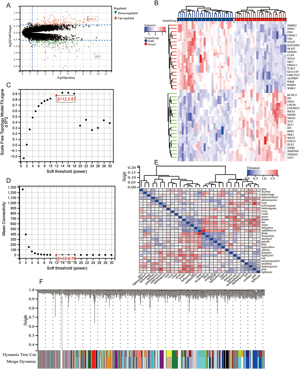 |
Figure 1 Differential analysis of transcriptome data and modular genes: (A) Differential gene volcano plot of transcriptome data, green triangles represent down regulated genes, red triangles represent up regulated genes, and black dots represent genes with no significant differences. (B) Differential gene heatmap of transcriptome data, red represents upregulated genes, blue represents downregulated genes. (C) Scale Independence of modular genes, the red star symbol indicates the key soft threshold for filtering, β=12. (D) Mean linkage of modular genes, the red star symbol indicates the key soft threshold for filtering, β=12. (E) Modular feature vector clustering heatmap, (F) Factor clustering tree heatmap. |
Results of Screening for Genes Linked to Ferroptosis
Two hundred and five genes were found when ferroptosis upregulated genes and transcriptome expression profile data intersected. Following the PPI network’s construction and screening, four important genes—IL1B, TNF, ACL4, and NCOA4—that are increased by ferroptosis during intervertebral disc degeneration were found (Figure 2A and B). One hundred and sixty-two genes were obtained by combining ferroptosis downregulated genes with transcriptome expression profile data. Two important genes, GPX4 and FSP1, were shown to be downregulated by ferroptosis during intervertebral disc degeneration after the PPI network was constructed and screened (Figure 2C and D). Ten key genes were identified by combining the upregulated and downregulated ferroptosis genes found in degenerative intervertebral discs; the PPI network showed how these genes interacted with one another. The genes in question are TP53, IL6, HIF1A, SIRT1, TFRC, HMOX1, PIK3CA, SLC7A11, G6PD, and SIRT3 (Figure 2E and F). According to transcriptome data, these genes’ statistical significance was determined to be ACSL4, NCOA4, IL6, SIRT1, PIK3CA, and SLC7A11 (Figure 2G–I).
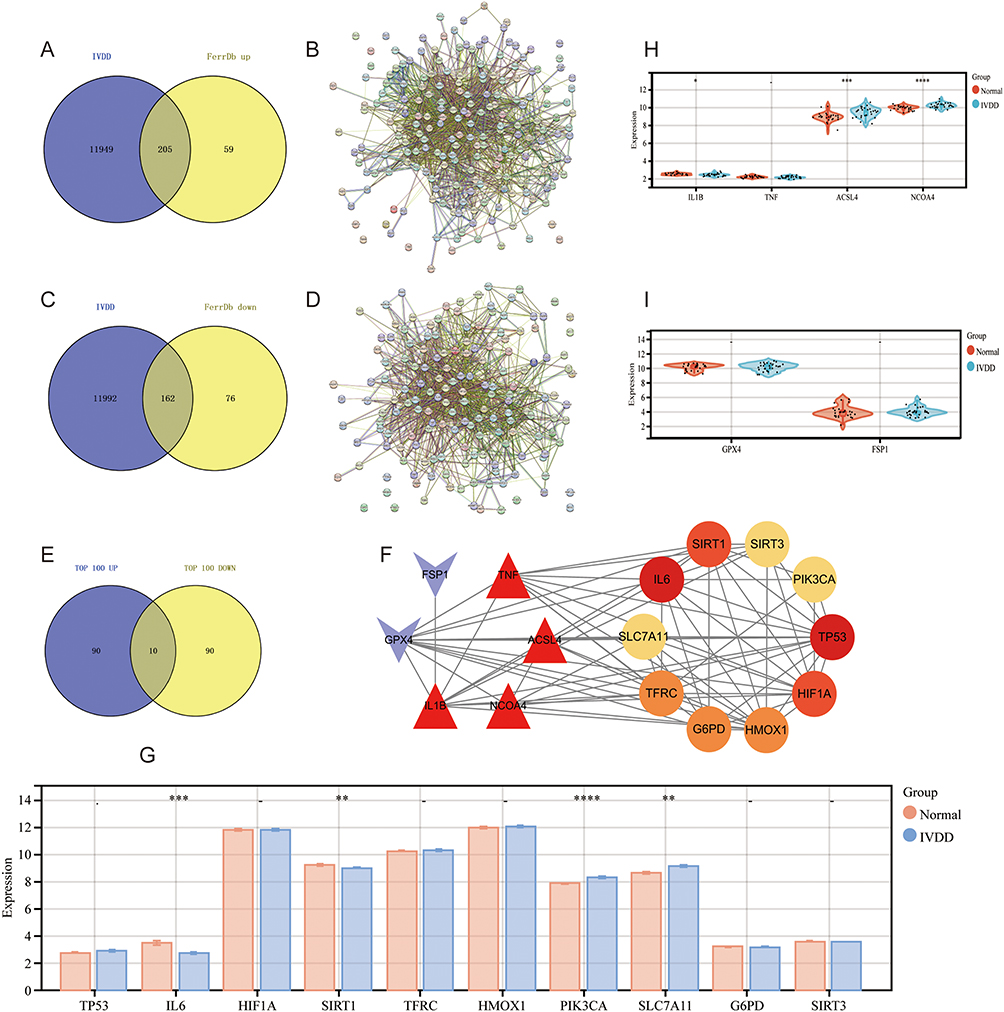 |
Figure 2 Intersection of transcriptome and Ferroptosis data: (A) Intersection of transcriptome data with Ferroptosis up-regulated genes, (B) Interaction plot of transcriptome data with Ferroptosis up-regulated genes and proteins, (C) Intersection of transcriptome data with Ferroptosis down-regulated genes, (D) Interaction plot of transcriptome data with Ferroptosis down-regulated genes and proteins, (E) Re-intersection of transcriptome data with Ferroptosis up-regulated and down-regulated genes, (F) Intervertebral disc degeneration Core genes of Ferroptosis during process, (G) Validation of core gene data of Ferroptosis during disc degeneration (**p<0.01, ***p<0.001, ****p<0.0001), (H) Validation of transcriptome data with core genes of Ferroptosis up-regulated genes (***p<0.001, ****p<0.0001), (I) Validation of transcriptome data with core genes of Ferroptosis down-regulated genes. (These are all applicable to (G–I) images, -, Indicates no statistical significance, *, **, Indicating statistical significance, ***, ****, Indicating significant statistical significance, ●, The black dots represent each sample, and there are as many samples as there are dots). |
Results of Core Gene Verification in Clinical Samples
The findings demonstrated that IVDD patients had considerably higher levels of IL1B, TNF-a, and IL6 (***P<0.001) (Figure 3A and B). Ferroptosis key indicators NCOA4 (*P<0.05) and ACSL4 (NS) increased in deteriorated tissue, while inflammatory factors IL1B (NS) and TNF-a (**P<0.01) rose during degeneration, according to Q-PCR detection based on nucleus pulposus tissue. Degenerated tissue showed a decrease in GPX4 (**P<0.01) and FSP1 (*P<0.05) (Figure 3C).
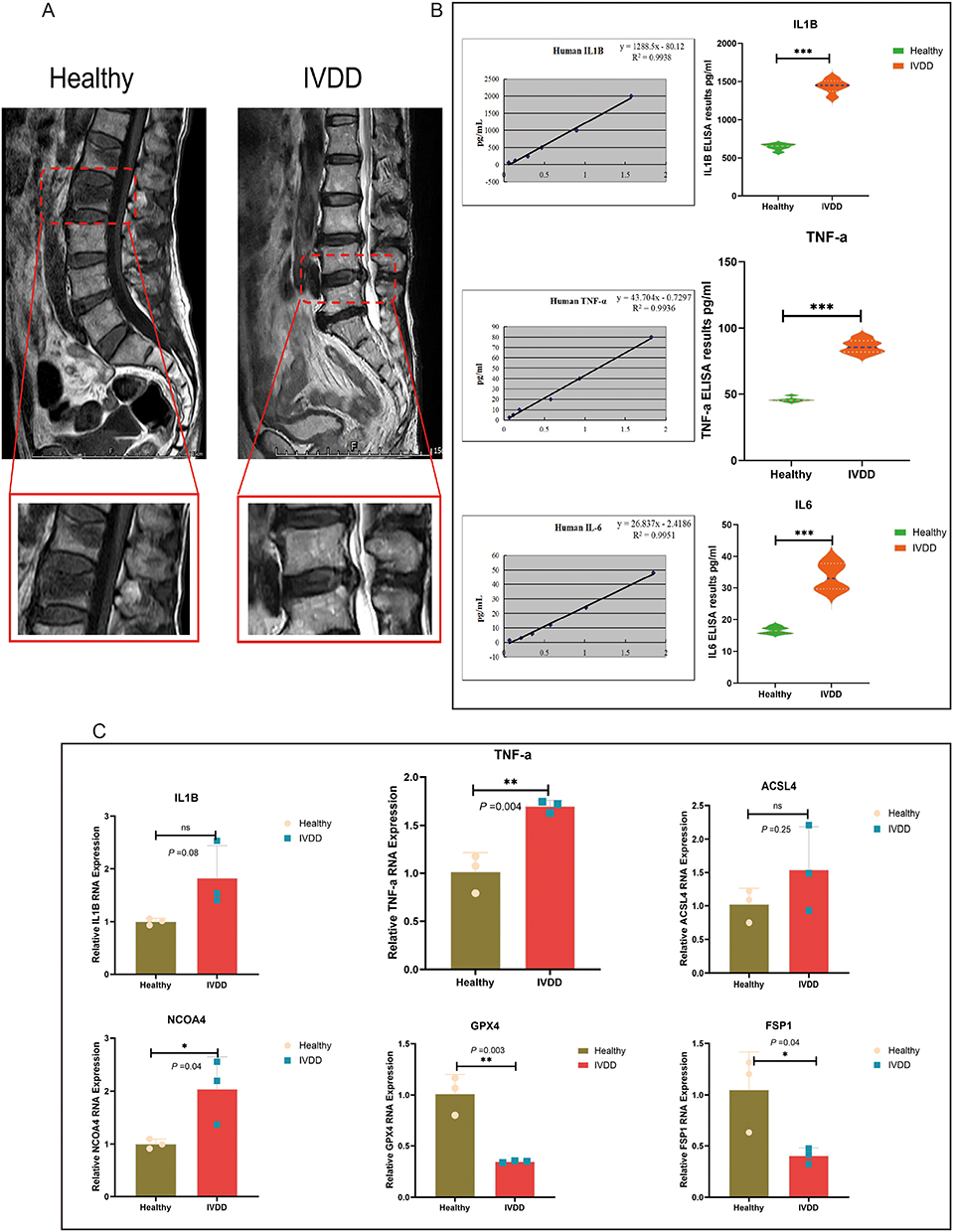 |
Figure 3 Clinical tissue validation of Ferroptosis and inflammatory factors: (A) MRI comparison between normal and degenerated discs, the red box represents the MRI image of the locally protruding nucleus pulposus tissue. (B) ELSA statistical analysis of inflammatory factors IL1B, TNF-a, IL6, (C) Tissue Q-PCR statistical analysis of inflammatory factors IL1B, TNF-a, Ferroptosis genes NCOA4, ACSL4, GPX4, FSP1. (*p<0.05, **p<0.01, ***p<0.001). Abbreviation: ns, no statistical significance. |
Results of Ferroptosis in Intervertebral Discs Using Bioinformatics
The IVDD process encompasses 286 signaling pathways, according to KEGG pathway analysis, and screening demonstrated that ferroptosis and autophagy are the primary cellular processes. The FoxO signaling pathway is primarily involved in environmental information processing (Figure 4A and B). GO was enriched with 3319 pieces of information based on our enrichment analysis. According to the first ten enrichment results, biological processes primarily include intracellular iron ion balance, circadian rhythm regulation, positive transcription regulation by RNA polymerase II, negative regulation of cell death, and cell responses to hypoxia. Cytoplasm, nucleoplasm, mitochondria, chromatin, nucleus, extracellular vesicles, and cytoplasmic perinuclear region are the primary cellular components implicated (Figure 4C).
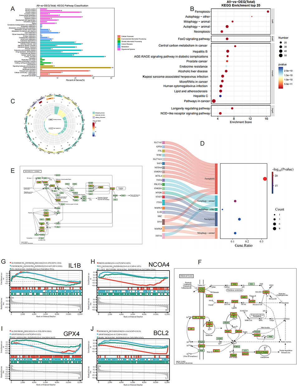 |
Figure 4 Bioinformatic analysis of intervertebral disc degeneration: (A) KEGG hierarchical information histogram of Ferroptosis hub genes during intervertebral disc degeneration, (B) KEGG signaling pathway bubble diagram of Ferroptosis hub genes during intervertebral disc degeneration, (C) GO chordogram of Ferroptosis hub genes during intervertebral disc degeneration, (D) Correlation diagram of Ferroptosis hub genes during intervertebral disc degeneration with autophagy and Ferroptosis signaling, (E) Correlation diagram of Ferroptosis hub genes with autophagy and Ferroptosis signaling, The protein marked with a red box is the core protein related to this study, (F) Ferroptosis signaling pathway and key proteins diagram, The protein marked with a red box is the core protein related to this study, (G) GSEA-enriched pathway diagram of IL1B, (H) GSEA-enriched pathway diagram of NCOA4, (I) GSEA-enriched pathway diagram of GPX4, (J) GSEA-enriched pathway diagram of BCL2. |
Ferroptosis, autophagy, and cross plots, as well as the primary targets of ferroptosis, demonstrated the important involvement of SLC3A2, GPX4, TFRC, NCOA4, and other proteins in the regulation of ferroptosis processes (Figure 4D). Lastly, we found by screening that ferroptosis and autophagy are tightly connected, and that NCOA4, a recently identified ferritinophagy autophagy regulation (referred to as ferritinophagy), may be important (Figure 4E and F). Furthermore, IL1B, NCOA4, GPX4, and BCL2 GSEA pathway analysis revealed that these targets are similarly implicated in autophagy, apoptosis, and P53 signaling regulation (Figure 4G–J).
Results of an Examination of Immune Infiltration
The stack plot revealed that the primary immune cells involved were CD4_naïve, CD8_nave, Cytotoxic, Exhausted, Trl, nTreg, iTreg, Th1, Th2, Th17, Monocyte, Macrophage, and 24 other immune cells (Figure 5A). We obtained the immune infiltration score of transcriptome genes using the CIBERSORT algorithm. Tfh, monocyte, and neutrophils penetrate degraded tissues, as the radar graphic illustrates. Additionally, the bar chart demonstrated a favorable relationship between intervertebral disc degeneration and monocytes, neutrophils, and macrophages (Figure 5A–C). These multi-level pieces of evidence collectively indicate that during the IVDD process, with the formation of new blood vessels and the release of chemotactic signals, the immune immune status of the intervertebral disc is disrupted, leading to specific infiltration of immune cells dominated by pro-inflammatory macrophages, thereby forming a pathological microenvironment that continuously amplifies inflammation and tissue destruction.
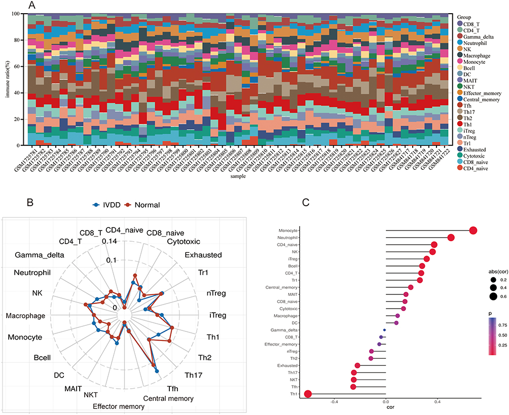 |
Figure 5 Immune infiltration analysis of transcriptome data: (A) Immune cell infiltration stacked plot in 24, (B) Immune cell infiltration radar plot, (C) Immune infiltration dot plot stick figure. |
Results of a Single-Cell Analysis
Cell clustering analysis was performed after single-cell data had been filtered and batch eliminated (Supplementary Figure 2A–C). Three moderately severe and three mildly deteriorated tissues were among the six tissues that were removed from this sample (Figure 6A and B). The distinctive genes of various subgroups are displayed on the marker gene point line distribution map (Figure 6C). We identified 11 cell subgroups using a literature search and TOP5 gene matching screening (Figure 6D). T-cells, macrophages, monocytes, B-cells, and other immune cells make up the majority of the cell population. Several NPC subtypes make up the tissue’s primary cells. Subsequent examination of developmental trajectories revealed that regulatory NPCs predominated in the early phases of intervertebral disc degeneration, but hypertrophic and fibrous NPCs emerged as the disease advanced (Figure 6E–I). Figure 7A–C illustrates how adhesive-like NPCs, fibroNPCs, homeostatic NPCs, and HT-CLNPs regulate the exchange of information between NPCs and different immune cells. Furthermore, IL6 and MIF signaling—particularly MIF signaling—may be crucial depending on the interaction between receptors and ligands (Figure 7D–H). These results are basically consistent with our previous research findings, especially the process of fibrosis in NPCs has been directly validated.1,3,33
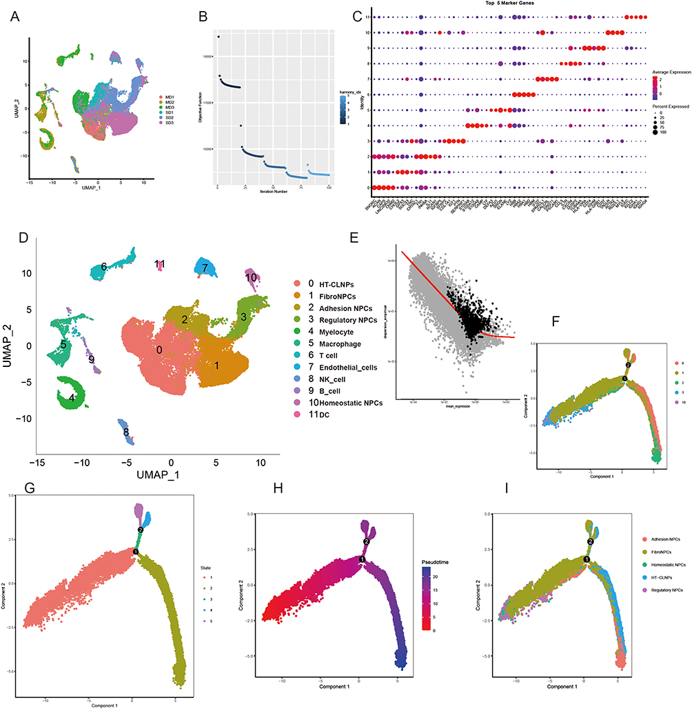 |
Figure 6 Analysis of single-cell data: (A) Tissue sample distribution of single cells, (B) De-batch QC plot, (C) Characteristic gene dot plot, (D) Single-cell subpopulation clustering annotation mapping, (E) Developmental trajectory dot plot, (F) Medullary cell developmental trajectory subtype categorization, (G) Medullary cell developmental trajectory state distribution, (H) Medullary cell developmental trajectory temporal heatmap, (I) Medullary cell developmental trajectory subtype annotation categorization. |
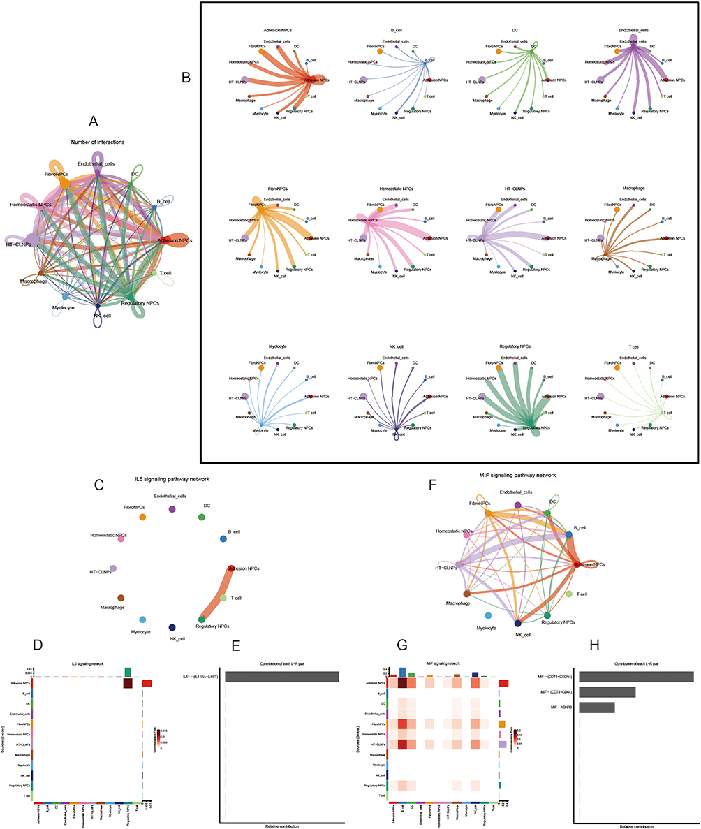 |
Figure 7 Single-cell data communication analysis: (A) Inter-cell communication links between all cells of a single-cell subpopulation, (B) Links between each type of cell of a single-cell subpopulation and other cells, (C) Subpopulations of cells predominantly involved in IL6 signaling, (D) Heatmap of subpopulations of cells involved in IL6 signaling, (E) Recipients and ligands of IL6 signaling, (F) Subpopulations of cells predominantly involved in MIF signaling, (G) MIF signaling involved in cells subpopulation heat map, (H) Ligands for MIF signaling. |
The enhancement of the IL6 signaling pathway mainly comes from stressed NPCs and cartilage endplate cells, with target cells including oneself and other NPCs, macrophages, etc, forming a positive feedback loop that promotes self amplification of inflammation. This finding is consistent with the upregulation of IL6 protein expression detected at the tissue level, but further clarifies its cellular origin, suggesting that this is an active, locally driven specific inflammatory dialogue. Of particular importance, the MIF signaling pathway mediates strong interactions between macrophages, NPCs, and cartilage endplate cells in degenerated tissues. MIF is a key factor known to drive macrophage polarization towards the classical pro-inflammatory (M1) phenotype. This analysis confirms that the activity of MIF signaling is highly co-localized with the expression of M1 macrophage markers. Due to the fact that M1 macrophages are the main source of reactive oxygen species and pro-inflammatory cytokines, which are the core upstream events inducing ferroptosis, the activity of the MIF signaling pathway specifically marks the establishment of an immune microenvironment that is prone to trigger ferroptosis in myeloid cells.
The above analysis indicates that specific reprogramming of intercellular communication occurs during intervertebral disc degeneration. The IL6 signal maintains and amplifies local inflammation, while the MIF signal directly bridges the link between immune cell polarization and the fate of NPCs (ferroptosis). These two pathways together form a specific signaling network that drives disease progression.
Results of a Combined Transcriptome and Single-Cell Analysis
We created bar charts and point graphs of several cell subpopulations in various deteriorated tissues based on single-cell data cell classification. The findings demonstrated that whilst regulatory NPCs, homestatic NPCs, and fibroNPCs were mostly expressed in slightly deteriorated tissues, T cells, monocytes, macrophages, DCs, and B cells were considerably expressed in moderately to severely degenerated tissues. This outcome is comparable to the cell’s developmental path (Figure 8A and B). Additionally, we created characteristic gene expression maps to better understand the roles played by the 10 ferroptosis hub genes in different cellular regulators. According to single gene UMAP analysis, monocytes and macrophages primarily regulate IL1B and TNF, but GPX4 is highly expressed in a variety of cells, including NPC and macrophage subtypes. The control of different NPC subtypes is also heavily influenced by ACSL4, SCL7A11, TFRC, SCL7A2, BCL2, and NCOA4; TFRC is also involved in the regulation of T cells, while NCOA4 is involved in the regulation of monocytes/macrophages and T cells. In conclusion, our finding raises the possibility that important proteins involved in ferroptosis and autophagy regulate NPC degeneration and immunological infiltration (Figure 8C–L).
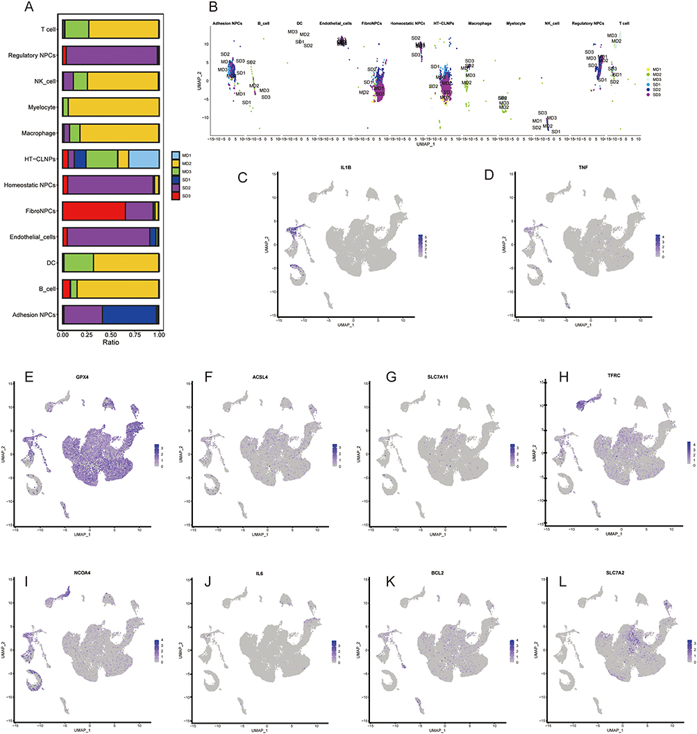 |
Figure 8 Combined analysis of transcriptome and single-cell data: (A) Histogram of the distribution of different cellular subpopulations in different degenerated disc tissues, (B) Hotspot map of the distribution of different cellular subpopulations in different degenerated disc tissues, (C–L) UMAP map of the distribution of the 10 Ferroptosis hub genes in single-cell subpopulations. |
Results of Resveratrol’s Improvement of Intervertebral Disc Degeneration Through Ferritin in Animal Experiments
The DHI index of intervertebral discs rose following modeling, and DHI recovered following resveratrol treatment with statistical significance, according to DR analysis conducted on animals (Figure 9A). Following acupuncture modeling, additional tissue section analysis showed that the model group rats’ central nucleus pulposus tissue had been considerably destroyed, and the organization of peripheral fibrosis had become disorganized. Following resveratrol treatment, the treatment group’s nucleus pulposus tissue saw a progressive partial recovery, a minor improvement in the organization of fibrosis, and a statistically significant tissue score (Figure 9B). Furthermore, we examined the inflammatory factors in the tissue and found that after modeling, inflammatory factors such as IL1B, TNF-a, and IL6 increased, while they decreased after treatment with resveratrol. The inhibition indicators of GPX4 and FSP1 ferroptosis suggest that ferroptosis occurs after modeling, and both indicators recover after treatment. Ferroptosis promotes an increase in the NCOA4 index after modeling, which improves after treatment (Figure 9C). In summary, from DR and tissue sections, we believe that resveratrol can improve intervertebral disc degeneration, and the possible mechanisms of action are related to reducing inflammatory factors, regulating ferroptosis, and autophagy. Our evaluation ensures objectivity through strict blinding. The therapeutic effect of resveratrol has statistical significance and consistency trend at the population level, with individual response differences within a reasonable range, and does not affect the validity of the core conclusion.
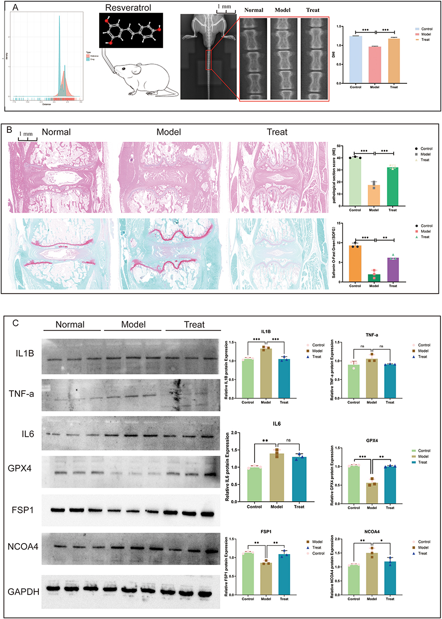 |
Figure 9 Animal experiments: (A) DR image analysis before and after resveratrol administration to rats by gavage, divided into normal, model, and treat groups, (B) Tissue section analysis before and after resveratrol administration to rats by gavage, divided into normal, model, and treat groups, (C) Tissue analysis of inflammatory factors and key proteins of Ferroptosis before and after resveratrol administration to rats by gavage, divided into normal, model, and treat groups. (*p<0.05, **p<0.01, ***p<0.001). Abbreviation: ns, no statistical significance. |
Results of Cell Experiments Showing That Resveratrol Improves Ferritinophagy-Mediated Intervertebral Disc Degeneration
Through CCK-8 assay, we analyzed that the optimal time and dosage for lipopolysaccharide and resveratrol intervention in NPCs were 10 μmol/L after 24 hours (Figure 10A). Through Western blot analysis of ferroptosis signaling pathway-related marker proteins, it was found that compared with the normal group, GPX4 and FSP1 decreased in the model group, while GPX4 increased in the ferroptosis inhibitor group and FSP1 did not show significant changes. After intervention with resveratrol, GPX4 and FSP1 increased in the model group. Compared with the normal group, NCOA4 and TFRC increased in the model group, while NCOA4 increased and TFRC decreased in the ferroptosis inhibitor group. After intervention with resveratrol, NCOA4 and TFRC decreased in the model group (Figure 10B). Only NCOA4 and IL1B exhibit statistical significance, despite the associated Q-PCR analysis trends being essentially constant. Furthermore, we identified important markers of P62, LC3, and BCL2 autophagy to confirm the pertinent mechanisms of autophagy. We discovered that these markers rose following modeling and fell following treatment, confirming the existence of autophagy in degenerated intervertebral discs and the ability of resveratrol to control the pertinent mechanisms (Figure 10C and D).
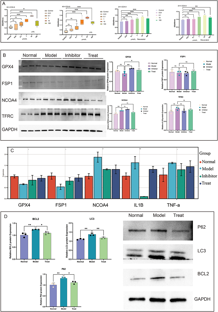 |
Figure 10 Results of cellular experiments: (A) Value-added results of lipopolysaccharide and resveratrol at 24 and 48 hours, (B) Protein expression of key proteins for Ferroptosis in normal, model, inhibitor and Treat groups, (C) RNA expression of key genes for Ferroptosis in normal, model, inhibitor and treatment groups, and (D) Protein expression of key markers for autophagy in normal, model and treatment groups. (*p<0.05, **p<0.01, ***p<0.001, These all indicate statistical significance; ns and -, no statistical significance, ●, ▲, The small dots and triangles in the image represent the sample data, n=3). |
To learn more about NCOA4’s role in autophagy regulation, we gathered cell confocal localization information for P62, LC3, BCL2, and NCOA4 from a human protein database. According to the findings, NCOA4 was mostly found in the cytoplasm, Golgi apparatus, and vesicles of cells, P62 was primarily found in the cytoplasm of cells, and LC3 and BCL2 were found in the vesicles of cells. These findings imply that BCL2, NCOA4, P62, and LC3 regulate autophagy via cellular vesicles and mitochondria. Lastly, immunofluorescence provided additional evidence of the elevated expression of NCOA4 in deteriorated tissues, whereas it declined following resveratrol treatment (Figure 11A and B).
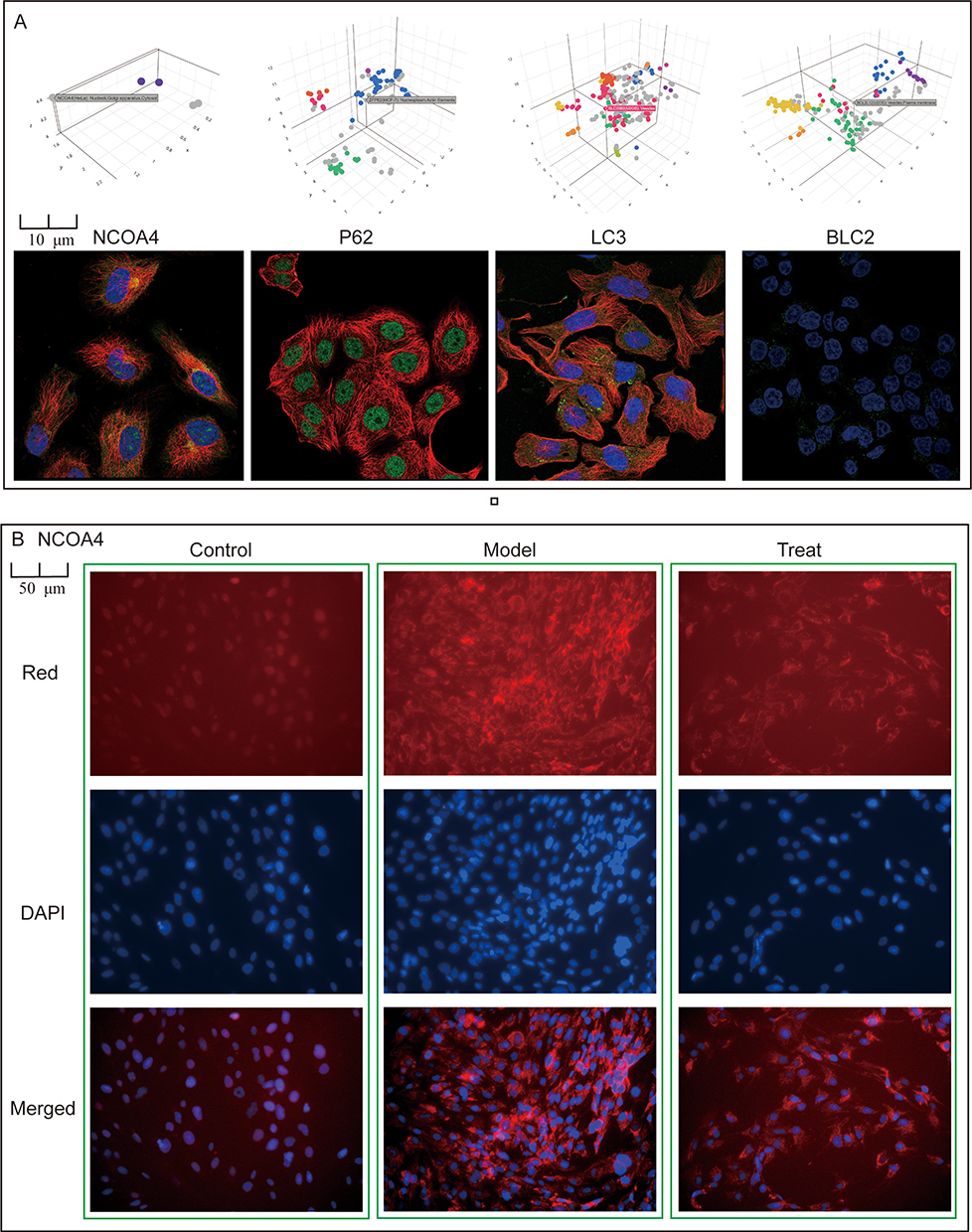 |
Figure 11 Analysis of key proteins of iron autophagy: (A) Confocal results of NCOA4, P62, LC3, BCL2, (B) Immunofluorescence of NCOA4 in normal group, model group, treatment group, The green box represents the same set of data. |
Discussion
IVDD is one of the major causes of chronic low back pain, and its pathogenesis involves multiple biological processes such as inflammatory response, cell death, and extracellular matrix metabolic disorders.34 In recent years, ferroptosis has received attention because it is closely related to lipid peroxidation, and the role of autophagy, as an important mechanism for cellular maintenance of homeostasis, has been progressively revealed in IVDD.35–37 However, the specific mechanism of the interaction between ferroptosis and autophagy (ie “ferritinophagy”) in IVDD is not yet clear. On the basis of confirming the involvement of ferroptosis, this study specifically focuses on its cross-linking mechanism with autophagy. By integrating multiple omics and experimental validations, we systematically elucidated that NCOA4 mediated ferritinophagy is a key upstream event driving ferroptosis in NPCs and confirmed that resveratrol can improve degeneration by precisely regulating this axis. This provides a direct basis for developing intervertebral disc protection strategies targeting iron homeostasis.
This study revealed through core data that NCOA4 is upregulated in degenerated intervertebral disc tissue, and its changes are closely related to ferroptosis, autophagy flow changes, and structural degeneration degree. Therefore, an innovative molecular hypothesis of the “NCOA4 ferritinophagy axis” as a novel and coherent mechanism in the process of IVDD was proposed. Animal experiments have shown that the therapeutic effect of resveratrol is accompanied by the regulation of this axis, providing a new perspective for its protective effect. However, the deepening of scientific understanding requires us to go beyond relevance and conduct a comprehensive and cautious analysis of the complexity of mechanisms, internal consistency, and transformation prospects.
Firstly, it is necessary to objectively evaluate the role positioning of NCOA4 in this study. Although multiple layers of evidence have jointly constructed a strong association between NCOA4 and disease progression, and functional acquisition experiments suggest that its overexpression is sufficient to induce degenerative phenotypes, the strictest causal relationship – that is, directly reversing or reproducing degeneration in in vitro and in vivo models through genetic manipulation specific to NPCs – still needs to be confirmed by future research. Therefore, the current work establishes NCOA4 as a highly promising core candidate target, and its position as a “hub” connecting ferroptosis and autophagy is still in the hypothesis verification stage, which is the primary scientific proposition derived from this study. Secondly, we fully recognize that resveratrol was selected as a concept validation tool compound in this study, and its known upstream pathways (such as SIRT1, AMPK, NF-κB) may independently or synergistically contribute to overall efficacy. The existing data cannot accurately analyze whether these classical signals converge upstream of NCOA4 or act in parallel with it. This suggests that the NCOA4 axis may be an important downstream effect link for resveratrol to exert its benefits. Clarifying its hierarchical relationship in the drug action network is the key to extracting precise targeting strategies from pleiotropic natural products.
Based on existing findings, we attempt to construct an integrated pathological network framework to reconcile different observations. We believe that they form a mutually nourishing driving cycle. On the one hand, the inflammatory microenvironment created by M1 macrophages recruited by signals such as MIF is a classic upstream factor in inducing ferroptosis. On the other hand, the specific damage related molecular patterns released by ferroptosis cells (such as oxidized phospholipids) can strongly activate immune responses, thereby forming a self amplifying loop of “inflammation ferroptosis chronic inflammation”. This may be one of the intrinsic driving forces for the sustained progression of IVDD once it is initiated. Under this framework, the transformation of NPCs into fibrotic phenotype can be regarded as the downstream structural output of this pathological network. The loss of cellular functionality caused by ferroptosis, as well as the lipid peroxidation signals and intracellular iron overload generated during the process, collectively disrupt tissue homeostasis, which may prompt residual cells to initiate dysregulated repair programs and shift towards a myofibroblast-like-phenotype that secretes abnormal matrix. Therefore, our intervention study can be interpreted as: by regulating the NCOA4 node, it may simultaneously act on multiple levels of ferroptosis, inflammatory vicious cycle, and abnormal repair, thereby producing a synergistic protective effect. This perspective also preliminarily integrates immune microenvironment signals (such as MIF) with intracellular execution events (ferroptosis) through logical cascades rather than direct molecular connections, although precise signal transduction bridges between the two are still directions that need to be explored in the future.
Previous studies have found that RBX1 inhibits NCOA4 mediated ferritinophagy to alleviate ferroptosis, Polydatin improves asthma by inhibiting ferritinophagy, Resveratrol activates autophagy to protect intervertebral discs, and the interaction between ferroptosis and mitochondrial autophagy plays an important role in IVDD.7,19,38 However, it has not been found yet that resveratrol can improve IVDD by regulating ferroptosis in NPCs through NCOA4. Therefore, this study innovatively confirmed in the IVDD model that there is a significant upregulation of NCOA4 in degenerated nucleus pulposus tissue, which systematically drives ferroptosis of NPCs by activating iron autophagy leading to excessive degradation of ferritin and accumulation of toxic iron ions. More importantly, we found that the natural compound resveratrol can effectively inhibit this pathological axis. This discovery not only deepens our understanding of the cell death mechanism in IVDD, but also extends the protective effect of resveratrol from regulating mitochondrial autophagy and ferroptosis to precise regulation of iron homeostasis, providing new experimental evidence for the development of intervertebral disc protection strategies targeting “ferritinophagy”. This series of findings prompts us to rethink our therapeutic strategies for autophagy. This study proposes to inhibit NCOA4 mediated ferritin autophagy to generate benefits, which may seem contradictory to the universal protective function of autophagy, but in fact reveals the fate bifurcation of the autophagy pathway under pathological conditions. In the specific microenvironment of IVDD, the protective autophagy function that clears damaged organelles may be impaired, while the specific branch of ferritin autophagy dependent on NCOA4 is abnormally activated, becoming a key driver of ferroptosis. Therefore, the essence of our strategy is to pursue precise regulation of autophagy types, rather than comprehensive inhibition of autophagy levels. The data also supports this distinction, that is, no general blockade of autophagy flow was observed during the intervention. This provides a conceptual basis for future treatment development to shift from extensive regulation to precise targeting.
Limitations and Future Perspectives
Although this study proposes insightful new hypotheses, there are still multi-level limitations that point the way for future research.
Model and evidence depth: The study mainly relies on LPS induced acute inflammation models, which differ from the chronic degeneration process in the human body. In the future, further validation is needed in models that are closer to physiological and pathological states, such as aging, mechanical loading, or surgical induction models. At the mechanistic level, although support has been provided through protein expression profiling and inhibitor experiments, there is still a lack of direct detection of lipid peroxidation end products and intracellular free iron concentration, which are key direct evidence for validating this pathway. Meanwhile, the study mainly used the ferroptosis inhibitor Ferrostatin-1 as a mechanism control, and in the future, other known protective drugs need to be introduced for “head to head” comparison to objectively evaluate the therapeutic potential of targeting this axis.
Data and statistical aspects: Heterogeneity in retrospective public databases may lead to potential biases. Although the sample size of animal experiments (n=10 per group) conforms to common norms in the field, it may limit the statistical power of detecting small effect values. In the future, a more optimal sample size can be determined through formal efficacy analysis. The multiple potential genes identified in early exploratory transcriptome screening were subsequently validated by focusing on a few key targets without conducting genome-wide multiplex testing correction. Therefore, the proposed mechanism network should be considered as a framework for generating hypotheses, and its core conclusions have been specifically validated through subsequent independent molecular biology experiments.
Transformation challenges and pathways: The low oral bioavailability and fast metabolism of resveratrol are bottlenecks in clinical applications, but the core value of this study lies in verifying the effectiveness of the targeted iron autophagy ferroptosis axis strategy. The future transformation pathway should focus on: firstly, screening or designing new small-molecule compounds with better pharmacokinetics based on this target; second, develop local delivery systems (such as injectable hydrogels and drug loaded nanoparticles) for the relatively closed anatomical structure of intervertebral discs to completely avoid the absorption and metabolism of systemic drug delivery, which may be the most promising path to achieve clinical transformation. In addition, the potential impact of long-term systemic intervention with NCOA4 on systemic iron homeostasis, determination of the optimal treatment window, and search for relevant biomarkers are all core issues that must be systematically addressed before moving towards clinical application.
In summary, this study systematically proposed the central hypothesis of NCOA4 ferritin autophagy axis in the IVDD pathological network, and logically integrated it with key links such as ferroptosis, chronic inflammation, and fibrosis remodeling. At the same time, it preliminarily correlated the therapeutic effect of resveratrol, opening up a new dimension for understanding the mechanism of intervertebral disc degeneration and proposing novel therapeutic intervention ideas. Future research should be conducted along two main lines: one is to deepen towards the essence of the mechanism, establish strict causal relationships through tools such as genetics, and draw a complete signal map; the second is to advance towards the reality of transformation, systematically addressing core obstacles such as target safety, delivery technology, and precision medicine strategies.
Conclusion
This study investigated the interaction between ferroptosis and autophagy in IVDD by integrating transcriptome and single-cell sequencing analyses. A set of ten core genes associated with this interaction, including GPX4 and NCOA4, were identified. The data suggest that NCOA4-mediated ferritin autophagy, evidenced by its colocalization with LC3/P62, may contribute to lipid peroxidation and the disruption of cellular homeostasis. Single-cell analysis implied a potential role for IL6/MIF signaling in modulating NPCs subtype transitions and immune cell infiltration, processes that appeared enhanced during degeneration. In vitro and in vivo experiments indicated that resveratrol, potentially via modulating the ferritinophagy pathway, could alleviate IVDD phenotypes, ameliorate aberrant ferroptosis-related markers (GPX4/NCOA4), and reduce the levels of inflammatory factors (IL1B/TNF-α/IL6). Collectively, this preclinical study highlights the concept of “ferritinophagy” as a potential novel target for IVDD treatment, while acknowledging that its precise mechanism and clinical translation require substantial future exploration.
Data Sharing Statement
All data are in the manuscript and/or supporting information files. If you need raw data, please contact the corresponding author.
Ethics Approval and Consent to Participate
After obtaining informed consent from patients and approval from the Ethics Committee of Ruikang Hospital, Guangxi University of Traditional Chinese Medicine, intervertebral disc tissue and blood samples were used in this study. All of our studies comply with the Helsinki Declaration and have provided comprehensive local clinical ethics applications and obtained approval. The ethical approval number for this study is GJJ19041. This experiment has been approved by the Medical Laboratory Animal Management Committee, with ethics code: DW20250319-065.
Acknowledgments
Thank you KEGG online platform for providing the raw data. We thank the BioBean (Sheng-Xin-Dou-Ya-Cai) team for providing the user-friendly bioinformatics platform (http://www.sxdyc.com/), which has significantly streamlined and accelerated our research process. Thank Sichuan Vista Medical Devices Co., Ltd.
Author Contributions
All authors made a significant contribution to the work reported, whether that is in the conception, study design, execution, acquisition of data, analysis and interpretation, or in all these areas; took part in drafting, revising or critically reviewing the article; gave final approval of the version to be published; have agreed on the journal to which the article has been submitted; and agree to be accountable for all aspects of the work.
Funding
The present study was supported in part by research grants from 2025 Traditional Chinese Medicine (TCM) Scientific Research Special Project of Sichuan Provincial Administration of Traditional Chinese Medicine (25ZDIZX029), Science and Technology Strategic Cooperation between Xuyong County People’s Hospital and Southwest Medical University (2024) (No. 2024XYXNYD06), Southwest Medical University 2025 College Student Innovation and Entrepreneurship Training Program (202510632082), the Construction Plan for Specialized Disease and Specialty Peak Projects (Document No. 99 of Southwest Medical University Hospital of Traditional Chinese Medicine (2024)), 2025 Luzhou Medical Association Research Project (2025-YXX-KY-M-072), Sichuan Vista Medical Devices Co., Ltd. and the Affiliated Hospital of Traditional Chinese Medicine of Southwest Medical University Collaborative Research Project, Southwest Medical University (SWMU) School-level Scientific Research Program (nos. 2024ZKZ009). All the funding’s funder was Zongchao Liu, the funder had role in study conceptualization, methodology, supervision, funding acquisition.
Disclosure
The authors declare that they have no conflicts of interest in this work.
References
1. Song C, Wu X, Chen C, et al. Single-cell analysis integrated with machine learning elucidates the mechanisms of nucleus pulposus cells apoptosis in intervertebral disc degeneration and therapeutic interventions. JOR Spine. 2025;8(1):e70036. doi:10.1002/jsp2.70036
2. Wang X, Song C, Zhou D, et al. Exploring the therapeutic potential of puerarin on intervertebral disc degeneration by regulating apoptosis of nucleus pulposus cells. JOR Spine. 2024;7(4):e70020. doi:10.1002/jsp2.70020
3. Song C, Liu F, Wu X, et al. Molecular mechanism of macrophage polarization regulating the cell senescence of nucleus pulposus during intervertebral disc degeneration. Int Immunopharmacol. 2025;149:114131. doi:10.1016/j.intimp.2025.114131
4. Song C, Liu F, Wu X, et al. ASIC1a mediated nucleus pulposus cells pyroptosis and glycolytic crosstalk as a molecular basis for intervertebral disc degeneration. Inflamm Res. 2025;74(1):29. doi:10.1007/s00011-025-02003-w
5. Guo D, Cheng K, Song C, et al. Mechanisms of inhibition of nucleus pulposus cells pyroptosis through SDF1/CXCR4-NFkB-NLRP3 axis in the treatment of intervertebral disc degeneration by Duhuo Jisheng Decoction. Int Immunopharmacol. 2023;124(Pt A):110844. doi:10.1016/j.intimp.2023.110844
6. Zhou D, Mei Y, Song C, et al. Exploration of the mode of death and potential death mechanisms of nucleus pulposus cells. Eur J Clin Invest. 2024;54(9):e14226. doi:10.1111/eci.14226
7. Zhou LP, Kang L, Zhang Z-G, et al. RBX1 mitigates ferroptosis by inhibiting NCOA4-mediated ferritinophagy and contributes to the attenuation of intervertebral disc degeneration. J Transl Med. 2025;23(1):514. doi:10.1186/s12967-025-06412-7
8. Mei Y, Wang L, Chen T, et al. Ferroptosis: a new direction in the treatment of intervertebral disc degeneration. Cell Biochem Biophys. 2025;83(1):33–26. doi:10.1007/s12013-024-01468-6
9. Liu S, Yao S, Yang H, et al. Autophagy: regulator of cell death. Cell Death Dis. 2023;14(10):648. doi:10.1038/s41419-023-06154-8
10. Klionsky DJ, Petroni G, Amaravadi RK, et al. Autophagy in major human diseases. EMBO J. 2021;40(19):e108863. doi:10.15252/embj.2021108863
11. Sun K, Shi Y, Yan C, et al. Glycolysis-derived lactate induces ACSL4 expression and lactylation to activate ferroptosis during intervertebral disc degeneration. Adv Sci. 2025;12:e2416149. doi:10.1002/advs.202416149
12. Zhang F, Cui D, Wang Z, et al. NOX4 regulates NLRP3 by inhibiting the ubiquitination of LRRC8A to promote ferroptosis in nucleus pulposus cells. Inflammation. 2025;48:3111–3129. doi:10.1007/s10753-025-02253-0
13. Lin J, Zheng X, Xiong Z, et al. DJ-1-mediated p62 degradation delays intervertebral disc degeneration by inhibiting apoptosis of nucleus pulposus cells. Apoptosis. 2023;28(9–10):1357–1371. doi:10.1007/s10495-023-01862-0
14. Fang Y, Chen X, Tan Q, et al. Inhibiting ferroptosis through disrupting the NCOA4-FTH1 interaction: a new mechanism of action. ACS Cent Sci. 2021;7(6):980–989. doi:10.1021/acscentsci.0c01592
15. Santana-Codina N, Mancias JD. The role of NCOA4-mediated ferritinophagy in health and disease. Pharmaceuticals. 2018;11(4):114. doi:10.3390/ph11040114
16. Ao X, Jiang T, Li Y, et al. n-3 polyunsaturated fatty acids delay intervertebral disc degeneration by inhibiting nuclear receptor coactivator 4-mediated iron overload. iScience. 2024;27(2):108721. doi:10.1016/j.isci.2023.108721
17. Chen J, Luo Y, Li F, et al. The pivotal role and intervention strategies of BMAL1 mediated circadian clock dysregulation in intervertebral disc degeneration. Inflamm Res. 2026;75(1):10. doi:10.1007/s00011-025-02167-5
18. Chen C, Wu X, Shen B, et al. Crosstalk between reactive oxygen species mediated programmed cell death of nucleus pulposus cells. Tissue Cell. 2025;97:103054. doi:10.1016/j.tice.2025.103054
19. Zhang B, Xu L, Zhuo N, et al. Resveratrol protects against mitochondrial dysfunction through autophagy activation in human nucleus pulposus cells. Biochem Biophys Res Commun. 2017;493(1):373–381. doi:10.1016/j.bbrc.2017.09.015
20. Li W, Tang Y, Liu W, et al. Polydatin ameliorates ovalbumin-induced asthma in a rat model through NCOA4-mediated ferroautophagy and ferroptosis pathway. FEBS Open Bio. 2025;15(11):1800–1813. doi:10.1002/2211-5463.70090
21. Kazezian Z, Gawri R, Haglund L, et al. Gene expression profiling identifies interferon signalling molecules and IGFBP3 in human degenerative annulus fibrosus. Sci Rep. 2015;5:15662. doi:10.1038/srep15662
22. He J, Xue R, Li S, et al. Identification of the potential molecular targets for human intervertebral disc degeneration based on bioinformatic methods. Int J Mol Med. 2015;36(6):1593–1600. doi:10.3892/ijmm.2015.2389
23. Huang J, Zhang J-L, Ang L, et al. Proposing a novel molecular subtyping scheme for predicting distant recurrence-free survival in breast cancer post-neoadjuvant chemotherapy with close correlation to metabolism and senescence. Front Endocrinol. 2023;14:1265520. doi:10.3389/fendo.2023.1265520
24. Zhou N, Yuan X, Du Q, et al. FerrDb V2: update of the manually curated database of ferroptosis regulators and ferroptosis-disease associations. Nucleic Acids Res. 2022;51(D1):D571–D582. doi:10.1093/nar/gkac935
25. Zhao C, Sahni S. String correction using the Damerau-Levenshtein distance. BMC Bioinf. 2019;20(Suppl 11):277. doi:10.1186/s12859-019-2819-0
26. Doncheva NT, Morris JH, Gorodkin J, et al. Cytoscape StringApp: network analysis and visualization of proteomics data. J Proteome Res. 2019;18(2):623–632. doi:10.1021/acs.jproteome.8b00702
27. Kanehisa M, Furumichi M, Sato Y, et al. KEGG for taxonomy-based analysis of pathways and genomes. Nucleic Acids Res. 2023;51(D1):D587–d592. doi:10.1093/nar/gkac963
28. Tian K, Qi W, Yan Q, et al. Signature constructed by glycolysis-immune-related genes can predict the prognosis of osteosarcoma patients. Invest New Drugs. 2022;40(4):818–830. doi:10.1007/s10637-022-01228-4
29. Stuart T, Satija R. Integrative single-cell analysis. Nat Rev Genet. 2019;20(5):257–272. doi:10.1038/s41576-019-0093-7
30. Song C, Liu F, Mei Y, et al. Integrated metagenomic and metabonomic mechanisms for the therapeutic effects of Duhuo Jisheng decoction on intervertebral disc degeneration. PLoS One. 2024;19(10):e0310014. doi:10.1371/journal.pone.0310014
31. Inoue H, Ohmori K, Miyasaka K, et al. Radiographic evaluation of the lumbosacral disc height. Skeletal Radiol. 1999;28(11):638–643. doi:10.1007/s002560050566
32. Colwill K, Gräslund S. A roadmap to generate renewable protein binders to the human proteome. Nat Methods. 2011;8(7):551–558. doi:10.1038/nmeth.1607
33. Song C, Zhou D, Chen C, et al. Integrating multiple omics analyses to elucidate the macrophage-driven “immune cellular senescence fibrosis” axis mechanism in intervertebral disc degeneration. Int J Surg. 2026. doi:10.1097/JS9.0000000000004695
34. Song C, Zhou Y, Cheng K, et al. Cellular senescence - Molecular mechanisms of intervertebral disc degeneration from an immune perspective. Biomed Pharmacother. 2023;162:114711. doi:10.1016/j.biopha.2023.114711
35. Song C, Xu Y, Peng Q, et al. Mitochondrial dysfunction: a new molecular mechanism of intervertebral disc degeneration. Inflamm Res. 2023;72(12):2249–2260. doi:10.1007/s00011-023-01813-0
36. Song C, Cai W, Liu F, et al. An in-depth analysis of the immunomodulatory mechanisms of intervertebral disc degeneration. JOR Spine. 2022;5(4):e1233. doi:10.1002/jsp2.1233
37. Liu F, Chao S, Yang L, et al. Molecular mechanism of mechanical pressure induced changes in the microenvironment of intervertebral disc degeneration. Inflamm Res. 2024;73(12):2153–2164. doi:10.1007/s00011-024-01954-w
38. Zhou Y, Mei Y, Wang H, et al. Targeting mitophagy and ferroptosis: a new direction for the treatment of intervertebral disc degeneration. Tissue Cell. 2026;98:103227. doi:10.1016/j.tice.2025.103227
 © 2026 The Author(s). This work is published and licensed by Dove Medical Press Limited. The
full terms of this license are available at https://www.dovepress.com/terms
and incorporate the Creative Commons Attribution
- Non Commercial (unported, 4.0) License.
By accessing the work you hereby accept the Terms. Non-commercial uses of the work are permitted
without any further permission from Dove Medical Press Limited, provided the work is properly
attributed. For permission for commercial use of this work, please see paragraphs 4.2 and 5 of our Terms.
© 2026 The Author(s). This work is published and licensed by Dove Medical Press Limited. The
full terms of this license are available at https://www.dovepress.com/terms
and incorporate the Creative Commons Attribution
- Non Commercial (unported, 4.0) License.
By accessing the work you hereby accept the Terms. Non-commercial uses of the work are permitted
without any further permission from Dove Medical Press Limited, provided the work is properly
attributed. For permission for commercial use of this work, please see paragraphs 4.2 and 5 of our Terms.