
Back to Journals » Journal of Blood Medicine » Volume 16
Respiratory Syncytial Virus Vaccine Induced Thrombotic Microangiopathy
Authors Seby R  , McCormick BJ, Wolf E , Kuhlman J , Jhawar N, Oman SP , Kase AM , Christenson C, Shaikh M
Received 17 August 2024
Accepted for publication 7 July 2025
Published 22 July 2025 Volume 2025:16 Pages 331—335
DOI https://doi.org/10.2147/JBM.S478230
Checked for plagiarism Yes
Review by Single anonymous peer review
Peer reviewer comments 2
Editor who approved publication: Dr Martin H Bluth
Robert Seby,1 Benjamin J McCormick,1 Emily Wolf,2 Justin Kuhlman,2 Nikita Jhawar,1 Sven Peter Oman,1 Adam M Kase,2 Chancey Christenson,3 Marwan Shaikh2
1Department of Internal Medicine, Mayo Clinic, Jacksonville, FL, USA; 2Department of Hematology/Oncology, Mayo Clinic, Jacksonville, FL, USA; 3Department of Pathology and Transfusion Medicine, Mayo Clinic, Jacksonville, FL, USA
Correspondence: Robert Seby, Email [email protected]
Abstract: Microangiopathic hemolytic anemia with associated multiorgan failure is a medical emergency. The differential diagnosis for microangiopathic hemolytic anemia is broad and requires a systematic, focused approach at ruling out serious causes. However, with the rise of new vaccines and sporadic reports of vaccine-induced microangiopathic hemolytic anemia, it is essential for providers to include vaccines as a potential cause on their differential diagnosis. Here, we report the first case of respiratory syncytial virus vaccine-induced microangiopathic hemolytic anemia, anuric renal failure, and metabolic encephalopathy in the world.
Keywords: microangiopathic hemolytic anemia, respiratory syncytial virus, renal failure, metabolic encephalopathy
Introduction
New onset microangiopathic hemolytic anemia with anuric renal failure and metabolic encephalopathy is a medical emergency and requires concomitant swift medical decision-making and prompt treatment of suspected etiology. The differential diagnosis for microangiopathic hemolytic anemia is broad and include etiologies like thrombotic thrombocytopenia purpura (TTP), atypical hemolytic uremic syndrome (aHUS), and disseminated intravascular coagulation (DIC). In May 2023, the Food and Drug Administration (FDA) approved Arexvy as the first respiratory syncytial virus (RSV) vaccine in patients above age 60. Current Arexvy RSV vaccine data sheets do not report thrombocytopenia or microangiopathic hemolytic anemias as known adverse events. Here, we present the case of a 91-year-old male who developed new microangiopathic hemolytic anemia, anuric renal failure and metabolic encephalopathy within 24 hours of administration of the new Arexvy RSV vaccine.
Case Presentation
Initial Presentation
A 91-year-old retired male presented to an outside hospital with severe low back pain and urinary retention. He received the new adjuvant RSV vaccine, known as Arexvy, the day prior at a local pharmacy. His past medical history included coronary artery disease status post bypass grafting, bovine aortic valve replacement, sick sinus syndrome status post pacemaker placement, hypertension, paroxysmal atrial fibrillation, heart failure with preserved ejection fraction, chronic thoracic aortic aneurysm, chronic anemia, and stage IV prostate cancer status post retro pelvic prostatectomy followed by radiation therapy with subsequent recurrence with extensive bony metastases. Pertinent outpatient medications included abiraterone acetate, prednisone (5 mg daily), and leuprolide acetate. He had not recently started any other new medications or herbal supplements. He had received the coronavirus-19 (COVID-19) Moderna vaccine booster 1 month prior to admission and he had received the Influenza vaccine 3 weeks prior to admission. He had baseline Karnofsky and Eastern Cooperative Oncology Group performance scores of 90% and 0, respectively. At the outside institution, he was found to have metabolic encephalopathy and renal failure. His initial labs showed white blood cell count of 7.7×10⁹/L, hemoglobin 12.4 g/dL, and platelet count of 112×109/L. His complete metabolic profile was normal, except for a creatinine 1.3 mg/dL, and total bilirubin of 2.2 mg/dL. Baseline labs from seven weeks prior were normal including a hemoglobin of 13 g/dL and a platelet count 201×109/L. His last available total bilirubin three months prior was normal. During his hospitalization, his renal failure (Cr 4.1 mg/dL) and encephalopathy worsened, his hemoglobin and platelets continued to decrease, and his AST increased to 95 U/L with normal ALT at 36 U/L. Due to clinical deterioration with no clear diagnosis, he was transferred to our institution for further evaluation.
Upon arrival at our institution, he was afebrile with pulse rate 60 bpm, respiratory rate 19/min, blood pressure 191/108 mmHg, and acute hypoxic respiratory failure requiring 5 L Oxymask to maintain SpO2 > 90%. The physical exam was significant for bibasilar crackles in the lungs, jugular venous distention, trace pedal edema, asterixis, ecchymosis in the left upper extremity, and acute metabolic encephalopathy (Glascow Coma Score 9).
Laboratory findings include the following (reference ranges listed parenthetically): hemoglobin, 9 g/dL (13.2–16.6 g/dL); platelet count, 10×109/L (135–317×109/L); white blood cell count, 7.1×10⁹/L (3.4–9.6 × 10⁹/L); creatinine, 5.15 mg/dL (0.75–1.34 mg/dL); total bilirubin, 2.5 mg/dL (<1.2 mg/dL); direct bilirubin, 1.0 mg/dL (0–0.3 mg/dL); lactate dehydrogenase, 3080 U/L (122–222 U/L); D-dimer, 33,652 ng/mL (<500 ng/mL); haptoglobin, <14 mg/dL (30–200 mg/dL); immature platelet fraction, 15.7% (1–7%). Prothrombin time, activated partial thromboplastin time, and fibrinogen were within normal limits. Soluble fibrin monomer was positive. Complement levels on admission included a normal C3 and C4. Chest x-ray revealed bilateral pulmonary edema. Transthoracic echocardiography (TTE) was obtained to assess for the Waring Blender effect, which revealed no prosthetic or periprosthetic regurgitation.
Differential Diagnosis
The differential diagnosis on admission for this patient with new-onset microangiopathic hemolytic anemia with anuric renal failure and metabolic encephalopathy included TTP, aHUS, disseminated intravascular coagulation (DIC), Waring-Blender syndrome secondary to paravalvular regurgitation, vaccine-induced thrombotic microangiopathy (TMA), or other causes of TMA. Non-TMA etiologies to explain the thrombocytopenia included vaccine-induced immune thrombotic thrombocytopenia (VITT) and heparin-induced thrombocytopenia (HIT).
Heparin platelet factor 4 (PF4) antibody and Coombs test were negative. Peripheral smear (Figure 1) revealed schistocytes (>10/hpf) and burr cells. ADAMTS13 and aHUS/TMA panels were obtained.
 |
Figure 1 Peripheral smear on day of admission reveals significant schistocytosis (>10 schistocytes per high power field). |
Management
Due to concern for TTP with multiorgan failure, plasma exchange (PLEX) and hemodialysis were promptly initiated. Given his stable respiratory status and no severe electrolyte abnormalities, PLEX initiation was prioritized over hemodialysis. After the initial PLEX session, the patient demonstrated significant improvement in mental status. Intermittent hemodialysis and prednisone (60 mg daily) were started subsequently. Rituximab therapy was held pending the ADAMTS13 result. The patient received eight sessions of PLEX in total with complete recovery of mental status to baseline within 48 hours. Prednisone (60 mg daily) was administered daily with PLEX until platelets reached 150×109/L for 3 days, after which steroids were tapered over the next few weeks.
ADAMTS13 activity results sent before PLEX returned at 67% (normal > 70%) essentially ruling out a diagnosis of TTP, so rituximab was not administered. AHUS/TMA/complement panel returned with the following lab values: total complement 60 (normal 30–75 U/mL), C3 84 (normal 75–175 mg/dL) C4 28 (normal 14–40 mg/dL), factor B complement antigen 36 (normal 30–75 mg/dL), factor H complement antigen 24.7 (normal 18.5–40.8 mg/dL), alternate complement pathway function >110% (normal > 46%), SC5b-9 complement 597 (normal <251 ng/dL), CBb complement >6 (normal <1.7 mcg/dL), and C4d complement 2.7 (normal <9.9 mcg/dL). Since this panel was not consistent with active alternative complement pathway activation and given the clinical improvement with PLEX, the patient was not started on eculizumab. On the day of discharge (day 11), platelet count was normal at 267×109/L. By discharge, his renal function had not improved, and he still required hemodialysis.
After discharge, his atypical HUS gene panel results showed a double heterozygote status for CFHR1 exon 2–6 deletion, and CFHR3 exon 1–6 deletion. These specific mutations are not well-characterized and are of unclear clinical significance. After discharge, his clinical status continued to improve. He completed his course of prednisone and his renal function improved to the point that he came off hemodialysis. His most recent labs at 9 weeks post-discharge (as seen in Table 1) showed LDH within normal limits at 188 U/L, Hb of 11.4 g/dL, and platelets of 289×109/L. ALT and AST within normal limits at 27 U/L and 33 U/L, respectively. Total bilirubin within normal limits at 0.5 mg/dL with no indirect hyperbilirubinemia. Creatinine normalized at 0.65 mg/dL.
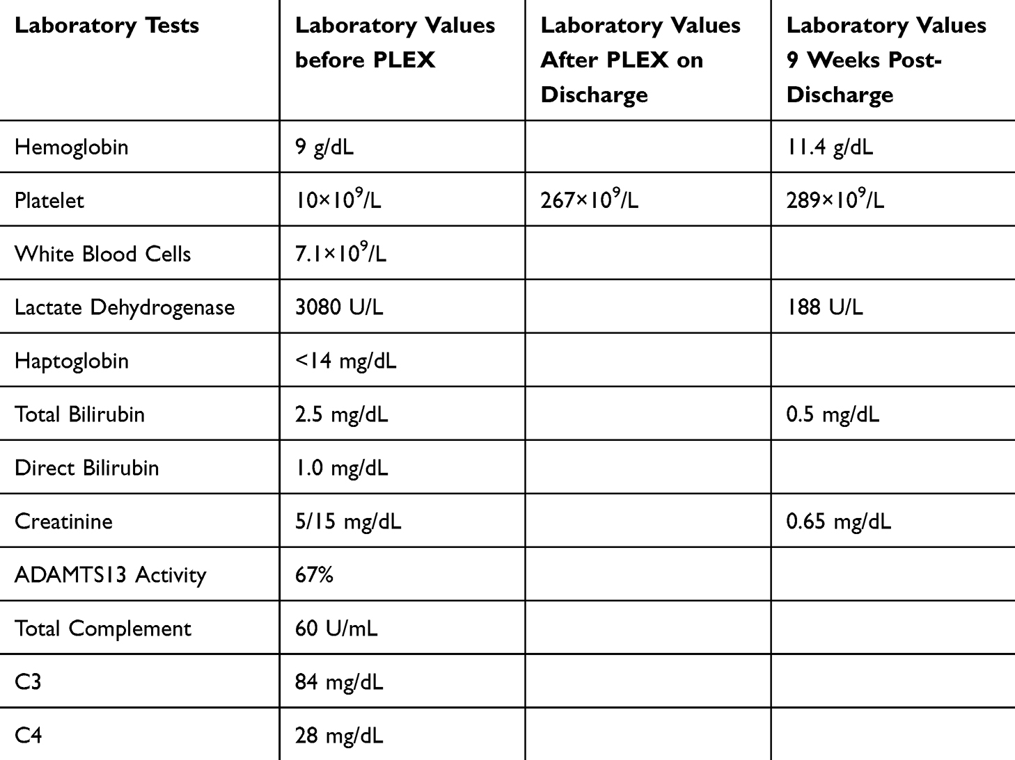 |
Table 1 Laboratory Values Pre- and Post-PLEX |
Discussion
We present the case of a patient with microangiopathic hemolytic anemia, anuric renal failure and metabolic encephalopathy within 24 hours of administration of the new Arexvy RSV vaccine. Arexvy RSV vaccine data sheets do not report thrombocytopenia or microangiopathic hemolytic anemias to be known adverse events.
TTP affects 3–10 adults per million population per year and is defined by excessive deficiency (<10%) of ADAMTS13 (a disintegrin and metalloproteinase), a cleavage protein for von Willebrand factor (vWF) polymers.1 ADAMTS13 cleaves vWF polymers within the vasculature, and without ADAMTS13, these ultra-long polymers accumulate and cause platelet clumping with resultant microthrombi formation. Historically, TTP has been characterized by the classic pentad of microangiopathic hemolytic anemia, thrombocytopenia, changes in neurological status, fever, and renal dysfunction.2 PLEX is a first-line treatment for TTP and works both by removing the antibody to the ADAMTS13 and repleting physiologic levels of ADAMTS13 with fresh plasma infusion. Due to the high rate of mortality, PLEX is often begun before confirmatory testing.
Complement-mediated thrombotic microangiopathy, also referred to as aHUS, may present in a similar manner to TTP, although often the renal function is typically worse in aHUS. It follows a different disease process, wherein the alternative pathway of the complement system undergoes complement dysregulation, leading to overactivation of the system. Several complement mutations have been reported including loss-of-function mutations in factor H, factor I, membrane cofactor protein (MCP), and thrombomodulin and gain-of-function mutations in C3 and factor B.3 Anti-factor-H, associated with deletions in genes encoding complement factor-H related proteins CFHR1 and CFHR3 are present in 5–7% of aHUS patients. Genetic abnormalities may be detected in up to half of patients with aHUS, while the remaining half may have complement dysregulation but no identifiable complement gene abnormality. Alternatively, a number of patients with AHUS may have identified genetic abnormalities but normal complement levels.4
VITT and HIT were ruled out given that both stem from a PF4 antibody which was negative in this patient. Although the clinical syndrome and rapid improvement following PLEX was suggestive of TTP, the ADAMTS13 level of 67% was not sufficiently low to confirm the diagnosis. A temporizing measure for TTP is the infusion of fresh frozen plasma (FFP) until definitive care can be obtained, and this can result in a falsely elevated ADAMTS13 level if the lab is drawn after the FFP. However, there is no record of FFP infusion at the outside facility.
Additionally, in patients with a history of prosthetic valve replacement who present with findings concerning for hemolysis, it is essential to obtain a TTE to assess for any paravalvular regurgitation that may be shearing the patient’s red blood cells.5 TTE was appropriately obtained in this patient with a history of bovine aortic valve replacement and demonstrated no prosthetic or periprosthetic regurgitation.
The patient was heterozygous for a large deletion involving the CFHR1 and CFHR3 genes, strongly suggesting the presence of a contiguous deletion of the full CFHR1 and CFHR3 genes. Specifically, it was noted that the complement factor H (CFH)-CFHR gene cluster is prone to structural variation resulting in large deletions, duplications, and hybrids of genes within the cluster. Currently, the clinical significance of heterozygous copy number variation impacting the CFHR1 and CFHR3 genes is not well-characterized, and the clinical significance of deletions and duplications impacting other genes within the CFHR cluster is not well-delineated. The patient’s complement levels were within normal range, with the exception of elevations in CBb and SC5b-9. These complement-level abnormalities indicate activation of the alternative complement pathway, and in the absence of other complement abnormalities this is most likely secondary to post-blood draw activation. Of note, the expected a-HUS lab findings (low factor H, normal C4 and low AH50) were not present. However, prior studies have reported that complement levels may remain normal in aHUS.4 Classically, aHUS is not effectively treated with PLEX, although it is a first-line treatment for Factor H deficiency.6
The concept of a TMA like picture post-vaccination is not new. Rare instances of TMAs have been reported after influenza, pneumococcal, H1N1, and rabies vaccines within two weeks of vaccination.6 In addition, several cases of COVID-19 vaccine-related TMAs have been reported recently that responded to PLEX. While the patient had received both the COVID-19 vaccine and the influenza vaccine 3–4 weeks prior to presentation, the combination of the low-normal ADAMTS13 levels, more recent RSV vaccine administration, and the classical syndrome of microangiopathic hemolytic anemia, renal failure and encephalopathy that responded quickly to PLEX made the most likely diagnosis RSV vaccine-induced thrombotic microangiopathy.
Conclusion
RSV vaccine-induced thrombotic microangiopathy with associated multi-organ failure is a diagnosis of exclusion. High dose corticosteroids with PLEX resolved the patient’s microangiopathic hemolytic anemia and thrombocytopenia and, in conjunction with intermittent hemodialysis, resolved the patient’s acute renal failure. This adverse event has been reported to the Food and Drug Administration as a potential complication of RSV vaccine administration. In the future, all clinicians counseling their elderly patients on the risks and benefits of the Arexvy vaccine should inform their patients of the potential complications outlined in this case report and should have a low threshold for further workup should a similar clinical presentation develop.
Informed Consent
Written informed consent has been obtained from the patient to publish this case report. No institutional approval from the Institutional Review Board was needed for this case report.
Disclosure
The authors report no conflicts of interest in this work.
References
1. Blombery P, Scully M. Management of thrombotic thrombocytopenic purpura: current perspectives. J Blood Med. 2014;5:15–23. doi:10.2147/JBM.S46458
2. Sarode R, Bandarenko N, Brecher ME, et al. Thrombotic thrombocytopenic purpura: 2012 American Society for Apheresis (ASFA) consensus conference on classification, diagnosis, management, and future research. J Clin Apher. 2014;29:148–167. doi:10.1002/jca.21302
3. Bouwmeester RN, Bormans EMG, Duineveld C, et al. COVID-19 vaccination and Atypical hemolytic uremic syndrome. Front Immunol. 2022;13:1056153. doi:10.3389/fimmu.2022.1056153
4. Ma Q, Xu G. New-onset and relapsed thrombotic microangiopathy post-COVID-19 vaccination. J Med Virol. 2023;95:e28946. doi:10.1002/jmv.28946
5. Arunthamakun J, Thorton K, Rafael A, et al. Waring blender syndrome - the mishaps of prosthetic heart valve. J Am Coll Cardiol. 2021;77:2381. doi:10.1016/S0735-1097(21)03736-0
6. Saluja P, Gautam N, Yadala S, et al. Thrombotic thrombocytopenic purpura (TTP) after COVID-19 vaccination: a systematic review of reported cases. Thromb Res. 2022;214:115–121. doi:10.1016/j.thromres.2022.04.020
 © 2025 The Author(s). This work is published and licensed by Dove Medical Press Limited. The
full terms of this license are available at https://www.dovepress.com/terms
and incorporate the Creative Commons Attribution
- Non Commercial (unported, 4.0) License.
By accessing the work you hereby accept the Terms. Non-commercial uses of the work are permitted
without any further permission from Dove Medical Press Limited, provided the work is properly
attributed. For permission for commercial use of this work, please see paragraphs 4.2 and 5 of our Terms.
© 2025 The Author(s). This work is published and licensed by Dove Medical Press Limited. The
full terms of this license are available at https://www.dovepress.com/terms
and incorporate the Creative Commons Attribution
- Non Commercial (unported, 4.0) License.
By accessing the work you hereby accept the Terms. Non-commercial uses of the work are permitted
without any further permission from Dove Medical Press Limited, provided the work is properly
attributed. For permission for commercial use of this work, please see paragraphs 4.2 and 5 of our Terms.