Back to Journals » OncoTargets and Therapy » Volume 12
Research progress of cystatin SN in cancer
Received 12 November 2018
Accepted for publication 15 February 2019
Published 6 May 2019 Volume 2019:12 Pages 3411—3419
DOI https://doi.org/10.2147/OTT.S194332
Checked for plagiarism Yes
Review by Single anonymous peer review
Peer reviewer comments 2
Editor who approved publication: Dr Federico Perche
Yanfang Liu, Jing Yao
Department of Oncology, The Union Hospital, Tongji Medical College, Huazhong University of Science and Technology, Wuhan, Hubei Province, People’s Republic of China
Abstract: Cystatin SN, belonging to the type 2 cystatin superfamily, is widely expressed and distributed in mammals. Cystatin SN is involved in inflammation, cell cycle, cellular senescence, tumorigenesis, and metastasis. Cystatin SN is also known to participate in signaling pathways like Wnt signaling pathway, GSK3 signaling pathway, AKT signaling pathway, and IL-6 signaling pathway. Cystatin SN was found to be highly expressed in peritumoral normal tissues in esophageal squamous cell carcinoma (ESCC); however, low cystatin SN expression was found in ESCC cancer tissues. Conversely, in other cancer types such as lung cancer, breast cancer, gastric cancer, pancreatic cancer, and colorectal cancer, high cystatin SN expression in cancer tissues but low cystatin SN expression in peritumoral normal tissues was found. Survival analyses showed that high cystatin SN expression benefited ESCC patients but did harm to other types of cancer patients. Univariate and multivariate analyses indicated that cystatin SN possibly acts as a marker for cancer prognosis. Here, we provide a brief introduction about the role of cystatin SN in cancer and discuss the different prognostic effects of cystatin SN on different tumors. Cystatin SN might be a potential marker for cancer prognosis and a target for cancer therapy.
Keywords: cystatin, cancers, signaling pathway, inflammation
Introduction
Cystatin belongs to a class of protease inhibitors, and in human cells and tissues, the number of cystatin forms appear to be 12.1 In the center of their sequence is present a highly conserved segment called “cystatin motif”, which is crucial for their functions as protease inhibitors.1 According to differences in the distribution of cystatin in the organism, 12 kinds of cystatin are identified and divided into three types: type 1, type 2, and type 3.2 Type 1 cystatin is an intracellular type and includes cystatin A and cystatin B, with the simplest structure among the three different types of cystatins.2 Type 1 cystatin is a single-chain protein containing 100 amino acid residues, with no disulfide bond and sugar chain.3 Type 2 cystatin is extracellular and includes cystatin C, cystatin D, cystatin E/M, cystatin F, cystatin G, cystatin S, cystatin SN, and cystatin SA.3 Type 2 cystatin is composed of non-glycosylated proteins which may be phosphorylated.2,3 Type 3 cystatin comprises intravascular inhibitors and includes L-kininogen and H-kininogen; this is also a multi-domain protein containing three cysteine protease inhibitors that exhibit different activities and structures.2,3
The type 2 cystatin superfamily was widely expressed and distributed in animals and microorganisms.1,4 Among animals, type 2 cystatin is found in body fluids and tissue fluids.5 All forms of human type 2 cystatin presented two conserved disulfide bridges at the C-terminal end of the sequence.6 Cystatin SN belongs to the type 2 cystatin superfamily, and it is a type of secretory protein consisting of 121 amino acids and with a relative molecular mass of ~14.5 kDa.6 Cystatin SN is encoded by CST1 gene which contains three exons and two introns and is located on chromosome 20p11.2.6 With regard to the function of cystatin SN, it is widely involved in determining cell fate and disease development, such as cell cycle, cellular senescence, tumorigenesis, and metastasis.7–14 Also, cystatin SN is involved in inflammation and functioned in signaling pathways like Wnt signaling pathway, GSK3 signaling pathway, AKT signaling pathway, and IL-6 signaling pathway.18,19,27,36 Cystatin SN was found to be an independent prognostic factor through univariate and multivariate analyses and was greatly related to survival of cancer patients.7,8,18,19,26,27,36 In this study, we are going to provide a detailed review of the function and role of cystatin SN in different cancers.
Function of cystatin SN and its role in non-neoplastic disease
Cystatin SN in human saliva has the function of antimicrobial activity and plays a crucial role in maintaining the homeostasis of the oral cavity.8 In healthy individuals, cystatin SN acts as the inhibitor of cysteine proteases in the oral environment and is involved in the control of the proteolytic events such as inhibiting the activity of papain in vivo.15,16
Studies showed that the expression of cystatin SN was related to the sensitivity of individual oral cavity to caffeine bitterness.14 For individuals who were not sensitive to caffeine bitterness, the expression level of cystatin SN was increased, while the sensitive individuals showed a decreased expression of cystatin SN.14 Studies also showed that caffeine intake may increase cystatin SN abundance and caused possible consequences on taste sensitivity.15
Cystatin SN was upregulated in patients with allergic rhinitis, and cystatin SN could amplify inflammation and may contribute to the severity and recurrence of intractable chronic rhinosinusitis with nasal polyps.10,16,17
Cystatin SN and inflammation
Cystatin SN is also involved in inflammation.10,16–18 In patients with seasonal allergic rhinitis, cystatin SN expression in the nasal mucosa was upregulated by allergen exposure through activation acquired Th2-type immune responses.10 Another study showed increased expression of cystatin SN in seasonal allergic rhinitis patients and that cystatin SN may contribute to inactivation of protease allergens and help to re-establish homeostasis of the nasal membranes through inhibiting histamine release.16 Cystatin SN could amplify eosinophilic infiltration and T-helper cell type 2 inflammation by interacting with epithelial-derived cytokines and fibroblasts on nasal polyps.17 IL-6 is an important inflammatory factor related to immune response.12 In MDA-MB-231 and SW480 cancer cells, CST1 knockdown lead to IL-6 accumulation,18 which affected the growth of cancer cells. Abovementioned studies showed that cystatin SN is a potential inflammation-related factor.
Cystatin SN and cell cycle
In SW480 colon cancer cells and MDA-MB-231 breast cancer cells, CST1 knockdown exhibited G1-phase cell cycle arrest, which was caused by the expression of cell cycle inhibitors p21 and p16 and the downregulation of cyclin D1 and p-Rb.18 In pancreatic cancer cell lines, CST1 mRNA overexpression contributed to the expression of cell cycle associated proteins like cyclin D1, cyclin A2, and cyclin E.19 All these studies showed that cystatin SN played critical roles in cell cycle.18,19
Cystatin SN and cellular senescence
Most healthy human tissues expressed little or no detectable levels of cystatin SN; however, significant amounts of cystatin SN were detected inside the senescent cells, which could make it an excellent candidate biomarker for identifying senescent cells in vivo.20
One study showed that during the progress of cellular senescence, human fibroblasts exhibited consistent and strong upregulation of cystatin SN, which showed cystatin SN was greatly related to cellular senescence.20 However, another study showed CST1 knockdown also contributed to cellular senescence and inhibited cancer cell proliferation and tumor growth.18
Cystatin SN and tumor
Cystatin SN was originally isolated as a neutral salivary cystatin,21 and in vitro studies suggested that cystatin SN might restrict the proteolytic activities of cysteine proteases.22–25 Cystatin SN is expressed differently in cancer, as shown in Figure 1. In esophageal squamous cell carcinoma (ESCC), cystatin SN showed higher expression in peritumoral tissues than in the ESCC tissues; however, in other types of cancer, tumor tissues usually showed higher cystatin SN expression than the normal tissues.7,9,13,19,26,27 Numerous studies have implied that cystatin SN played important roles in the development of several kinds of human cancer, such as pancreatic malignant neoplasm, gastric cancer, ESCC, and colorectal cancer (CRC).11,19,26,27 In different types of cancers, cystatin SN functioned in various ways, and its function mechanisms in cancer types are summarized in Table 1. Univariate and multivariate analyses of cystatin SN expression indicated that cystatin SN was an independent prognostic factor in several types of cancers, as shown in Table 2. In ESCC, high expression of cystatin SN was a profitable survival factor; however, other cancer types showed completely opposite results. The results of the survival analyses of cystatin SN expression in patients are presented in Figure 2.
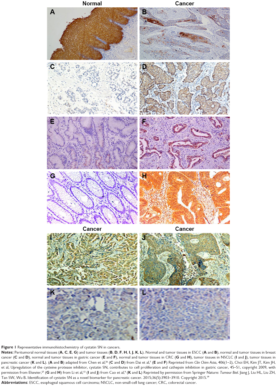 | Figure 1 Representative immunohistochemistry of cystatin SN in cancers. |
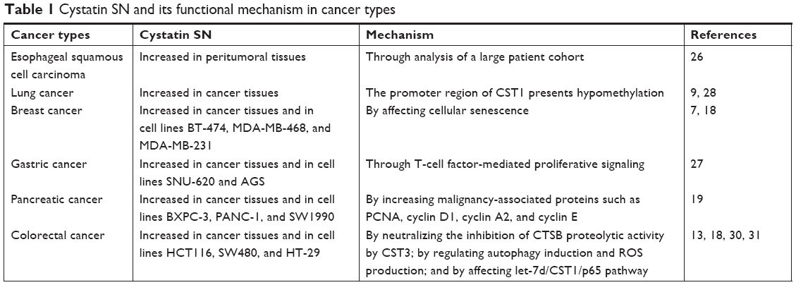 | Table 1 Cystatin SN and its functional mechanism in cancer types |
 | Table 2 Univariate and multivariate analyses of cystatin SN expression |
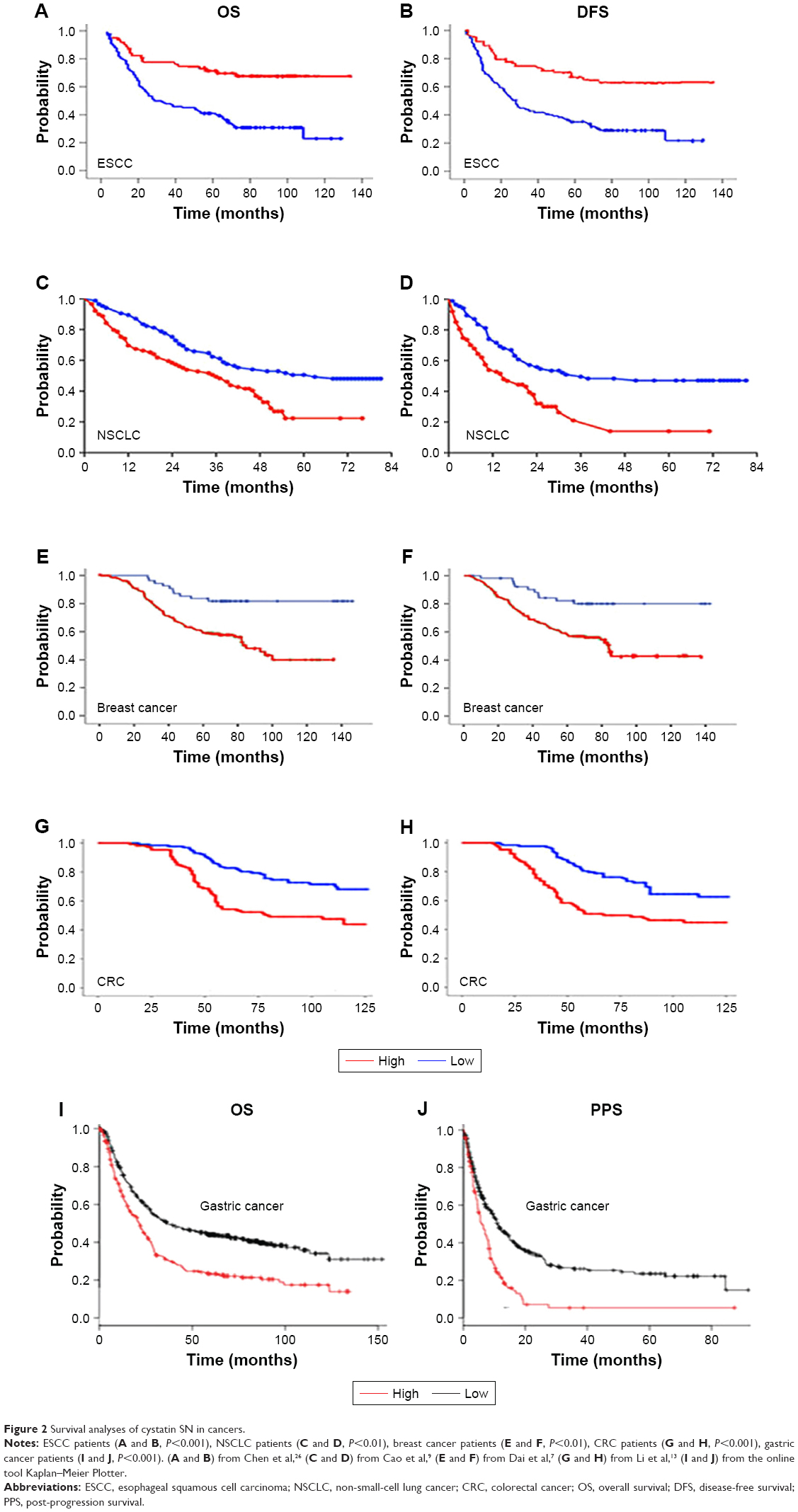 | Figure 2 Survival analyses of cystatin SN in cancers. |
ESCC
Peritumoral tissues showed higher cystatin SN expression than ESCC tissues (Figure 1A and B). Patients with high level of cystatin SN expression had more favorable survival time after surgical treatment than those with low expression, (Figure 2A and B). Besides, cystatin SN was believed to be an independent predictor of the 5-year survival rate of patients with surgically resected ESCC, based on the analysis of a large patient cohort.26 In this study, univariate and multivariate analyses demonstrated that cystatin SN expression was an independent prognostic factor, as shown in Table 2.
Lung cancer
In lung cancer, the promoter region of CST1 presents hypomethylation, which played an important role in the upregulation of cystatin SN.28 High expression level of cystatin SN was observed in non-small-cell lung cancer (NSCLC) patients (Figure 1), and patients with high level of cystatin SN showed more serious recurrence and poorer survival times compared to those with lower expression (Figure 2C and D). Through univariate and multivariate analyses, we could infer that high expression of cystatin SN was a significant prognostic indicator for a higher rate of recurrence, metastatic risk, and poor survival in patients with surgically resected NSCLCs,9 as summarized in Table 2.
Breast cancer
CST1 was generally upregulated in breast cancer at both mRNA and protein levels and played an important role in breast cancer patients (Figure 1C and D). Studies showed cystatin SN could promote cell proliferation and clone formation, migration, and invasion in BT-549 and MDA-MB-415 cells.7 Survival curves showed that the low cystatin SN expression group appeared to have higher survival probability than the high expression group, which indicated that the expression level of cystatin SN was closely correlated to the survival risk in breast cancer patients (Figure 2E and F). Table 2 shows that by univariate and multivariate analyses, CST1 expression was demonstrated as an independent prognostic factor. CST1 knockdown in BT-474 and MDA-MB-468 cells significantly inhibited cell migration capability.7 In MDA-MB-231 cells, CST1 knockdown increased phosphorylation of glycogen synthase kinase 3β at serine 9, leading to induction of glycogen accumulation associated with cellular senescence and inhibition of tumor proliferation,18 which indicated cystatin SN might affect the growth of cancer cells by affecting the GSK3 signaling pathway (Figure 3A). Furthermore, CST1 knockdown suppressed cancer cell proliferation and tumor growth in a xenograft model and led to IL-6 accumulation,18 which indicated that cystatin SN might also affect the IL-6 signaling pathway (Figure 3A). Another study in MDA-231 cell lines showed that the expression of CST1 gene could be suppressed significantly by CAPC, a tumor suppressor gene that played important roles in the suppression of tumor growth and metastasis.29
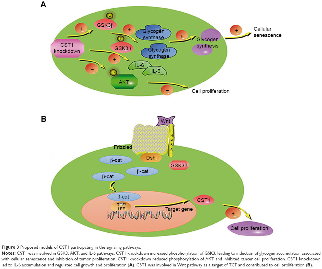 | Figure 3 Proposed models of CST1 participating in the signaling pathways. |
Gastric cancer
Cystatin SN expression was found to be upregulated in gastric tumor tissues (Figure 1E and F) and cell lines (SNU-620 and AGS). Upregulation of cystatin SN contributed to cell proliferation in gastric cancer.27 Classical Wnt signaling pathway was widely involved in a variety of cancers and played diverse roles in governing cell fate, proliferation, migration, polarity, and death.33–35 In AGS and SNU638 gastric cancer cells, cystatin SN contributed to the proliferation of cancer cells by affecting the expression of two main important Wnt signaling pathway molecules, β-catenin and TCF,27 which indicated CST1, as a target of TCF,36 may regulate the proliferation of cancer cells through affecting Wnt signaling pathway (Figure 3B). Study also showed that cystatin SN was significantly correlated with pTNM stage.27 Survival analyses from the online tool Kaplan–Meier plotter (http://kmplot.com/analysis/index.php?p=service&cancer=gastric) indicated that patients with high cystatin SN expression showed worse survival probabilities than those with low cystatin SN expression (Figure 2). Univariate analyses indicated that cystatin SN may be an independent prognostic factor for gastric cancer patients, the results for which are summarized in Table 2.
Pancreatic cancer
Cystatin SN was a potential biomarker for early diagnosis of malignant pancreatic neoplasms. Immunohistochemistry confirmed that cystatin SN was upregulated in tumor tissues (Figure 1). In several pancreatic cancer cell lines (BXPC-3, PANC-1, SW1990), CST1 was upregulated at both protein and mRNA levels, and was correlated closely with malignancy-associated proteins such as PCNA, cyclin D1, cyclin A2, and cyclin E. Consistent with this finding, CST1 knockdown reduced the expression of the proliferation-related proteins p-AKT and PCNA significantly, as well as colony formation and xenograft development in vitro.19 In conclusion, cystatin SN possibly contributed to the proliferation of cancer cells by participating in the AKT signaling pathway (Figure 3A).
Colorectal cancer
Cystatin SN was highly expressed in colon tumor tissues (Figure 1),13,18,30–32 and cystatin SN overexpressing cell line HCT116 exhibited increased tumor growth and tumor metastasis in vitro. Survival analysis has demonstrated that high cystatin SN expression was closely associated with poor clinical status in colon cancer patients (Figure 2).13 Cystatin SN acted by neutralizing the inhibition of CTSB proteolytic activity by CST3, which might be involved in colorectal tumorigenesis and poor prognosis.31 In human SW480 cancer cells, cystatin SN enhanced tumor development by affecting the GSK3 signaling pathway,18 which was similar to the mechanism observed in MDA-MB-231 cells (Figure 3A). Furthermore, CST1 knockdown suppressed tumor growth in vivo and led to IL-6 accumulation,18 which indicated cystatin SN might affect the IL-6 signaling pathway (Figure 3A). In HCT116 CRC cells, the let-7d/CST1/p65 pathway modulated CRC cell proliferation, and also, the let-7d/CST1/p65 pathway may prove to be useful in the prevention and treatment of CRC.32 A previous study in HT-29 and SW480 cells showed that cystatin SN was highly expressed at both the mRNA and protein levels.30 Increased cystatin SN expression inhibited auranofin-induced cell death by regulating autophagy induction and ROS production.30 Univariate and multivariate analyses demonstrated that cystatin SN possibly acted as an independent prognostic factor for CRC prognosis,13,18,30–32 as shown in Table 2.
Conclusion
Studies showed that the expression level of cystatin SN was related to the survival of cancer patients.7,9,13,26 High expression of cystatin SN was closely related to poor prognosis such as recurrence and metastasis in several cancer types like gastric cancer, CRC, pancreatic cancer, and lung cancer; however, in ESCC, high cystatin SN expression patients showed more favorable survival times after surgical treatment. The conclusion was presented based on the analysis of a large patient cohort and the different roles that cystatin SN plays in these several types of cancers. We concluded that the possible causes for functional differences among cancers may be related to factors such as tissue specificity and pathological types. As we all know esophageal cancer mainly includes esophageal adenocarcinoma and ESCC, and the later accounts for the majority of esophageal cancers. At present, cystatin SN is mainly studied in ESCC, but rarely studied in esophageal adenocarcinoma. There may be completely opposite expression level of cystatin SN in the two types of esophageal cancer. In other types of cancer, similar phenomenon may also exist among different pathological types. Therefore, basic research in different pathological types of tumors is necessary, which will help to clearly clarify the function of cystatin SN in cancer.
In different types of cancers and cell lines, cystatin SN functions in various ways. In gastric cancer, cystatin SN contributed to gastric tumorigenesis through T-cell factor-mediated proliferative signaling. In pancreatic cancer, cystatin SN acted by increasing the expression of malignancy-associated proteins such as PCNA, cyclin D1, cyclin A2, and cyclin E. In CRC, cystatin SN was found to play a vital role in cell proliferation by regulating autophagy and by affecting let-7d/CST1/p65 pathway. Though some basic studies have been done in this regard, the reasons why cystatin SN functions in different ways among different cancers is still not very clear. Univariate and multivariate analyses of cystatin SN expression indicated that cystatin SN was an independent prognostic factor in several types of cancers. In ESCC, high expression of cystatin SN was a profitable prognostic factor; however, in other types of cancers like gastric cancer, CRC, pancreatic cancer, and lung cancer, high expression of cystatin SN was a poor prognostic factor, which indicated cystatin SN could be a potential target in therapy of cancer patients. In terms of its different prognosis in different cancers, upregulating cystatin SN in ESCC and downregulating cystatin SN in other cancers may be a good approach for treatment. Keeping in mind the strong relationship between expression of cystatin SN and survival probability, as well as generally observed high expression of cystatin SN in cancers, cystatin SN may be taken as a diagnostic factor.
Cystatin SN is expected to become a biomarker for tumor diagnosis and prognosis prediction. Cystatin SN regulates proliferation of cancer cells by affecting signaling pathways like Wnt signaling pathway, GSK3 signaling pathway, AKT signaling pathway, and IL-6 signaling pathway. In the process of studying cystatin SN, trying to figure out why cystatin SN functions so differently between ESCC and other cancers and determine the specific molecular mechanisms involved in normal and cancer tissues would be exciting and challenging research goals. If the mechanisms are well researched, specific targeted intervention could be made to treat cancer patients.
Acknowledgments
This work was supported by the National Natural Science Foundation of China (No 81202460) and the Provincial Natural Science Foundation of Hubei (Nos 2015BCA270, 2014CFB404). In addition, Yanfang Liu especially wishes to thank her friend Wei Chen, who had given valuable suggestions and powerful spiritual support during the process of paper writing.
Disclosure
The authors report no conflicts of interest in this work.
References
Abrahamson M, Alvarez-Fernandez M, Nathanson CM. Cystatins. Biochem Soc Symp. 2003;70:179–199. doi:10.1042/bss0700179 | ||
Dickinson DP, Thiesse M, Hicks MJ. Expression of type 2 cystatin genes CST1-CST5 in adult human tissues and the developing submandibular gland. DNA Cell Biol. 2002;21(1):47–65. doi:10.1089/10445490252810311 | ||
Magister S, Kos J. Cystatins in immune system. J Cancer. 2013;4(1):45–56. doi:10.7150/jca.5044 | ||
Nandy SK, Bhuyan R, Seal A. Modelling family 2 cystatins and their interaction with papain. J Biomol Struct Dyn. 2013;31(6):649–664. doi:10.1080/07391102.2012.706403 | ||
Abrahamson M, Barrett AJ, Salvesen G, Grubb A. Isolation of six cysteine proteinase inhibitors from human urine. Their physicochemical and enzyme kinetic properties and concentrations in biological fluids. J Biol Chem. 1986;261(24):11282–11289. | ||
de Sousa-Pereira P, Amado F, Abrantes J, Ferreira R, Esteves PJ, Vitorino R. An evolutionary perspective of mammal salivary peptide families: cystatins, histatins, statherin and PRPs. Arch Oral Biol. 2013;58(5):451–458. doi:10.1016/j.archoralbio.2012.12.011 | ||
Dai DN, Li Y, Chen B, et al. Elevated expression of CST1 promotes breast cancer progression and predicts a poor prognosis. J Mol Med (Berl). 2017;95(8):873–886. doi:10.1007/s00109-017-1537-1 | ||
de Sousa-Pereira P, Abrantes J, Pinheiro A, Colaco B, Vitorino R, Esteves PJ. Evolution of C, D and S-type cystatins in mammals: an extensive gene duplication in primates. PLoS One. 2014;9(10):e109050. doi:10.1371/journal.pone.0109050 | ||
Cao X, Li Y, Luo RZ, et al. Expression of cystatin SN significantly correlates with recurrence, metastasis, and survival duration in surgically resected non-small cell lung cancer patients. Sci Rep. 2015;5:8230. doi:10.1038/srep08230 | ||
Fukuoka A, Matsushita K, Morikawa T, et al. Human cystatin SN is an endogenous protease inhibitor that prevents allergic rhinitis. J Allergy Clin Immunol. 2019;143(3):1153–1162.e12. | ||
Nakajima M. Identification of cystatin SN as a novel tumor marker for colorectal cancer. Int J Oncol. 2009;35(1):33–40. doi:10.3892/ijo_00000310 | ||
Heinrich PC, Behrmann I, Muller-Newen G, Schaper F, Graeve L. Interleukin-6-type cytokine signalling through the gp130/Jak/STAT pathway. Biochem J. 1998;334(Pt 2):297–314. | ||
Li T, Xiong Q, Zou Z, Lei X, Jiang Q, Liu D. Prognostic significance of cystatin SN associated nomograms in patients with colorectal cancer. Oncotarget. 2017;8(70):115153–115163. doi:10.18632/oncotarget.23041 | ||
Dsamou M, Palicki O, Septier C, et al. Salivary protein profiles and sensitivity to the bitter taste of caffeine. Chem Senses. 2012;37(1):87–95. doi:10.1093/chemse/bjr070 | ||
Dsamou M, Morzel M, Le Corre L, Severin I, Chagnon MC. Caffeine increases the expression of cystatin SN in human submandibular acinar-like HSG cells. Arch Oral Biol. 2013;58(10):1511–1516. doi:10.1016/j.archoralbio.2013.06.005 | ||
Imoto Y, Tokunaga T, Matsumoto Y, et al. Cystatin SN upregulation in patients with seasonal allergic rhinitis. PLoS One. 2013;8(8):e67057. doi:10.1371/journal.pone.0067057 | ||
Kato Y, Takabayashi T, Sakashita M, et al. The expression and functional analysis of CST1 in intractable nasal polyps. Am J Respir Cell Mol Biol. 2018;59(4):448–457. doi:10.1165/rcmb.2017-0325OC | ||
Oh SS, Park S, Lee KW, et al. Extracellular cystatin SN and cathepsin B prevent cellular senescence by inhibiting abnormal glycogen accumulation. Cell Death Dis. 2017;8(4):e2729. doi:10.1038/cddis.2017.153 | ||
Jiang J, Liu HL, Liu ZH, Tan SW, Wu B. Identification of cystatin SN as a novel biomarker for pancreatic cancer. Tumour Biol. 2015;36(5):3903–3910. doi:10.1007/s13277-014-3033-3 | ||
Keppler D, Zhang J, Bihani T, Lin AW. Novel expression of CST1 as candidate senescence marker. J Gerontol A Biol Sci Med Sci. 2011;66(7):723–731. doi:10.1093/gerona/glr033 | ||
Isemura S, Saitoh E, Sanada K. Characterization of a new cysteine proteinase inhibitor of human saliva, cystatin SN, which is immunologically related to cystatin S. FEBS Lett. 1986;198(1):145–149. | ||
Tseng CC, Tseng CP, Levine MJ, Bobek LA. Differential effect toward inhibition of papain and cathepsin C by recombinant human salivary cystatin SN and its variants produced by a baculovirus system. Arch Biochem Biophys. 2000;380(1):133–140. doi:10.1006/abbi.2000.1909 | ||
Baron A, DeCarlo A, Featherstone J. Functional aspects of the human salivary cystatins in the oral environment. Oral Dis. 1999;5(3):234–240. | ||
Baron A, Barrett-Vespone N, Featherstone J. Purification of large quantities of human salivary cystatins S, SA and SN: their interactions with the model cysteine protease papain in a non-inhibitory mode. Oral Dis. 1999;5(4):344–353. | ||
Hiltke TR, Lee TC, Bobek LA. Structure/function analysis of human cystatin SN and comparison of the cysteine proteinase inhibitory profiles of human cystatins C and SN. J Dent Res. 1999;78(8):1401–1409. doi:10.1177/00220345990780080501 | ||
Chen YF, Ma G, Cao X, et al. Overexpression of cystatin SN positively affects survival of patients with surgically resected esophageal squamous cell carcinoma. BMC Surg. 2013;13:15. doi:10.1186/1471-2482-13-15 | ||
Choi EH, Kim JT, Kim JH, et al. Upregulation of the cysteine protease inhibitor, cystatin SN, contributes to cell proliferation and cathepsin inhibition in gastric cancer. Clin Chim Acta. 2009;406(1–2):45–51. doi:10.1016/j.cca.2009.05.008 | ||
Mullapudi N, Ye B, Suzuki M, et al. Genome wide methylome alterations in lung cancer. PLoS One. 2015;10(12):e0143826. doi:10.1371/journal.pone.0143826 | ||
Liu XF, Xiang L, Zhang Y, Becker KG, Bera TK, Pastan I. CAPC negatively regulates NF-kappaB activation and suppresses tumor growth and metastasis. Oncogene. 2012;31(13):1673–1682. doi:10.1038/onc.2011.355 | ||
Oh BM, Lee SJ, Cho HJ, et al. Cystatin SN inhibits auranofin-induced cell death by autophagic induction and ROS regulation via glutathione reductase activity in colorectal cancer. Cell Death Dis. 2017;8(3):e2682. doi:10.1038/cddis.2017.100 | ||
Kim JT, Lee SJ, Kang MA, et al. Cystatin SN neutralizes the inhibitory effect of cystatin C on cathepsin B activity. Cell Death Dis. 2013;4:e974. doi:10.1038/cddis.2013.485 | ||
Jiang J, Liu HL, Tao L, et al. Let7d inhibits colorectal cancer cell proliferation through the CST1/p65 pathway. Int J Oncol. 2018;53(2):781–790. doi:10.3892/ijo.2018.4419 | ||
Daniels DL, Eklof Spink K, Weis WI. Beta-catenin: molecular plasticity and drug design. Trends Biochem Sci. 2001;26(11):672–678. | ||
Katoh M, Katoh M. Comparative genomics on Wnt5a and Wnt5b genes. Int J Mol Med. 2005;15(4):749–753. | ||
Li F, Chong ZZ, Maiese K. Winding through the WNT pathway during cellular development and demise. Histol Histopathol. 2006;21(1):103–124. doi:10.14670/HH-21.103 | ||
Schwartz DR, Wu R, Kardia SL, et al. Novel candidate targets of beta-catenin/T-cell factor signaling identified by gene expression profiling of ovarian endometrioid adenocarcinomas. Cancer Res. 2003;63(11):2913–2922. |
 © 2019 The Author(s). This work is published and licensed by Dove Medical Press Limited. The
full terms of this license are available at https://www.dovepress.com/terms
and incorporate the Creative Commons Attribution
- Non Commercial (unported, 3.0) License.
By accessing the work you hereby accept the Terms. Non-commercial uses of the work are permitted
without any further permission from Dove Medical Press Limited, provided the work is properly
attributed. For permission for commercial use of this work, please see paragraphs 4.2 and 5 of our Terms.
© 2019 The Author(s). This work is published and licensed by Dove Medical Press Limited. The
full terms of this license are available at https://www.dovepress.com/terms
and incorporate the Creative Commons Attribution
- Non Commercial (unported, 3.0) License.
By accessing the work you hereby accept the Terms. Non-commercial uses of the work are permitted
without any further permission from Dove Medical Press Limited, provided the work is properly
attributed. For permission for commercial use of this work, please see paragraphs 4.2 and 5 of our Terms.