
Back to Journals » Clinical Ophthalmology » Volume 19
Repeatability of a Dual-Scheimpflug Placido Disc Corneal Tomographer/Topographer in Eyes with Keratoconus
Authors Poulsen A  , Jang DH, Khan M, Al-Mohtaseb ZN, Chen M, Banerjee K, Scott IU, Pantanelli SM
Received 17 April 2025
Accepted for publication 6 August 2025
Published 14 August 2025 Volume 2025:19 Pages 2751—2758
DOI https://doi.org/10.2147/OPTH.S530011
Checked for plagiarism Yes
Review by Single anonymous peer review
Peer reviewer comments 5
Editor who approved publication: Dr Scott Fraser
Anne Poulsen,1 Diane Haeji Jang,2 Mahmood Khan,3 Zaina Nabil Al-Mohtaseb,4 Michael Chen,5 Kalins Banerjee,6 Ingrid U Scott,6,7 Seth M Pantanelli7
1St. Paul Eye Clinic, St. Paul, MN, USA; 2Virginia Mason Medical Center, Seattle, WA, USA; 3Department of Ophthalmology, Weill Cornell Medical College, New York, NY, USA; 4Whitsett Vision Group, Houston, TX, USA; 5Department of Ophthalmology, Denver Health, Denver, CO, USA; 6Department of Biostatistics, University of Michigan, Ann Arbor, MI, USA; 7Department of Ophthalmology, Penn State College of Medicine, Hershey, PA, USA
Correspondence: Seth M Pantanelli, Department of Ophthalmology, Penn State College of Medicine, 500 University Drive, HU19, Hershey, PA, 17033, USA, Tel +1717-531-5690, Fax +1717-531-5009, Email [email protected]
Purpose: To investigate the repeatability of a combined Dual-Scheimpflug placido disc corneal tomographer/topographer (Ziemer Galilei G4) with respect to keratometric indices used to monitor progression of keratoconus (KCN).
Methods: Patients with KCN were prospectively enrolled. For each eye lacking history of corneal surgery, 5 measurements were taken in succession. Eyes in which 3 or more measurements could be obtained (defined by the device’s 4 image quality metrics) were included in the analysis. The repeatability limits (RL) and interclass correlation coefficients (ICC) were calculated for various parameters.
Results: Thirty-two eyes from 25 patients met all image quality metrics, and 54 eyes from 38 patients met at least 3/4 criteria (all except the placido image quality metric). RLs for key parameters when 4/4 or ≥ 3/4 image quality metrics were met included: 0.37 and 0.77 diopters (D) for steep simulated keratometry, 0.79 and 1.65 D for maximum keratometry, 13.80 and 13.88 degrees for astigmatism axis, 0.64 and 0.56 μm for vertical coma magnitude, and 3.76 and 3.84 μm for thinnest pachymetry, respectively. The ICCs for all parameters were excellent (above 0.87) except for spherical aberration (0.77), which was still considered good.
Conclusion: The dual-Scheimpflug placido disc corneal tomographer/topographer is highly repeatable in quantifying parameters used in monitoring KCN. Excellent placido images are difficult to capture in eyes with KCN, but when available, increase the reliability of the measurements. When clinicians find that a topographic index changes by more than the RLs defined herein, they can have confidence that this represents real change and may appropriately recommend interventions such as corneal cross-linking or intrastromal corneal ring segments.
Keywords: keratoconus, topography, tomography, keratoconus progression, Scheimpflug imaging
Introduction
Keratoconus is the most common corneal degeneration, with an estimated prevalence between 120 and 265 per 100,000 in the general population.1,2 The degeneration results in significant visual impairment secondary to irregular astigmatism and high myopia.3 In the age of corneal cross-linking, early diagnosis and detection of progression are essential elements of management that limit morbidity associated with the disease.4 Achieving a consensus in the definition of progression in keratoconus is limited by the requirements that such indices be based upon high-quality data (which is often difficult to obtain in keratoconic corneas) and long-term follow-up in eyes that progress but do not have a surgical intervention (which may be impractical or unethical). One study by Gupta et al evaluated the change in several keratometric indices over time and determined that variation in best fit sphere radius was one of the most sensitive predictors of progression.5 Another study by Martinez-Abad followed patients with keratoconus for 6 years and proposed a “Progression Index” equation, but this study was limited in that it was based only upon anterior surface indices, which limits its sensitivity when only posterior ectatic disease is present.6
The Global Consensus on Keratoconus and Ectatic Disease defined ectatic progression by a consistent change, where the magnitude of the change is above the noise of the testing system.7 More recently, Belin et al proposed the ABCD Staging and Classification System,8 which emphasized the importance of both anterior and posterior keratometric indices for detecting early progression, and interprets these indices in the context of the sensitivity of the measurement device.9 In conforming to these guidelines, a study was published by de Luis Eguileor et al, which investigated the repeatability of measurements pertinent to the monitoring of progression in eyes with keratoconus using a Scheimpflug system (Pentacam HR).10 This study found that sequential measurements differing by more than 1.54 D, 1.08 D, and 0.71 µm in the steep keratometry, maximum keratometry, and vertical coma, respectively, likely represented real change. Of note, this study only included eyes for which high-quality data were available in the analysis. The study results are interesting and immediately applicable for eye care providers who have a Pentacam HR available. However, at least two previous studies have shown that the measured indices from one device are not interchangeable with those of another, like the Orbscan II (Bausch & Lomb, Rochester, NY, USA) or Galilei Dual Scheimpflug Analyzer (Ziemer, Port, Switzerland).11,12 It is therefore unclear how these repeatability limits (RL) reported by de Luis Eguileor et al would translate for use with a different device, particularly a device employing placido-based or a combination of keratometric technologies. Furthermore, repeatability data in keratoconus that relies on ideal imaging conditions is not representative of clinical reality. As such, it is unclear whether such RLs can be applied in the setting of sub-optimal image quality, which commonly occurs when measuring keratoconic eyes.
In contrast to Pentacam HR, which uses Scheimpflug imaging only, the Ziemer Galilei G4 is a corneal tomographer/topographer combining both Scheimpflug imaging and placido videokeratography. It is plausible that the RLs previously described by de Luis Eguileor do not apply to this alternative device. In order to properly apply Belin’s ABCD Progression Index to the Ziemer Galilei, the RLs, as a function of image quality, must be elucidated. As such, the objective of the current study was to assess the repeatability of measurements obtained using the Galilei Dual Scheimpflug Analyzer (Ziemer, Port, Switzerland) in keratoconic eyes and to determine which of these measurements have RLs that may allow accurate monitoring of keratoconus progression over time. Since the Galilei captures placido images and the Pentacam HR does not, we also sought to investigate the contribution good placido images have on reliability of these measurements.
Methods
We conducted a prospective observational study of patients seen at either the Penn State Eye Center (Penn State College of Medicine, Hershey, PA) or the Cullen Eye Institute (Baylor College of Medicine, Houston, TX). The study was approved by the Institutional Review Board at both academic institutions (STUDY00007951) and registered at clinicaltrials.gov (NCT03511495) before the first patient was enrolled. The study conformed to the tenets of the Declaration of Helsinki and integrity of the data was maintained in accordance with the Health Insurance Portability and Accountability Act (HIPPA). Deidentified demographics and data on individual subjects are available upon written request by contacting the corresponding author for up to 3 years after publication.
Patients with keratoconus being seen in routine follow-up between September 2017 and November 2018 were provided with information about the study and consent was obtained prior to inclusion. All keratoconus patients over the age of 18 were offered the opportunity to participate. A diagnosis of keratoconus was made with slit lamp examination and topographic confirmation, and met criteria for at least Stage I Amsler-Krumeich classification.13 Exclusion criteria included history of any corneal or intra-ocular surgery (ie corneal cross linking, LASIK, corneal transplant, or cataract surgery). If a surgery had been performed in one eye, the fellow eye was still eligible for inclusion.
The Galilei G4 is a combined dual-Scheimpflug tomographer/placido disc topographer that employs monochromatic light sources at 470 nm and 750 nm for Scheimpflug and placido disc illumination, respectively. Each scan generates over 122,000 data points. The placido and Scheimpflug data are integrated using a proprietary algorithm to generate information about the anterior surface of the cornea, while only the Scheimpflug data are used to generate information about the posterior surface.
Before starting the study, the Galilei G4 was calibrated at both institutions. Each eye was aligned to the visual axis via the fixation light on the machine. Patients were instructed to blink between measurements. Five consecutive measurements were obtained in each eligible eye, using the standard resolution setting. All images for an individual patient were captured by a single examiner, but given the multi-center nature of this study, multiple examiners were used to obtain data from different patients. The instrument defines 4 image quality metrics to qualify the integrity of the data collected. Satisfactory metrics include a motion compensation value >85%, placido image quality >85%, Scheimpflug image quality >90%, and motion distance >70%.
For each captured image, the following keratometric indices were assessed: flat and steep simulated keratometry (SimK) magnitudes, steep SimK axis, maximum keratometry values in the central zone (Kmax), anterior (BFS_anterior) and posterior (BFS_posterior) radius best fit sphere of the cornea with the diameter set to 8 mm, maximum anterior (max_BFS_ant) and posterior elevations (max_BFS_post), area of thinnest pachymetry (TCT), corneal asphericity (Q) at 6 mm, spherical aberration (Z40), vertical (Z3−1) and horizontal (Z31) coma, coma axis, root-mean-square (RMS) of coma measurements, and root-mean-square of higher order aberrations (HOA RMS) at 6 mm.
RLs for the above indices were calculated, and represent the value over which a change is likely to be real and not due to noise or variability inherent to the machine.14 The intraclass correlation coefficient (ICC) was also calculated for each index, which estimates the percentage of the total variability in the results that can be attributed to real differences between measurements. The RLs and ICCs were calculated for two different conditions: 1) eyes with at least 3 measurements that met all four of the devices image quality metrics and 2) eyes with at least 3 measurements that met 3 or more of the devices 4 image quality metrics (an excellent placido image was not required). In the instance where 4 or 5 high-quality images were obtainable, all of the available data was used in the analysis. Required sample size for the study was based upon the recommendations of McAlinden et al.14 Note that, in the instance of keratoconus, which is an asymmetric disease with poor correlation between the two eyes, it is accepted to use both eyes from the same subject without control for non-independence. Statistical analyses were carried out using R programming language.
Results
Fifty-one patients were enrolled in the study. Mean age was 37.8 ± 13.87 (range 18–89). Twenty were female and 31 were male. Of 102 potentially measurable eyes, 86 met inclusion/exclusion criteria for measurement. Of these, 32 eyes (16 right and 16 left) from 25 patients had at least 3 measurements that met all four image quality metric criteria (15 eyes had 5 measurements, 13 had 4 measurements, and 4 had 3 measurements; mean = 4.3). This sample size corresponds to a confidence in the repeatability limit estimate within 10–15%.14 Fifty-four eyes (27 right and 27 left) from 38 patients met at least 3 of 4 image quality metric criteria (35 eyes had 5 measurements, 15 had 4 measurements, and 4 had 3 measurements; mean = 4.6). This sample size corresponds to a confidence in the repeatability limit estimate within 5–10%.14 The severity of keratoconus, as assessed by Amsler-Krumeich classification,13 for eyes included in each of the analyses is detailed in Table 1.
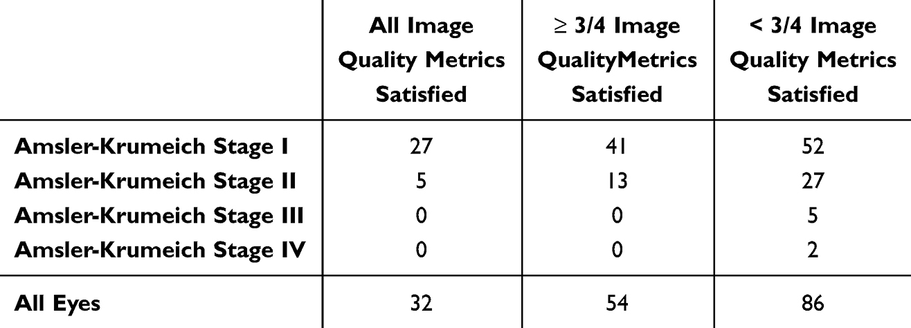 |
Table 1 Amsler-Krumeich Classification of Eyes Included in Each Analysis |
The RLs and ICCs for all parameters are shown in Table 2. We found that obtaining measurements that met all four image quality metrics was challenging – the placido image criterion was the most difficult to meet. Dropping the requirement for a good placido image and forcing the device to rely more heavily upon the Scheimpflug data lead to higher (worse) RLs. The ICC was found to be excellent (above 0.90) for most parameters, and good (above 0.75) for vertical coma and spherical aberration.15
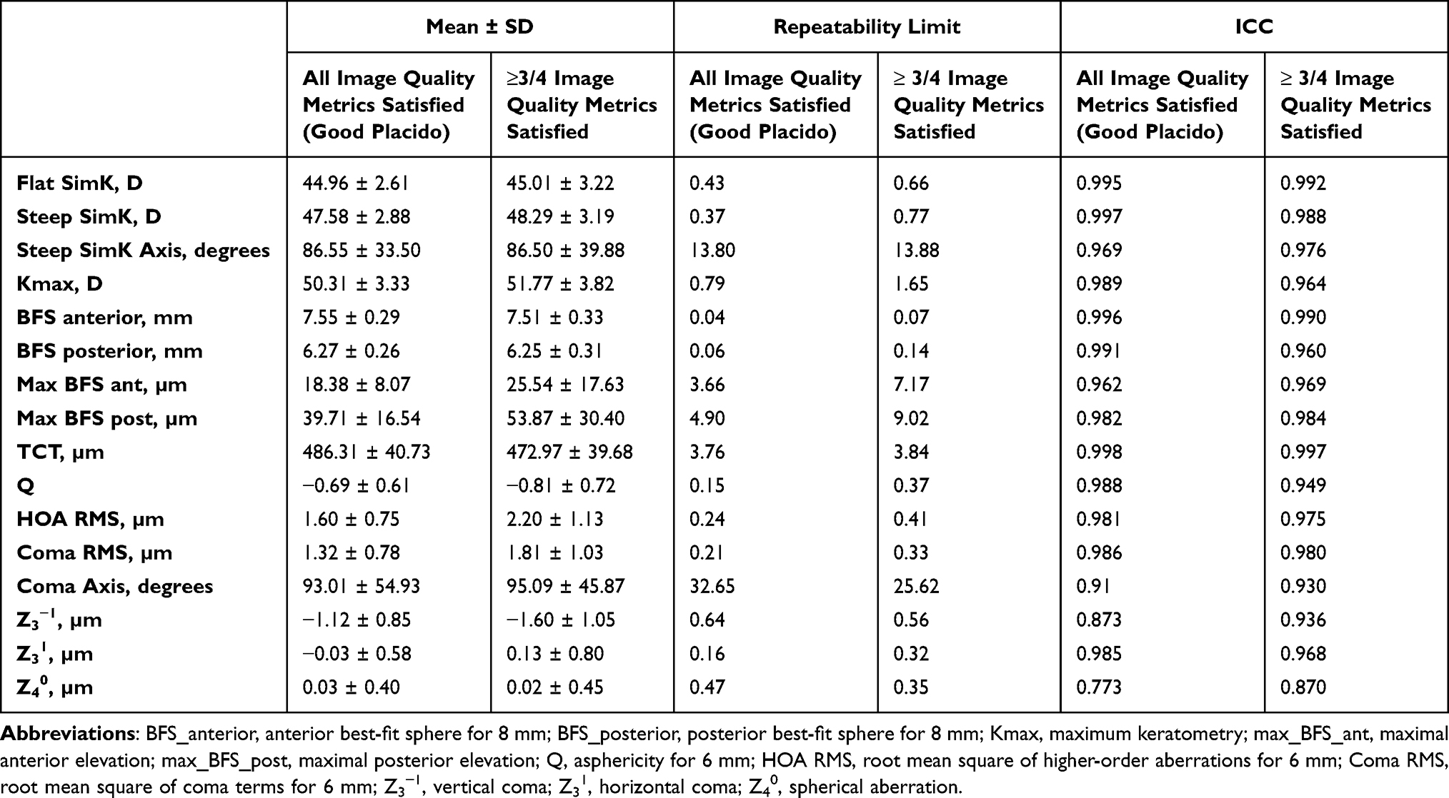 |
Table 2 Means, Repeatability Limits and Interclass Correlation Coefficients (ICCs) Obtained with Galilei G4 System |
For the 54 eyes that had less than 3 images captured that met all four of the image quality metrics, the Kmax was 56.02 ± 6.44. Of these, all 54 had poor quality placido images and 27 also had poor Scheimpflug images. A strong and highly significant inverse relationship was found between Kmax and placido image quality (Figure 1a; Pearson r = −0.83; p ≤ 0.001). A much weaker and barely significant inverse relationship was found between Kmax and Scheimpflug image quality (Figure 1b; Pearson r = −0.29; p = 0.04). This suggests that the placido image was more sensitive to degradation from corneal steepening in moderate or advanced keratoconic disease.
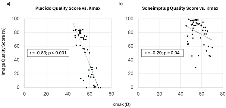 |
Figure 1 Placido (a) and Scheimpflug (b) image quality scores (%) as a function of maximum keratometry [Kmax (D)]. There was a stronger correlation between increasing Kmax and degradation of the placido image than the Scheimpflug image. |
Discussion
There are at least two previous studies that have investigated the repeatability of the Galilei in a keratoconic population. Shetty et al evaluated 55 keratoconic eyes on three different devices and found that the Galilei was the least repeatable when compared to the Pentacam HR and the Sirius (Costruzione Strumenti Oftalmici, Florence, Italy).12 However, these investigators only compared performance with respect to three keratometric indices (anterior keratometry, thinnest corneal thickness, and posterior keratometry). More recently, Meyer et al evaluated 50 keratoconic eyes and found the Pentacam to be superior to the Galilei with respect to keratometry measurements, but found the reverse to be true with respect to pachymetry measurements.11 It is important to note, however, that all eyes, including those for which high-quality measurements could not be obtained, were included in the Meyer study analysis, and that 25% of eyes on the Galilei and 12% on the Pentacam HR were reported to have poor quality scans. The rationale for including these data is understandable; especially in abnormal eyes, high-quality measurements may not be obtainable. However, the disparate proportions of poor-quality data included in the analysis could also skew the results in favor of one device over the other. It is well understood that poor quality data leads to less repeatable measurements, but the present study is the first to quantify how much these RLs are influenced by image quality.
In the present study, RLs and ICCs were excellent when all four of the image quality metrics were met. However, when a good placido image was not necessarily available, the device was still able to use available Scheimpflug data to reliably report on various keratometric indices. Dropping the requirement for a good placido image appeared to affect the RLs for the magnitude of various keratometric indices more than it did the axes. For example, the RLs for steep keratometry in the central zone (SimK; 0.37 and 0.77 D), maximum keratometry (Kmax; 0.79 and 1.65 D), and maximum anterior elevation (max_BFS_anterior; 3.66 and 7.17 µm) all worsened when dropping the requirement for a good placido image; in contrast, RLs for steep keratometry axis in the central zone (SimK axis; 13.80 and 13.88 degrees) and coma axis (32.65 and 25.62 degrees) remained relatively unchanged. We therefore propose that poor placido images do not warrant dismissal of the measurement altogether, assuming Scheimpflug and other image quality metrics are met. The data must just be interpreted in a different context.
Many of the RLs found in the present study were lower than those in the de Luis Eguileor study.10 This was especially true when all four of the devices image quality metrics were met. We hypothesize that this is due to the Pentacam HR’s reliance upon a single Scheimpflug camera. In contrast, the Galilei G4 employs placido disc video keratography and combines it with two Scheimpflug cameras. Having two Scheimpflug cameras might also make the Galilei less susceptible to motion artifact and parallax, since every point on the cornea is being viewed from two different perspectives at any given time. Alternatively, the differences in RLs and ICCs reported in the de Luis Eguileor study and current study may be due to differences in the severity of keratoconus in the two study populations. A relationship between keratoconus severity and measurement reliability was previously investigated by Hashemi et al, who found that reliability of measurements was acceptable for all tested devices when mild keratoconics were considered, but that reliability dropped off significantly when the maximum K reading was greater than 55.0 D.16 Many moderate and severe keratoconic eyes were imaged in the present study, but were excluded from the analysis due to poor image quality. It would follow that inclusion of these eyes would have increased RLs and decreased ICCs.
The intraclass correlation coefficient (ICCs) is a statistical measure used to assess the agreement among multiple measurements of the same variable and ranges from 0 to 1. In the present study, the ICCs were above 0.87 for all evaluated keratometric indices, except for spherical aberration, which had an ICC 0.77 when both placido and Scheimpflug imaging was considered and 0.87 when based only on Scheimpflug. Because of this, we hypothesize that spherical aberration may not be an ideal metric for evaluating progression in keratoconus, and suggest that clinicians instead rely on metrics that are more consistent from measurement to measurement. The fact that the ICC was higher without the placido image than with it suggests that this particular metric may have less variability when assessed using Scheimpflug imaging only than when augmented by placido imaging, in eyes with keratoconus.
The Global Consensus on Keratoconus and Ectatic Disease previously defined ectatic progression by a consistent change in 2 of the 3 following parameters, where the magnitude of the change is above the noise of the testing system: 1) steepening of the anterior corneal surface, 2) steepening of the posterior corneal surface, or 3) thinning and/or an increase in the rate of corneal thickness change from the periphery to the thinnest point.7 With regard to steepening of the anterior corneal surface, multiple authors have suggested a magnitude of change in Kmax greater than 1.0 D as indicative of progression.17–19 Since the present study found RLs for Steep SimK, Kmax, and BFS_anterior of 0.37 D, 0.79 D, and 0.04 mm (~0.25 D change from mean BFS_anterior), respectively, we conclude that these three parameters may be used to diagnose progression with the tested device. Similarly, the RL for the BFS_posterior was 0.06 (~0.5 D change from mean BFS_posterior), and thus may also be a valid metric used to satisfy the second criterion. With regard to thinning, Vinciguerra and Caporossi suggested a change in thinnest corneal thickness (TCT) of 20 and 10 µm, respectively, as a criterion for defining progression after cross-linking.20,21 Since the present study’s RL for this metric was 3.76 µm, this metric can be used to satisfy the third criterion. It should be emphasized that these metrics likely cannot be applied to poor quality images or those from Amsler-Krumeich Stage III/IV keratoconus,13 where RLs are likely higher.
The present study results also have implications with regard to the implantation of ICRS. Alfonso et al previously proposed criteria for ICRS implantation which included 1) a difference between the flat axis of the corneal cylinder measured with a Javal keratometer and with an Orbscan IIz of less than 30 degrees and 2) a coma axis within 30 degrees of the flat topographic axis.22 The RL for the coma axis in the present study was high at 32.65 degrees, suggesting that this metric cannot be relied upon to satisfy the Alfonso criteria when planning ICRS orientation.
This study is not without limitations. First, it includes a relatively small sample size, though similar to that of previous works in the same domain.11,12 It is important to keep in mind that, while previous works included only 3 scans per eye, most eyes in the present study had 4 or even 5 scans on which to base RL calculations. This dramatically increases the confidence estimate of the data and mitigates the impact of the small sample size.14 Second, in some cases, both eyes from the same patient were included in the analysis. While this practice typically requires an adjustment for inter-eye correlation (such as use of generalized estimating equations), keratoconus is an asymmetric disease and, as such, is an exceptional population in which compensation may not be needed.14 Third, it is worth noting that the present study included some older individuals [37.8 ± 13.87 (range 18–89)], an age-range that, no doubt, includes many keratoconus patients that are no longer progressing. While it would make little sense to include such patients in a longitudinal prospective study monitoring for progression, we argue that their inclusion actually strengthens the results and applicability of the present study. As laid out, the present study is focused on identifying the amount of noise in measurements from eyes with keratoconus, such that any amount of variation across time that exceeds the noise thresholds must undeniably reflect real change. Whether the eye is actively progressing at the moment of study participation is immaterial. Had the study focused only on young individuals (12–25 years old), the severity of the keratoconus within the population would have undeniably been milder. As such, image quality might have been higher, leading to lower repeatability limits. By including older patients, and thus more advanced disease states, we sought to define repeatability limits that can be applied to both mild (those eyes that met all four image quality metrics) and more moderate/severe disease (those eyes in which only Scheimpflug data was available). The data obtained from older individuals can be used to evaluate for progression of disease in younger individuals with more advanced disease states. This is not something that would have been achievable had only young individuals been included. Lastly, many eyes with Amsler-Krumeich Stage III/IV keratoconus were scanned but not included in the analysis since neither good placido nor Scheimpflug images were unobtainable. As such, the results of this study are most applicable to eyes with Amsler-Krumeich Stage I and II keratoconus. However, detection of progression is also most important in this sub-population, in whom consideration for ICRS or corneal cross-linking is most often applicable. It is much less important to know whether progression is occurring in an eye with Stage III or IV keratoconus, where the only option remaining for vision restoration is cornea transplant surgery.
In conclusion, we found that the Galilei Dual Scheimpflug Analyzer was highly repeatable in quantifying parameters typically used in monitoring for mild and moderate keratoconus. Parameters with especially low RLs included Steep SimK, anterior and posterior best fit sphere radius of curvatures, and thinnest corneal thickness, suggesting that these may be particularly useful metrics to detect progression of disease. Axis measurements and aberrometric parameters (ie coma and spherical aberration) were less reliable, but may still be used to detect progression when interpreted in the context of high image quality and milder keratoconic disease. While the placido videokeratography information likely aids in the Galilei’s sensitivity to keratometric indices, it is also responsible for quickly degrading the quality of the data in more advanced disease states. Poor quality placido images do not warrant dismissal of the measurement altogether, assuming Scheimpflug and other image quality metrics are met. The data must just be interpreted in the context of more relaxed RLs. With RLs described herein in hand, clinicians are now better equipped to evaluate for changes by comparing various metrics across two measurements taken at different time points, and evaluating these differences in the context of image quality. When the change in a particular index is greater than the corresponding RL, there is only a 5% chance that this difference represents noise; in other words, there is a 95% chance it represents real change or progression of disease.
Data Sharing Statement
Deidentified demographics and data on individual subjects are available upon written request by contacting the corresponding author for up to 3 years after publication.
Acknowledgments
The authors would like to acknowledge Drs. Tara O’Rourke and Donald Williams for their assistance with patient recruitment. This paper has been uploaded to Medrixiv as a preprint: https://www.medrxiv.org/content/10.1101/2020.05.13.20067710v1.full.
Disclosure
Dr Seth Pantanelli reports grants, personal fees from Bausch & Lomb, grants, personal fees from Carl Zeiss Meditec, outside the submitted work. The authors report no other conflicts of interest in this work.
References
1. Chan E, Chong EW, Lingham G, et al. Prevalence of keratoconus based on Scheimpflug imaging: the Raine study. Ophthalmology. 2021;128(4):515–521. doi:10.1016/j.ophtha.2020.08.020
2. Godefrooij DA, de Wit GA, Uiterwaal CS, et al. Age-specific incidence and prevalence of keratoconus: a nationwide registration study. Am J Ophthalmol. 2017;175:169–172. doi:10.1016/j.ajo.2016.12.015
3. Rabinowitz YS. Keratoconus. Surv Ophthalmol. 1998;42:297–319. doi:10.1016/s0039-6257(97)00119-7
4. Saleh S, Koo EB, Lambert SR, et al. Outcomes after corneal crosslinking for keratoconus in children and young adults. Cornea. 2022;41:408–416. doi:10.1097/ICO.0000000000002730
5. Gupta N, Trindade BL, Hooshmand J, et al. Variation in the best fit sphere radius of curvature as a test to detect keratoconus progression on a Scheimpflug-based corneal tomographer. J Refract Surg. 2018;34:260–263. doi:10.3928/1081597X-20180206-03
6. Martínez-Abad A, Piñero DP, Chorro E, et al. Development of a reference model for keratoconus progression prediction based on characterization of the course of nonsurgically treated cases. Cornea. 2018;37:1497–1505. doi:10.1097/ICO.0000000000001673
7. Gomes JA, Rapuano CJ, Belin MW, et al. Global consensus on keratoconus diagnosis. Cornea. 2015;34:e38–39. doi:10.1097/ICO.0000000000000623
8. Belin MW, Kundu G, Shetty N, et al. ABCD: a new classification for keratoconus. Indian J Ophthalmol. 2020;68:2831–2834. doi:10.4103/ijo.IJO_2078_20
9. Belin MW, Alizadeh R, Torres-Netto EA, et al. Determining progression in ectatic corneal disease. Asia Pac J Ophthalmol. 2020;9:541–548. doi:10.1097/APO.0000000000000333
10. de Luis Eguileor B, Escudero Argaluza J, Pijoán Zubizarreta JI, et al. Evaluation of the reliability and repeatability of Scheimpflug system measurement in keratoconus. Cornea. 2018;37:177–181. doi:10.1097/ICO.0000000000001373
11. Meyer JJ, Gokul A, Vellara HR, et al. Repeatability and agreement of Orbscan II, Pentacam HR, and Galilei tomography systems in corneas with keratoconus. Am J Ophthalmol. 2017;175:122–128. doi:10.1016/j.ajo.2016.12.003
12. Shetty R, Arora V, Jayadev C, et al. Repeatability and agreement of three Scheimpflug-based imaging systems for measuring anterior segment parameters in keratoconus. Invest Ophthalmol Vis Sci. 2014;55:5263–5268. doi:10.1167/iovs.14-15055
13. Krumeich JH, Daniel J, Knülle A. Live-epikeratophakia for keratoconus. J Cataract Refract Surg. 1998;24:456–463. doi:10.1016/s0886-3350(98)80284-8
14. McAlinden C, Khadka J, Pesudovs K. Precision (repeatability and reproducibility) studies and sample-size calculation. J Cataract Refract Surg. 2015;41:2598–2604. doi:10.1016/j.jcrs.2015.06.029
15. Bobak CA, Barr PJ, O’Malley AJ. Estimation of an inter-rater intra-class correlation coefficient that overcomes common assumption violations in the assessment of health measurement scales. BMC Med Res Methodol. 2018;18:93. doi:10.1186/s12874-018-0550-6
16. Hashemi H, Yekta A, Khabazkhoob M. Effect of keratoconus grades on repeatability of keratometry readings: comparison of 5 devices. J Cataract Refract Surg. 2015;41:1065–1072. doi:10.1016/j.jcrs.2014.08.043
17. Henriquez MA, Izquierdo L, Bernilla C, et al. Riboflavin/Ultraviolet A corneal collagen cross-linking for the treatment of keratoconus: visual outcomes and Scheimpflug analysis. Cornea. 2011;30:281–286. doi:10.1097/ICO.0b013e3181eeaea1
18. Magli A, Forte R, Tortori A, et al. Epithelium-off corneal collagen cross-linking versus transepithelial cross-linking for pediatric keratoconus. Cornea. 2013;32:597–601. doi:10.1097/ICO.0b013e31826cf32d
19. Poli M, Cornut PL, Balmitgere T, et al. Prospective study of corneal collagen cross-linking efficacy and tolerance in the treatment of keratoconus and corneal ectasia: 3-year results. Cornea. 2013;32:583–590. doi:10.1097/ICO.0b013e31825e8414
20. Vinciguerra R, Romano MR, Camesasca FI, et al. Corneal cross-linking as a treatment for keratoconus: four-year morphologic and clinical outcomes with respect to patient age. Ophthalmology. 2013;120:908–916. doi:10.1016/j.ophtha.2012.10.023
21. Caporossi A, Mazzotta C, Paradiso AL, et al. Transepithelial corneal collagen crosslinking for progressive keratoconus: 24-month clinical results. J Cataract Refract Surg. 2013;39:1157–1163. doi:10.1016/j.jcrs.2013.03.026
22. Alfonso JF, Lisa C, Merayo-Lloves J, et al. Intrastromal corneal ring segment implantation in paracentral keratoconus with coincident topographic and coma axis. J Cataract Refract Surg. 2012;38:1576–1582. doi:10.1016/j.jcrs.2012.05.031
 © 2025 The Author(s). This work is published and licensed by Dove Medical Press Limited. The
full terms of this license are available at https://www.dovepress.com/terms
and incorporate the Creative Commons Attribution
- Non Commercial (unported, 4.0) License.
By accessing the work you hereby accept the Terms. Non-commercial uses of the work are permitted
without any further permission from Dove Medical Press Limited, provided the work is properly
attributed. For permission for commercial use of this work, please see paragraphs 4.2 and 5 of our Terms.
© 2025 The Author(s). This work is published and licensed by Dove Medical Press Limited. The
full terms of this license are available at https://www.dovepress.com/terms
and incorporate the Creative Commons Attribution
- Non Commercial (unported, 4.0) License.
By accessing the work you hereby accept the Terms. Non-commercial uses of the work are permitted
without any further permission from Dove Medical Press Limited, provided the work is properly
attributed. For permission for commercial use of this work, please see paragraphs 4.2 and 5 of our Terms.