
Back to Journals » Clinical Ophthalmology » Volume 20
Refractive Accuracy of Intraoperative Aberrometry for a New-Material Trifocal Intraocular Lens: A Multicenter, Prospective, and Observational Study
Authors Fuchigami A, Matsumoto Y, Morii Y, Kato Y, Oka Y
Received 17 October 2025
Accepted for publication 12 March 2026
Published 21 April 2026 Volume 2026:20 565869
DOI https://doi.org/10.2147/OPTH.S565869
Checked for plagiarism Yes
Review by Single anonymous peer review
Peer reviewer comments 4
Editor who approved publication: Dr Bharat Gurnani
Aki Fuchigami,1 Yukihiro Matsumoto,2,3 Yusuke Morii,4 Yuji Kato,5 Yoshitaka Oka1,6
1Senshinkai Eye Clinic Iizuka, Iizuka, Japan; 2Eye Care Clinic Soka, Soka, Japan; 3Eye Care Clinic Tokyo, Chuo-ku, Japan; 4Morii Eye Clinic, Otsu, Japan; 5Sapporo Kato Eye Clinic, Sapporo, Japan; 6Senshinkai Eye Clinic, Osaka, Japan
Correspondence: Aki Fuchigami, Senshinkai Eye Clinic Iizuka, Iizuka, Japan, Tel +81 948 22 5155, Fax +81 948 22 6655, Email [email protected]
Purpose: We evaluated the refractive accuracy of the Optiwave Refractive Analysis (ORA) system in eyes implanted with a new-material trifocal intraocular lens (IOL), CNWT (Clareon PanOptix), and compared it to preoperative IOL power calculation formulas.
Patients and Methods: In this multicenter, prospective, observational study, CNWTT0 was implanted based on intraoperative ORA measurements. The absolute refractive prediction error (RPE) and the percentage of eyes with the absolute RPE were compared between ORA and three formulas: Sanders–Retzlaff–Kraff/Theoretical (SRK/T), Barrett Universal II (Barrett U II), and Haigis. Monocular uncorrected distance (UCVA), intermediate (UIVA), and near visual acuity (UNVA) were evaluated.
Results: Seventy-eight eyes (78 patients) were included. Mean absolute RPE for ORA, SRK/T, Barrett U II, and Haigis were 0.24 ± 0.20, 0.28 ± 0.20, 0.23 ± 0.19, and 0.35 ± 0.24 diopters (D), respectively. ORA showed significantly lower absolute RPE than Haigis (p< 0.001), but no significant difference from SRK/T and Barrett U II. Significant differences were also found between Barrett U II and SRK/T (p< 0.004) and between Barrett U II and Haigis formulas (p< 0.0001). The percentage of eyes with an absolute RPE ≤ 0.25 D was significantly higher with ORA (61.5%) and Barrett U II (65.4%) than those with SRK/T and Haigis (p< 0.05 for both comparisons). No significant differences were observed for the absolute RPE ≤ 0.5 D among the groups. The mean ± standard deviation UCVA, UIVA, and UNVA were − 0.01 ± 0.07, 0.08 ± 0.12, and 0.06 ± 0.10 logMAR, respectively.
Conclusion: The ORA system provides accurate refractive prediction and favorable clinical outcomes for the new trifocal IOL. Whereas its accuracy was comparable to that of the Barrett Universal II formula, the ORA system achieved more favorable outcomes than conventional formulas. Integrating intraoperative validation with preoperative calculations offers a robust strategy to minimize refractive surprises and optimize clinical results.
Keywords: CNWT, refractive prediction error, IOL power calculation, presbyopia correction
Introduction
Phacoemulsification surgery with intraocular lens (IOL) implantation is the standard treatment for age-related cataracts. Although monofocal IOLs offer good visual acuity at a single focal point, most patients who receive monofocal IOL require spectacles. Presbyopia-correcting IOLs, such as multifocal IOLs, have been reported to reduce spectacle dependence compared with monofocal IOLs.1,2 Among them, trifocal IOLs are the latest generation and can provide good visual acuity at near, intermediate, and distance ranges, thereby offering a high potential for spectacle independence;3–5 however, uncorrected distance visual acuity (UCVA) in eyes implanted with trifocal IOL may still be affected by small refractive errors, even as minimal as ± 0.5 diopters (D) of myopia or hyperopia.6
To minimize refractive error, several new IOL power calculation formulas, such as the KANE, O formula, and Pearl-DGS, have been developed, demonstrating improved refractive accuracy.7–9 In contrast, Barrett Universal II (Barrett U II), Sanders–Retzlaff–Kraff/Theoretical (SRK/T), and Haigis formulas remain widely used in clinical practice in Japan and are, therefore, important comparators in real-world settings. In addition to these formulas, other advanced technologies have been developed to achieve good refractive accuracy, including the intraoperative wavefront aberrometer and Optiwave Refractive Analysis (ORA) system (Alcon Vision LLC, Fort Worth, TX, USA).10 The ORA system measures ocular refraction intraoperatively after the removal of the crystalline lens. Conceptually, this offers a fundamental advantage owing to the direct measurement of the aphakic refraction that incorporates the total corneal power, including posterior corneal astigmatism, in the actual surgical state rather than reliance solely on preoperative anatomical measurements and predictive algorithms. Indeed, Reitblat et al11 reported that in eyes with high posterior corneal astigmatism (> 0.80 D), utilizing measured posterior corneal astigmatism using ORA yields higher accuracy than predicted posterior corneal astigmatism. Similarly, in previous studies, such optimization based on real-time ORA measurements significantly reduced residual astigmatism in toric IOL implantation compared with the conventional IOL calculation formulas.12,13 Furthermore, its integrated analyzer database equipped with the ORA system can store the pre-, intra-, and postoperative data of the patient and globally optimize surgical factors to improve refractive accuracy.14 Although the benefits of ORA have been well-established, it is worth validating whether its high accuracy is maintained with new material platforms.
Recently, a new trifocal IOL made of hydrophobic acrylic, known as CNWT (Clareon PanOptix, Alcon Vision LLC, Fort Worth, TX, USA), was developed. CNWT replaces phenylethyl methacrylate with hydroxyethyl methacrylate, resulting in a material with 1.5% water content. This material change is expected to improve optical clarity and reduce the risk of long-term glistening and surface light scattering. The CNWT IOL also features a 4.5-mm non-apodized diffractive zone and incorporates ENLIGHTEN™ optical technology, which redistributes light energy to enhance distance vision and contrast sensitivity. Although CNWT and TFNT (AcrySof PanOptix, Alcon Vision LLC, Fort Worth, TX, USA) differ in material composition, they share the same optical design, offering +2.17 D of intermediate add power and +3.25 D of near add power at the IOL plane. CNWT is now used worldwide, and some studies have reported that CNWT offers significantly better contrast sensitivity than TFNT.15,16
Several studies have shown that, in eyes implanted with TFNT, the refractive accuracy of the ORA system is significantly higher than that of traditional preoperative IOL power calculation formulas, such as the SRK/T and Barrett U II.17–19 However, no studies have yet compared the refractive accuracy of the ORA system and conventional IOL formulas in eyes implanted with CNWT. Therefore, it is important for cataract surgeons to determine whether the ORA system provides equivalent or superior refractive prediction in CNWT.
Whereas newer-generation formulas such as Kane and Pearl-DGS have recently achieved excellent precision, Barrett U II and SRK/T remain widely used in Japanese clinical practice owing to their integration into standard biometers. The purpose of this prospective, multicenter study was to evaluate the refractive accuracy of the ORA system compared with these conventional preoperative formulas in eyes implanted with a new trifocal IOL material, CNWT.
Materials and Methods
Participants
This multicenter (five-facility), prospective, observational study was conducted between October 2022 and November 2023. The study protocol was approved by the Institutional Review Board of the Advanced Ophthalmological Medical Management Society (Hyogo, Japan) on July 28, 2022, and was conducted in accordance with the tenets of the Declaration of Helsinki and Ethical Guidelines for Medical and Biological Research involving human participants in Japan. All patients were properly counseled and provided written informed consent.
Eligible patients were aged ≥ 20 years and scheduled for cataract surgery with implantation of the Clareon PanOptix IOL model CNWTT0. Additional inclusion criteria were an expected postoperative corrected distance visual acuity (CDVA) of 20/30 or better (Snellen notation). Exclusion criteria consisted of preoperative and intraoperative factors. Preoperative exclusion criteria included regular corneal astigmatism of ≥ 1.25 D, irregular corneal astigmatism on corneal topography, history of corneal refractive surgery, additional ocular surgery within 3 months, ocular diseases other than cataracts, and the use of medications that could affect postoperative vision. Intraoperative exclusion criteria were corneal pathology interfering with intraoperative ORA measurements and intraoperative complications, such as posterior capsule rupture.
To avoid intra-subject correlation, only one eye per patient was included in the analysis. For patients who underwent bilateral surgery, the first-operated eye was selected.
Surgery
Preoperative examinations were performed using a swept-source optical coherence tomography (SS-OCT) biometer ARGOS® (ARGOS, Alcon Vision LLC, Fort Worth, TX, USA). Preoperative IOL power calculations using the SRK/T, Barrett U II, and Haigis formulas were performed using the ARGOS planning module. Optimized IOL constants for each IOL power calculation formula were used for each study site.
Cataract surgery with phacoemulsification and IOL implantation was performed at five surgical sites by four experienced surgeons (Y.O., Y.M., Y.K., and Y.M). The surgery was performed through a clear 2.4-mm corneal incision, and a continuous curvilinear capsulorhexis was created. After removing the cataract using the phacoemulsification and aspiration technique with the Centurion® Vision System (Alcon Vision LLC, Fort Worth, TX, USA), the anterior chamber was pressurized using a cohesive ophthalmic viscosurgical device (OVD; ProVisc®, Alcon Vision LLC, Fort Worth, TX, USA); intraoperative wavefront measurements were performed using the ORA system.
To maximize measurement precision and reproducibility, a strict standardized protocol was followed in accordance with the manufacturer’s instructions. First, an intraocular pressure of 21 mmHg was confirmed using a Barraquer tonometer. To eliminate potential sources of error, surgeons strictly confirmed that the capsular bag and anterior chamber were uniformly filled with the OVD without air bubbles or corneal folds. They also confirmed the absence of excessive edema at the incision site and that the lid speculum and eyelids were positioned at least 1 mm away from the corneal limbus to avoid any mechanical distortion of the globe. During capture, the microscope was focused on the corneal vertex with the surgical illumination turned off, and patients were instructed to fixate on the red light. Stable corneal hydration was maintained while avoiding excess moisture on the conjunctiva to ensure a clear, stable red reflex. Sequential measurements were performed at least three times to confirm stability, and the median value was adopted for intraoperative IOL power selection. These operational steps were standardized across all surgical sites to minimize inter-institutional variability.
CNWTT0 trifocal IOLs were implanted into the capsular bag. The implanted IOLs targeted emmetropia or slight myopia. The final IOL power was determined at the discretion of each surgeon, based on a comprehensive assessment of both the preoperative IOL power calculated using formulas and the intraoperative ORA recommendations. Three of the five centers used the LenSx® femtosecond laser system (Alcon Vision LLC, Fort Worth, TX, USA) for anterior capsulotomy and lens fragmentation, whereas two centers performed these steps manually. The remaining surgical steps were standardized across all centers.
Endpoints and Postoperative Examination
The primary endpoint was the absolute refractive prediction error (RPE) in all eyes. Additional endpoints included the percentage of eyes with an absolute RPE of ≤ 0.25 D, ≤ 0.50 D, ≤ 0.75 D, and ≤ 1.0 D, as well as monocular CDVA, UCVA, uncorrected intermediate visual acuity (UIVA), and uncorrected near visual acuity (UNVA).
RPE for the ORA and each IOL power calculation formula was defined as the difference between the predicted refractive spherical equivalent (SE) and the actual SE measured by subjective refraction at 3 months postoperatively. Visual acuity and refraction were assessed using a Landolt ring chart at 5 m. Monocular CDVA, UCVA, corrected intermediate visual acuity (CIVA) and UIVA at 60 cm, corrected near visual acuity (CNVA) and UNVA at 40 cm, and subjective refraction were evaluated at 3 months postoperatively.
Statistical Analysis
Statistical analysis was performed using JMP® 18 software (SAS Inc, Cary, NC, USA). Based on a post-hoc assessment, the final sample size of 78 eyes was adequate to achieve a statistical power of 92.4% to detect a mean difference of 0.07 D in absolute RPE between groups, assuming a standard deviation (SD) of 0.18 D and using a paired t-test with a two-sided significance level of 5%.
Following a previously reported method, the mean RPE was adjusted to zero for the ORA and each IOL calculation formula to eliminate systematic errors from the chosen IOL constant.20 In addition, monocular CDVA, CIVA, CNVA, UCVA, UIVA, and UNVA were converted to logarithms of the minimum angle of resolution (logMAR) visual acuity, and the mean and SD values were calculated.
Absolute RPE values for the ORA system and for the IOL power calculation formulas (SRK/T, Barrett U II, and Haigis) were reported as mean, SD, mode, and median. The rate of absolute RPE (≤ 0.25 D, ≤ 0.50 D, ≤ 0.75 D, and ≤ 1.0 D) was described as percentages.
The paired t-test and Wilcoxon signed-rank test were used to compare the mean and absolute RPE between the groups. McNemar’s test was used to compare the rate of absolute RPE between groups. Additionally, heteroscedasticity was evaluated to compare the variability of RPE between the ORA system and each formula.
To further assess the distribution of RPE and identify outliers, frequency distribution histograms were constructed in 0.25 D increments to show the percentage of eyes and determine the mode. Furthermore, scatterplots were generated to evaluate the relationship between predicted and achieved SE values, as well as the correlation between RPE and axial length (AL), to evaluate predictive accuracy and trends in prediction errors for each formula. In all cases, p < 0.05 was considered statistically significant.
Results
Patient Demographics
Overall, 78 eyes from 78 participants underwent CNWTT0 IOL implantation following cataract removal at the five study sites. The mean age of participants was 65.6 ± 10.1 years. Of the participants, 65.4% were female and 34.6% were male. Additional demographic characteristics are presented in Table 1.
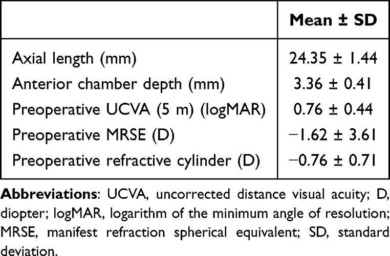 |
Table 1 Patient Demographics |
Absolute RPE
The mean ± SD values of the absolute RPE using the ORA, SRK/T, Barrett U II, and Haigis formulas were 0.24 ± 0.20, 0.28 ± 0.20, 0.23 ± 0.19, and 0.35 ± 0.24 D, respectively. The absolute RPE with ORA was significantly lower than Haigis (p = 0.001) but not significantly different from that with SRK/T and Barrett U II. Significant differences were also observed between Barrett U II and SRK/T (p = 0.004) and between Barrett U II and Haigis (p = 0.0001) (Figure 1). The median absolute RPE for ORA was 0.16 D, the lowest among all formulas (SRK/T: 0.25 D, Barrett U II: 0.18 D, Haigis: 0.30 D).
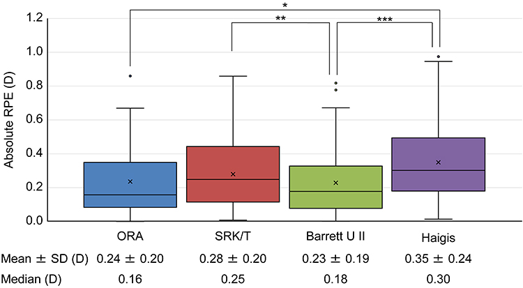 |
Figure 1 Absolute RPE. * p = 0.001, ** p = 0.004, *** p = 0.0001. Abbreviations: RPE, refractive prediction error; SD, standard deviation; Barrett U II, Barrett Universal II; D, diopter. |
Figure 2 shows the percentage of eyes with an absolute RPE of ≤ 0.25 D, ≤ 0.50 D, ≤ 0.75 D, and ≤ 1.0 D. The percentage of eyes with absolute RPE within 0.25 D for ORA, SRK/T, Barrett U II, and Haigis were 61.5%, 50.0%, 65.4%, and 42.3%, respectively, and those within 0.5 D were 87.2%, 85.9%, 87.2%, and 76.9%, respectively. The percentage of eyes with the absolute RPE within 0.25 D was significantly higher with ORA than with the SRK/T (p = 0.028) and Haigis formulas (p = 0.004), whereas no significant differences were observed between ORA and Barrett U II formulas. In addition, the Barrett U II formula showed a higher percentage of the absolute RPE within 0.25 D than the SRK/T (p = 0.003) and Haigis (p = 0.002) formulas. No significant differences were observed in the rate of RPE within 0.5 D, 0.75 D, and 1.0 D among the groups.
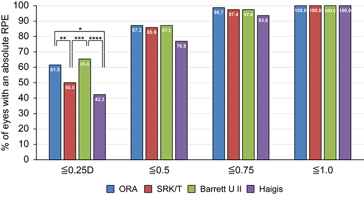 |
Figure 2 Percentage of eyes with an absolute RPE of ≤ 0.25 D, ≤ 0.50 D, ≤ 0.75 D, and ≤ 1.0 D. * p = 0.004, ** p = 0.028, *** p = 0.003, **** p = 0.002. Abbreviations: D, diopter; RPE, refractive prediction error; Barrett U II, Barrett Universal II. |
In the analysis of heteroscedasticity, both the ORA (SD: 0.31 D) and the Barrett U II (SD: 0.30 D) demonstrated significantly lower variability in prediction errors than the Haigis (SD: 0.42 D) (p = 0.005 and p = 0.002, respectively). No significant heteroscedasticity was observed among the other formulas.
Distribution of RPE and Scatterplots
Figure 3 shows the percentage of eyes with RPE in 0.25 D increments. The mode for ORA (41.0%), Barrett U II (38.5%), and SRK/T (26.9%) was located in the 0 D bin. In contrast, the mode for the Haigis was observed in the 0.25 to 0.50 D range (23.5%). When comparing the distribution patterns, ORA and Barrett U II demonstrated a higher degree of concentration in the 0 D bin, forming a sharper peak than SRK/T. Regarding outliers, ORA, Barrett U II, and SRK/T showed no eyes (0.0%) with a large refractive miss of ≤ −1.00 D. However, the Haigis demonstrated a notable myopic shift, with an RPE of ≤ −0.75 D for 13.5% of the eyes.
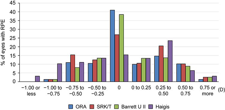 |
Figure 3 Percentage of eyes with a refractive prediction error in 0.25 D increments. Abbreviations: Barrett U II, Barrett Universal II; D, diopter; RPE, refractive prediction error. |
The scatterplots showing the relationship between predicted and achieved SE values are presented in Figure 4A–D. Data points for ORA tended to cluster relatively tightly around the identity line. Barrett U II and SRK/T exhibited similar distribution patterns, although both showed slightly more dispersion from the identity line than ORA. In contrast, the Haigis formula demonstrated a wider horizontal distribution of predicted values, with a broader range of variability among individual cases than the other methodologies.
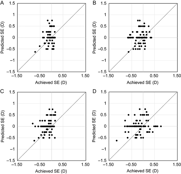 |
Figure 4 Scatterplots showing the relationship between predicted and achieved spherical equivalent. (A) ORA, (B) SRK/T, (C) Barrett U II, (D) Haigis. Abbreviations: Barrett U II, Barrett Universal II; D, diopter; SE, spherical equivalent. |
The scatterplots showing the relationship between AL and RPE are presented in Figure 5A–D. SRK/T and Haigis tended toward a hyperopic shift as AL increased, showing larger prediction errors in long eyes compared with ORA and Barrett U II. In contrast, ORA and Barrett U II demonstrated a trend toward a myopic shift in longer eyes.
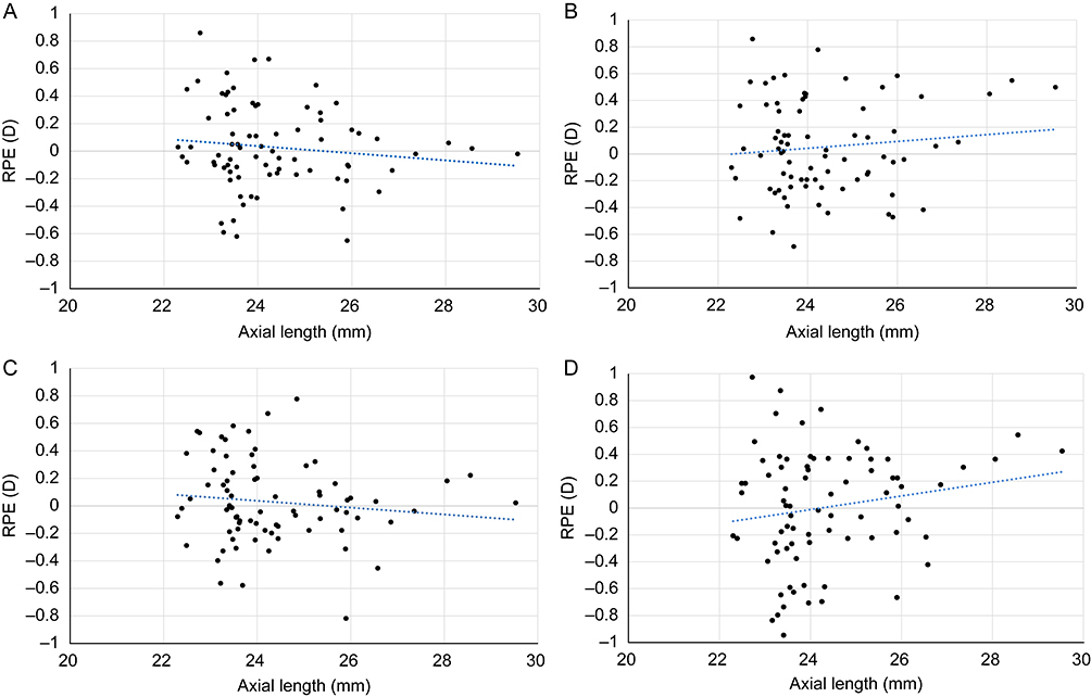 |
Figure 5 Scatterplots showing the relationship between axial length and refractive prediction error. (A) ORA, (B) SRK/T, (C) Barrett U II, (D) Haigis. Abbreviations: Barrett U II, Barrett Universal II; D, diopter; RPE, refractive prediction error. |
Visual Acuities
Table 2 shows visual acuity and refraction at 3 months after surgery. The mean monocular CDVA, UCVA, UIVA, and UNVA were −0.06 ± 0.07, −0.01 ± 0.07, 0.08 ± 0.12, and 0.06 ± 0.10 logMAR, respectively. The mean residual astigmatism was −0.34 ± 0.45 D. The manifest refraction SE (MRSE) was 0.03 ± 0.29 D. The MRSE was within ± 0.5 D for 94.9% of the eyes (74 of 78 eyes) and within ± 0.25 D for 73.1% of the eyes (57 of 78 eyes).
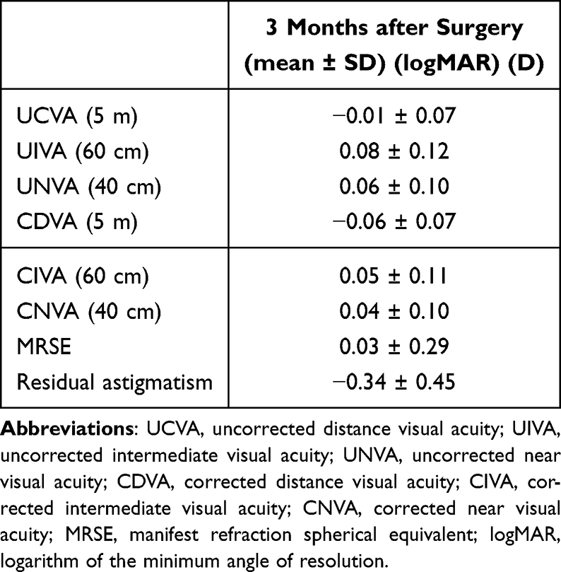 |
Table 2 Monocular Visual Acuity and Refraction at 3 Months After Surgery |
Discussion
In recent years, refractive expectations after cataract surgery have increased. A highly accurate IOL power calculation is required, especially for eyes that are implanted with a presbyopia-correcting IOL; however, the refractive outcomes in the new-material IOLs are sometimes unclear immediately after they are launched. This prospective multicenter study was aimed at comparing the accuracy of refraction in eyes implanted with the new trifocal IOL material using ORA and preoperative IOL calculations. In this study, the median absolute RPE for ORA was 0.16 D, and the rate of absolute RPE within 0.5 D was 87.2%. Both the frequency distribution (Figure 3) and the scatterplots (Figure 4) consistently demonstrated that the outcomes were highly concentrated around the 0.00 D mark with no significant outliers.
According to the report by Melendez et al, which showed the refractive accuracy of ORA for eyes with CNWT toric IOL implantation, the mean absolute RPE for ORA was 0.43 ± 0.36 D, and the absolute RPE rate within 0.5 D was 72.5% (using SS-OCT).17 In addition, a previous study on the refractive accuracy of ORA in eyes implanted with TFNT IOL, which is a trifocal IOL and has the same optical design as CNWT, showed that the median absolute RPE for ORA was 0.19 D, and the rate of absolute RPE within 0.5 D was highest with ORA (87.8%) (using SS-OCT).18 Blaylock et al reported that the mean ± SD of absolute RPE for ORA in the eyes with TFNT was 0.213 ± 0.219 D, and the rate of absolute RPE within 0.5 D was 88% (using partial coherence interferometry (PCI)).21 The current results in eyes implanted with the CNWTT0 trifocal IOL were equivalent to or better than the previous results.
Some studies have compared ORA and preoperative IOL power calculation formulas. Ma et al reported that there were no significant differences in the rate of absolute RPE within 0.5 D in the eyes with monofocal, multifocal, or toric IOLs between ORA (82%) and IOL power calculation formulas (Barrett U II, Hill RBF, SRK/T, 70–81%) (using SS-OCT and PCI).22 In a study by Cionni et al comparing ORA to preoperative IOL power calculation formulas in the eyes with AcrySof IQ monofocal, IQ toric, and Restor IOLs, they showed a significantly higher rate of absolute RPE within 0.5 D with ORA (82.4%) compared with preoperative IOL power calculation formulas (76%).23 In the present study, the refractive prediction accuracy within ±0.50 D was 87.2% for ORA alone and 87.2% for the Barrett U II formula alone, demonstrating comparable performance. In patients with long eyes, Raufi et al reported that there was no statistical difference in eyes with a > 26.25-mm AL between the ORA and preoperative IOL power calculation formulas (Barrett U II and Hill RBF) (using optical low-coherence reflectometry).24 Although some studies have reported that the ORA has superior refractive accuracy compared with preoperative IOL power calculation formulas, others have concluded that the ORA is the same as or inferior to preoperative IOL power calculation formulas.11,18,21–27 The RPEs of both ORA and preoperative IOL formulas are influenced by constant optimization, IOL type, and patient demographics; therefore, the superiority of one system over the other remains inconclusive.
In the current study, regarding the relationship between AL and RPE (Figure 5), the RPE in eyes with longer ALs was smaller with ORA and Barrett U II than those with the SRK/T and Haigis formulas. Notably, whereas the Haigis formula exhibited a slightly larger variance, no extreme outliers were observed across the entire range of ALs. This might be explained by the use of the ARGOS biometer, which performs IOL power calculations using segmented AL. Previous studies have reported that calculations based on segmented AL are less affected by AL variability.28,29
In this study, high refractive accuracy was consistent with good visual acuity outcomes from distance to near. These visual acuities are consistent with previous studies on eyes with TFNT.30–32 Nicula et al showed that monocular UCVA, UIVA at 60 cm, and UNVA at 40 cm were 0.07 ± 0.14, 0.08 ± 0.14, and 0.07 ± 0.14 logMAR, respectively.30 Another study reported that monocular UCVA, UIVA, and UNVA were 0.06 ± 0.07, 0.20 ± 0.10, and 0.05 ± 0.07 logMAR, respectively.31 The CNWT trifocal IOL can provide good distance, intermediate, and near visual acuity, similar to the TFNT trifocal IOL. Ullrich et al reported that the Clareon IOL possesses an axial stability equivalent to that of the AcrySof IQ IOL, indicating that the proven stability of the AcrySof platform has been inherited.33 In addition, a recent study by Kohnen et al reported excellent visual acuity and contrast sensitivity with CNWT, along with no glistening, at 12 months.34 While the water content of the material has been increased with CNWT, this modification is primarily intended to enhance optical clarity and contributes to improved visual quality without directly affecting refractive outcomes. As demonstrated in the current study, the new IOL retaining the stable physical behavior of the proven platform allowed ORA to maintain high precision, despite the change in material.
Trifocal IOLs are particularly sensitive to refractive errors, and even minor refractive errors can adversely affect visual acuity.6 Therefore, in addition to improving mean prediction accuracy, it is desirable to suppress variability in outcomes and minimize unexpected refractive surprises. One approach to evaluating variability is to analyze the SD for each formula. Heteroscedasticity analysis has been reported as an appropriate method for assessing SD differences.35 The analysis of heteroscedasticity in the current study revealed no significant difference in variance between ORA and the Barrett U II; however, ORA demonstrated significantly lower variability than the conventional Haigis (P = 0.005). This finding is consistent with a report by Watanabe, which suggested that ORA can reduce the variance of prediction errors and stabilize refractive outcomes compared with certain conventional formulas.36
From the perspective of clinical convenience, the Barrett U II formula is efficient and well-suited for daily surgical workflows, as it relies solely on preoperative measurements. Conversely, the role of the ORA system lies in providing real-time validation of power selection based on intraoperative wavefront aberrometry, a mechanism fundamentally distinct from preoperative calculation formulas. The observation that our final postoperative outcomes reached 94.9% is attributable to the adoption of a “hybrid approach” that integrated preoperative calculations with intraoperative ORA data rather than relying on a single method. Utilizing two tools with distinct measurement principles for “mutual validation” may be a viable strategy to suppress outliers and further stabilize clinical outcomes for trifocal IOLs.36,37
This study had some limitations. First, the sample size was relatively small, and the number of eyes with long and short ALs was limited. Second, as this study was non-randomized, the clinical outcomes reflect a “hybrid approach” integrating both preoperative data and ORA measurements. However, the “within‑eye comparison” design is a major strength of this study, as it eliminates the influence of anatomical differences or variations in background characteristics across different patients and allows for a more accurate assessment of the pure predictive performance of each calculation method. Furthermore, as emphasized in previous real-world studies, combining preoperative formulas with ORA is currently the most common clinical practice for ORA users, and the primary objective of this study was to evaluate the performance in such a real-world setting. ORA accuracy is highly sensitive to intraoperative conditions. While we established and followed a strict protocol to ensure precision, this may not be reproducible in routine clinical practice owing to the difficulty of maintaining such stringent conditions. The high cost and limited accessibility of ORA may restrict the generalizability of our results to global surgical settings. Therefore, future studies, including randomized controlled trials, will require verification using large sample sizes encompassing a wider variety of cases, such as those with extreme long and short ALs.
Conclusion
For the new CNWTT0 trifocal IOL, the refractive accuracy of the ORA system yielded more favorable outcomes than the SRK/T and Haigis formulas, whereas it was comparable to the Barrett U II formula. Integrating intraoperative validation with preoperative calculations provides a robust strategy to minimize refractive surprises and further optimize clinical outcomes.
Abbreviations
CDVA, corrected distance visual acuity; CIVA, corrected intermediate visual acuity; CNVA, corrected near visual acuity; D, diopter; logMAR, logarithm of the minimum angle of resolution; IOL, intraocular lens; MRSE, manifest refraction spherical equivalent; ORA, optiwave refractive analysis; RPE, refractive prediction error; SD, standard deviation; SE, spherical equivalent; SRK/T, Sanders–Retzlaff–Kraft/Theoretical; SS-OCT, swept-source optical coherence tomography; UCVA, uncorrected visual acuities; UIVA, uncorrected intermediate visual acuity; UNVA, uncorrected near visual acuity.
Data Sharing Statement
The datasets used and/or analyzed during the current study are available from the corresponding author on reasonable request.
Ethics Approval and Consent to Participate
The study protocol was approved by the Institutional Review Board of the Advanced Ophthalmological Medical Management Society (Hyogo, Japan) on July 28, 2022, and was conducted in accordance with the tenets of the Declaration of Helsinki and Ethical Guidelines for Medical and Biological Research involving human participants in Japan. All patients were properly counseled and provided written informed consent.
Acknowledgments
The authors would like to thank Apex LLC for editorial assistance in the preparation of the manuscript with funding from Alcon Japan Ltd.
Author Contributions
All authors made a significant contribution to the work reported, whether that is in the conception, study design, execution, acquisition of data, analysis and interpretation, or in all these areas; took part in drafting, revising, or critically reviewing the article; gave final approval of the version to be published; have agreed on the journal to which the article has been submitted; and agree to be accountable for all aspects of the work.
Funding
This study was supported by a grant for an Investigator-Initiated Trial from Alcon Japan Ltd. (#73398427). The funder had no role in the conceptualization, design, data collection, analysis, decision to publish, or preparation of the manuscript.
Disclosure
Y. Oka and Y. Matsumoto were supported by grants from Alcon Japan Ltd. The other authors declare that they have no competing interests for this work.
References
1. Shah S, Peris-Martinez C, Reinhard T, Vinciguerra P. Visual outcomes after cataract surgery: multifocal versus monofocal intraocular lenses. J Refract Surg. 2015;31(10):658–12. doi:10.3928/1081597X-20150611-01
2. Schallhorn JM, Pantanelli SM, Lin CC, et al. Multifocal and accommodating intraocular lenses for the treatment of presbyopia: a report by the American Academy of Ophthalmology. Ophthalmology. 2021;128(10):1469–1482. doi:10.1016/j.ophtha.2021.03.013
3. Bissen-Miyajima H, Ota Y, Hayashi K, Igarashi C, Sasaki N. Results of a clinical evaluation of a trifocal intraocular lens in Japan. Jpn J Ophthalmol. 2020;64(2):140–149. doi:10.1007/s10384-019-00712-4
4. Modi S, Lehmann R, Maxwell A, et al. Visual and patient-reported outcomes of a diffractive trifocal intraocular lens compared with those of a monofocal intraocular lens. Ophthalmology. 2021;128(2):197–207.
5. Kohnen T, Herzog M, Hemkeppler E, et al. Visual performance of a quadrifocal (trifocal) intraocular lens following removal of the crystalline lens. Am J Ophthalmol. 2017;184:52–62. doi:10.1016/j.ajo.2017.09.016
6. Hayashi K, Sato T, Igarashi C, Yoshida M. Effect of spherical equivalent error on visual acuity at various distances in eyes with a trifocal intraocular lens. J Refract Surg. 2019;35(5):274–279. doi:10.3928/1081597X-20190404-01
7. Connell BJ, Kane JX. Comparison of the Kane formula with existing formulas for intraocular lens power selection. BMJ Open Ophthalmol. 2019;4(1):e000251. doi:10.1136/bmjophth-2018-000251
8. Goto S, Maeda N, Ohnuma K, et al. Preliminary demonstration of a novel intraocular lens power calculation: the O formula. J Cataract Refract Surg. 2022;48(11):1305–1311. doi:10.1097/j.jcrs.0000000000000983
9. Debellemanière G, Dubois M, Gauvin M, et al. The PEARL-DGS Formula: the development of an open-source machine learning–based thick IOL calculation formula. Am J Ophthalmol. 2021;232:58–69.
10. Wiley WF, Bafna S. Intra-operative aberrometry guided cataract surgery. Int Ophthalmol Clin. 2011;51(2):119–129. doi:10.1097/IIO.0b013e31820f226d
11. Reitblat O, Levy A, Megiddo Barnir E, Assia EI, Kleinmann G. Toric IOL calculation in eyes with high posterior corneal astigmatism. J Refract Surg. 2020;36(12):820–825. doi:10.3928/1081597X-20200930-03
12. Woodcock MG, Lehmann R, Cionni RJ, Breen M, Scott MC. Intraoperative aberrometry versus standard preoperative biometry and a toric IOL calculator for bilateral toric IOL implantation with a femtosecond laser: one-month results. J Cataract Refract Surg. 2016;42(6):817–825. doi:10.1016/j.jcrs.2016.02.048
13. Hatch KM, Woodcock EC, Talamo JH. Intraocular lens power selection and positioning with and without intraoperative aberrometry. J Refract Surg. 2015;31(4):237–242. doi:10.3928/1081597X-20150319-03
14. Spekreijse LS, Bauer NJC, van den Biggelaar FJHM, et al. Predictive accuracy of an intraoperative aberrometry device for a new monofocal intraocular lens. J Cataract Refract Surg. 2022;48(5):542–548. doi:10.1097/j.jcrs.0000000000000791
15. Lee YW, Choi CY, Moon K, et al. Clinical outcomes of new multifocal intraocular lenses with hydroxyethyl methacrylate and comparative results of contrast sensitivity, objective scatter, and subjective photic phenomena. BMC Ophthalmol. 2022;22(1):379. doi:10.1186/s12886-022-02600-x
16. Yamashita K, Hayashi K, Hata S. Clinical performance and shape analysis of trifocal intraocular lenses via scanning electron microscopy. BMC Ophthalmol. 2024;24(1):86.
17. Melendez RF, Nguyen TH, Solis AI, Ortiz D, Moezzi C, Hall B. Outcomes after implantation of a trifocal toric intraocular lens using intraoperative aberrometry, digital image tracking, and femtosecond laser. Clin Ophthalmol. 2024;18:2033–2039. doi:10.2147/OPTH.S460060
18. Watanabe K. Evaluation of refractive accuracy of ORA and the factors impacting residual astigmatism in patients implanted with trifocal IOLs during cataract surgery: a retrospective observational study. Clin Ophthalmol. 2022;16:2491–2503. doi:10.2147/OPTH.S371555
19. Blaylock JF, Hall B. Astigmatisc results of a diffractive trifocal toric IOL following intraoperative aberrometry guidance. Clin Ophthalmol. 2020;14:4373–4378. doi:10.2147/OPTH.S285711
20. Wang L, Koch DD, Hill W, Abulafia A. Pursuing perfection in intraocular lens calculations: III. Criteria for analyzing outcomes. J Cataract Refract Surg. 2017;43(8):999–1002. doi:10.1016/j.jcrs.2017.08.003
21. Blaylock JF, Hall B. Clinical outcomes of a diffractive trifocal intraocular lens with femtosecond laser, digital tracking, and intraoperative aberrometry. Can J Ophthalmol. 2022;57(5):291–296.
22. Ma J, El-Defrawy S, Lloyd J, Rai A. Prediction accuracy of intraoperative aberrometry compared with preoperative biometry formulae for intraocular lens power selection. Can J Ophthalmol. 2023;58(1):2–10.
23. Cionni RJ, Dimalanta R, Breen M, Hamilton C. A large retrospective database analysis comparing outcomes of intraoperative aberrometry with conventional preoperative planning. J Cataract Refract Surg. 2018;44(10):1230–1235. doi:10.1016/j.jcrs.2018.07.016
24. Raufi N, James C, Kuo A, Vann R. Intraoperative aberrometry vs modern preoperative formulas in predicting intraocular lens power. J Cataract Refract Surg. 2020;46(6):857–861. doi:10.1097/j.jcrs.0000000000000173
25. Soifer M, Passi SF, Wisely CE, et al. Refractive outcomes using intraoperative aberrometry for highly myopic, highly hyperopic, and post-refractive eyes. J Refract Surg. 2021;37(9):609–615. doi:10.3928/1081597X-20210609-03
26. Davison JA, Potvin R. Preoperative measurement vs intraoperative aberrometry for the selection of intraocular lens sphere power in normal eyes. Clin Ophthalmol. 2017;11:923–929. doi:10.2147/OPTH.S135659
27. Pantanelli SM, Hatch K, Lin CC, et al. Intraoperative aberrometry versus preoperative biometry-based formulas for intraocular lens power calculation: a report by the American Academy of Ophthalmology. Ophthalmology. 2025;132(2):238–252. doi:10.1016/j.ophtha.2024.08.007
28. Tamaoki A, Kojima T, Hasegawa A, et al. Clinical evaluation of a new swept-source optical coherence biometer that uses individual refractive indices to measure AL in cataract patients. Ophthal Res. 2019;62(1):11–23. doi:10.1159/000496690
29. Shammas HJ, Shammas MC, Jivrajka RV, Cooke DL, Potvin R. Effects on IOL power calculation and expected clinical outcomes of axial length measurements based on multiple vs single refractive indices. Clin Ophthalmol. 2020;14:1511–1519.
30. Nicula CA, Popescu R, Rednik AM, Nicula D, Bulboaca AE, Stanescu I. Refractive lens exchange in hyperopic presbyopes with the Acrysof IQ Panoptix intraocular lens: one-year results and analysis of the literature. Ther Clin Risk Manag. 2020;16:1125–1137. doi:10.2147/TCRM.S279065
31. Torky MA, Nokrashy AE, Metwally H, Abdelhameed AG. Visual performance following implantation of presbyopia correcting intraocular lenses. Eye. 2025;39(1):79–87. doi:10.1038/s41433-022-02188-y
32. Rementería-Capelo LA, Contreras I, García-Pérez JL, Blázquez V, Ruiz-Alcocer J. Visual quality and patient satisfaction with a trifocal intraplantation of presbyopia correcting intraocular lenses. J Cataract Refract Surg. 2019;45(11):1584–1590. doi:10.1016/j.jcrs.2019.06.014
33. Ullrich M, Ruiss M, Hienert J, et al. Anterior chamber depth variability between 2 hydrophobic acrylic 1-piece intraocular lenses: randomized trial. J Cataract Refract Surg. 2021;47(11):1460–1465.
34. Kohnen T, Kaiser KP, Biller M, et al. Visual performance after implantation of a new trifocal IOL with an advanced hydrophobic acrylic biomaterial. J Cataract Refract Surg. 2025;51:777–783. doi:10.1097/j.jcrs.0000000000001688
35. Holladay JT, Wilcox RR, Koch DD, Wang L. Review and recommendations for univariate statistical analysis of spherical equivalent prediction error for IOL power calculations. J Cataract Refract Surg. 2021;47(1):65–77. doi:10.1097/j.jcrs.0000000000000370
36. Watanabe K. Accuracy of predicted refraction using two swept-source optical coherence biometers and an intraoperative aberrometer. J Cataract Refract Surg. 2025;51(6):461–467. doi:10.1097/j.jcrs.0000000000001628
37. Greenwood MD, Hutchison JW, Gorham RA, Kramer BA. The use of intraoperative aberrometry in normal eyes: an analysis of intraocular lens selection in scenarios of disagreement. J Refract Surg. 2022;38(5):304–309. doi:10.3928/1081597X-20220331-01
 © 2026 The Author(s). This work is published and licensed by Dove Medical Press Limited. The
full terms of this license are available at https://www.dovepress.com/terms
and incorporate the Creative Commons Attribution
- Non Commercial (unported, 4.0) License.
By accessing the work you hereby accept the Terms. Non-commercial uses of the work are permitted
without any further permission from Dove Medical Press Limited, provided the work is properly
attributed. For permission for commercial use of this work, please see paragraphs 4.2 and 5 of our Terms.
© 2026 The Author(s). This work is published and licensed by Dove Medical Press Limited. The
full terms of this license are available at https://www.dovepress.com/terms
and incorporate the Creative Commons Attribution
- Non Commercial (unported, 4.0) License.
By accessing the work you hereby accept the Terms. Non-commercial uses of the work are permitted
without any further permission from Dove Medical Press Limited, provided the work is properly
attributed. For permission for commercial use of this work, please see paragraphs 4.2 and 5 of our Terms.