
Back to Journals » International Medical Case Reports Journal » Volume 19
Recurrent Pyoderma Gangrenosum with Initial Onset in Early Pregnancy: A PARACELSUS-Supported Clinical Diagnosis in a Resource-Limited Setting in South-Western Uganda
Authors Olwit G  , Tumwesige C, Nek AJ , Chris-Uchendu HC , Tumwijukye S , Omalla ID, Mugisha AI , Atuhaire A , Mutekanga A, Odongo L
Received 6 May 2026
Accepted for publication 17 July 2026
Published 21 July 2026 Volume 2026:19 622264
DOI https://doi.org/10.2147/IMCRJ.S622264
Checked for plagiarism Yes
Review by Single anonymous peer review
Peer reviewer comments 2
Editor who approved publication: Professor Thomas E Hutson
Gerald Olwit,1,* Charles Tumwesige,2 Arthur Jonathan Nek,2 Hans Chinomso Chris-Uchendu,2 Sam Tumwijukye,1 Ivan Darcy Omalla,2 Aime Ishimwe Mugisha,3 Arnold Atuhaire,2 Andrew Mutekanga,1 Leo Odongo4,5,*
1Department of Internal Medicine, Kabale University School of Medicine, Kabale, Uganda; 2Department of General Surgery, Kabale University School of Medicine, Kabale, Uganda; 3Department of Internal Medicine, University of Rwanda, College of Medicine and Health Sciences, School of Medicine and Pharmacy, Kigali, Rwanda; 4Department of Dermatology, Allergology and Venereology, Kabale University School of Medicine, Kabale, Uganda; 5Department of Obstetrics and Gynaecology, Kabale University School of Medicine, Kabale, Uganda
*These authors contributed equally to this work
Correspondence: Gerald Olwit, Department of Internal Medicine, Kabale University School of Medicine, P.O.Box 317, Kabale, Uganda, Tel +256 776822110, Email [email protected]
Background: Pyoderma gangrenosum (PG) is a rare neutrophilic dermatosis characterized by rapidly progressive, painful cutaneous ulceration and remains a diagnosis of exclusion because no single laboratory or histopathological test is diagnostic. Establishing a clinically well-supported diagnosis is particularly challenging in resource-limited settings where histopathology and other advanced investigations are often unavailable, necessitating reliance on structured clinical assessment and validated diagnostic frameworks.
Case Presentation: A 26-year-old multiparous woman from South-Western Uganda presented with recurrent, progressively painful multifocal ulcerations involving both lower limbs and upper extremities. The initial episode developed during the first month of pregnancy as pruritic pustules that rapidly evolved into ulcerative lesions following minor trauma, with recurrence occurring two years later from a persistent non-healing ulcer. Comprehensive clinical evaluation found no evidence suggestive of inflammatory bowel disease (IBD) or other major associated systemic conditions, while wound cultures were sterile, supporting a non-infectious process. Because histopathology, gastrointestinal endoscopy, and other advanced investigations were unavailable, diagnosis relied on careful clinical assessment, systematic exclusion of important differential diagnoses, characteristic lesion morphology, pathergy, previous response to immunosuppressive therapy, and a PARACELSUS score supportive of PG. The patient was treated with oral prednisolone, cyclosporine, structured wound care, supportive therapy, and physiotherapy, resulting in marked clinical improvement at three-month follow-up.
Conclusion: This case demonstrates how structured clinical reasoning and validated diagnostic frameworks can support a clinically well-substantiated diagnosis of probable PG when confirmatory investigations are inaccessible. It also describes a temporal association between disease onset during early pregnancy and pregnancy loss, an observation that should be interpreted cautiously and requires further investigation rather than implying causality. Careful exclusion of alternative causes of ulceration remains fundamental before initiating immunosuppressive therapy.
Keywords: pyoderma gangrenosum, pregnancy-associated dermatosis, PARACELSUS criteria, Delphi consensus criteria, case report, resource-limited settings, Uganda
Introduction
Pyoderma gangrenosum is a rare, non-infectious neutrophilic dermatosis characterized by rapidly progressive, painful cutaneous ulcerations with undermined violaceous borders and surrounding erythema.1–5 It belongs to the spectrum of neutrophilic dermatoses, a group of disorders driven by dysregulated innate immune responses and aberrant neutrophil activity.1,6 Despite its striking clinical appearance, PG remains a diagnostic challenge due to the absence of pathognomonic laboratory or histopathological features, as well as its ability to mimic a wide range of infectious, vascular, malignant, and inflammatory conditions.3,7–9 The disease most commonly affects the lower extremities and often follows a relapsing–remitting course, contributing to significant morbidity and impaired quality of life.10–12
The pathogenesis of PG is incompletely understood but is thought to involve a complex interplay between genetic susceptibility, immune dysregulation, and environmental triggers.1,7,9,13 Abnormal neutrophil chemotaxis, altered cytokine profiles including tumor necrosis factor-α and interleukin-mediated pathways and dysregulation of both innate and adaptive immunity have been implicated.14–16 PG is frequently associated with systemic diseases, most notably IBD, inflammatory arthritis, and hematologic disorders, with up to 50% of cases occurring in the context of an underlying condition.6,9,13 Consequently, a thorough evaluation to identify associated comorbidities is a critical component of patient assessment.3
Pregnancy represents a unique immunological state characterized by dynamic modulation of the maternal immune system, which may predispose susceptible individuals to the development or exacerbation of immune-mediated conditions such as PG.6,10,13,17 Although PG occurring during pregnancy has been documented in the literature, it remains uncommonly reported in low-resource settings, and its clinical course, triggers, and outcomes are not fully characterized.6 The immunological shifts toward a neutrophil-dominant and pro-inflammatory milieu during certain stages of pregnancy have been proposed as potential contributors to disease onset or progression. However, the relationship between pregnancy and PG is complex and not yet clearly defined.6,18,19
The diagnosis of PG is primarily clinical and relies on the recognition of characteristic lesion morphology, exclusion of alternative causes of ulceration, and supportive clinical features such as pathergy and response to immunosuppressive therapy.3,13,16 In recent years, standardized diagnostic frameworks have been developed to improve diagnostic accuracy, most notably the Delphi Consensus Criteria proposed by Maverakis et al and the PARACELSUS score.13,20–22 While the Delphi criteria combine histopathology with clinical features, the PARACELSUS score emphasizes clinical characteristics and exclusion of differential diagnoses, making both valuable adjuncts when evaluating suspected pyoderma gangrenosum.21–23 While both tools have demonstrated value in supporting diagnosis, the PARACELSUS score is particularly advantageous in resource-limited settings where access to skin biopsy, dermatopathology, gastrointestinal endoscopy, and advanced immunological investigations may be unavailable.7,21,23 In such environments, careful clinical reasoning supported by validated clinical diagnostic frameworks becomes essential for establishing a well-substantiated diagnosis of probable pyoderma gangrenosum.3,8,24
Although pregnancy-associated pyoderma gangrenosum has been described previously, published reports have largely originated from resource-rich settings where histopathology, advanced laboratory investigations, vascular assessment, and gastrointestinal endoscopy are readily available to support diagnosis. Consequently, there remains limited evidence describing how clinicians can confidently evaluate and manage suspected PG when these investigations are inaccessible. Furthermore, few reports have systematically applied validated clinical diagnostic frameworks to substantiate a diagnosis in such settings. This case therefore addresses an important knowledge gap by demonstrating how meticulous exclusion of differential diagnoses, structured application of the PARACELSUS and Delphi diagnostic criteria, and careful clinical reasoning can support a clinically well-substantiated diagnosis of probable PG despite substantial diagnostic constraints. By detailing the diagnostic work-up, clinical decision-making process, and pragmatic management in a resource-limited Ugandan setting, this report provides an educational framework that may assist clinicians facing similar diagnostic challenges where confirmatory investigations are unavailable.
Case Presentation
A 26-year-old multiparous woman from South-Western Uganda presented to our emergency department with recurrent, progressively painful ulcerative skin lesions involving both lower limbs and upper extremities. She reported a two-year history of recurrent painful skin ulcerations that initially began during the first month of her third pregnancy. Her obstetric history was significant for two miscarriages: the first occurred 6 months prior to the onset of the skin disease, while the second occurred shortly (one month) after the initial appearance of the lesions during early pregnancy. The disease initially started as intensely pruritic pustular lesions over both ankles during early pregnancy. Following minor trauma from scratching, the pustules rapidly evolved into painful ulcers that progressively enlarged, extending to the legs, knees, and elbows. The lesions were associated with severe pain, seropurulent discharge, surrounding erythema, and progressive difficulty in walking due to pain and limited mobility. At the time of the initial presentation two years earlier, she had been treated with systemic corticosteroids and supportive wound care, resulting in marked improvement of all lesions except for one persistent non-healing ulcer on the right leg. Approximately one month prior to the current admission, she developed worsening pain and recurrence of multiple ulcerations originating from the previously persistent ulcer on the right leg, with rapid progression to involve both ankles, both legs, both knees, and both elbows. She denied preceding trauma, recent surgery, insect bites, or use of new medications before the recurrence.
Because PG is strongly associated with IBD, a detailed gastrointestinal history was obtained despite the inability to perform advanced investigations such as colonoscopy, upper gastrointestinal endoscopy, intestinal biopsy, fecal calprotectin testing, inflammatory marker profiling, and skin biopsy due to financial limitations and lack of availability in our resource-limited setting. The patient denied history of chronic diarrhea, bloody stools, mucoid stools, abdominal pain, abdominal cramping, tenesmus, urgency, nocturnal diarrhea, unexplained weight loss, recurrent oral ulcers, perianal pain, fistula formation, rectal bleeding, abdominal distension, or previous diagnosis of Crohn’s disease or ulcerative colitis. There was also no family history of IBD, autoimmune disease, or chronic dermatologic conditions. She further denied history of chronic cough, night sweats, fever, genital ulcers, recent sexually transmitted infections, known tuberculosis exposure, diabetes mellitus, HIV-related symptoms, or previous malignancy. There was no history suggestive of vasculitis, connective tissue disease, or hematologic malignancy.
Given the patient’s history of two miscarriages, thrombotic and autoimmune vasculopathies, particularly antiphospholipid syndrome, were also considered during the diagnostic evaluation because these conditions may present with chronic lower-extremity ulceration and adverse pregnancy outcomes. However, specialized investigations including coagulation studies, antiphospholipid antibody testing (lupus anticoagulant, anticardiolipin antibodies, and anti-β2-glycoprotein I antibodies), and other thrombophilia assessments were not performed because they were unavailable and financially inaccessible in our resource-limited setting. Nevertheless, the clinical presentation was considered less consistent with vasculopathy, given the characteristic rapidly progressive pustule-to-ulcer evolution, pathergy following minor trauma, multifocal lesions with undermined violaceous borders, marked pain, sterile wound cultures, and previous favorable response to immunosuppressive therapy.
On general examination, she was fully conscious, oriented, and in severe painful distress, with difficulty ambulating due to pain. Her vital signs were within normal limits. She had mild conjunctival pallor but no jaundice, cyanosis, lymphadenopathy, finger clubbing, or pedal edema. Dermatological examination revealed multiple deep, extremely tender ulcerations involving both ankles, circumferentially extending over the lower legs, with additional lesions over both knees and both elbows. The ulcers had irregular undermined violaceous borders with erythematous surrounding skin and necrotic slough at the base. Some lesions showed seropurulent discharge and crusting, while others demonstrated granulation tissue with areas of healing. The lesions were sharply demarcated and exhibited the characteristic violaceous undermined edge typical of PG (Figure 1). Marked tenderness was elicited on palpation, and movement of the affected joints was significantly limited by pain. No features suggestive of venous insufficiency, arterial disease, diabetic ulcers, or pressure sores were identified.
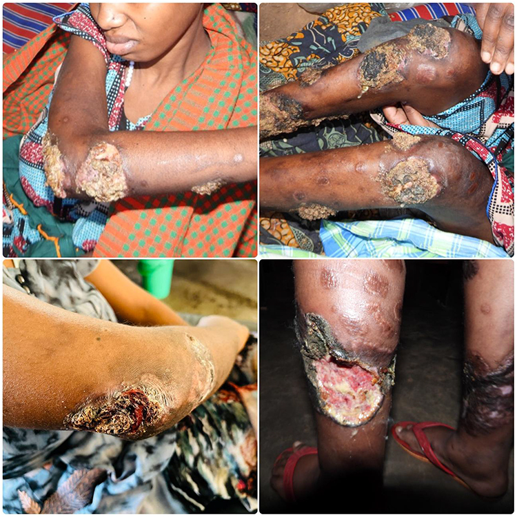 |
Figure 1 Clinical photographs obtained at the time of hospital admission before initiation of systemic immunosuppressive therapy, demonstrating multifocal, painful ulcerative lesions involving both lower limbs and upper extremities. The lesions exhibit characteristic irregular ulcers with undermined violaceous borders, surrounding erythema, necrotic slough, and areas of seropurulent discharge, features that supported a clinical diagnosis of probable pyoderma gangrenosum after exclusion of major differential diagnoses. |
Initial laboratory investigations demonstrated normocytic normochromic anemia with hemoglobin of 7.4 g/dL, mild leukocytosis with neutrophilia and monocytosis, and thrombocytosis, consistent with active inflammation. Liver function tests showed hypoalbuminemia and elevated aspartate aminotransferase, while renal function remained preserved (Table 1). Serum electrolytes were within normal ranges. An ulcer swab was obtained for microscopy, culture, and sensitivity to exclude infectious causes of chronic ulceration. No bacterial growth was isolated on culture, supporting a sterile inflammatory rather than infectious etiology. This negative culture was particularly important in excluding bacterial pyoderma and other infective ulcerative dermatoses before initiation of immunosuppressive therapy. Although skin biopsy and histopathological confirmation would have been ideal to further exclude neoplastic and vasculitic causes, this was not feasible due to financial constraints and limited access to dermatopathology services. Likewise, colonoscopy and gastrointestinal endoscopy to definitively exclude IBD were unavailable and unaffordable for the patient.
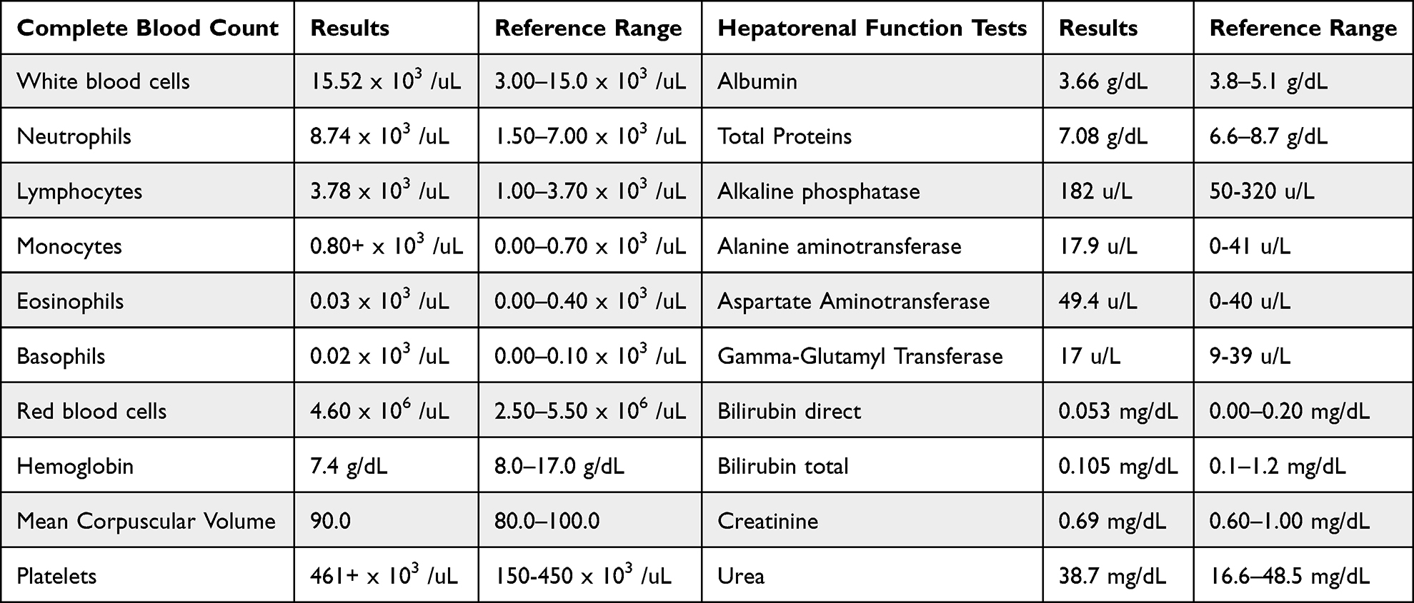 |
Table 1 Baseline Complete Blood Count and Hepatorenal Function Test Results Obtained on Admission |
A clinically well-supported diagnosis of probable recurrent ulcerative PG was reached based on the characteristic history, recurrent nature of disease, lesion morphology, pathergy-like progression from pustules following minor trauma, negative wound cultures, exclusion of major differential diagnoses through detailed history and examination, and previous favorable response to systemic corticosteroids.
Using the PARACELSUS score for PG, the patient strongly fulfilled the diagnostic criteria: rapidly progressive disease (3 points), exclusion of relevant differential diagnoses (3 points), reddish-violaceous wound border (3 points), extreme pain (2 points), lesion occurring at sites of trauma/pathergy following scratching (2 points), irregular ulcer shape (2 points), and improvement with immunosuppressive therapy during the previous episode (2 points), giving a total score of 17 points, which strongly supported a clinical diagnosis of probable PG.
Application of the Delphi Consensus Criteria further strengthened the clinical assessment supporting a diagnosis of probable PG. Although the major criterion of biopsy-confirmed neutrophilic infiltrate could not be assessed due to the unavailability of histopathological services, the patient fulfilled several minor criteria, including exclusion of infection through negative wound cultures, pathergy following minor trauma, rapidly progressive painful ulceration evolving from pustules, characteristic morphology with undermined violaceous borders, multifocal involvement of the anterior lower legs, and a favorable response to systemic corticosteroids and cyclosporine.
She was commenced on daily wound cleansing using diluted potassium permanganate solution for two weeks to promote antisepsis, reduce bacterial colonization, dry excessive wound exudate, decrease odor, and facilitate healthy granulation tissue formation while minimizing secondary bacterial infection. Intravenous ceftriaxone 2 g once daily for seven days was administered not as primary treatment for pyoderma gangrenosum, but to prevent and manage possible secondary bacterial infection of the open ulcerative wounds. Oral prednisolone 40 mg and cyclosporine 200mg once daily were initiated as the primary immunosuppressive therapy for two weeks to control the underlying neutrophilic inflammatory process. She also received oral lansoprazole 30 mg once daily for gastric protection during corticosteroid therapy, oral calcium and vitamin D (Calcivita tablets) supplementations for one month to reduce steroid-associated bone demineralization risk, oral morphine syrup for severe pain control during the first week, and intramuscular diclofenac for additional short-term analgesia during the initial 48 hours.
To prevent the development of limb contractures, the patient was taught to administer active physiotherapy. Adequate feeding and fluid intake were also encouraged. Three months after treatment initiation, the patient demonstrated noticeable clinical improvement, with significant reduction in pain, decreased surrounding inflammation, reduced purulent discharge, and improved mobility, although she still required support with a walking stick for ambulation. Several ulcers showed progressive granulation and reduction in inflammatory activity, though some persistent lesions continued to discharge minimally (Figure 2).
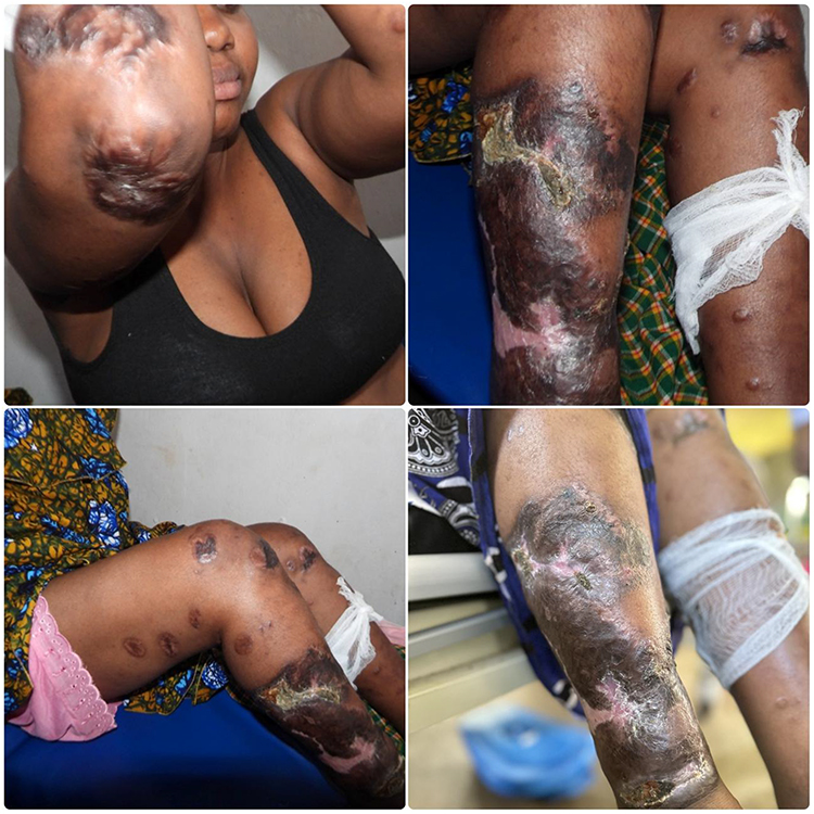 |
Figure 2 Clinical photographs obtained three months after initiation of treatment with oral prednisolone, cyclosporine, structured wound care, and supportive therapy, demonstrating marked clinical improvement. Compared with baseline, the lesions show reduced surrounding inflammation, decreased purulent discharge, progressive granulation tissue formation, contraction of ulcer size, and early re-epithelialization, consistent with a favorable clinical response to therapy. |
Discussion
This case highlights how a clinical diagnosis of suspected recurrent ulcerative PG was established in a resource-limited setting through careful synthesis of history, examination, exclusion of differentials, and structured application of the PARACELSUS and the Delphi Consensus Criteria. A central criticism in prior reviews of PG case reports is diagnostic uncertainty, particularly when histopathology and advanced investigations are lacking.8 In this patient, multiple converging features support PG: rapid evolution from pustules to painful ulcers following minor trauma (pathergy), characteristic undermined violaceous borders, severe pain out of proportion to examination findings, multifocal distribution, chronic relapsing course, negative wound cultures, and a clear therapeutic response to systemic corticosteroids. The PARACELSUS score of 17 and several minor components of the Delphi Consensus Criteria further strengthens diagnostic confidence and directly addresses concerns regarding reliance on clinical impression alone. While biopsy and endoscopic evaluation would have been ideal, their absence does not preclude diagnosis when a high-probability clinical framework is rigorously applied, particularly in low-resource environments.
Recent reviews recommend a structured diagnostic work-up for suspected PG incorporating comprehensive clinical assessment, systematic exclusion of mimicking conditions, microbiological evaluation, histopathological examination whenever feasible, assessment for associated systemic diseases, and application of validated diagnostic frameworks.22 Our diagnostic approach was aligned with these principles within the constraints of our resource-limited setting, although histopathology and several specialized investigations were unavailable.
A key aspect of this case is the systematic exclusion of alternative causes of chronic ulceration prior to initiation of immunosuppression. Infectious etiologies including bacterial, mycobacterial, and deep fungal infections were considered and reasonably excluded by negative culture results and lack of systemic features such as fever, lymphadenopathy, or constitutional symptoms. Vascular causes such as arterial insufficiency, venous stasis ulcers, and vasculitis were not supported clinically by lesion morphology, distribution, or associated signs. Malignancy and pyoderma-like neoplastic processes were also considered unlikely given the rapid inflammatory progression, pain severity, and rapid response to oral steroids and cyclosporine. Importantly, IBD, one of the most common systemic associations of PG,1,4,9 was carefully evaluated through detailed history in the absence of colonoscopy and histological assessment. The patient denied experiencing hallmark features of IBD, including chronic diarrhea, hematochezia, abdominal pain, weight loss, and perianal disease. While the inability to perform endoscopic evaluation remains a limitation, the absence of clinical indicators significantly lowers the pre-test probability of IBD and supports a clinically well-substantiated diagnosis of probable PG in the absence of clinical evidence suggestive of IBD.
The temporal relationship between disease onset and early pregnancy in this patient aligns with existing literature suggesting pregnancy as a potential trigger for PG, although reported cases remain rare.6,17,18,25 Prior studies have described PG arising during pregnancy often in the second trimester with variable lesion distribution and severity, and with or without recurrence in subsequent pregnancies.6,10 In contrast, our patient developed PG in the first month of pregnancy, which is relatively uncommon and may reflect early immunological shifts that favor neutrophil activation. Pregnancy is characterized by complex immune modulation, including alterations in cytokine profiles and enhanced neutrophil activity, which may predispose susceptible individuals to neutrophilic dermatoses.18,25 Furthermore, the recurrence of PG in this patient two years later from a persistent ulcer supports the concept that incomplete resolution or treatment interruption may serve as a nidus for reactivation, a phenomenon also described in previous case series.14,15
The use of antibiotics in this patient warrants clarification because wound cultures were sterile. Antibiotics were not prescribed to treat pyoderma gangrenosum, which is a sterile neutrophilic dermatosis, but rather to reduce the risk of secondary bacterial infection in extensive open ulcerative wounds prior to and during initiation of systemic immunosuppressive therapy. Patients with large chronic ulcers remain vulnerable to subsequent bacterial colonization despite initially negative cultures,3,5,26 particularly in resource-limited settings where close microbiological surveillance may not be feasible. Consequently, antimicrobial therapy served as an adjunct to immunosuppression and comprehensive wound care rather than definitive treatment of the underlying disease, a concept described in literature.3
This case contributes to the limited literature describing the diagnostic evaluation and management of probable PG in resource-limited settings, where histopathology and advanced investigations are unavailable. More importantly, this report illustrates how a systematic diagnostic approach incorporating meticulous clinical assessment, careful exclusion of major differential diagnoses, microbiological evaluation, and complementary application of the PARACELSUS and Delphi diagnostic frameworks can support a clinically well-substantiated diagnosis of probable pyoderma gangrenosum when confirmatory investigations are not feasible. The case also demonstrates a pragmatic management strategy in which systemic immunosuppression, structured wound care, empiric measures to reduce the risk of secondary bacterial infection, pain control, and rehabilitation were integrated within the realities of a low-resource healthcare setting. By transparently acknowledging the limitations imposed by unavailable diagnostic investigations while detailing the clinical reasoning underpinning the diagnosis and management, this report provides an educational framework that may assist clinicians encountering similar diagnostic challenges in comparable resource-constrained environments.
This case has several important limitations that should be acknowledged. Histopathological examination, the major criterion of the Delphi Consensus Criteria, could not be performed because dermatopathology services were unavailable and financially inaccessible. Similarly, gastrointestinal endoscopy, vascular imaging, coagulation studies, antiphospholipid antibody testing, and other specialized immunological investigations could not be undertaken, preventing complete exclusion of IBD, thrombotic vasculopathies, and other less common mimickers of pyoderma gangrenosum. Consequently, despite rigorous clinical evaluation, systematic exclusion of major differential diagnoses, negative microbiological findings, and supportive PARACELSUS and Delphi diagnostic criteria, a definitive diagnosis could not be established. Instead, the findings support a clinically well-substantiated diagnosis of probable PG, consistent with current recommendations that PG remains a diagnosis of exclusion. These limitations reflect the realities of clinical practice in many resource-constrained settings and underscore the importance of interpreting this diagnosis within the context of the available evidence.
Conclusion
This case highlights the challenges of diagnosing PG in resource-limited settings where histopathology, advanced immunological investigations, vascular studies, and gastrointestinal endoscopy may not be readily available. Through comprehensive clinical evaluation, meticulous exclusion of major differential diagnoses, negative microbiological findings, and structured application of the PARACELSUS and Delphi diagnostic frameworks, a clinically well-supported diagnosis of probable PG was reached. Although the absence of histopathological confirmation and certain specialized investigations, including evaluation for thrombotic vasculopathies, precludes a definitive diagnosis, the characteristic clinical presentation, pathergy, recurrent disease course, and favorable response to immunosuppressive therapy supported PG as the most likely diagnosis. This case underscores the value of structured diagnostic reasoning in low-resource settings while emphasizing that pyoderma gangrenosum remains a diagnosis of exclusion requiring careful consideration of alternative causes before initiating immunosuppressive treatment. Finally, the temporal association between disease onset during early pregnancy and pregnancy loss should be interpreted cautiously as an observation that warrants further investigation rather than implying a causal relationship.
Ethics Approval
Ethical approval was not required for this case report in accordance with Kabale University Research Ethics Committee (KAB-REC).
Consent for Publication
Written informed consent was obtained from the patient for publication of the case details and accompanying clinical images.
Acknowledgment
We sincerely express our deepest gratitude to the patient and her husband for their willingness to share her clinical journey and for granting informed consent to publish this case for educational and scientific purposes. Their cooperation and trust made this report possible and contribute meaningfully to advancing understanding of this rare condition. We also extend our appreciation to the Departments of Dermatology, Allergology and Venereology and General Surgery of Kabale University School of Medicine and the clinical team at Kabale Regional Referral Hospital for their invaluable support in the care of this patient. Their commitment to patient-centered care and clinical excellence, particularly within a resource-limited setting, was instrumental in the diagnosis, management, and documentation of this case.
Author Contributions
All authors made a significant contribution to the work reported, whether that is in the conception, study design, execution, acquisition of data, analysis and interpretation, or in all these areas; took part in drafting, revising or critically reviewing the article; gave final approval of the version to be published; have agreed on the journal to which the article has been submitted; and agree to be accountable for all aspects of the work.
Funding
The authors received no funding for this work.
Disclosure
The authors declare that they have no competing interests in this work.
References
1. David F, Lopes Freitas R, Brás-Cruz R, Rocha J, Rosário C. A case of recurrent idiopathic pyoderma Gangrenosum. Cureus. 2022. doi:10.7759/cureus.25112
2. Suvirya S, Pathania S, Singhai A. A case of bullous pyoderma gangrenosum. BMJ Case Rep. 2019;12(3):e228772. doi:10.1136/bcr-2018-228772
3. Ye M, Ye J, Wu L, Keating C, Choi WT. A challenging diagnosis: case report of extensive pyoderma gangrenosum at multiple sites. Clin Cosmet Invest Dermatol. 2014;105. doi:10.2147/CCID.S60229
4. Mohtadi M, Alocha H, Mahmoud A, Perez C, Lovaas C. From unassuming to unbelievable: a case report of Pyoderma Gangrenosum. Cureus. 2024. doi:10.7759/cureus.53491
5. Langan SM, Groves RW, Card TR, Gulliford MC. Incidence, mortality, and disease associations of Pyoderma Gangrenosum in the United Kingdom: a retrospective cohort study. J Invest Dermatol. 2012;132(9):2166–9. doi:10.1038/jid.2012.130
6. Ohmaru-Nakanishi T, Goto H, Maehara M, Oishi H, Ueoka Y. Perineal pyoderma gangrenosum in pregnancy: a case report. Case Rep Womens Health. 2019;22:e00102. doi:10.1016/j.crwh.2019.e00102
7. Daniel Su WP, Davis MDP, Weenig RH, Powell FC, Perry HO. Pyoderma gangrenosum: clinicopathologic correlation and proposed diagnostic criteria. Int J Dermatol. 2004;43(11):790–800. doi:10.1111/j.1365-4632.2004.02128.x
8. Weenig RH, Davis MDP, Dahl PR, Daniel Su WP. Skin ulcers misdiagnosed as pyoderma gangrenosum. N Engl J Med. 2002;347(18):1412–1418. doi:10.1056/NEJMoa013383
9. Rukhshan R, Eliogu C, Mistry A, Brar S, Mohd AA, Ruck L. Pyoderma gangrenosum: a case highlighting the importance of timely diagnosis. Cureus. 2024. doi:10.7759/cureus.57762
10. Steele RB, Nugent WH, Braswell SF, Frisch S, Ferrell J, Ortega‐Loayza AG. Pyoderma gangrenosum and pregnancy: an example of abnormal inflammation and challenging treatment. Br J Dermatol. 2016;174(1):77–87. doi:10.1111/bjd.14230
11. Zhang XQ, Tang ZW, Jing J. Progressive Facial Ulcer: a Case Report of Pyoderma gangrenosum. J Inflamm Res. 2024;17:687–691. doi:10.2147/JIR.S441751
12. Brunsting LA. Pyoderma (Echthyma) gangrenosum: Clinical And Experimental Observations In Five Cases Occurring In Adults. Arch Dermatol Syphilol. 1930;22(4):655. doi:10.1001/archderm.1930.01440160053009
13. Skopis M, Bag-Ozbek A. Pyoderma gangrenosum: a review of updates in diagnosis, pathophysiology and management. Multidisc Scient J. 2021;4(3):367–375. doi:10.3390/j4030028
14. Becker SL, Velasco R, Ortega-Loayza AG. Recalcitrant Pyoderma Gangrenosum: clinical burden and unmet needs. Clin Cosmet Invest Dermatol. 2023;16:2143–2152. doi:10.2147/CCID.S381490
15. Clark AL, Williams B. Recurrence of Pyoderma Gangrenosum potentially triggered by COVID-19 vaccination. Cureus. 2022. doi:10.7759/cureus.22625
16. Bardazzi F, Maltoni L, Clarizio G, et al. Pyoderma gangrenosum: a retrospective case series of 44 patients. Dermatol Pract Concept. 2024;14(4):e2024265. doi:10.5826/dpc.1404a265
17. Wanberg LJ, Fletcher KM, Goldfarb N, Alavi A. Treatment of pyoderma gangrenosum in pregnancy with certolizumab pegol. JEADV Clin Pract. 2024;3(2):696–698. doi:10.1002/jvc2.346
18. Wanberg LJ, Gorman BG, Theis‐Mahon N, Goldfarb N, Alavi A. Management of pyoderma gangrenosum during pregnancy and breastfeeding: a systematic review. Int J Dermatol. 2025;64(1):11–14. doi:10.1111/ijd.17402
19. Horth D, Assouyat N, Auger I. Successful treatment of refractory pyoderma gangrenosum with risankizumab in a 57-year-old patient: a case report. SAGE Open Med Case Rep. 2025;13:2050313X251352130. doi:10.1177/2050313X251352130
20. Maverakis E, Ma C, Shinkai K, et al. Diagnostic criteria of ulcerative pyoderma gangrenosum: a delphi consensus of international experts. JAMA Dermatol. 2018;154(4):461. doi:10.1001/jamadermatol.2017.5980
21. Jockenhöfer F, Wollina U, Salva KA, Benson S, Dissemond J. The
22. Moltrasio C, Romagnuolo M, Tavoletti G, Maronese CA, Marzano AV. Pyoderma gangrenosum: pathogenetic mechanisms and their implications for treatment. Semin Immunopathol. 2025;47(1):38. doi:10.1007/s00281-025-01064-7
23. Maronese CA, Pimentel MA, Li MM, Genovese G, Ortega-Loayza AG, Marzano AV. Pyoderma gangrenosum: an updated literature review on established and emerging pharmacological treatments. Am J Clin Dermatol. 2022;23(5):615–634. doi:10.1007/s40257-022-00699-8
24. Chen B, Li W, Qu B. Practical aspects of the diagnosis and management of pyoderma gangrenosum. Front Med. 2023;10:1134939. doi:10.3389/fmed.2023.1134939
25. Tan MG, Singh N, Kirshen C, Tolkachjov SN. Pregnancy‐associated pyoderma gangrenosum: a systematic review. Int J Dermatol. 2025;64(1):201–203. doi:10.1111/ijd.17474
26. Schøsler L, Fogh K, Bech R. Pyoderma gangrenosum: a retrospective study of clinical charac-teristics, comorbidities, response to treatment and mortality related to prednisone dose. Acta Derm Venereol. 2021;101(4):adv00431. doi:10.2340/00015555-3776
 © 2026 The Author(s). This work is published and licensed by Dove Medical Press Limited. The
full terms of this license are available at https://www.dovepress.com/terms
and incorporate the Creative Commons Attribution
- Non Commercial (unported, 4.0) License.
By accessing the work you hereby accept the Terms. Non-commercial uses of the work are permitted
without any further permission from Dove Medical Press Limited, provided the work is properly
attributed. For permission for commercial use of this work, please see paragraphs 4.2 and 5 of our Terms.
© 2026 The Author(s). This work is published and licensed by Dove Medical Press Limited. The
full terms of this license are available at https://www.dovepress.com/terms
and incorporate the Creative Commons Attribution
- Non Commercial (unported, 4.0) License.
By accessing the work you hereby accept the Terms. Non-commercial uses of the work are permitted
without any further permission from Dove Medical Press Limited, provided the work is properly
attributed. For permission for commercial use of this work, please see paragraphs 4.2 and 5 of our Terms.