
Back to Journals » International Journal of Nanomedicine » Volume 20
Recent Trends in Biomedical Applications of Cu2MX4-Based Nanocomposites: An Updated Review
Authors Gangadhar L, Sana SS  , Mishra V, Venkatesan R , Kim SC, Al-Tabakha MM
Received 23 June 2025
Accepted for publication 18 September 2025
Published 26 September 2025 Volume 2025:20 Pages 11895—11939
DOI https://doi.org/10.2147/IJN.S548959
Checked for plagiarism Yes
Review by Single anonymous peer review
Peer reviewer comments 5
Editor who approved publication: Prof. Dr. RDK Misra
Lekshmi Gangadhar,1,* Siva Sankar Sana,2,* Vijayalaxmi Mishra,2,* Raja Venkatesan,2,3 Seong-Cheol Kim,2 Moawia M Al-Tabakha4,5
1Department of Nanotechnology, Nanodot Research Private Limited, Nagercoil, Kanyakumari, Tamil Nadu, India; 2School of Chemical Engineering, Gyeongsan, Yeungnam University, Gyeongsan, Gyeongbuk, Republic of Korea; 3Department of Biomaterials, Saveetha Dental College and Hospitals, SIMATS, Saveetha University, Chennai, Tamil Nadu, India; 4Department of Pharmaceutical Sciences, College of Pharmacy and Health Sciences, Ajman University, Ajman, United Arab Emirates; 5Centre of Medical and Bio-Allied Health Sciences Research Centre, Ajman University, Ajman, United Arab Emirates
*These authors contributed equally to this work
Correspondence: Seong-Cheol Kim, Email [email protected] Moawia M Al-Tabakha, Email [email protected]
Abstract: Recent advancements in Cu2MX4 (CMX)-based nanocomposites have garnered significant attention in the biomedical field due to their exceptional structural, optical, electrical, and catalytic properties. In this review, recent developments regarding the synthesis, properties, and applications of CMX nanostructures in biomedicine, along with their high versatility and functionality, are discussed in detail. The various synthesis techniques, such as hydrothermal, solvothermal, and chemical vapour deposition methods and their influence on the properties of nanomaterials for therapeutic and diagnostic applications are also discussed. CMX-based nanocomposites cover highly important biomedical applications, including drug delivery, photothermal and photodynamic therapies, bioimaging, and antimicrobial activity. For the applications in targeted and controlled drug delivery, CMX, therefore, provides an efficient pathway to improve therapeutic efficiency while reducing adverse effects. The high photothermal conversion efficiency also makes this material beneficial for cancer therapies. The inherent fluorescence and magnetic properties of these agents may be beneficial in advanced bioimaging techniques. The good antimicrobial efficacy of CMX materials opens new avenues for combating microbial resistance. Mechanistic insights into cellular interactions, oxidative stress induction, and catalytic activities help provide a deeper understanding of the functions of these nanostructures in biological systems. Along with many future awaiting applications, toxicity, scalability, physico-stability, and regulatory issues are critical hurdles that need to be addressed for clinical translation to occur with CMX-based nanocomposite. The future aspects of enhancing the synthesis route, biocompatibility, and leveraging interdisciplinary approaches to optimize these materials for biomedical applications are also discussed. The unique multifunctionality of Cu2MX4 positions it as a next-generation nanomaterial, and this review provides timely insights to accelerate its translation from laboratory research to real-world biomedical applications.
Keywords: Cu2MX4, drug delivery systems, photothermal therapy, biocompatibility
Introduction
Over the past few years, nanotechnology studies have made significant progress, not just contributing to material science and electronics but also to biomedicine. Nanomaterials have transformed the healthcare domain by providing superior nanoscale properties along with distinctive optical, chemical, and biological properties that can be employed in various therapeutic and diagnostic purposes.1 To this end, copper chalcogenide compounds of the type Cu2MX4, where M is a transition metal such as Fe, Co, Ni, or Zn and X represents the presence of a chalcogen like S, Se, or Te, have been widely sought after. The Cu2MX4 compounds consist of very versatile structural as well as functional properties, hence emerging as a potent class of nanomaterials, especially intriguing for biomedical research.2 Cu2MX4 compounds were first of interest in applications in energy storage, catalysis, and optoelectronics. The compounds have striking electronic structures, high surface area, and tunable conductivity, which serve as benefits.3 Nevertheless, as scientists delved deeper into the possibility of these compounds, they were able to find areas of application in biomedicine. Advancements in synthetic chemistry and nanofabrication methods have made it possible to have a precise level of control over their properties, and these materials are poised as strong contenders for uses in drug delivery, photothermal therapy, photodynamic therapy, and bioimaging.4 These materials have superior photothermal and photoluminescent properties and can be seen as potential contenders for targeted cancer treatment and imaging applications. Their catalytic ability facilitates the construction of novel strategies for antimicrobial interventions and biosensing technology.5 Figure 1 shows the overview of Cu2MX4 (CMX)-based nanocomposites. Such responses are a reflection of the philosophy of nano architectonics, which is defined as a converging methodology combining nanotechnology, supramolecular chemistry, and material science aimed at designing and structuring functional materials at the nanoscale for biomedical and other high-tech applications.
 |
Figure 1 Biomedical applications of Cu2MX4. |
The structure of Cu2MX4 compounds is a complex network of crystalline frameworks in which transition metals and chalcogens are accommodated, coordinating with copper ions. Flexibility within the choice of “M” and “X” elements allows fine-tuning of the physical and chemical properties of compounds with adaptability that extends to various biomedical uses. The magnetic or catalytic properties are provided by transition metal atoms like Fe, Co and Zn, whereas optical and electronic properties are provided by chalcogen atoms such as S, Se, and Te.6 This kind of compositional flexibility allows the preparation of Cu2MX4 nanomaterials with a specific electronic bandgap and minimal variation, which is important for applications in which a particular optical response is necessary, such as bioimaging or photothermal therapy. The typical crystalline structure of Cu2MX4 compounds is usually tetragonal or cubic, allowing for efficient electron or phonon transfer across the material, which is beneficial for biomedical applications.7 Copper atoms are generally in a +1 oxidation state, whereas the oxidation state of the transition metal may vary, balancing the compound’s overall charge and enhancing its stability and reactivity. Also, surface properties can be tailored by functionalizing them with organic molecules, polymers, or biomolecules.8 Thus, their application range can be further expanded in biomedicine. Because functionalization results in selective interactions with cell types or molecules present within the body, such materials can be used in specific applications, such as drug delivery and biosensing.9 The structure of the CMX nanomaterial is presented in Figure 2. Figure 2 depicts (A, B) the chemical structure of CMX architectures in side and top views, and (C) the layered structure of ternary Cu2MX4, where the P-phase and I-phase are along the a, b, and c axes. The I-phase (intermediate tetragonal) and P-phase (kesterite/polyhedral) differ in atomic arrangements and stability, influencing optical/electronic properties. The green ball represents the M site atom in the Cu2MX4 lattice.
 |
Figure 2 Schematics showing the (A and B) chemical structure of CMX architectures, with side view and top view. (C) The layered structure of ternary Cu2MX4 with P-phase and I-phase form a, b and c-axis. |
Cu2MX4 compounds possess several features that make them excellent candidates for therapeutic and diagnostic applications. One of their most compelling characteristics is their ability to convert photonic energy into heat, which forms the basis for photothermal therapy (PTT) for cancer treatment.10 This photothermal efficiency, particularly at longer wavelengths, can be tuned by adjusting the particle size, morphology, and composition of Cu2MX4 nanomaterials, enabling absorption to extend into the near-infrared (NIR) range. In PTT, nanoparticles made from the Cu2MX4 are administered directly to the cancerous tissue, followed by exposure to specific wavelengths of light. These particles absorb and convert light into localized heat, killing cancer cells without affecting the surrounding healthy tissue. The targeted nature of this treatment has shown promise in experimental cancer models, leading to growing interest in optimizing Cu2MX4 compounds for even more efficient photothermal responses.11 In addition to photothermal therapy, Cu2MX4 compounds are utilized in photodynamic therapy (PDT), which selectively induces oxidative damage to cancer cells through the generation of reactive oxygen species (ROS). PDT can occur through two mechanisms: Type I, electron transfer to produce radical species (superoxide, hydroxyl radicals) regardless of oxygen concentration, and Type II, energy transfer to molecular oxygen to produce singlet oxygen (¹O2). Cu2MX4 primarily occurs through Type II oxygen-dependent pathways; however, methods like co-delivery of oxygen carriers or with oxygen-evolving systems can increase the efficacy of PDT under hypoxia. Notably, in contrast to most organic photosensitizers, Cu2MX4 shows very little aggregation-induced quenching (AIQ) as a result of its stable inorganic lattice structure, which maintains photoluminescence and ROS formation even at high concentration of particles. Some compositions of Cu2MX4 nanomaterials have high photosensitivity and generate ROS upon exposure to light. Targeted ROS generation can cause damage or destruction of cancer cells without damaging adjacent healthy tissue. The advantage of PDT with Cu2MX4 nanomaterials is that, in addition to reducing systemic toxicity, it provides a noninvasive alternative treatment with fewer side effects compared to traditional treatments.12
Cu2MX4 nanostructures also offer significant potential for bioimaging, serving as fluorescent probes or magnetic resonance imaging (MRI) agents. This is because of their exceptional optical qualities, which can guarantee bright image quality and accurate diagnosis of medical diseases.13 Some Cu2MX4 compounds emit near-infrared fluorescence that penetrates deeper into the tissue while producing clearer images for diagnostic purposes. In addition, since Cu2MX4 materials are metallic, they can be used as MRI contrast agents through magnetic interactions. Therefore, these materials can be used in both diagnostic and therapeutic fields. One of the most important biomedical applications of Cu2MX4 compounds is their potential use in antimicrobial treatment.14,15 These nanomaterials are very effective antimicrobial agents; thus, they can be used for the development of novel infection treatments or as a basis for the development of antimicrobial-coated devices for medical purposes. The Cu2MX4-based nanoparticles exhibit strong antibacterial properties, including effectiveness against drug-resistant bacteria, by disrupting cell walls or generating ROS to eliminate pathogens.15 This property is highly relevant to the healthcare field because of global antimicrobial resistance; the Cu2MX4 compounds may represent assets against difficult-to-treat infections. Several distinct properties confer biomedical value to the Cu2MX4 compounds and clearly distinguish them from other nanomaterials. It makes it possible to adjust the composition to fine-tune properties such as the bandgap, magnetic susceptibility and photothermal efficiency for specific biomedical needs. Moreover, Cu2MX4 materials are typically nontoxic and exhibit suitable biocompatibility, especially compared with metallic nanoparticles. Copper is an essential trace element in the human body. Consequently, due to variations in size, surface characteristics and dosage adjustments in the size of nanoparticles, Cu2MX4 nanomaterials are capable of delivering therapeutic benefits without much negative impact. The potential synthesis of nanoforms such as nanoparticles, nanorods and nanosheets of Cu2MX4 compounds, the application of these materials will further increase versatility. Such diversification allows for an extensive range of potential applications, from injectable formulations to local applications and even coatings for medical devices. In addition, surface modification makes Cu2MX4 nanomaterials suitable for specific biomedical applications, such as targeting certain cell types or therapeutic agent delivery in response to changes in the environment, pH and temperature. In drug delivery, nanoparticles of Cu2MX4 may be functionalized to be selectively adsorbed by cancer cells, which allows local release of the drug and thus reduces side effects.16 The multifunctionality of Cu2MX4 nanomaterials, combining photothermal, photodynamic and imaging modalities within one particle, makes them especially promising candidates as theranostics: platforms that integrate therapeutic and diagnostic functions. This property aligns particularly well with the goals of personalized medicine, in which integrated diagnostics and targeted therapies streamline care, reduce costs and improve patient outcomes. In addition, the combined potential of photothermal and photodynamic therapies can be used to treat cancer, which is a complicated disease.
Thus, Nanoarchitectures based on Cu2MX4 show promise for biomedical applications. Their essence is to represent tunable physical properties, multifunctionality and adaptation through surface modification. These features make them highly promising for biomedical applications, such as treating cancer, treating multiresistant microbes and bioimaging. The problems associated with biocompatibility, stability and large-scale clinical practice are not posed by these compounds. In future, continued developments in nanotechnology and materials science may provide potential applications in surgery, where such compounds are expected to play an enormously significant role in future innovations. This review captures the trend of the past year for Cu2MX4 based nanocomposite concerning their synthesis, properties and applications, as well as challenges in current times and directions into the future with biomedical applications.
History of Biomedical Applications of CMX-Based Nanocomposite
Early investigations in the first half of the 2010s investigated the specific optical, electronic and structural characteristics of Cu2MX4 compounds and identified their possible biomedical applications. In the mid-2010s, scientists began to consider PTT and PDT applications while also considering their biocompatibility and antimicrobial potential. The materials soon found application in multifunctional theranostic in which imaging and the therapeutic aspects of treatment converge at a targeted site. In the near past, there has been a vast advancement in the improvement of photothermal efficiency. This allows them to be used in combination therapies and targeted drug delivery. They are now in their best stages of optimizing synthesis for scalability and initiating preclinical trials, thus moving closer to clinical implementation in cancer therapy and antimicrobial applications. Table 1 summarizes the history of biomedical applications of CMX-based nanocomposite. The biomedical applications of CMX-based nanocomposite are presented in Figure 3.
 |
Figure 3 CMX properties and bio functional attributes of various CMX-based nanocomposite. |
 |
Table 1 History of Biomedical Applications of CMX-Based Nanocomposite |
Properties of Cu2MX4
Nanostructures of Cu2MX4, where M is a variable metal element and X refers to a chalcogen: sulfur, selenium and tellurium. These nanostructures have shown high interest in biomedical applications because of their unique electronic, optical and catalytic properties.14 These nanostructures exhibit excellent electronic, optical and catalytic properties, making them versatile materials for applications in bioimaging, drug delivery and biosensing. Their morphologies are also easily tunable in addition to their high surface area, which enhances their interaction with biological systems; therefore, they can be efficiently functionalized and targeted toward specific tissues or cellular components. Cu2MX4 nanostructures have outstanding photothermal and photodynamic activity, making them suitable candidates for non-invasive therapeutic applications, especially PTT and PDT.
Biocompatibility and degradability also significantly contribute to their biocompatibility, as they can notably reduce long-term risks related to toxicity, an important parameter for ensuring safe integration into biomedical applications. Moreover, the intrinsic conductivity and electron transfer properties of the Cu2MX4 compounds are likely to render them suitable for their controlled drug release and responsive behaviour in therapeutic systems. The stability of the Cu2MX4 compound in biological environments also recommends its use for longer periods and safety.14 In summary, the multifunctional properties of the nanostructures of Cu2MX4 and their amenability to surface modification make them excellent candidates for advanced, personalized medical applications. Table 2 highlights the structural, optical and chemical properties of Cu2MX4 nanostructures, along with their relevance to biomedical applications of Cu2MX4 nanostructures.
 |
Table 2 The Structural, Optical and Chemical Properties of Cu2MX4 Nanostructures |
Comparison of Cu2MX4 Nanoarchitectures with Other Nanomaterials
CMX-based nanoarchitectures have proven to be excellent candidates for biomedical applications owing to their high antibacterial, anticancer and photothermal capabilities. In contrast to conventional nanomaterials like gold nanoparticles (AuNPs), MXenes and carbon-based nanomaterials, Cu2MX4 exhibits high therapeutic efficacy mainly because of its copper-ion-mediated reactive oxygen species (ROS) production.15 This property renders it especially potent against bacterial infections and cancer cells, with 80–95% killing of tumour cells and 90–99% inhibition of bacteria in studies. Despite this, biocompatibility issues persist due to cytotoxicity, as Cu ion release can trigger oxidative stress in normal tissues. Biocompatibility-wise, AuNPs are superior to Cu2MX4 because of their established safety record and FDA-approved products with cell viability >90% at therapeutic levels.16 MXenes are moderately biocompatible, with surface terminations and oxidation affecting their toxicity. Carbon nanomaterials, including graphene and CNTs, are variable in biocompatibility, where pristine materials tend to be toxic but functionalized derivatives have 85–95% cell viability.31
One of the primary strengths of Cu2MX4 compared to conventional organic photosensitizers (like porphyrins or phthalocyanines) is that there is minimal aggregation-induced quenching (AIQ) as a result of the stable inorganic lattice, guaranteeing reliable photoluminescence and ROS generation even at concentrated levels.19 Furthermore, Cu2MX4 has higher photothermal conversion efficiency, wider absorption in the near-infrared (NIR) window by bandgap tuning, and higher catalytic activity, while most organic agents have limited absorption windows, poor stability, and decreased effectiveness under hypoxic tumour conditions. Such inherent advantages render Cu2MX4 more dependable and multifunctional for long-term therapeutic use.
Scalability is also an important consideration in nanomaterial development. AuNPs and carbon nanomaterials both have large-scale synthesis routes and are thus very scalable.32 Cu2MX4, however, has moderate scalability issues because of cost-expensive synthesis pathways and reproducibility issues, like MXenes, which are plagued by stability problems in large-scale production. Regulatory issues are also a major challenge for Cu2MX4 nanoarchitectures. AuNPs have moderate regulatory issues, with some FDA-approved products on the market, whereas Cu2MX4, MXenes and carbon-based nanomaterials have greater regulatory issues owing to sparse clinical data and toxicity issues.33 The Cu2MX4 nanoarchitectures hold strong therapeutic promise, anticancer applications, but need to be studied more in terms of biocompatibility, scalability and regulatory acceptance before reaching the clinic. Their biomedical applicability can be further optimized by surface modifications and the mitigation of toxicity through future research studies. Table 3 shows a comparison of Cu2MX4 nanoarchitectures with other nanomaterials for biocompatibility, scalability and therapeutic efficacy, scalability and therapeutic efficacy.
 |
Table 3 A Comparison of Cu2MX4 Nanoarchitectures with Other for Biocompatibility, Scalability and Therapeutic Efficacy. Nanomaterials |
Synthesis and Fabrication of Cu2MX4 (CMX)-Based Nanocomposite
CMX-based nanocomposite is known to hold great promise for applications in a wide range of fields, including drug delivery, biosensing and phototherapy and their synthesis and fabrication have attracted considerable interest. The unique structural, electronic and catalytic features can be identified and engineered through specific synthesis and fabrication methods. This would be very challenging because the final nanoarchitectures of CMX materials are highly sensitive to their structures and properties.31 Precise control over morphology, particle size distribution, stability and functionalization are essential for optimizing the performance of these materials in biomedical and environmental applications. Precise control of the structural and functional properties of the CMX nanostructures was achieved by different synthesis techniques, such as the use of a combination of solvothermal/hydrothermal processes, chemical vapour deposition (CVD), electrochemical deposition and the sol-gel methods.19 These materials are particularly useful for template-assisted synthesis, molecular layer deposition (MLD) and atomic layer deposition (ALD), which prove to be advantageous for structures that offer layered or composite structures in developing the material’s performance and its range of applications. Other techniques, such as sonochemical and microwave-assisted synthesis, are very rapid and energy-saving and produce products that may have higher surface areas and active sites.32
Each synthesis technique has its advantages and limitations, and the choice of method depends on the specific application. Cost, scalability, environmental compatibility and facility determine the easy functionalization of the chosen method.33 Ongoing innovations in CMX synthesis methods have focused on improving the uniformity, scalability and stability of these nanoarchitectures. These advances open them to various biomedical and technological fields. Here, the most commonly applied synthesis and fabrication methods for creating CMX nanoarchitectures are discussed, summarizing their mechanisms, advantages and suitability for biological applications.
Solvothermal and Hydrothermal Synthesis
In the last decade, solvothermal and hydrothermal synthesis methods is the common method, and have gained increasing popularity in the synthesis of nanoarchitectures based on CMX, which control the size, morphology and crystallinity of the particles. The reactions used in solvothermal and hydrothermal synthesis occur in sealed high-pressure vessels at very high temperatures, usually with water as a medium (hydrothermal) or with other solvents (solvothermal).25 High pressure and temperature favour fast nucleation and growth, allowing precise control over nanostructure formation. These approaches are advantageous for the preparation of uniform and stable CMX nanostructures, which are critical for biomedical applications. However, these methods often require long reaction times and are not scalable to produce large quantities.
Increased research interest has been paid to the synthesis of CMX-based nanocomposite for the distinctive and versatile applications of copper-based nanomaterials. Early investigations of copper oxide nanomaterials have revealed some promising properties; CuO is a monoclinic p-type semiconductor. The remarkable thermal conductivity, photovoltaic properties and antimicrobial properties of CuO trigger its use in catalysis, gas sensing and conversion of solar energy.17 Other environmentally friendly syntheses have been explored for the synthesis of copper-based chalcogenide nanocrystals, such as Cu2FeSnS4 as a low-cost, non-toxic and mechanochemically synthesized copper-rich sulphide. This demonstrates the structural properties of Cu2FeSnS4 with enhanced thermoelectric performance.39
The further synthesis of two-dimensional copper nanomaterials has been motivated by the potential of copper as a low-cost, conductive alternative material that can replace other materials and has applications in energy storage where surface morphology could enhance light absorption. The synthesis of two-dimensional nanomaterials by both “bottom-up” and “top-down” methods is critically discussed in line with the evolving manufacturing landscape of nanomaterials.18 The size-controlled Cu2SnS3 quantum dots whose scalable growth promises exciting applications in infrared photodetectors, expand the utility of copper-based materials.28 Green synthesis methods for 2D Cu nanosheets focus on replacing toxic chemicals with the need for sustainable nanomaterial production. Further characterization of these nanosheets shows impressive conductivity and catalytic efficiency with significant roles of Cu in advanced nanomaterial designs.40 Another application involves microwave-assisted hydrothermal synthesis, which has produced high-quality copper-substituted spinels, thus giving evidence to this method’s versatility and effectiveness for the production of nanomaterials.
Moreover, green synthesis routes based on plant extracts not only offer an eco-friendly approach because wastes containing toxic products are minimized and synthesis mediated through plants appears to be the panacea for today’s challenges in research.41 Therefore, propagating these methods toward biotechnological applications requires further improvements to enhance green synthesis techniques in terms of their applicability for industrial purposes. Hence, nanoarchitectonics based on CMX can be considered as a new and green frontier in which various methods for synthesis and possibilities for application can be observed regarding copper-based nanomaterials that correspond to the needs of recent research priorities. High-quality Cu2MoS4 single-crystal nanosheets may be easily and sustainably generated utilizing the solvothermal process, which uses Cu2O nanocrystals as the sacrificial template. Other Cu2MX4 compounds, including the Cu2WS4, can also be synthesized using this method. According to experimental findings, our Cu2MoS4 nanosheets have the potential to be both electrocatalysts for the HER and photocatalysts at visible wavelengths.42 The schematic representation of the solvothermal synthesis steps of ternary CMX nanosheets is schematically illustrated in Figure 4.
 |
Figure 4 The schematic representation of solvothermal synthesis steps of ternary CMX nanosheets (Image is created by Biorender.com). |
Chemical Vapour Deposition (CVD) Techniques
The Chemical Vapour Deposition technique can be used with great versatility for the synthesis of high-purity and uniform nanoarchitectonics of CMX. In the CVD process, gaseous precursors are allowed to enter a reactor where they decompose thermally or react chemically on the substrate at elevated temperatures, eventually depositing a thin CMX film or nanostructure layer-by-layer. This process proves to be useful for precise control of the nanostructure thickness, composition and morphology, thus being used in applications requiring very uniform coatings. CVD techniques, including thermal and plasma-enhanced CVD, imply better tailoring of CMX properties, but use specialized equipment for many applications and, hence, may have higher operational costs.28
In recent times, critical studies have been followed by a surge of research activities focused on the development of CVD techniques for synthesizing CMX-based nanoarchitectonics. The basis for future studies on the reusability of nanoparticles and particle size adjustment: Both are important parameters for optimizing Cu-based materials in applications. Seeding-nanocrystal synthesis route to β-Cu2V2O7 emphasizes that the size of the seed particles strongly controls the grain size and photoelectrochemical performance of the final material.43 This method not only includes sol-gel and solid-state chemistries but also opens new horizons for synthesizing complex materials with enhanced properties, making the importance of CVD techniques in the development of advanced nanoarchitectures evident. The multi-walled carbon nanotube (CNTs) CVD process shows the utility of the CVD method for obtaining high-purity CNTs on desired substrates, which is necessary for integrating these nanostructures into electronic devices. The discussion of different CVD methods, both at atmospheric pressure and low pressure, illustrates the flexibility of CVD synthesis processes for a wide variety of nanomaterials, such as those based on Cu2MX4.44 Challenges in bilayer graphene synthesis through atmospheric pressure CVD, stressing the understanding of growth mechanisms to improve quality and scalability. The results of this study are crucial for refining the CVD processes, particularly for the synthesis of high-quality graphene that can be integrated with Cu2MX4 materials.45 Here, an innovative water vapor-assisted CVD technique is reported for synthesizing WS2-MoS2 heterostructures and overcoming the reproducibility problems associated with the growth of two-dimensional transition metal dichalcogenides (MX2). The results point out the critical influence of environmental factors on the growth process and could be highly useful in developing similar methods for Cu2MX4-based materials, where control of the growth conditions becomes pivotal for achieving the desired properties.46 The developments in CVD processes for silicon carbide (SiC) films with low-temperature processes and atomic layer deposition (ALD) emphasizes that material properties should be controlled for use in micro- and nanoelectromechanical systems, similar to the demand for proper control over the synthesis of materials when synthesizing the Cu2MX4 materials.47
The growth of single-crystal transition-metal dichalcogenide seeds in S-CVT establishes the possibility of obtaining high-quality optoelectronic materials through this method. The possibility of large-scale crystal growth with S-CVT makes it particularly relevant for the synthesis of Cu2MX4 materials, which opens promising prospects for future research.48 Synthesis and application of CVD-fabricated graphene: significance of substrate choice and growth conditions. The results show that transfer techniques are essential for graphene-based applications, which may be reflected in the preparation of devices with CMX samples.49 Studies on MXene derivatives are summarized to present various synthetic routes and some applications in energy conversion and storage. Insight into the control of the morphology and structural design of MXenes will be useful for synthesizing Cu2MX4 materials, especially for optimizing their performance in electronic applications.24 A systematic study on the solution/ammonolysis synthesis of copper(I) nitride nanostructures, indicating the flexibility of synthetic methods to yield various nanostructures with specific surface areas, emphasizes the potential of methods in the context of Cu2MX4 nanocomposite, where tailored properties are critical for specific applications.50,51
Thus, the CVD of MoS2 flakes, highlighting key parameters determining growth reproducibility and material quality, focusing on growth protocol optimization, aligns with more general objectives for enhancing synthesis methods for Cu2MX4 materials, which are both likely to produce high-quality reproducible output.52 Several studies on the importance of wafer-scale production of two-dimensional transition metal chalcogenides and their assessment of various growth methods, including CVD, support their applicability in achieving a uniform thickness and large crystal domains, features crucial for the industrial application of Cu2MX4 materials are still interesting.53 The schematic representation of the CVD method of preparation for CMX is presented in Figure 5.
 |
Figure 5 Schematic representation of CVD method of preparation for CMX (Image is created by Biorender.com). |
Electrochemical Deposition Methods
Electrochemical deposition is an efficient and controlled means of fabricating nanoarchitectures based on CMX with a specified thickness, composition and morphology. Electrodeposition involves the use of an electric current that passes through a solution containing metal precursors to deposit CMX layers onto a conductive substrate. This method allows for the layer-by-layer construction of nanostructures necessary for any application that requires specific surface features and active sites. Electrochemical deposition is particularly useful for forming uniform, adhesive coatings, especially on complex geometries. Although it is an inexpensive and scalable method, its potential application in biomedical applications requires careful optimization of the electrolyte composition and deposition parameters to achieve desirable structural properties.
Studies on the latest developments in electrochemical deposition techniques used for the preparation of CMX-based nanocomposite reveal a highly dynamic field based on innovative techniques and material designs that are directed toward improving the performance of electrochemical devices. The work by Tóth et al underscores the importance of optimizing the deposition potential of Cu during the electrochemical deposition of multilayer structures. These studies lead to the expectation that the deposition must be carefully controlled to avoid unwanted electrochemical reactions and to maximize the magnetoresistance properties of the resulting materials in an attempt to understand the subtle interactions between the deposition conditions and material properties in multilayer systems.54 The scientists working on this basis have further studied the usage of MXenes, two-dimensional materials that are gaining popularity because of their high surface area and ion transport capability. The development of MXene-based materials for lithium-ion capacitors is promising for energy storage applications. For the structure of materials, such importance is manifested in the synthesis of MXenes and their performance as electrode materials, which is often repeated throughout subsequent studies.54 The synthesis and application of MXene derivatives in energy conversion and storage have demonstrated the unique properties, such as high surface area and excellent conductivity, of MXenes to fit a range of electrochemical applications. Also, the current progress made in the research on MXene identifies challenges that should be addressed to fully exploit those potentials in energy storage technologies.24 A quasi-2D ultra-thin liquid layer is electrochemically deposited to create a Cu2O/SnO2 periodic heterostructure sheet. Research on this material’s photoresponsivity revealed the response behaviours under various lighting situations. According to the tunnelling modulation process, it has a respectable UV photoresponsivity.55,56 Figure 6 shows the various steps in the electrochemical deposition methods of (CMX) and represents the schematic diagrams of the process of electrochemical deposition.
 |
Figure 6 Various steps in the electrochemical deposition methods of CMX (Image is created by Biorender.com). |
The study on the synthesis of three-dimensional interconnected conductive networks based on CuOx demonstrated that such architectures can lead to improved electrochemical characteristics of electrode materials through enhanced ionic and electronic transport. The unique properties of CuOx-based materials combined with novel structural approaches may provide dominant improvements in the performance of supercapacitors and glucose sensors.23 Specifically, through the demonstration of Cu species efficacy in enhancing electrochemical activity inside asymmetrical supercapacitors, the importance of structural design to achieve superior electrochemical properties possesses much importance. This further supports the concept that the integration of different materials often leads to promising results for energy-storage applications.57 Some strengths of other techniques, arguably cost and scalability considerations in the versatility of electrodeposition as a method to synthesize nanostructured materials.30 Finally, recent works on the optimization of energy storage properties through the in-situ electrodeposition of nickel-cobalt sulphide composites are demonstrated as examples of how strategic design and synthesis methods can lead to enhanced electrochemical performance, representing the potential of hierarchical structures within energy storage applications.58
Microwave-Assisted Synthesis
Microwave-assisted synthesis is a fast and energy-efficient approach to directly synthesizing CMX-based nanocomposite with atomically precise control over particle size and morphology. Rapid, uniform heating facilitated by microwaves can enhance reaction rates and support the formation of highly crystalline nanostructures with desired properties, including increased surface area.20 The reaction time is markedly reduced, and thus, the synthesis times usually fall in the range of minutes under microwave synthesis conditions rather than hours in traditional synthesis. Mass production is highly efficient and scalable using microwave-assisted synthesis. However, there is an optimization need at the scientific level, to optimize microwave power, reaction time and precursor concentration for nanoparticle agglomeration to avoid quality degradation in CMX nanostructures.59
Studies on microwave-assisted synthesis of CMX-based nanocomposite have exemplified progressive evolution in methodologies and applications, with much emphasis on advantages related to microwave technology in the fabrication of nanomaterials. The exploration begins with the use of the microwave irradiation method as a transformative approach for synthesizing nanomaterials, especially copper oxides. These studies demonstrate the efficiency, energy saving and environmental advantage of the proposed method as a basis for the rapid and uniform production of nanostructures via microwave-assisted approaches.17 This being the basis, new research works further explain the method by illustrating the microwave irradiation application in the synthesis of MOFs, emphasizing the ability of the method in terms of promoting growth with controlled particle morphology and size. The kinetic and thermodynamic perspectives presented here highlight the flexibility of microwave-assisted synthesis in material preparation, which has great implications for gas storage and catalytic applications.60 The mechano-synthetic introduction of mechanochemical forces into the toolbox, complementing nanomaterial synthesis, suggests that the combination of mechanochemical methods with microwave techniques could provide better material properties for titanium dioxide photocatalysts. This synergy offers a much wider synthesis optimization scope using innovative approaches.61 The latest studies introduced a new aspect of microwave synthesis by demonstrating how electric discharges derived from metal particles can be used for the rapid production of inorganic nanomaterials, which discusses the capabilities of microwave-induced arcs for the generation of nanoparticles and demonstrates an alternate microwave technology dimension that may also be used for CMX-based nanoarchitectonics synthesis.62 Microwave-assisted hydrothermal synthesis of nanoparticles, particularly copper-substituted spinels, has reinforced the fact that microwave techniques can be impressively applied for precursors targeting advanced catalytic applications and further solidifies the role of microwave synthesis within the framework.29 Because there are currently no effective treatments for bacterial biofilm-related wound infections, human health is at risk. Thus, there is an urgent need to create a unique approach to wound infection care.56
The importance of microwave solution combustion synthesis, describing the efficiencies and versatility of producing metal oxide nanomaterials, articulating the unique heating mechanisms of microwave synthesis and contributing to uniform heat distribution and rapid material preparation. It is regarded in terms of nanomaterials with tailored properties and enhanced potential for applications in various fields.63 Finally, the synthesis of bimetallic Au-Cu nanostructures, mainly focusing on the promising applications of catalysis and photonics, underscores the optical and electronic benefits to be gained from the combination of copper with gold, adding light to the possibility of elaborating advanced nanoarchitectures that exploit the strength points of both metals.64 Hence, microwave-assisted synthesis techniques with a transformative nature and changes in the product and functionality of Cu2MX4-based nanoarchitectonics are very innovative. The introduction of such innovative methodologies opens new avenues toward the creation of advanced materials with various applications, which could be applied in catalysis, energy storage, or both.
Template-Assisted Synthesis and Self-Assembly
Template-assisted synthesis and self-assembly techniques are highly valuable for building CMX-based nanocomposite with precisely defined shapes, sizes and hierarchical structures. Template-assisted synthesis relies on templates, which can include porous materials or nanostructured moulds, to guide the development of CMX nanostructures so that their morphology and uniformity can be controlled. In contrast, self-assembly techniques use organizational processes between molecules and construct ordered architectures for CMX nanoparticles without the use of moulds. Both methods can produce complex and functional nanostructures of particular value for targeted biomedical applications. However, the removal of templates and optimized conditions in self-assembly introduces additional complexity and expense, making large-scale production problematic.65
The field of microwave-assisted synthesis has seen extensive growth recently, particularly in the synthesis of copper-based nanomaterials. A new microwave-assisted polyol route for the production of copper nanocrystals (CuNCs) without the need for supplementary protective or reducing agents has focused attention on these 2 nm CuNCs because of their low resistance and superior catalytic properties for use in printed electronics and catalytic materials.66 This innovative approach of using a non-aqueous solvent was minimized in terms of oxidation, providing a platform for further studies that aim to optimize synthesis conditions to obtain improved material properties. In 2014, the introduction of copper oxide nanomaterials, with special attention to the distinctive properties and potential applications of CuO nanostructures, pointed to the significance of chemical synthetic strategies and their factors in the synthesis process, which is important for realizing practical applications of CuO in various technological fields. This provides a foundation for further explorations of the synthesis techniques that are to follow.17 Discussions were held on the development of microwave synthesis techniques involving electric discharges generated during the microwave irradiation of metal particles. This highlights how microwave synthesis can drastically reduce the reaction time and increase product density and mechanical properties compared to conventional methods. The results demonstrate the effectiveness of microwave-assisted techniques in obtaining high-quality nanomaterials.62 The synthesis of nanostructured Cu-substituted ZnM2O4 spinels via microwave-assisted hydrothermal synthesis was investigated in 2022. This showed the potential of microwave techniques for synthesizing thermally stable Cu catalysts, and this aspect contributed to the discussion on optimizing the synthesis conditions to achieve the desired material characteristics.29 The findings of biogenic synthesis methods, especially plant-mediated approaches for producing copper-based nanomaterials, have underscored the benefits of green synthesis methods and set researchers in new research directions, namely sustainable practices in the production of nanomaterials. This perspective is critical as the field moves toward more environmentally friendly synthesis techniques.41 A novel one-step method for synthesizing copper oxide nanoparticles by arc discharge plasma in liquid, where it demonstrated the possibility of controlling the Cu/O ratio by varying the discharge currents, indicates that this method permits possible tuning of material properties, which are essential for enhancing the photocatalytic applications of copper oxide nanoparticles.67 Anodic aluminium oxide (AAO) was used as a hard template in a simple solvothermal method to create an array of highly ordered quaternary semiconductor Cu2ZnSnS4 nanowires. The nanowires are single-crystalline and homogeneous when prepared. They can grow in either the crystalline [11̅0] or [111̅] direction, and the resulting nanowire array can have comparable structural properties.68,69 These studies have catalysed a robust trajectory of innovation in microwave-assisted synthesis, especially concerning the development and application of Cu2MX4-based nanoarchitectonics.
Sonochemical Synthesis
Sonochemical synthesis is highly effective for the realization of nanoarchitectonics based on copper iodide CMX using ultrasonic waves, where the reaction chemical processes are accelerated by acoustic cavitation. Ultrasound creates cavitation bubbles in a reaction medium at high energies that finally collapse and, therefore, raise localized high temperature and pressure, which in turn accelerate rapid nucleation as well as the growth of nanostructure-related CMX. This usually results in superior surface area and reactivity. Sonochemical synthesis is highly advantageous, particularly through its rapid rate of synthesis and the ability to produce particles of comparable size. However, strict control over ultrasound frequency, power and duration also has to be carried out to not allow agglomeration in biomedical applications and to ensure material quality.70 Because of its many uses in the scientific and medicinal domains; metal complex synthesis and characterisation have attracted a lot of attention. The biological activity, spectroscopic analysis and sonochemical synthesis of novel copper (Cu) complexes were examined. The results indicate that the produced Cu2+-complexes have a great deal of potential for use in cancer treatment and medication administration. This study advances the field of supramolecular chemistry and creates multipurpose materials for a range of scientific and therapeutic uses.71
Recent advances in the sonochemical synthesis of nanoarchitectonics of Cu2MX4 well reflect the versatility and effectiveness of sonochemistry for fabricating functional nanomaterials. Therefore, the nanoparticle synthesis of Cu2SnS3 was possible. The solvothermal methods for the preparation of quantum dots are optimized for near-infrared photodetection, indicating a growing interest in size control and reusability for practical applications.28 Examples include the synthesis of nanomaterials with improved structural and optical properties, such as titania nanoparticles in photocatalysts, demonstrating how novel synthesis methods can lead to favourable material properties.61 Ultrasonic sonochemical aluminium crystal growth and graphitization of PVP demonstrate ultrasound’s ability to increase the quality features of nanostructures and offer a more efficient method of production than classical techniques, underlining the environmental advantages of sonochemistry.72,73 Similarly, sonochemical routes are utilized to synthesize complex Cu(II) and Zn(II) metal-organic frameworks that have potential applications as catalysts in various catalytic applications; this has further demonstrated the versatility of sonochemical synthesis for all sorts of nanomaterials.74 The preparation of MXene derivatives further unifies the relationship between morphology and functionality, demonstrating that controlled size and shape are directly related to the performance of energy storage and conversion.24 Advances in environmental applications for metal and metal oxide nanostructures have driven the emphasis on solid-state methods for synthesizing metal and metal oxide nanostructures. This implies that low-energy, solvent-free syntheses are a cornerstone of sustainable chemistry.75 Microwave-assisted hydrothermal synthesis is an eco-friendly route that generates energy-efficient nanomaterials. Hence, techniques combining sonochemistry probably point toward the development of high-quality, energy-efficient nanomaterials.29 Honeycomb-layered oxides, particularly copper-based ones, are especially promising materials whose properties can be invaluable to photocathodes and transparent conducting, illustrated by coordination chemistry’s impact on the material properties.76 Additionally, a more environmentally friendly synthesis of copper nanomaterials from a biogenic synthesis process using plant extracts has been reported and is further called green nanotechnology.41
Sol-Gel Method
The sol-gel synthesis route provides an excellent pathway for highly versatile and efficient pathways for preparation of nanoarchitectures based on CMX, where the particle size, shape and porosity are focused at perfect control. Initially, a liquid “sol” transforms through a sequence of hydrolysis and polycondensation reactions into a solid “gel”, obtained using metal precursors, and this gel, when dried and calcined, gives CMX nanostructures that are highly pure and stable It provides homogeneous and very finely dispersed particles, which have made the sol-gel process useful for drug delivery and imaging. However, long processing times and strict regulation of reaction conditions are required to achieve the desired material properties. In recent years, sol-gel processing has emerged as an indispensable method for synthesizing nanostructured materials. Among them, the preparation of nanoarchitectures based on Cu2MX4 (CMX) is of particular interest. The sol-gel process has been widely used to synthesize hollow spheres and 1D structures with high-purity products and fine particle sizes with chemical uniformity. These materials have wide applications in energy storage and environmental remediation.77 Various synthetic routes have been used to obtain MXene derivatives, most notably two-dimensional transition metal carbides and nitrides, like Ti3C2Tx. These structures enable zero-dimensional quantum dots and three-dimensional nanoflowers to be used in energy conversion and storage. The study of the formation mechanisms of such nanostructures is of paramount importance for optimizing their shape and performance.24
The Cu2ZnSnS4 (CZTS) absorber is made utilizing the sol-gel process and the spray deposition approach to synthesize an absorber crystalline layer of the solar cell device. The goal of this work is to create stoichiometric CZTS thin films without the need for a hazardous atmosphere or the sulfurization procedure. Given the differences in the observed microstructure at various substrate temperatures, the structural and optical characteristics of all generated CZTS thin films were examined.78
In addition to this, during research on solid-state synthesis to obtain metal and metal oxide nanostructures, eco-friendly synthesis methods were conducted, wherein a new approach in the solid-state method was implemented, which is to produce metallic nanostructures for use in environmental remediation. This method supports the sol-gel technique because it offers various alternative routes for the preparation of nanostructures in many industries.75 The combined energy-saving chemical synthesis and improvement of nanoscale material properties by hydrothermal synthesis under microwave irradiation, along with the sol-gel combine and improve. This step is important for making production sustainable.29 The effectiveness of creating nanostructures with precise accuracy was a showcase of the trends in sol-gel synthesis, according to studies conducted in China. The flexibility of sol-gel processing in designing nanostructures with specific electrochemical properties, particularly for electrodes of lithium-ion batteries, has been emphasized and pointed toward the growing role of materials derived from sol-gel synthesis in meeting modern energy challenges.79 The preparation of Cu2O nanorods using the SILAR method shows that their electrochemical properties are highly dependent on the concentration of the electrolyte used. These findings form excellent cases for Cu2O as a material for photocatalytic applications because the material overcomes a significant challenge in the inconsistency of nanostructured materials.22
Research on honeycomb-layered oxides with the inclusion of copper atoms confirms that such materials can be well-suitable for application in either photocathodes or as transparent conducting oxides to further demonstrate the various capabilities that can be achieved depending on the correct formulation of the material.76,80 The synthesis, properties and applications of MXenes have shown their relevance to energy-conversion and storage systems, particularly supercapacitors and lithium-ion batteries. It deserves further research because further knowledge of its capabilities could lead to massive solutions to energy and environmental challenges.26 Figure 7 shows various steps involved in the synthesis of CMX.
 |
Figure 7 Various steps in the sol-gel synthesis of CMX (Image is created by Biorender.com). |
Molecular Layer Deposition (MLD) and Atomic Layer Deposition (ALD)
Advanced construction methods for nanoarchitectures based on Cu2MX4 or CMX enable precise atomic control. Atomic Layer Deposition (ALD) is a method that sequentially introduces metal precursors and reactants to form conformal layers on substrates. MLD is an extension of this process, with organic layers included to form hybrid or composite structures. These two techniques provide potential pathways to tailor the surface and structural characteristics of CMX materials, specifically for biomedical applications. These techniques are costly and time-consuming and require expensive equipment.
The scopes of ALD and MLD in CMX nanoarchitectronics. In ALD, in-situ QMS is advantageous for establishing the mechanism of TMA decomposition on copper oxide surfaces, showing how surface interactions enhance the quality of alumina films for nanoarchitectonic applications.81 ALD can also be used to produce materials with tailored morphologies—an important criterion for use in applications for energy storage and conversion, whereas the study has concentrated on optimization of crystallization temperatures and surface coverage to get better quality of deposition.77 MLD has proven to be a route to create ultrathin microporous metal oxide layers. Mass spectrometry and atomic force microscopy were applied in the work toward an understanding of growth dynamics that puts the potential of MLD into the improvement of polymer surfaces for advanced materials.80 Additionally, MLD demonstrates its potential for exact regulation of film properties through the use of bifunctional monomers but indicates difficulties when deposited on particles in contrast to flat substrates, which become crucial for electronic as well as solar applications.80
The complexity of ALD is also related to military compound deposition. In this case, the system of Cu-In-S is used for in situ microgravimetric investigations to clarify the mechanisms of ion exchange and demonstrate the difficulties in obtaining congruent layers of sulphides. This work particularly emphasizes the necessity of better refinement of deposition recipes to gain more insight into reactions at surfaces, mainly in complex materials.82 ALD has proven to be very useful for synthesizing two-dimensional transition metal dichalcogenide alloys, where the control of the atomic structure dictates the electronic behaviour and thus enables bandgap tuning for applications in electronics and catalysis.83 Hybrid approaches combining ALD with sputtering have also been investigated, where aluminium oxide-copper nanocomposites with remarkable optical properties can be useful for photonics and optoelectronics applications.72 ALD has also enabled significant progress in alkali-metal rechargeable batteries by enabling the controlled growth of thin layers of inorganic materials, making it critical for devices in energy storage. ALD precision is important for fabricating nanomechanical resonators on suspended two-dimensional materials for precise thickness control and conformal layering, which supports scaling in nanoelectromechanical systems.84 Much of the focus research also aims directly to apply ALD to monolayer MoS2 by devising routes to optimize dielectric growth. Of course, high-quality dielectric films are needed for electronics to advance, and this makes the optimization of the ALD parameters very significant.85 Figure 8 shows various steps in the MLD and ALD. Table 4 presents various synthesis techniques for creating Cu2MX4 (CMX)-based nanoarchitectonics, including parameters such as reaction time, temperature, cost, scalability, particle control and biocompatibility for a broad range of methods.
 |
Figure 8 Various steps in the MLD and ALD (CMX) (Image is created by Biorender.com). |
 |
Table 4 Various Synthesis Techniques for Creating Cu2MX4 (CMX)-Based Nanoarchitectonics |
Mechanisms of Action of Cu2MX4 (CMX)-Based Nanoarchitectonics
The multifunctional and multi-dimensional action mechanism of Cu2MX4 nanoarchitectonics attracts them a lot for their use in various biomedical applications, ranging from therapeutic to diagnostic applications. The flexible structures, high surface area and versatile surface chemistry of these nanoparticles indicate their potential for dynamic interaction with various molecular and cellular targets in the body. The action mechanisms of Cu2MX4 nanoarchitectonics can broadly be categorized into two ways, namely, molecular/cellular interactions and thermal, oxidative and catalytic effects. Figure 9 discusses the mechanisms of action of Cu2MX4 (CMX)-based nanoarchitectonics.
 |
Figure 9 Mechanisms of action of Cu2MX4 (CMX)-based nano architectonics. |
Molecular-Cellular Interactions
The cell activity of Cu2MX4 based nanomaterials is primarily determined by endocytosis. The cells ingest materials from their surroundings through this process. The particle size, surface charge and functionalization of Cu2MX4 nanostructures significantly affect the uptake and distribution of the structures into cells. In general, nanostructures of sizes smaller than 100 nm can easily penetrate cells, where they can be localized within different organelles, such as lysosomes, mitochondria, or even the nucleus, according to their surface properties.14
Upon entering cells, Cu2MX4 nanomaterials may affect cellular responses through the generation of ROS, which is dependent on their intrinsic properties and surface functionalization. Such ROS can induce apoptosis, which is beneficial for anticancer therapy because enhanced oxidative stress triggers selective cytotoxicity. In general, the cytotoxicity of Cu2MX4 is primarily dose-dependent. Higher nanoparticle concentrations may cause oxidative stress, leading to cell damage. Surface modification with increased biocompatibility can minimize or control cytotoxicity by minimizing the risk to healthy cells. Conversely, Cu2MX4 nanoparticles have been shown to affect cellular signalling pathways that, in turn, influence cell responses. These nanomaterials can interact with signalling pathways related to proliferation and inflammation. This allows them to change the rate of tumour growth or the inflammatory conditions. Such nanomaterials extensively interact with cell receptors because they possess a high surface-to-volume ratio, which facilitates targeted therapy under suitable applications.21
Thermal, Oxidative and Catalytic Effects
These nanomaterials, the Cu2MX4, have unique thermal and oxidative properties that can be exploited for potential therapeutic uses such as PDT and photothermal therapy. In photothermal therapy, near-infrared irradiation excites the nanoparticles of Cu2MX4, which are then expected to soak up this energy and convert it into heat. This localized heat can then be used to form hyperthermia, an elevated temperature that selectively kills cancer cells without disturbing adjacent healthy tissue. Therefore, the photothermal conversion efficiency of Cu2MX4 materials is one of the most useful advantages, making them highly potent agents for cancer treatment using minimally invasive approaches.88 Cu2MX4 nanomaterials can catalyze reactions in the cellular environment, including the generation of ROS in PDT. These NPs enhance the generation of singlet oxygen and other toxic ROS in cancer cells when exposed to visible light. The oxidative damage they cause in cellular components, including proteins, lipids and DNA, results in cell death. The catalytic activity of these NPs expresses “nanozyme” behaviour, mimicking the action of natural enzymes that accelerate the reaction generating ROS, thus potentially amplifying the therapeutic effect.89
The nanomaterials can cause oxidative stress and heat to disrupt the bacterial membranes, thus acting dually and consequently reducing the likelihood of the bacteria becoming resistant. Thus, nanoarchitectonics based on Cu2MX4 hold tremendous promise as versatile tools for infection and cancer treatment. Hence, owing to the diversity of mechanisms for cellular uptake and response modulation through thermal and oxidative effects, Cu2MX4 nanostructures have proved to be highly effective in targeted therapy and imaging applications as well as antimicrobial treatment.
Biomedical Applications of Cu2MX4 (CMX)-Based Nanoarchitectonics
The nanoarchitectonics area of Cu2MX4 (CMX) has the potential to be exploited due to its unusual structural, optical and catalytic behaviour, thus making it applicable as a biomedicine material. Such nanomaterials are very valuable because of their multifunctionality, which allows them to work like drugs and diagnostic agents, in which such materials can be theranostic. These particles can produce ROS and convert near-infrared light to heat, making them advantageous for several cancer treatments, including PTT and PDT.
The antimicrobial activity of Cu2MX4 nanoparticles is achieved by embedding them in coatings or wound dressings to handle the adverse effects associated with the use of bacteriocide agents. Nanoarchitectures Cu2MX4 are offered with good contrast, which is conducive to early diagnosis and timely monitoring of the treatment for imaging. Their potential extends even to drug and gene delivery systems in which the structure of the system allows for efficient loading and controlled release of therapeutic agents. From targeted cancer therapy and regenerative medicine to antioxidant therapy and biosensing, Cu2MX4-based nanoarchitectonics offers bright prospects for health care and medicine and a unique blend of therapeutic and diagnostic capabilities.14 Figure 10 shows the biomedical Applications of Cu2MX4 (CMX)-based nanoarchitectonics.
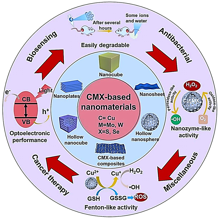 |
Figure 10 Biomedical Applications of Cu2MX4 (CMX)-based nano architectonics (Image is created by Biorender.com). |
Cancer and Drug Therapies
Nano architectonics based on Cu2MX4 are promising candidates for the development of cancer therapy and drug delivery because of their unique structural, optical and thermal properties. These copper-based chalcogenides are photothermally and photodynamically active. Also, in PTT, Cu2MX4 nanostructures convert NIR light into localized heat that kills cancer cells without killing healthy tissues. In PDT, in response to light activation, these materials produce ROS that induce oxidative stress and selectively kill cancer cells. Furthermore, Cu2MX4 nanoparticles can be surface-functionalized and tailored for targeted drug delivery, opening pathways to accurate and controlled drug release directly at the tumour site with minimal systemic toxicity. Their substantial surface area enables efficient loading of chemotherapeutic agents, and these nanoparticles may act as suitable delivery vehicles for drugs that are often used in conjunction with PTT or PDT to exploit synergies of theragnostic. Moreover, these nanoarchitectures can overcome MDR in cancer cells because their delivery mechanism bypasses the cellular defences that confer resistance. In general, Cu2MX4 nanoarchitectures can be a very effective tool for personalized medicine because they are multifaceted, integrating capabilities for therapeutic applications in cancer, drug delivery and diagnostic imaging.
Thus, the multifunctional nanoarchitectonics of Cu2MX4-based nanomaterials for cancer therapy have gained increased attention during the past few years. Typically, researchers give a broad theme to the use of Cu2MX4 nanoparticles for different therapeutic approaches, mainly PTT and PDT and drug delivery mechanisms. Research has been conducted on the thermal properties of Cu2MX4 nanoparticles, which can convert near-infrared light into heat and hence kill cancer cells through hyperthermia. The discussion of surface modification through conjugation with polyethene glycol (PEG) is used to improve biocompatibility and circulation in vivo for more successful therapeutic applications. This suggests the possibility of these nanoparticles reducing intrinsic toxicity while maximizing treatment efficacy.86 These findings add to the concept of the multifunctionality of Cu2MX4 nanoparticles in cancer therapy. Their research recapitulates the dual functions of these compounds in both PTT and PDT, indicating that they can produce ROS in the presence of light exposure, which leads to cell damage in cancer.
The research further emphasizes the versatility of Cu2MX4 nanostructures, especially for controlled drug delivery inside tumour cells, thus elevating the therapeutic potential of these nanoparticles.90 Chemo dynamic therapy (CDT), starvation therapy, phototherapy and immunotherapy are used in combination to treat cancer in a hollow mesoporous Cu2MoS4 (CMS) multifunctional cascade bioreactor loaded with glucose oxidase (GOx). The multivalent elements Cu1+/2+ and Mo4+/6+ found in CMS exhibit Fenton-like, glutathione (GSH) peroxidase-like and catalase-like action. The PEGylated CMS@GOx-based synergistic therapy may effectively eradicate primary tumours and stop cancer metastases when combined with checkpoint blockade therapy.27 Figure 11a represents the schematic illustration of cancer therapy using Cu2MX4 (CMX)-based nano architectonics, and Figure 11b is the mechanism of antitumor immune responses induced by PEGylated CMS@GOx-based phototherapy in combination with checkpoint blockade therapy.
 |
Figure 11 (a) Schematic illustration of cancer therapy using Cu2MX4 (CMX)-based nano architectonics, (b) Mechanism of antitumor immune responses induced by PEGylated CMS@GOx-based phototherapy in combination with checkpoint blockade therapy. Reproduced with permission from.27 (c) Schematic illustration of biosensing using Cu2MX4 (CMX)-based nano architectonics, (d) mechanism of photothermal Fenton reaction assembly of CMS-PEG-EDTA for CEA immunoassay. Reproduced with permission from.91 (e) Schematic illustration of anti-bacterial action using Cu2MX4 (CMX)-based nano architectonics, (f) Schematic illustration of NIR-II light-responsive CMS nanoplates for bacteria-killing effect. Reproduced with permission from.92 (g) Schematic illustration of anti-oxidant action using Cu2MX4 (CMX)-based nano architectonics, (h) Schematic illustration of fabrication and mechanism of antitumor immune responses induced by PEGylated CMS@GOx-based phototherapy. Reproduced with permission from.27 |
A novel application with ultrathin Cu-TCPP metalorganic framework nanosheets that can demonstrate effective heating under 808-nm laser irradiation and be exploited as agnostic platforms for magnetic resonance and near-infrared thermal imaging. Such progress suggests a quantum leap in the integration of diagnostic and therapeutic functions into single nanoplatforms and thus represents an all-inclusive approach to cancer therapy.87 These studies examined the PTT and PDT abilities of Cu2MX4 nanoparticles, with an emphasis on their potential role as a responsive platform for improving cancer theranostics. Figure 11c shows the illustration of biosensing using Cu2MX4 (CMX)-based nanoarchitectonics. This agrees with previous findings as it strongly upholds the notion that these types of nanoparticles can be used strategically to increase the loading capability of chemotherapeutic agents to increase their potential therapeutic effects via a controlled release mechanism.93 Thus, support for established Cu2MX4 nanoparticle functionalities in combinational cancer therapy can be generated in PTT, PDT and drug delivery applications as developed in treatment methods related to cancer as relevant nanosized drug-delivering particles.94 The multifunctionality of Cu2MX4 nanoparticles, reiterating their capacities for the conversion of near-infrared light to heat and the generation of ROS, underscores their considerable importance in the current cancer therapy, which highlights the great relevance of these nanoparticles in diagnostic and therapeutic applications.95
The application of 3D CNT/MXene microspheres, which possess photothermal, photodynamic and chemotherapeutic modalities, demonstrates synergistic potential that can improve treatment efficacy through the integrated application of these forms of therapy and firmly places Cu2MX4 nanoparticles in comprehensive cancer treatment strategies.96 Figure 11d shows the mechanism of photothermal Fenton reaction assembly of CMS-PEG-EDTA for CEA immunoassay.91 Figure 11e is the illustration of anti-bacterial action using Cu2MX4 (CMX)-based nanoarchitectonics, and Figure 11f shows the NIR-II light-responsive CMS nanoplates for bacteria-killing effect.92 The anti-oxidant action using Cu2MX4 (CMX)-based nanoarchitectonics is shown in Figure 11g, while Figure 11h shows the fabrication and mechanism of antitumor immune responses induced by PEGylated CMS@GOx-based phototherapy.27 Recent progress in the field of nanomedicine, including impediments and approaches for the practical use of Cu2MX4 nanoparticles. Further progress within the field has been described as insightful into the different methods of effective use of photothermal agents in cancer treatment, highlighting the demand for innovative strategies that optimize the best outcomes.97
Bioimaging and Diagnostic Imaging
The nanoarchitectonics of Cu2MX4 are increasingly being applied in bioimaging and diagnostic imaging because of their particular optical and magnetic properties. In fluorescence imaging, the Cu2MX4 nanoparticles can be engineered to display bright fluorescence signals, which can be visualized by researchers and clinicians. These are useful for tracking cellular processes and molecular anomalies. In photoacoustic imaging, Cu2MX4 nanostructures produce an acoustic signal upon NIR irradiation. Such structures provide high-resolution images deep within tissues without requiring any invasive procedures to resolve anatomy with great resolution. In addition, the nanomaterials can also be doped with other metals and work as excellent MRI contrast agents. Their magnetic properties enhance the contrast of MRI, thereby providing clear images of soft tissues and abnormalities, which is essential in the diagnosis of diseases like tumours and cardiovascular diseases. Altogether, Cu2MX4 nanoarchitectures provide a multi-functional approach to bioimaging and diagnostics, integrating high resolution, minimal invasiveness and multimodal capabilities that greatly enhance the accuracy and detail of imaging in medical diagnostics.
Cu2MX4 (CMX)-based nanoarchitectonics has drawn enormous attention in applications related to bioimaging and diagnostic imaging because of its multi-functionality. The changeability of Cu2MX4 nanoparticles due to modification for detecting fluorescence emission allowed better visualization of the cellular structures. The researchers have considered the generation of an acoustic signal via near-infrared (NIR) irradiation for high-resolution imaging via these nanoparticles. The versatility of these nanoparticles is further supported by their ability to serve as MRI contrast agents when combined with other metals, thereby enhancing the contribution of diagnostic imaging techniques.98 By employing cuprous oxide (Cu2O) as a self-sacrificing template that can perform efficient mass transfer in the Fenton-like reaction, a novel kind of nanoprobe for the detection of carcinoembryonic antigen (CEA) is created utilizing molybdenum copper sulphide (Cu2MoS4) with peroxidase activity. EDTA (ethylene diamine tetra acetic acid) can absorb the generated Cu2+. This work not only offers the first instance of Fenton metal sulphide nanoparticles being used in biosensors, but it also proposes an easy-to-manufacture immunosensor with promising clinical diagnostic potential.91
The reinforcement of multifunctionality of Cu2MX4 nanoparticles in theranostic indicated that these two types of nanoparticles are suitable for MRI as well as fluorescence imaging, which is crucial for cancer treatment applications. The utility of manganese (II) chelate-functionalized Cu2S4 nanoparticles for providing dual-modal imaging, thus validating the fact that Cu2MX4 nanoparticles may be used suitably for guided photothermal therapy.99,100 The use of ion-doped melanin nanoparticles as an imaging agent constitutes applications of Cu2MX4 nanoparticles. It has been proven that fluorescence and acoustic signal generation aid imaging techniques based on research findings.101 The discussion on controlled drug release and MRI contrast enhancement by augmenting MnO2-gated nanoplatforms furthered the scope of biomedical applications of Cu2MX4 nanoparticles.102
The latest study synthesis of luminescent lanthanide-based bimodal nanoprobes where size control in nanoparticles, especially for bimodal nanoparticles, can improve imaging performance of how different functionalities can be integrated into nanoparticles, therefore crucial for enhancing diagnostic imaging using nanoparticles.103 Investigation of the potential role of nanoparticles in neuro-oncology and the relevance of Cu2MX4 nanoparticles in various therapeutic scenarios.38 In terms of magnetic nanoparticles in theranostics, once again, the multifunctionality of such materials calls for special attention to Cu2MX4 nanoparticles for modulating imaging techniques.104 Recent efforts have focused on translating nanoparticles for use in biosensing and bioimaging, focusing on early disease detection and treatment.105,106
Antibacterial and Antimicrobial Applications
The most promising frontier area directly applied in bioimaging and diagnostic imaging is represented by Cu2MX4 (CMX) in nano architectonics. Here, the optical, electronic and structural properties that combine various advantageous features in its compounds, such as adjustable photoluminescence and excellent biocompatibility with a very high surface area, make this Cu-based material versatile. CMX nanostructures enable real-time tracking of biological processes, which is particularly useful for early disease detection and cellular monitoring. As such, they provide the possibility of tailoring the photophysical properties toward diverse imaging modalities, such as fluorescence, magnetic resonance imaging (MRI) and computed tomography (CT) imaging approaches; thus, a multifunctional diagnostic methodology is now in place. The second is the flexibility of design in nanoarchitectures of CMX, where precise engineering of size, shape and surface chemistries is made to improve their stability in biological environments and enhance target specificity.36 Functionalization with biomolecules or contrast agents improves biodistribution and cellular uptake and is critical for precise targeting in diagnostic imaging. However, toxicity and biodistribution issues, as well as regulatory hurdles, are challenges that need to be addressed. These limitations need to be overcome if their promise is to be fully realized in clinical practice.
One of the most significant research studies has been conducted in the last few years, particularly in the context of MXenes, its applications and their relevance in biomedicine, concerning nanoarchitectonics for anti-oxidant and anti-inflammatory therapy based on CMX or Cu2MX4. Research on the synthesis and toxicity assessment of MXenes underscores the need for modern nanotoxicology methods, especially organ-on-chip technologies and computational approaches, but claims that, under these conditions, great potential could be placed in MXenes as breakthrough materials in biomedical applications, pointing to more experimental data to understand the mechanisms and behaviour of their toxicity.107 The biocompatibility and multifunctional properties of MXenes, especially for breast cancer diagnosis and treatment, with the exploration of surface modifications for better biodegradability and reduced cytotoxicity, underpin the versatility of MXenes in targeted therapies, reinforcing their applicability in therapeutic contexts. This is in line with the broader integration of nanomaterials into cancer treatment frameworks.95 In addition, this study further focuses on copper-based nanomaterials to discuss the biogenic synthesis of copper nanomaterials using plant extracts. This highlights their eco-friendly approach and their eventual effects on the development of nanomaterials with antimicrobial and antioxidant properties. Findings from such studies suggest that plant-mediated synthesis could provide new avenues for nanotechnology applications, especially for antioxidant therapy applications.41
The application of MXenes in drug delivery systems with their low toxicity and excellent biocompatibility will identify the challenge of effective drug absorption and the significance of the modification of the composites based on MXenes for improving therapeutic efficacy. Such a view is critical for comprehending how MXenes can be tailored for targeted delivery in cancer therapy and add value to the current debate in nanoarchitectonics CMX.108 Studies on systematic assessment regarding the safety of the compound for clinical use illustrate the promising nature of MXenes but require appropriate comprehensive analysis regarding the aspect of biosafety over an extended period and potential toxic effects to gain successful incorporation into medical usage.109 The new application of carboxymethyl chitosan-modified nanoparticles in the oral delivery of astaxanthin, where the compound has antioxidant and anti-inflammatory activity, emphasizes problems in the oral drug delivery of astaxanthin and offers a new concept to enhance its bioavailability using the potential applications of CMX-based treatments to treat inflammatory bowel disease.110 To effectively eradicate multidrug-resistant (MDR) bacteria, a novel near-infrared II (NIR-II) light-responsive nanozyme (Cu2MoS4 nanoplates, CMS NPs) was introduced. CMS NPs can catalyse the production of reactive oxygen species (ROS) due to their inherent dual enzyme-like properties. Crucially, CMS NPs exhibit improved oxidase and peroxidase-like enzymatic activity in response to NIR-II light, which enhances ROS production for extremely effective bacterial death. By combining the catalytic production of ROS with the NIR-II photothermal impact of enzymes, this study offers a unique antibacterial method for the effective treatment of infections caused by MDR bacteria.92
Antioxidant and Anti-Inflammatory Therapy
The nanoarchitectonics of Cu2MX4 (CMX) have recently emerged as an extremely effective agent for the therapeutic management of oxidative stress through antioxidant and anti-inflammatory therapy. CMX is characterized by specific redox properties, large surface areas and modulation of reactive oxygen species (ROS) in disease. High ROS levels are associated with cellular damage, ageing and inflammatory diseases. CMX nanostructures act as ROS scavengers, which minimize oxidative stress at inflammation sites and induce cellular recovery. The neutralization of free radicals can be remarkably achieved using these nanoarchitectures through their Cu content and by tuning their electronic properties, which deliver persistent antioxidant activity. Modulation in inflammatory pathways also results from interactions with signalling molecules and cellular receptors that reduce the release of inflammatory cytokines. Functionalization with biomolecules enhances their biocompatibility and stability, making them effective for long-term therapy. Although promising to be less damaging during oxidation and inflammation, CMX-based nanoarchitectures have to be evaluated for biocompatibility, biodistribution and therapeutic efficacy through comprehensive in vivo studies. Therefore, antioxidant and anti-inflammatory therapy modalities based on CMX open avenues for the use of nanomaterials for treating chronic inflammatory diseases and many related oxidative stress conditions.
The development of nano architectonics based on Cu2MX4 over the last decade has progressed dramatically and demonstrated a broad range of applications and deep insights into the biocompatibility and functionality of copper and MXene nanomaterials. Early work laid down crucial background knowledge about the cellular responses of mouse macrophages to copper and copper oxide nanoparticles, with relevant emphasis on the specificity of proteomic changes associated with responses to oxidative stress in these treatments. Their findings showed that these nanoparticles influenced cellular mechanisms that required further investigation, especially inflammatory responses.111 From this point of view, the study related to Cu ions in nanotheranostics aims to understand the role of Cu ions in angiogenesis and wound healing. This indicated that the Cu nanoparticles have multiple functions, such as photothermal effects coupled with catalytic activity, for enhancing oxidative monotherapy. Cu-based biomaterial nano systems are highly promising in clinical medicine and can expand the range of applications of CMX-based materials.112 Further research during the following years was mainly focused on the oxidation stability of the material properties, which would define the MXene performance and further expand the ability to apply them in the majority of fields, among them biomedical applications. This is essential because it relates to oxidative degradation challenges, which are crucial in determining the sustainability and efficacy of MXene-based therapies.113
Discussion about the synthesis, toxicity assessment, and potential biomedical applications of MXenes has paved modern approaches in nanotoxicology and thereby revealed the promising scope for MXenes in “smart diagnostics” and targeted drug delivery systems.107 This consideration is crucial for enhancing the safety and efficacy of MXene-based medicines. As demonstrated by their multifunctionality and potential for targeted therapy, the application of MXenes to breast cancer diagnosis and treatment highlights the kind of biomedical applications that MXenes can use to improve drug delivery systems through the modification of their surfaces.95 As such, the enhancement of biocompatibility and pharmacokinetics of systems is an important aspect that needs to be addressed to favourably direct drug delivery through targeted action mechanisms within the context of cancer treatment. These studies emphasize MXene functionalization as imperative for achieving effective therapeutic applications that help reduce possible side effects.108 Finally, the study of the toxicity and environmental risks related to MXenes, therefore requiring the systemic evaluation of their biocompatibility and cytotoxicity analyses, highlights the need for comprehensive toxicological analysis to ensure the safe application of enzymes based on MXenes in biomedicine.27,109 Figure 11g represents the schematic illustration of anti-oxidant action using Cu2MX4 (CMX)-based nano architectonics, and (h) shows the schematic illustration of fabrication and mechanism of antitumor immune responses induced by PEGylated CMS@GOx-based phototherapy. Thus, it can be concluded that this dynamic and rapidly changing environment of nanoarchitectonics based on CMX underscores the potential of these materials in advancing antioxidant and anti-inflammatory therapies while addressing important issues concerning safety and efficacy in their applications.
Wound Healing and Tissue Regeneration
Cu2MX4-based nano architectonics provide potentially transformative technology for wound healing and tissue regeneration via the use of their bioactivity, excellent antimicrobial properties and compatibility with biological tissues for the healing process. Because wound healing is a multiscale complex process, it further complements the prospects of materials that possess both antimicrobial and regenerative properties, such as CMX nanostructures. The natural inherent antimicrobial properties of copper make the nanoarchitectures of CMX particularly effective at preventing infection and creating an environment favourable for tissue repair. CMX nanostructures have recently been found to promote collagen synthesis and induce angiogenesis-important components involved in wound healing and tissue remodelling. Functionalization with growth factors or bioactive molecules allows for the modulation of these structures to enhance cellular adhesion, migration and proliferation, thereby promoting the healing process. Nevertheless, careful design is necessary for any successful clinical translation that will not become cytotoxic or lose its biocompatibility over time. This introduction highlighted the possibility of the use of CMX nanoarchitectures as potential sources for wound care, where their multifunctional properties could be an effective, advanced pathway for rapid tissue regeneration and healing.
The research surrounding CMX-based nanoarchitectonics for wound healing and tissue regeneration has seen significant advancements in recent years, particularly focusing on the unique properties of MXenes and their composites. The exploration of 2D MXenes has emerged as a promising area in the biomedical field, highlighting their potential as drug or protein carriers because of their high specific surface area and tunable structures.114 This foundational work underscores the versatility of MXenes in various biomedical applications, paving the way for further research into their synthesis and functionalization. Studies on MXene nanocomposites, emphasizing their diverse chemistries and superior electrical conductivities, illustrate the potential of MXenes when combined with other materials, suggesting that their surface properties can be optimized to enhance their biological applications, particularly in drug delivery and cancer therapy. The need for safer synthesis methodologies for bare MXenes has also been identified, which is crucial for advancing their practical applications.115 The synthesis and toxicity of MXenes were explored by revealing their ability to treat tumours resistant to conventional chemotherapy. The development of a 2D MXene-based composite nanoplatform for targeted therapy demonstrated significant potential for tumour eradication, highlighting the importance of biocompatibility and osteoinductive properties for tissue engineering applications. This study emphasizes the dual role of MXenes in both therapeutic contexts and tissue regeneration.107
Advanced MXene-based micro and nano systems for targeted drug delivery, demonstrating their multifunctionality in biomedicine. The studies showed the adjustable mechanical properties and photothermal conversion efficiency of MXenes, which are essential for enhancing drug delivery and tissue engineering applications. This highlights the necessity of controlling the physicochemical characteristics of MXenes to unlock their full potential in biomedical technologies.108 The expansive growth reflects the ongoing innovation and potential of MXenes in medical applications, particularly in addressing the challenges of human translation of these technologies.116 The integration of MXenes into polymeric nanofibers highlights their promising characteristics for biomedical applications. The ability to enhance the properties of polymeric materials through MXene incorporation is critical for advancing tissue engineering strategies, reinforcing the importance of understanding the interactions between these materials.117 A novel composite hydrogel incorporating antimicrobial peptides and MXene nanoparticles for effective skin wound healing properties. This study highlights the synergistic effects of the composite in promoting cellular infiltration and tissue regeneration, making it a valuable treatment option for skin injuries.118 Figure 12a shows the schematic illustration of wound healing action using CMX-based nano architectonics, and Figure 12b shows the mechanism of wound healing of photosensitive GelMA hydrogel. A multifunctional nanozyme that integrates antioxidant, antimicrobial and pro-vascularity properties, showcasing its potential in skin-wound management, suggests that such multifunctional materials could revolutionize wound-healing strategies, although further investigation into their biosafety and mechanisms of action is warranted.119
 |
Figure 12 (a) Schematic illustration of wound healing action using Cu2MX4 (CMX)-based nanoarchitectonics, (b) Mechanism of wound healing of photosensitive GelMA hydrogel. Reproduced with permission from.118 (c) Schematic illustration of gene therapy and DNA delivery, (d) Mechanism of multifunctional carboxymethyl cellulose nano hydrogel carriers based on near-infrared DNA-templated quantum dots for tumour theranostics. Reproduced with permission from.120 (e) Schematic illustration of osteogenesis and (f) SEM images and working mechanism of copper-containing mesoporous bioactive glasses for bone defects therapy. Reproduced with permission from.121 |
Gene Therapy and DNA Delivery
The strong structural flexibility of Cu2MX4-based nano architectonics, high loading capacity and protection against nucleic acid degradation make them good candidates for use in gene therapy and DNA delivery. Gene therapy requires safe and effective delivery systems for genetic material into target cells; therefore, the CMX nanoarchitecture shows promise in this regard. Their tunable surface chemistry and high surface area enable them to efficiently carry DNA or RNA, while structural stability in CMX protects the genetic material against degradation by enzymes in biological environments. The Cu content in the nanoarchitectures of CMX might also help in cell uptake and endosomal escape for successful gene transfection. Functionalization of targeting ligands or cell-penetrating peptides enhances the specificity and efficacy of gene delivery across nanoarchitectures based on CMX. Although these nanoarchitectures hold great promise, they face many challenges, among which immune responses and minimal cytotoxicity are considerable.
The nanoarchitectonics of CMX for gene therapy and DNA delivery has been a very popular area of research in the last few years, mirroring the breathtaking development of nanotechnology and its biomedical applications. In 2011, the foundation of Magnetic Drug Targeting (MDT) and magnetofection by Schwerdt et al highlighted the points that magnetic nanoparticles (MNPs) are used to improve the efficiency of gene delivery. Their research underlined that by applying external magnetic fields, MNPs can be highly efficient in concentrating therapeutic complexes in the desired areas to be treated, which is quite efficient in cancer therapy. The authors provided information about MNP-gene vector complexes with the ability to mediate stable and nontoxic gene transfer, and such research opened the way for the development of methodologies of magnetic gene targeting (MGT).122 Based on these basic concepts, the clinical development of nanoparticles for gene delivery overcame the issues that have been observed during the transference of gene therapeutics from bench to bedside, proving the potential of nanoparticles as good vehicles for gene therapy and capable of overcoming the limitations related to safety and efficiency. Some of the nanoparticle candidates identified to have clinical relevance, including cationic polymers based on polypeptides and cyclodextrins, have shed light on how engineered nanoparticles could improve gene delivery systems.123
In 2017, discussions on mesoporous carbon nanomaterials (MCNs) expanded widely, with their various applications in drug delivery. It is noted that sp2 carbon-based materials, such as graphene and carbon nanotubes, have favourable biomedical properties. Following this rationale, the authors reported that MCNs are among the next generation of drug delivery platforms that offer high surface areas, biocompatibility and drug release control on the versatility of carbon nanomaterials for gene transfection and drug delivery systems.124 The developments of inorganic nanocarriers for gene therapy by the surface modifications of gold nanoparticles, carbon nanotubes and other nanomaterials show the potential to significantly increase the level of transfection and reduce toxicity.120,121 Thus, these novel strategies include the production of RGD peptide-targeted AuNPs and pH-responsive strontium sulfate nanoparticles to demonstrate the feasibility of these nanocarriers in the efficient delivery of genetic material into target cells.
The therapeutic applications of cell membrane-coated nanocarriers and chemically modified DNA nanostructures for drug delivery will be developed by 2022.125,126 Such work has placed chemically modified DNA on a promising track for developing smart nanocarriers. Indeed, research on MXene-based micro- and nanosystems, including biocompatibility, aims at targeted drug delivery and cancer therapy. They suggested more research on the functionalization and optimization of MXene materials to improve their efficiency and reduce their hazardous content.126 Multifunctional carboxymethyl cellulose nanohydrogels can integrate imaging functionality with drug delivery to enhance the precision of anticancer treatments.120 Figure 12c shows the schematic illustration of gene therapy and DNA delivery and Figure 12d illustrates the mechanism of multifunctional carboxymethyl cellulose nano hydrogel carriers based on near-infrared DNA-templated quantum dots for tumour theranostics. These studies, the opportunity for aptamer-functionalized hydrogels to be used in site-targeted delivery, allowing for enhanced therapeutic efficacy and reduced risks. Finally, the new generation of synergistic delivery systems for tumour therapy underlined the importance of multifunctional nanocarriers capable of delivering multiple therapeutic agents in real-time.127 These contributes to a better understanding of how nanotechnology can revolutionize genetic delivery systems and improve the efficacy and safety of gene therapies.
Osteogenesis and Bone Regeneration
CMX-based nano architectonics have extensive potential in osteogenesis and bone regeneration, owing to their biocompatibility, osteoinductive properties and structural flexibility. Bone regeneration refers to materials that enable cell attachment and proliferation but further stimulate the activity of osteoblasts and mineralization. Copper ions significantly contribute to the enhanced proliferation and differentiation of bone cells through CMX nanoarchitectures, and their high surface area and porosity support the adhesion of cells and the transportation of nutrients. CMX-based nanoarchitectures can also release ions in a controlled manner, which promotes the process of angiogenesis as well as depositing bone matrix that leads to effective regeneration. Further functionalization with osteoinductive agents or growth factors will improve the bioactivity of CMX nanoarchitectures for bone grafts and scaffold applications.121 Figure 12e shows the schematic illustration of osteogenesis, and Figure 12f shows the SEM images and working mechanism of copper-containing mesoporous bioactive glasses for bone defects therapy. Despite these advantages, issues such as the long-term stability of CMX in physiological environments and the control of degradation rates remain problematic for application in clinical practice. The field of bone tissue engineering has seen significant advancements in recent years, particularly with the integration of nanotechnology and innovative materials aimed at enhancing osteogenesis and bone regeneration. The application of nanotechnology in bone tissue engineering highlights the limitations of traditional bone grafts and the potential of synthetic materials to bridge bone defects. This analysis emphasizes the need for biomaterials that mimic the natural extracellular matrix (ECM) of bone, highlighting the importance of nanofibers and nanoparticles in developing more effective implants.128
Based on this background, the research further focuses on the application of carbon nanostructures in bone tissue engineering: a variety of carbon-based materials, ranging from multi-walled carbon nanotubes (MWCNTs) to nanodiamonds, have been demonstrated to exhibit osteoinductive properties as well as to enhance the proliferation and differentiation ability of bone marrow stem cells. Findings show that, although many in vitro studies exist, an urgent need is to conduct in vivo tests before establishing the full potential of these materials in clinical practice.129 Guided bone regeneration (GBR) using multilayered films of titanium carbide MXene. Results summarize the features of MXene possessing structural integrity along with osteoinductivity that is capable of overcoming the limitations of conventional GBR membranes. This study demonstrated the promising application of MXenes for enhancing the healing process around dental implants and further explored bone tissue applications.130 Biodegradable tissue engineering: Bio-inspired mineralized collagen scaffolds for bone repair. They claim that the chemical composition, as well as the structure of a scaffold, is vastly important because the natural bone environment is best mimicked by mineralized collagen scaffolds, thereby improving osteogenesis. Such studies indicated the requirement for scaffold modification concerning physical and chemical properties to enhance their functions for better bone regeneration.131 A novel inorganic nanomaterial-based therapy for bone tissue regeneration offers the synergistic effects of mesoporous silica nanoparticles (MSNs) combined with natural components such as hydroxyapatite (nHA), which enhance cell adhesion and osteogenic differentiation. Finally, doping with metal ions, like magnesium and copper, promotes greater bioactivity of the materials and therefore ascribes a multifaceted approach to optimizing the regeneration strategies for bones.132
In the same category of research, studies on copper-doped mesoporous bioactive glasses (MBGs) and their potential for bone defect treatment have been conducted. This study showed that copper oxides introduced into MBGs significantly influence the biological behaviour of MBGs, including osteogenesis and angiogenesis. Thus, this study sheds light on the structural nature of MBGs and their interaction with biological environments. Material design in bone regeneration applications has also been underscored in this context.121 The osteogenic capability of mesoporous glass scaffolds doped with zinc ions and the peptide osteostatin demonstrates the synergy between these components in boosting osteoblast differentiation, suggesting great promise for increasing the osteogenic capability of biosynthetic bone grafts.133 A novel ternary nanofibrous scaffold consisting of MXene nanoparticles that support spontaneous osteogenic differentiation was demonstrated, further highlighting its structural and functional superiority. However, this indicates that a promising microenvironment exists that can support the essential cellular functions required for bone regeneration. These studies represent the progression of the evolution of materials toward efficient solutions to bone defects.134 Integrate nanotechnology, innovative materials and a deeper understanding of the biological interplay in osteogenesis and bone regeneration: this will help bring future breakthroughs toward applications into clinical practice. Table 5 highlights the diverse biomedical applications of Cu2MX4 (CMX)-based nanoarchitectonics, including parameters such as composition, target properties, efficiency parameter values and potential applications, making it suitable for comprehensive analysis.
 |
Table 5 The Diverse Biomedical Applications of Cu2MX4 (CMX)-Based Nanoarchitectonics Highlight Different Compositions, Target Properties, Efficiency Parameter Values and Potential Applications, Making It Suitable for Comprehensive Analysis |
Challenges and Limitations
CMX- based nano architectonics are considered promising routes toward biomedical applications. This is largely due to the versatility of the structural and electronic properties that can be exploited in drug delivery, imaging and therapeutic interventions. However, biocompatibility is the primary challenge in clinical applications. The release of Cu ions from CMX materials tends to have cytotoxic effects, which subsequently increase the vulnerability to tissue damage or cellular dysfunction.
These materials may accumulate in organs due to insufficient knowledge of the mechanisms of their long-term clearance. Hence, the repeated administration of CMX nanoparticles may lead to chronic toxicity. Furthermore, CMX particles can trigger immunological responses, resulting in inflammation or other toxic effects that significantly hinder repeated or systemic use. Scalability and stability are other concerns: Laboratory-scale synthesis of CMX nanoarchitectures may achieve the desired properties, but scaling up these methods for industrial production often yields particles that lack uniformity, purity and structural stability. An important challenge is the stability of CMX-based materials in complex biological fluids, such as blood or intracellular fluids, where aggregation or premature degradation may impede their effectiveness and pose safety hazards. In addition, costs associated with the development of scalable, stable production processes can be high barriers to widespread availability.
The regulatory landscape of CMX nano architectonics remains challenging. Rigorous testing should be conducted before these materials qualify for clinical use to confirm a satisfactory safety profile that encompasses not only biocompatibility and toxicity studies but also biodistribution and long-term effects. Each aspect requires thorough data because it involves different behaviours in a biological system. Such rigorous regulatory requirements are both cost-intensive and scientifically challenging, so an appropriate assessment needs to be conducted for CMX-based materials in biomedical research before they can be implemented fully in any healthcare application. Table 6 explores the various challenges and limitations of using CMX-based nano architectonics for biomedical applications.
 |
Table 6 Challenges and Limitations of Using Cu2MX4 (CMX)-Based Nanoarchitectonics for Biomedical Applications |
Future Perspectives
CMX-based nano architectonics is promising to drive the development of biomedical applications, and future developments are expected to involve innovation synthesis, clinical translation and interdisciplinary integration. These copper chalcogenides exhibit unique photothermal and photodynamic capabilities, biocompatibility and tunable surface characteristics, making them ideal for targeted drug delivery, cancer treatment, antimicrobial therapy and diagnostic imaging. The most promising approach toward maximal biomedical utility is the development of novel synthetic and functionalization techniques. Such new eco-friendly methods could help to produce more sustainable and reproducible, biocompatible Cu2MX4 nanoparticles, where advanced surface modification allows enhanced targeting and controlled release while reducing toxicity. These methods would enable morphology with controlled precision and responsive coatings, which could enhance the safety and effectiveness of drug delivery systems to allow specific release based on environmental triggers like pH or temperature. Of critical consideration is the transition of Cu2MX4 nanoarchitectures from laboratory research to clinical applications: scale-up synthesis, biocompatible coating development, comprehensive in-depth toxicity and pharmacokinetic studies, regulatory compliance with clinical-grade manufacturing protocols that will be required to ensure successful translation and to eventually tailor dosage forms to the needs of personalized medicine.
Finally, the biomedical applications of CMX are probably facilitated by interdisciplinary approaches. Interdisciplinary collaborations in bioinformatics, materials science and nanomedicine will likely play key roles in predictive modelling, enhancing drug-target interactions and extending the applicability of computational designs for material creation. Such an integration may rapidly speed up development cycles and open up new applications, from tissue engineering to lab-on-a-chip diagnostics. Together, these strategies will fully realize the biomedicine potential in Cu2MX4 nanoarchitectures while pointing the way forward to future applications in next-generation personalized medicine therapies and diagnostic tools. Table 7 presents the future perspectives of biomedical applications of CMX-based nano architectonics, with a focus on synthesis innovation, clinical translation potential and interdisciplinary approaches.
 |
Table 7 Future Perspectives for Biomedical Applications of Cu2MX4 (CMX)-Based Nanoarchitectonics |
Conclusion
CMX-based nano architectonics can be revolutionary in a vast range of biomedical applications, from targeted therapeutics and imaging to smart drug delivery. One of the attractive properties of CMX materials is their unique electronic and structural properties, offering great promise for biomedical applications, particularly targeted therapy, in which selective delivery and controlled release are essential. While recent results demonstrate the versatility and effectiveness of materials based on CMX, a plethora of challenges must be overcome before their clinical translation. The biocompatibility of CMX nanoarchitectures is challenging. The release of Cu ions can cause cytotoxicity and even lead to cellular damage or disruption of normal physiological functions. The effects of such releases are therefore cell or tissue-dependent and detailed compatibility testing should be performed in diverse biological contexts. Bioaccumulation potential is another serious concern because accumulation in crucial organs after a period can result in chronic toxicity, especially following repeated or high dosages. The immune response is another critical factor; interactions with CMX materials with immune cells might provoke inflammation and other unfavourable reactions, restrict their application and place an improved risk to the safety of the patients involved. Hence, further development is needed for the safe interaction of CMX nanoarchitectures with biological systems.
Scalability and stability remain significant challenges. While many properties are favourable at the laboratory scale, scaling up the synthesis of CMX-based materials for industrial production has proven difficult. Large-scale synthesis necessitates maintaining uniformity, purity and particle stability for reproducible results as well as proper clinical performance. Moreover, the nanoarchitecture in CMX must be stable in complex biological fluids such as blood or intracellular fluids. Stability is important because it prevents degradation of the nanoarchitectures in advance or aggregation, which might lead to less effective therapy or adverse side effects. The high development costs associated with scalable, stable production processes further intensify these barriers to making CMX-based technologies accessible and economically viable. In addition to the challenges related to technology and biology; regulatory problems remain a major disadvantage for the application of CMX nanoarchitecture in clinics. Regulatory bodies require a lot of preclinical information to confirm the non-toxicity and high efficiency of the materials being considered. This includes assessments related to toxicity, biodistribution, immune response and long-term effects. Studies on nanomaterials in biological systems are usually more elaborate than those of traditional drugs or materials because of the unique characteristics of nanomaterials. Thus, more time and financial input is required for approval. In addition, since nanotechnology is a novel field, standard regulations are still adaptive and are thus often vague, which may consume more time for approval.
The successful development of safe and compliant CMX-based technologies will require extensive collaboration among researchers, manufacturers and policymakers to meet regulatory requirements. In the future, for the advancement of biomedical applications based on CMX-based nanoarchitectures, innovation and interdisciplinary collaboration will be in high demand. There is a need to target research to optimize the biocompatibility of CMX materials to avoid toxicity and immune responses. Scalable, efficient and cost-effective production methods that retain stability and function will be important in further developing their breadth of application. Proactive efforts to address regulatory requirements, including the generation of robust data and collaboration with regulatory bodies, will be essential for facilitating clinical translation. With continued effort and investment over time, CMX-based nanoarchitectures may indeed fulfil their promise of bringing unprecedented benefits to diagnostics, targeted therapies and regenerative medicines.
Acknowledgment
The authors extend their appreciation to the Deanship of Graduate Studies at Ajman University, Ajman, United Arab Emirates, for publication charges support.
Disclosure
The authors report no conflicts of interest in this work.
References
1. Riehemann K, Schneider S, Luger T, et al. Nanomedicine—Challenge and perspectives. Angew. Chem. Int. Ed. 2009;48(5):872–897. doi:10.1002/anie.200802585
2. Lohse SE, Murphy CJ. Applications of colloidal inorganic nanoparticles: from medicine to energy. J Am Chem Soc. 2012;134(38):15607–15620. doi:10.1021/ja307589n
3. Tao L, Zhang P, Qin C, et al. Recent progresses in the exploration of machine learning methods as in-silico ADME prediction tools. Adv. Drug Delivery Rev. 2015;86:83–100. doi:10.1016/j.addr.2015.03.012
4. Xia Y. Nanomaterials at work in biomedical research. Nature Mater. 2008;7(10):758–760. doi:10.1038/nmat2284
5. Sun T, Zhang YS, Pang B, Hyun DC, Yang M, Xia Y, et al. Engineered nanoparticles for drug delivery in cancer therapy. Nanomaterials Neoplasms. 2021;31–142.
6. Guan Y, Qian H, Guo J, et al. Synthesis of acidified palygorskite/BiOI with exceptional performances of adsorption and visible-light photoactivity for efficient treatment of aniline wastewater. Appl. Clay Sci. 2015;114:124–132. doi:10.1016/j.clay.2015.05.025
7. Kankala RK, Han Y-H, Na J, et al. Nanoarchitectured structure and surface biofunctionality of mesoporous silica nanoparticles. Adv. Mater. 2020;32(23):1907035. doi:10.1002/adma.201907035
8. Minuth WW, Denk L, Glashauser A. Cell and drug delivery therapeutics for controlled renal parenchyma regeneration. Adv. Drug Delivery Rev. 2010;62(7–8):841–854. doi:10.1016/j.addr.2010.03.002
9. Chen BQ, Xia H-Y, Mende LK, et al. Trends in layered double hydroxides‐based advanced nanocomposites: recent progress and latest advancements. Adv. Mater. Interfaces. 2022;9(22):2200373. doi:10.1002/admi.202200373
10. McNamara K, Tofail SA. Nanoparticles in biomedical applications. Adv. Phys. 2017;2(1):54–88. doi:10.1080/23746149.2016.1254570
11. Torney F, Trewyn BG, Lin VS-Y, et al. Mesoporous silica nanoparticles deliver DNA and chemicals into plants. Nature Nanotechnol. 2007;2(5):295–300. doi:10.1038/nnano.2007.108
12. Xia HY, Li BY, Zhao Y, et al. Nanoarchitectured manganese dioxide (MnO2)-based assemblies for biomedicine. Coord. Chem. Rev. 2022;464:214540. doi:10.1016/j.ccr.2022.214540
13. Han YH, Liu C-G, Chen B-Q, et al. Orchestrated tumor apoptosis (Cu2+) and bone tissue calcification (Ca2+) by hierarchical Copper/Calcium-ensembled bioactive silica for osteosarcoma therapy. Chem Eng J. 2022;435:134820. doi:10.1016/j.cej.2022.134820
14. Duan C, Yu M, Zhao Y, et al. Ternary Cu2MX4 (CMX)-based nanoarchitectonics: chemistry, synthesis and biomedical applications. Coord. Chem. Rev. 2024;518:216052. doi:10.1016/j.ccr.2024.216052
15. Sut TN, Yoon BK, Jeon W-Y, et al. Supported lipid bilayer coatings: fabrication, bioconjugation and diagnostic applications. Appl. Mater. 2021;25:101183. doi:10.1016/j.apmt.2021.101183
16. Margabandhu M, Raj SA, Jothi SP, Gajalakshmi D. Investigation of structural, morphological, magnetic properties and biomedical applications of Cu2+ substituted uncoated cobalt ferrite nanoparticles. Braz. Arch. Biol. Technol. 2016;59(spe2):e16161046. doi:10.1590/1678-4324-2016161046
17. Tran TH, Nguyen VT. Copper oxide nanomaterials prepared by solution methods, some properties and potential applications: a brief review. Int Scholarly Res Notices. 2014;2014:856592. doi:10.1155/2014/856592
18. Wang S. Novel 2D structure nanomaterials synthesis and IR absorption. 2018. doi:10.15760/etd.6423
19. Jiang B, Yang M, Huang L, Li Y. Nanoarchitectonics of metallene materials for electrocatalysis. ACS Nano. 2023;17(14):13017–13043. doi:10.1021/acsnano.3c05181
20. Yang K, Zhang Y, Sun J. NIR-responsive MoS2–Cu2WS4 nanosheets for catalytic/photothermal therapy of methicillin-resistant Staphylococcus aureus infections. Nanoscale. 2022;14(27):9796–9805. doi:10.1039/D2NR03560A
21. Peplow P. Properties and actions of non-steroidal anti-inflammatory drugs, including their effects on prostaglandin and macromolecular biosynthesis. Prostaglandins, Leukotrienes Essent Fatty Acids. 1988;33(3):239–252. doi:10.1016/0952-3278(88)90111-6
22. Hossain MA, Islam MS, Chowdhury Z. Facile synthesis of Cu2O nanorods in the presence of NaCl by SILAR method and its characterizations. arXiv preprint arXiv:2203.05760. 2022. doi:10.48550/arXiv.2203.05760
23. Chang HW, Lee YH, Chou YH, Liu F-J, Tsai Y-C. Constructing morphologically tunable copper oxide-based nanomaterials on Cu wire with/without the deposition of manganese oxide as bifunctional materials for glucose sensing and supercapacitors. Int J Mol Sci. 2022;23(6):3299. doi:10.3390/ijms23063299
24. Sui J, Yu H, Zhi C, Peng W, Zhang F, Fan X. MXene derivatives: synthesis and applications in energy conversion and storage. RSC Adv. 2021;11(26):16065–16082. doi:10.1039/D1RA01650K
25. Shin M, Choi H, Kim D. Nanoarchitectonics of three-dimensional carbon nanofiber-supported hollow copper sulfide spheres for asymmetric supercapacitor applications. Int J Mol Sci. 2023;24(11):9685. doi:10.3390/ijms24119685
26. Rahman UU, Khan AS, Bai X. MXenes as emerging materials: synthesis, properties and applications. Molecules. 2022;27(15):4909. doi:10.3390/molecules27154909
27. Chang M, Wang M, Wang M, et al. A multifunctional cascade bioreactor based on hollow-structured Cu 2 MoS 4 for synergetic cancer chemo-dynamic therapy/starvation therapy/phototherapy/immunotherapy with remarkably enhanced efficacy. Adv. Mater. 2019;31(51):1905271. doi:10.1002/adma.201905271
28. Dias S, Kumawat K, Biswas S, Krupanidhi S B. Solvothermal synthesis of Cu2SnS3 quantum dots and their application in near-infrared photodetectors. Inorg. Chem. 2017;56(4):2198–2203. doi:10.1021/acs.inorgchem.6b02832
29. Schmidt R, Prado-Gonjal J, Morán E. Microwave assisted hydrothermal synthesis of nanoparticles. arXiv preprint arXiv:2203 02394. 2022. doi:10.48550/arXiv.2203.02394
30. Ruiz-Gómez S, Fernández-González C, Perez L. Electrodeposition as a tool for nanostructuring magnetic materials. Micromachines. 2022;13(8):1223. doi:10.3390/mi13081223
31. Gupta D, Kumar A, Yadav S, Singh S, Gupta TK. Nanoarchitectonics: functional nanomaterials and nanostructures—a review. J Nanopart Res. 2022;24(10):196. doi:10.1007/s11041-022-03134-0
32. Alruwais RS, Adeosun WA. Electrochemical detection of hydrogen peroxide using copper-based metal–organic frameworks: nanoarchitectonics and sensing performance. J Inorg Organomet Polym Mater. 2024;34(1):14–37. doi:10.1007/s10904-023-02174-1
33. Huang P, Zhang Z, Liu X. Metal-organic framework-based nanoarchitectonics: a promising material platform for electrochemical detection of organophosphorus pesticides. Coord. Chem. Rev. 2024;501:215534. doi:10.1016/j.ccr.2024.215534
34. Ouahrani T, Boufatah RM, Benaissa M, Morales-García Á, Badawi M, Errandonea D. Effect of intrinsic point defects on the catalytic and electronic properties of Cu2WS4 single layer: ab initio calculations. Phys Rev Mater. 2023;7(2):025403. doi:10.1103/PhysRevMaterials.7.025403
35. Zhuang Y, Cheng H, Tian S, et al. Construction of Cu2MoS4/ZnO heterostructures and mechanism of photocatalytic hydrogen production. Langmuir. 2025. doi:10.1021/acs.langmuir.4c04325
36. Phung TLH, Vu NP, Nguyen TA, et al. Copper molybdenum sulfide (Cu2MoS4) nanoplates as a proficient electrocatalytic interface for enhancing the electrochemical redox signals of ofloxacin for detection in pharmaceutical samples. New J Chem. 2023;47(8):3714–3725. doi:10.1039/d3nj00090j
37. Cui Y, Niu Q, Hu Z, et al. Multifunctional nanozyme-hydrogel system in bacterial-infected wounds. Cell Biomaterials. 2025;1:100074. doi:10.1016/j.celbio.2025.100074
38. Klein AL, Nugent G, Cavendish J, et al. Nanoparticles as a tool in neuro-oncology theranostics. Pharmaceutics. 2021;13(7):948. doi:10.3390/pharmaceutics13070948
39. Baláž P, Malachová K, Sayagués M. Chalcogenide quaternary Cu2FeSnS4 nanocrystals for solar cells: explosive character of mechanochemical synthesis and environmental challenge. Crystals. 2017;7(12):367. doi:10.3390/cryst7120367
40. Lee S, Wang S, Wern C, et al. The green synthesis of 2D copper nanosheets and their light absorption. Materials. 2021;14(8):1926. doi:10.3390/ma14081926
41. Vincent J, Lau KS, Evyan YC-Y, et al. Biogenic synthesis of copper-based nanomaterials using plant extracts and their applications: current and future directions. Nanomaterials. 2022;12(19):3312. doi:10.3390/nano12193312
42. Chen W, Chen H, Zhu H, et al. Solvothermal Synthesis of Ternary Cu 2 MoS 4 Nanosheets: structural Characterization at the Atomic Level. Small. 2014;10(22):4637–4644. doi:10.1002/smll.201401238
43. Gadiyar C, Strach M, Schouwink P, et al. Chemical transformations at the nanoscale: nanocrystal-seeded synthesis of β-Cu 2 V 2 O 7 with enhanced photoconversion efficiencies. Chem. Sci. 2018;9(25):5658–5665. doi:10.1039/C8SC01592A
44. Sengupta J. Growth and characterization of multi-walled carbon nanotubes using chemical vapor deposition. IIT Kharagpur. arXiv preprint arXiv:1808.06596. 2010. doi:10.48550/arXiv.1808.06596
45. Phuong Pham V. Atmosphere pressure chemical vapor deposition of graphene. arXiv e-printsarXiv:1807 07774. 2018. doi:10.48550/arXiv.1807.07774
46. Zhao Y, Jin S. Controllable water vapor assisted chemical vapor transport synthesis of WS2–MoS2 heterostructure. ACS Mater Lett. 2019;2(1):42–48. doi:10.1021/acsmaterialslett.9b00412
47. Fraga M, Pessoa R. Progresses in synthesis and application of SiC Films: from CVD to ALD and from MEMS to NEMS. Micromachines. 2020;11(9):799. doi:10.3390/mi11090799
48. Li H, Liu J, Guo N, et al. Seeded growth of high-quality transition metal dichalcogenide single crystals via chemical vapor transport. CrystEngComm. 2020;22(46):8017–8022. doi:10.1039/D0CE01112A
49. Kataria S, Shinde SA, Sood K. Chemical vapor deposited graphene: from synthesis to applications. Phys Status Solidi. 2014;211(11):2439–2449. doi:10.1002/pssa.201431159
50. Szczęsny R, Kulińska A, Toboła D, Gregory DH. Solution/ammonolysis syntheses of unsupported and silica-supported copper (I) nitride nanostructures from oxidic precursors. Molecules. 2021;26(16):4926. doi:10.3390/molecules26164926
51. Kim SK, Yang HS, Lee SW, Ok KM, Shim I-W. Preparation of CuGaS2 thin films by two-stage MOCVD method. Solar Energy Mater Solar Cells. 2008;92(11):1311–1314. doi:10.1016/j.solmat.2008.07.010
52. Seravalli L, Bosi M. A review on chemical vapour deposition of two-dimensional MoS2 flakes. Materials. 2021;14(24):7590. doi:10.3390/ma14247590
53. Wang P, Yang D, Pi X. Towards wafer-scale production of two-dimensional transition metal chalcogenides. arXiv preprint arXiv:2203 03894. 2022. doi:10.48550/arXiv.2203.03894
54. Tóth B, Kovács A, Nagy B. Influence of Cu deposition potential on the giant magnetoresistance and surface roughness of electrodeposited Ni–Co/Cu multilayers. Electrochim. Acta. 2013;91:122–129. doi:10.1016/j.electacta.2013.01.140
55. Cui G, Zhang W, He Y, Zhang M. In situ electrodeposition of a Cu 2 O/SnO 2 periodical heterostructure film for photosensor applications. Phys Chem Chem Phys. 2016;18(16):10918–10923. doi:10.1039/C6CP00782A
56. Dong H, Liu T, Wang L. Photocatalytic Cu2WS4 nanocrystals for efficient bacterial killing and biofilm disruption. Int J Nanomed. 2022;17:2735–2750. doi:10.2147/IJN.S351915
57. Pathak M, Polaki S, Rout CS. High performance asymmetric supercapacitors based on Ti3C2Tx MXene and electrodeposited spinel NiCo2S4 nanostructures. RSC Adv. 2022;12(17):10788–10799. doi:10.1039/D2RA01587J
58. Lv S, Zhang C, Jiang X. Achieving self-supported hierarchical Cu(OH)2/nickel–cobalt sulfide electrode for electrochemical energy storage. Micromachines. 2023;14(1):125. doi:10.3390/mi14010125
59. Ravipati M, Badhulika S, Badhulika S. Hybridization of 2D MnO2 and MoS2 via single-step microwave synthesis for trace-level detection of thrombin in simulated blood serum samples. Microchem J. 2024;202:110831. doi:10.1016/j.microc.2023.110831
60. Sun Y, Zhou HC. Recent progress in the synthesis of metal–organic frameworks. Sci Technol Adv Mater. 2015;16(5):054202. doi:10.1088/1468-6996/16/5/054202
61. Giannakoudakis DA, Chatel G, Colmenares JC. Mechanochemical forces as a synthetic tool for zero- and one-dimensional titanium oxide-based nano-photocatalysts. Heterogeneous Photocatalysis. 2019;29–70. doi:10.1007/s41061-019-0262-3
62. Tripathi V, Mishra A, Joshi P, Panchakarla LS. Microwave-induced electric discharges on metal particles for the synthesis of inorganic nanomaterials under solvent-free conditions. Beilstein J. Nanotechnol. 2020;11(1):1019–1025. doi:10.3762/bjnano.11.87
63. Yin Z, Li Q, Zhang X. A review on the synthesis of metal oxide nanomaterials by microwave-induced solution combustion. RSC Adv. 2023;13(5):3265–3277. doi:10.1039/D2RA07388G
64. Mi X, Zhang Z, Lee J, Qiao H. Plasmonic Au–Cu nanostructures: synthesis and applications. Front Chem. 2023;11:1153936. doi:10.3389/fchem.2023.1153936
65. Cui Y, Liu Y, Pu T, et al. Cu2MoS4/CdS heterostructure photocatalyst to achieve highly efficient photocatalytic hydrogen production. Appl. Surf. Sci. 2025;710:Article163933. doi:10.1016/j.apsusc.2025.163933
66. Kawasaki H, Tanaka H, Ito T, Narushima T, Yonezawa T, Arakawa R. Microwave-assisted polyol synthesis of copper nanocrystals without using additional protective agents. Chem. Commun. 2011;47(27):7740–7742. doi:10.1039/C1CC11652C
67. Zhakypov AS, Maimankulov AR, Mun GS. Synthesis and in situ oxidation of copper micro- and nanoparticles by arc discharge plasma in liquid. Sci Rep. 2023;13(1):15714. doi:10.1038/s41598-023-42911-6
68. Shi L, Wang Z, Zhang Y, Li Q. Template-directed synthesis of ordered single-crystalline nanowires arrays of Cu 2 ZnSnS 4 and Cu 2 ZnSnSe 4. J Am Chem Soc. 2011;133(27):10328–10331. doi:10.1021/ja203078g
69. Pradeep V, Veerakumar P, Veeraraghavan VP. Facile microwave-assisted hydrothermal synthesis of copper oxide nanoneedle arrays for practical biomedical applications. Cureus. 2024;16(1). doi:10.7759/cureus.19845
70. Ul Ain N, Qasim M, Khan M, Butler IS, Rehman Z. Copper sulfide nanostructures: synthesis and biological applications. RSC Adv. 2022;12(12):7550–7567. doi:10.1039/D2RA01417J
71. Ghanbari H, Fattahi M, Mahdavi F. Synthesis, characterization and biological activity of a fresh class of sonochemically synthesized Cu2+ complexes. Sci Rep. 2024;14(1):21325. doi:10.1038/s41598-024-57384-2
72. Sullivan M, Tang P, Meng X. Atomic and molecular layer deposition as surface engineering techniques for emerging alkali metal rechargeable batteries. Molecules. 2022;27(19):6170. doi:10.3390/molecules27196170
73. Ouyang X, Huo B, Peng S, et al. Heterojunction CuS@Cu2MoS4 nanocubes for nonenzymatic electrochemical glucose sensing. ACS Appl. Nano Mater. 2025;8(14):7132–7139. doi:10.1021/acsanm.5c00378
74. Li Z, Zhang W, Liu L. Sonochemical fabrication of inorganic nanoparticles for applications in catalysis. Ultrason Sonochem. 2021;71:105384. doi:10.1016/j.ultsonch.2021.105384
75. Diaz C, Valenzuela ML, Laguna-Bercero MÁ. Solid-state preparation of metal and metal oxides nanostructures and their application in environmental remediation. Int J Mol Sci. 2022;23(3):1093. doi:10.3390/ijms23031093
76. Kanyolo GM, Tschentscher S, Wang S. Advances in honeycomb layered oxides: part I—Syntheses and characterizations of pnictogen-and chalcogen-based honeycomb layered oxides. arXiv Preprint arXiv:2207 06499. 2022. doi:10.48550/arXiv.2207.06499
77. Yang G, Dou M, Liu X, et al. A special coupling strategy: cu2MoS4 as a large-sized co-catalyst for promoting photocatalytic hydrogen production performance. J Colloid Interface Sci. 2025;678:134–142. doi:10.1016/j.jcis.2024.09.003
78. Khushaim M, Naeem M, Khan I, Jaber A, Alamri S. Study of kesterite Cu 2 ZnSnS 4 (CZTS) thin films deposited by spray technique for photovoltaic applications. J Taibah Univ Sci. 2021;15(1):329–339. doi:10.1080/16583655.2020.1858720
79. Yang H, Zhu M, Li Y. Sol–gel research in China: a brief history and recent research trends in synthesis of sol–gel derived materials and their applications. J Sol Gel Sci Techn. 2023;106(2):406–421. doi:10.1007/s10971-023-05979-3
80. Song Z. (2016). Ultrathin microporous metal oxide coatings: preparation by molecular layer deposition, characterization and application (Doctoral dissertation, University of South Carolina).
81. Gharachorlou A. (2014). Surface science approach to atomic layer deposition chemistry. (Doctoral dissertation, University of California).
82. Le Tulzo H, Schneider N, Donsanti F. In situ microgravimetric study of ion exchanges in the ternary Cu-In-S system prepared by atomic layer deposition. Materials. 2020;13(3):645. doi:10.3390/ma13030645
83. Schulpen JJ, Verheijen MA, Kessels WMM (Erwin), et al. Controlling transition metal atomic ordering in two-dimensional Mo 1−x W x S 2 alloys. 2D Mater. 2022;9(2):025016. doi:10.1088/2053-1583/ac51cf
84. Liu H, Basuvalingam SB, Lodha S, et al. Nanomechanical resonators fabricated by atomic layer deposition on suspended 2D materials. 2D Mater. 2023;10(4):045023. doi:10.1088/2053-1583/aca7eb
85. Schilirò E, Panasci SE, Mio AM, et al. Direct atomic layer deposition of ultra-thin Al2O3 and HfO2 films on gold-supported monolayer MoS2. Appl. Surf. Sci. 2023;630:157476. doi:10.1016/j.apsusc.2022.157476
86. Cherukula K, Manickavasagam Lekshmi K, Uthaman S, et al. Multifunctional inorganic nanoparticles: recent progress in thermal therapy and imaging. Nanomaterials. 2016;6(4):76. doi:10.3390/nano6040076
87. Li B, Wang X, Chen L, et al. Ultrathin Cu-TCPP MOF nanosheets: a new theragnostic nanoplatform with magnetic resonance/near-infrared thermal imaging for synergistic phototherapy of cancers. Theranostics. 2018;8(15):4086–4098. doi:10.7150/thno.24887
88. Li S, et al. Rational design of cuprofullerene bipyridine nanozyme with high peroxidase-like activity for colorimetric sensing of bleomycin. Anal. Bioanal.Chem. 2024;416:1–11. doi:10.1007/s00216-024-04683-3
89. Owusu EG. (2020). Light-activated antimicrobial surfaces containing quantum dots for the prevention of hospital-acquired infections (Doctoral dissertation, University College London). Available from: https://www.ucl.ac.uk.
90. Hussein EA, Zagho M, Nasrallah G, et al. Recent advances in functional nanostructures as cancer photothermal therapy. Int J Nanomed. 2018;13:2897–2906. doi:10.2147/IJN.S169070
91. Ma C, Zhao C, Zhang L, et al. Construction of a “signal on” electrochemical immunosensor based on light induction. ACS Sustainable Chem. Eng. 2021;9(41):13788–13797. doi:10.1021/acssuschemeng.1c01767
92. Shan J, Yang K, Xiu W, et al. Cu 2 MoS 4 Nanozyme with NIR-II Light Enhanced Catalytic Activity for Efficient Eradication of Multidrug-Resistant Bacteria. Small. 2020;16(40):2001099. doi:10.1002/smll.202001099
93. Huang X, Deng G, Han Y, et al. Right Cu 2− x S@MnS core–shell nanoparticles as a Photo/H2O2-responsive platform for effective cancer theranostics. Adv. Sci. 2019;6(20):1901461. doi:10.1002/advs.201901461
94. Wang J, Wu X, Shen P, et al. Applications of inorganic nanomaterials in photothermal therapy based on combinational cancer treatment. Int J Nanomed. 2020;15:1903–1914. doi:10.2147/IJN.S275541
95. Ranjbari S, Darroudi M, Hatamluyi B, et al. Application of MXene in the diagnosis and treatment of breast cancer: a critical overview. Front Bioeng Biotechnol. 2022;10:984336. doi:10.3389/fbioe.2022.984336
96. Gao W, Zhang W, Yu H, et al. 3D CNT/MXene microspheres for combined photothermal/photodynamic/chemo for cancer treatment. Front Bioeng Biotechnol. 2022;10:996177. doi:10.3389/fbioe.2022.996177
97. Taheri-Ledari R, Ganjali F, Zarei-Shokat S, et al. Plasmonic porous micro-and nano-materials based on Au/Ag nanostructures developed for photothermal cancer therapy: challenges in clinicalization. Nanoscale Adv. 2023;5(24):6768–6786. doi:10.1039/D3NA00652A
98. Getzin M, Gjesteby L, Chuang YJ, et al. A pilot study on coupling CT and MRI through use of semiconductor nanoparticles. arXiv preprint arXiv:1412 7554. 2014.
99. Walia S, Acharya A. Silica micro/nanospheres for theranostics: from bimodal MRI and fluorescent imaging probes to cancer therapy. Beilstein J. Nanotechnol. 2015;6(1):546–558. doi:10.3762/bjnano.6.59
100. Liu R, Jing L, Peng D, et al. Manganese (II) chelate functionalized copper sulfide nanoparticles for efficient magnetic resonance/photoacoustic dual-modal imaging guided photothermal therapy. Theranostics. 2015;5(10):1144–1155. doi:10.7150/thno.12478
101. Ha SW, Cho H-S, Yoon YI, et al. Ions doped melanin nanoparticle as a multiple imaging agent. J Nanobiotechnol. 2017;15:1–9. doi:10.1186/s12951-017-0337-7
102. Shi Y, Guenneau F, Wang X, et al. MnO 2 -gated nanoplatforms with targeted controlled drug release and contrast-enhanced MRI properties: from 2D cell culture to 3D biomimetic hydrogels. Nanotheranostics. 2018;2(4):403–419. doi:10.7150/ntno.26797
103. Mnasri W, Parvizian M, Ammar-Merah S. Design and synthesis of luminescent lanthanide-based bimodal nanoprobes for dual magnetic resonance (MR) and optical imaging. Nanomaterials. 2021;11(2):354. doi:10.3390/nano11020354
104. Coene A, Leliaert J. Magnetic nanoparticles in theranostic applications. J Appl Phys. 2022;131(16). doi:10.1063/5.0080391
105. Babu Busi K, Palanivel M, Kanta Ghosh K, et al. The multifarious applications of copper nanoclusters in biosensing and bioimaging and their translational role in early disease detection. Nanomaterials. 2022;12(3):301. doi:10.3390/nano12030301
106. Ganguly S, Margel S. Bioimaging probes based on magneto-fluorescent nanoparticles. Pharmaceutics. 2023;15(2):686. doi:10.3390/pharmaceutics15020686
107. Vasyukova IA, Zakharova OV, Kuznetsov DV, et al. Synthesis, toxicity assessment, environmental and biomedical applications of MXenes: a review. Nanomaterials. 2022;12(11):1797. doi:10.3390/nano12111797
108. Mohajer F, Ziarani GM, Badiei A, et al. Advanced MXene-based micro- and nanosystems for targeted drug delivery in cancer therapy. Micromachines. 2022;13(10):1773. doi:10.3390/mi13101773
109. Iravani S, Varma RS. MXene-based composites as nanozymes in biomedicine: a perspective. Nano-Micro Lett. 2022;14(1):213. doi:10.1007/s40820-022-00857-2
110. Zhang W, Zhang X, Lv X, et al. Oral delivery of astaxanthin via carboxymethyl chitosan-modified nanoparticles for ulcerative colitis treatment. Molecules. 2024;29(6):1291. doi:10.3390/molecules29061291
111. Triboulet S, Aude-Garcia C, Carrière M, et al. Molecular responses of mouse macrophages to copper and copper oxide nanoparticles inferred from proteomic analyses. Mol Cell Proteomics. 2013;12(11):3108–3122. doi:10.1074/mcp.M113.032919
112. Dong C, Feng W, Xu W, et al. The coppery age: copper (Cu)-involved nanotheranostics. Adv. Sci. 2020;7(21):2001549. doi:10.1002/advs.202001549
113. Iqbal A, Hong J, Ko TY, et al. Improving oxidation stability of 2D MXenes: synthesis, storage media and conditions. Nano Convergence. 2021;8(1):9. doi:10.1186/s12951-021-00812-3
114. Lin H, Chen Y, Shi J. Insights into 2D MXenes for versatile biomedical applications: current advances and challenges ahead. Adv. Sci. 2018;5(10):1800518. doi:10.1002/advs.201800518
115. Damiri F, Rahman MH, Zehravi M, et al. MXene (Ti3C2Tx)-embedded nanocomposite hydrogels for biomedical applications: a review. Materials. 2022;15(5):1666. doi:10.3390/ma15051666
116. Garg R, Vitale F. Latest advances on MXenes in biomedical research and health care. MRS Bulletin. 2023;48(3):283–290. doi:10.1557/s43580-023-00079-3
117. Pant B, Park M, Kim AA. MXene-embedded electrospun polymeric nanofibers for biomedical applications: recent advances. Micromachines. 2023;14(7):1477. doi:10.3390/mi14071477
118. Liang C, Wang H, Lin Z, et al. Augmented wound healing potential of photosensitive GelMA hydrogel incorporating antimicrobial peptides and MXene nanoparticles. Front Bioeng Biotechnol. 2023;11:1310349. doi:10.3389/fbioe.2023.1310349
119. Jiang T, Chen W, Lu C, et al. A multifunctional nanozyme integrating antioxidant, antimicrobial and pro-vascularity for skin wound management. Int J Nanomed. 2024;19:3217–3232. doi:10.2147/IJN.S417651
120. Zheng Y, He S, Jin P, et al. Construction of multifunctional carboxymethyl cellulose nanohydrogel carriers based on near-infrared DNA-templated quantum dots for tumor theranostics. RSC Adv. 2022;12(49):31869–31877. doi:10.1039/D2RA06979A
121. Jiménez-Holguín J, Sánchez-Salcedo S, Vallet-Regí M, et al. Development and evaluation of copper-containing mesoporous bioactive glasses for bone defects therapy. Microporous Mesoporous Mater. 2020;308:110454. doi:10.1016/j.micromeso.2020.110454
122. Schwerdt JI, F. Goya G, Pilar Calatayud M, B. Herenu C, C. Reggiani P, G. Goya R. Magnetic field-assisted gene delivery: achievements and therapeutic potential. Current Gene Ther. 2012;12(2):116–126. doi:10.2174/156652312800129684
123. Chen J, Guo Z, Tian H, et al. Production and clinical development of nanoparticles for gene delivery. Mol Ther Meth Clin Develop. 2016;3:16024. doi:10.1016/j.omtm.2016.03.004
124. Zhao Q, Lin Y, Han N, et al. Mesoporous carbon nanomaterials in drug delivery and biomedical applications. Drug Delivery. 2017;24(2):94–107. doi:10.1080/10717544.2016.1262619
125. Pan X, Veroniaina H, Su N, et al. Applications and developments of gene therapy drug delivery systems for genetic diseases. Asian J. Pharm. Sci. 2021;16(6):687–703. doi:10.1016/j.ajps.2021.01.008
126. Lei W, Yang C, Wu Y, et al. Nanocarriers surface engineered with cell membranes for cancer targeted chemotherapy. J Nanobiotechnol. 2022;20(1):45. doi:10.1186/s12951-022-01242-0
127. Chen D, Liu X, Lu X, et al. Nanoparticle drug delivery systems for synergistic delivery of tumor therapy. Front Pharmacol. 2023;14:1111991. doi:10.3389/fphar.2023.1111991
128. Griffin M, Kalaskar DM, Seifalian A, et al. Suppl-3, M4: an update on the application of nanotechnology in bone tissue engineering. Open Orthop J. 2016;10:836. doi:10.2174/1874325001610010836
129. Perkins BL, Naderi N. Suppl-3, M7: carbon nanostructures in bone tissue engineering. Open Orthop J. 2016;10:877. doi:10.2174/1874325001610010877
130. Zhang J, Fu Y, Mo A. Multilayered titanium carbide MXene film for guided bone regeneration. Int J Nanomed. 2019;14:10091–10103. doi:10.2147/IJN.S219636
131. Li Z, Du T, Ruan C, et al. Bioinspired mineralized collagen scaffolds for bone tissue engineering. Bioact. Mater. 2021;6(5):1491–1511. doi:10.1016/j.bioactmat.2020.11.011
132. Fu Y, Cui S, Luo D, et al. Novel inorganic nanomaterial-based therapy for bone tissue regeneration. Nanomaterials. 2021;11(3):789. doi:10.3390/nano11030789
133. Heras C, Sanchez-Salcedo S, Lozano D, et al. Osteostatin potentiates the bioactivity of mesoporous glass scaffolds containing Zn2+ ions in human mesenchymal stem cells. Acta Biomater. 2019;89:359–371. doi:10.1016/j.actbio.2019.02.020
134. Lee SH, Jeon S, Qu X, et al. Ternary MXene-loaded PLCL/collagen nanofibrous scaffolds that promote spontaneous osteogenic differentiation. Nano Convergence. 2022;9(1):38. doi:10.1186/s12951-022-01602-4
135. Mao Q, Ma J, Chen M, Lin S, Razzaq N, Cui J, et al. Recent advances in heavily doped plasmonic copper chalcogenides: from synthesis to biological application. Adv. Sci. 2023;10(8):2206249. doi:10.1002/advs.202206249
136. Ai K, Huang J, Xiao Z, et al. Localized surface plasmon resonance properties and biomedical applications of copper selenide nanomaterials. Mater Today Chem. 2021;20:100402. doi:10.1016/j.mtchem.2021.100402
137. Webber BC. Investigation of the structure and dynamics of regioisomeric Eu3+ and Gd3+ chelates of NB-DOTMA: implications for MRI contrast agent design. Inorg. Chem. 2013;52(14):7731–7738. doi:10.1021/ic401179t
138. Regulacio MD, Wang Y, Seh ZW, et al. Tailoring porosity in copper-based multinary sulfide nanostructures for energy, biomedical, catalytic and sensing applications. ACS Appl. Nano Mater. 2018;1(7):3042–3062. doi:10.1021/acsanm.8b00302
139. Beltran-Huarac J. Photodynamic therapeutic effect of nanostructured metal sulfide photosensitizers for cancer treatment. Mater Sci Eng. 2022;128:112290. doi:10.1016/j.msec.2021.112290
140. Wang H, Hsu JC, Song W, et al. Nanorepair medicine for treatment of organ injury. Natl Sci Rev. 2024;11(9):nwae280. doi:10.1093/nsr/nwae280
141. Wang L, Yuan X, Cai Q, et al. Mitochondria-targeting Cu2_ₓSe-TPP with dual enzyme activity alleviates Alzheimer’s disease by modulating oxidative stress. Colloids Surf. B. 2025;245:114244. doi:10.1016/j.colsurfb.2023.114244
142. Jawore A. (2022). Nickel and zinc sulfide nanoparticles and thin films prepared from substituted thiourea based complexes (Unpublished master’s thesis). Vaal University of Technology, South Africa. https://hdl.handle.net/10352/717.
143. Zhao Y, Chen B-Q, Kankala RK, et al. Recent advances in combination of copper chalcogenide-based photothermal and reactive oxygen species-related therapies. ACS Biomater. Sci. Eng. 2020;6(9):4799–4815. doi:10.1021/acsbiomaterials.0c00862
144. Žalnėravičius R, Pakštas V, Grincienė G, et al. Antimicrobial particles based on Cu2ZnSnS4 monograins. Colloids Surf. B. 2023;225:113275. doi:10.1016/j.colsurfb.2022.113275
145. Zhou Y. (2023). Tunable gasotransmitter generation and delivery using nanozymes (Doctoral dissertation, UNSW Sydney). 10.26190/7z31-bk74.
146. Korowash SI, Keskin-Erdogan Z, Hemdan BA, et al. Selenium-and/or copper-substituted hydroxyapatite: a bioceramic substrate for biomedical applications. J. Biomater. Appl. 2023;38(3):351–360. doi:10.1177/08853282231153925
147. Vincely VD, Zhong X, Huda K, et al. Bornite (Cu5FeS4) nanocrystals as an ultrasmall biocompatible NIR-II contrast agent for photoacoustic imaging. Photoacoustics. 2024;40:100649. doi:10.1016/j.photoacs.2024.100649
148. Zhang S. Transition metal chalcogenides based molecular probes in biomedical applications. J. Biomed. Nanotechnol. 2017;13(2):183–192. doi:10.1166/jbn.2017.2334
149. Cao L, Ding Q, Liu M, et al. Biochar-supported cu 2+ /cu + composite as an electrochemical ultrasensitive interface for ractopamine detection. ACS Appl. Bio Mater. 2021;4(2):1424–1431. doi:10.1021/acsabm.0c01091
150. Escobar S, Velasco-Lozano S, Lu C-H, et al. Understanding the functional properties of bio-inorganic nanoflowers as biocatalysts by deciphering the metal-binding sites of enzymes. J Mat Chem B. 2017;5(23):4478–4486. doi:10.1039/C7TB01322D
151. Passos SG, Kunst TH, Freitas DV, et al. Paired electrosynthesis of ZnSe/ZnS quantum dots and Cu2+ detection by fluorescence quenching. J Lumin. 2020;228:117611. doi:10.1016/j.jlumin.2020.117611
152. Ignatova M, Anastasova I, Manolova N, et al. Bio-based electrospun fibers from chitosan Schiff base and polylactide and their Cu2+ and Fe3+ complexes: preparation and antibacterial and anticancer activities. Polymers. 2022;14(22):5002. doi:10.3390/polym14225002
153. Li X, Cui Y, Zhou T, et al. Nanomedicine targets endogenous copper ions for disease diagnosis and therapy. Chem Eng J. 2023;451:144951. doi:10.1016/j.cej.2023.144951
 © 2025 The Author(s). This work is published and licensed by Dove Medical Press Limited. The
full terms of this license are available at https://www.dovepress.com/terms
and incorporate the Creative Commons Attribution
- Non Commercial (unported, 4.0) License.
By accessing the work you hereby accept the Terms. Non-commercial uses of the work are permitted
without any further permission from Dove Medical Press Limited, provided the work is properly
attributed. For permission for commercial use of this work, please see paragraphs 4.2 and 5 of our Terms.
© 2025 The Author(s). This work is published and licensed by Dove Medical Press Limited. The
full terms of this license are available at https://www.dovepress.com/terms
and incorporate the Creative Commons Attribution
- Non Commercial (unported, 4.0) License.
By accessing the work you hereby accept the Terms. Non-commercial uses of the work are permitted
without any further permission from Dove Medical Press Limited, provided the work is properly
attributed. For permission for commercial use of this work, please see paragraphs 4.2 and 5 of our Terms.