Back to Journals » International Journal of Nanomedicine » Volume 21
Recent Advances in Nanocarrier-Based Drug Delivery Systems for Lung Cancer
Authors Li Z, Liu C ![]() , Di Z, Zhang G, You S, Chao S
, Di Z, Zhang G, You S, Chao S ![]() , Ren Y
, Ren Y
Received 28 October 2025
Accepted for publication 12 March 2026
Published 27 March 2026 Volume 2026:21 577151
DOI https://doi.org/10.2147/IJN.S577151
Checked for plagiarism Yes
Review by Single anonymous peer review
Peer reviewer comments 4
Editor who approved publication: Professor Eng San Thian
Zhengjun Li,1,* Chang Liu,1,* Zexi Di,2,* Guofeng Zhang,1 Sibo You,1 Siwei Chao,3 Yi Ren1
1Department of Thoracic Surgery, Shenyang Chest Hospital, Shenyang, 110044, People’s Republic of China; 2Central Hospital Affiliated to Shenyang Medical College, Shenyang Medical College, Shenyang, 110024, People’s Republic of China; 3Shenyang Medical College, Shenyang, 110034, People’s Republic of China
*These authors contributed equally to this work
Correspondence: Yi Ren, Email [email protected]
Abstract: Lung cancer (LC) remains the leading cause of cancer-related deaths globally. Conventional therapeutic strategies, including surgery, radiotherapy, and chemotherapy, are often hampered by limitations such as poor tumor selectivity, low bioavailability, and severe systemic toxicity, which compromise treatment efficacy. Nanocarriers are colloidal formulations characterized by abundant porosity and unique physicochemical properties, such as tunable size, high specific surface area, and stimuli-responsiveness. Nanocarrier-based drug delivery systems (NDDSs) have emerged as a promising solution to enable targeted delivery, controlled drug release, and theranostics. This review discusses the advantages, limitations, and clinical translation of three major classes of nanodelivery systems for LC therapy: organic, inorganic, and inorganic–organic hybrid nanosystems. Organic systems are characterized by high biocompatibility and versatile drug-loading capacity, whereas inorganic counterparts provide distinctive optical or magnetic functionalities that enable imaging and synergistic therapy. Hybrid designs integrate both material classes to improve stability and therapeutic performance. Future research is expected to focus on optimizing inhalation strategies for deep lung deposition, developing multi-targeted biomimetic carriers, advancing theranostic platforms, and employing computational tools to accelerate nanocarrier design and clinical translation. This review aims to offer critical perspectives on the development and clinical implementation of nanomedicines for LC.
Keywords: lung cancer, NDDSs, organic nano-delivery systems, inorganic nano-delivery systems, inorganic-organic hybrid nano-delivery systems, targeted therapy, theranostics, clinical translation
Introduction
Lung cancer (LC), as the most prevalent malignant tumor globally, stands as the leading cause of cancer-related mortality. According to the latest estimates from the International Agency for Research on Cancer (IARC), there were nearly 2.5 million newly diagnosed LC cases in 2022, accounting for one-eighth of all global cancer cases (12.4% of total global cancers).1 Histologically, LC is primarily classified into two major subtypes: small-cell lung cancer (SCLC) and non-small-cell lung cancer (NSCLC). Among these, NSCLC accounts for approximately 85% of all LC cases, while SCLC constitutes only 15%. Owing to the difficulty in early detection, high metastatic potential, and elevated recurrence risk of LC, its prognosis remains poor, with a 5-year survival rate of merely 23%. The current LC treatment paradigm is based on surgical resection, radiotherapy, and chemotherapy,2 integrated with targeted therapy and immunotherapy to form a multimodal treatment regimen. For advanced lung cancer, pharmacotherapy remains the mainstay of treatment. However, current conventional administration routes (eg, oral administration, intravenous injection) lack precision and selectivity for tumor cells, resulting in extremely low bioavailability.3 Concurrently, they induce toxic side effects on normal tissues. This scenario highlights the urgent need to develop novel drug delivery systems to address the aforementioned issues, thereby enhancing therapeutic efficacy.4
Nanomedicine represents a burgeoning frontier in pharmaceutical sciences. Since the U.S. Food and Drug Administration (FDA) first approved Doxil, the pioneering anticancer nanomedicine, in 1995,5 NDDSs have undergone remarkable development.6–8 NDDS are defined as drug delivery platforms constructed using nanomaterials (diameter < 1000 nm) as carriers. These systems exhibit superior physicochemical characteristics, such as efficient tissue-specific delivery capacity, a large specific surface area, and tunable surface chemical properties. These inherent properties confer upon NDDS distinct advantages, including precise targeting, deep tissue penetration, stimuli-responsive drug release, co-delivery of multiple therapeutic agents, and multimodal synergistic effects.9,10
Nanocarriers utilized for drug delivery are broadly classified into three distinct categories: organic, inorganic, and inorganic-organic hybrid systems.11 Organic nanocarriers comprise structures including lipid-based (such as liposomes and solid lipid nanoparticles (SLNs)), polymeric nanoparticles (PNPs)(natural polymeric and synthetic polymeric nanoparticles). These carriers are generally characterized by favorable biocompatibility, relatively low systemic toxicity, and the capacity for conjugation with diverse therapeutic agents and targeting ligands, which enhances their specificity and functionality in drug delivery applications. Inorganic nanocarriers encompass a diverse array of materials, main including metal nanoparticles, silica nanoparticles (SNPs), transition metal dichalcogenides (TMDs), graphene Nanomaterials, black phosphorus nanoparticles and so on. These systems often offer unique optical, magnetic, or structural properties that are advantageous for imaging, targeting, or controlled release. Hybrid nanocarriers represent an integrated class of composite nanosystems designed to synergistically combine the beneficial features of both organic and inorganic components. This integration often results in enhanced physicochemical stability, multifunctionality, and improved performance in biological environments. A prominent example of this category is metal-organic frameworks (MOFs), which exemplify the tailored design achievable through hybrid architectures.12 Currently, clinically approved nanodelivery systems for lung cancer treatment encompass a variety of carrier types, but they are mainly concentrated in albumin-bound nanomedicines (eg, albumin-bound paclitaxel)13,14 and liposomal nanomedicines (eg, doxorubicin liposomes).15
In the approaches for loading anticancer drugs into Nano-delivery systems, drugs can be primarily attached to the outer shell or corona of nanoparticles via covalent bonding or physical electrostatic adsorption16 (Figure 1). Drug binding through covalent bonds can enhance drug solubility and significantly increase the effective concentration of drugs at the target site.17–19 However, this binding method generally exhibits low stability and high sensitivity to pH values.20 Drugs can also be entrapped in the core of nanoparticles through hydrogen bonding, electrostatic interactions, or hydrophobic interactions. Hydrophobic drugs may form hydrophobic interactions with the core of nanoparticles, thereby improving their own solubility;21 nevertheless, premature release of drugs may occur before they reach the target site, which in turn induces toxicity.22
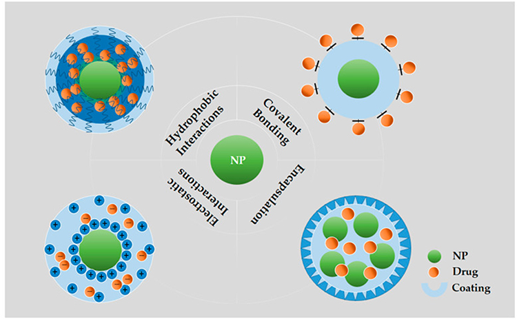 |
Figure 1 Illustration of various methods of loading/bonding therapeutics into NPs. Covalent bonding: Drugs directly to the surfaces of NPs through covalent bonds; Hydrophobic interactions: Partitioning hydrophobic drug molecules in an amphiphilic Corona layer; Electrostatic interactions: Loading drugs onto the surfaces of NPs by electrostatic layer-by-layer assembly; Encapsulation: Loading drugs into the hollow NPs. Reproduced with permission from ref,16 CC BY 4.0. Copyright© 2020 by the authors. |
Regarding controlled and targeted delivery, NDDS exert their effects on tumor cells via two primary mechanisms: passive targeting and active targeting23 (Figure 2). Passive targeting leverages the defective vascular architecture characteristic of solid tumors. Such vascular defects induce the formation of interendothelial gaps, thereby enhancing vascular permeability. This phenomenon is termed the enhanced permeability and retention (EPR) effect, a key mechanism that facilitates the accumulation of small-sized nanoparticles (NPs) in tumor tissues.24 In contrast, active targeting is an alternative strategy that achieves targeted delivery through surface modification of NPs. Targeting ligands, which specifically recognize overexpressed receptors on tumor cells, are conjugated to the NP surface, enabling active targeting of tumor sites while sparing healthy cells.25
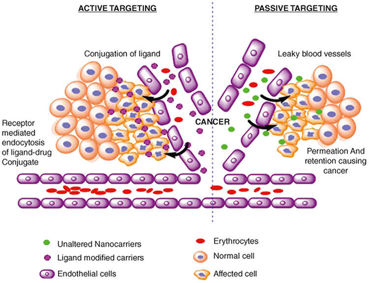 |
Figure 2 Active and passive targeting. Active targeting: ligand-modified carriers to deliver drugs to tumor cells through receptor-mediated endocytosis of ligand-drug conjugate; passive targeting: unaltered nanocarriers to deliver drugs to tumor cells through permeability and retention causing cancer. Reproduced with permission from ref,23 CC BY 4.0. Copyright © 2024 Informa UK Limited. |
In recent years, substantial advancements have been achieved in the research on NDDS applications for LC.26,27 The number of NP-based therapeutic products approved for clinical application has been steadily growing. Based on the type of carrier material, NDDS can be categorized into organic Nano-delivery systems, inorganic Nano-delivery systems, and inorganic-organic hybrid Nano-delivery systems.28 These Nano-delivery materials have exhibited promising therapeutic potential and clinical translation prospects in both preclinical and clinical studies of LC. Nevertheless, this field remains confronted with numerous challenges. Against this backdrop, this review will systematically summarize the advances in NDDS applications for LC, elaborate on the structural characteristics, mechanisms of action, and specific therapeutic applications of different types of NDDS in LC treatment, and aim to provide valuable insights for the research, development, and clinical translation of LC nanomedicines.
Organic Nano-Delivery Systems
Organic Nano-delivery systems refer to nanocarriers constructed from synthetic polymers, lipids, micelles, natural polymer derivatives, or their composite materials (typically with a particle size range of 10–200 nm).29 These systems have demonstrated considerable potential in lung cancer therapy research due to their high biocompatibility, superior targeting ability, low systemic toxicity, and capacity for co-loading multiple therapeutic agents.5 Specifically, their advantages include the following aspects: Efficient drug encapsulation—it solves the low solubility problem of hydrophobic anticancer drugs (eg, paclitaxel, docetaxel, curcumin) and significantly improves drug solubility and bioavailability.5,30–33 Targeted delivery—specific targeting to the tumor microenvironment or cancer cells is achieved through surface modification (eg, ligands, antibodies, peptides), reducing toxic side effects on normal tissues.34–36 Sustained and controlled release—it optimizes drug release kinetics, prolongs the local drug exposure time in tumors, and overcomes the defects of rapid metabolism and clearance in traditional chemotherapy.37 Efficacy enhancement and toxicity reduction—it improves cell uptake efficiency (eg, through receptor-mediated endocytosis), synergistically enhances anticancer activity, and reduces the risk of systemic toxicity.38,39 Multifunctional integration—it can simultaneously load chemotherapeutic drugs, genes (siRNA/mRNA), photosensitizers, or imaging probes, realizing the integration of therapy and diagnosis (Theranostics).40,41
Lipid-Based Nano-Delivery Systems
Lipid-based nanoparticles represent one of the most promising carriers for anticancer drugs. Their composition resembles that of cell membranes, enabling enhanced cellular uptake and reduced side effects. The core strength of this system lies in the effective integration of loading versatility and targeting precision42 (Figure 3), allowing for the encapsulation and stable delivery of diverse therapeutic agents, including DNA, peptides, RNAi, and chemotherapeutic drugs. By modifying the nanoparticle surface with RGD peptides (arginine-glycine-aspartic acid) or specific antibodies, active targeting capability can be further conferred, along with improved cellular internalization. Currently, lipid-based nanoplatforms have demonstrated significant potential in the diagnosis and treatment of non-small cell lung cancer, emerging as a cutting-edge research direction for enhancing targeted lung cancer therapy and overcoming drug resistance.43
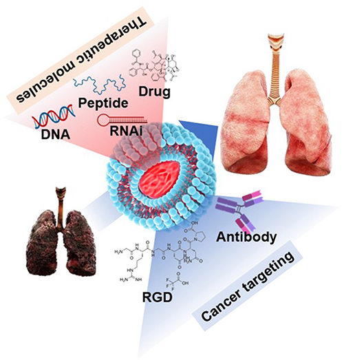 |
Figure 3 Schematic illustration of liposome-mediated LC treatment. Lipid-based nanocarriers encapsulating therapeutic molecules (eg, DNA, peptides, RNAi, and drugs), with surface modifications such as RGD peptides and antibodies for cancer targeting. Lipid-based nanocarriers encapsulated by therapeutic molecules including DNA, peptide, RNAi and drug, surface-modified by RGD and antibody for cancer-targeting. Reproduced with permission from ref,42 CC BY-NC 3.0 © 2023 Kim et al. |
Liposomes
Liposomes are spherical nanovesicles constructed from amphiphilic lipids such as natural or synthetic phospholipids and cholesterol. Their core structure consists of a lipid bilayer membrane enclosing an aqueous core. This membrane structure mimics biological cell membranes, endowing liposomes with high biocompatibility and the ability to seamlessly integrate with the pulmonary surfactant layer in the lungs. Meanwhile, liposomes possess both hydrophilic and hydrophobic domains, enabling efficient delivery of molecules with varying solubilities. As the most commonly used lipid-based nanocarriers, the pharmacokinetic behavior of liposomes mainly depends on key properties such as particle size, surface charge, membrane lipid packing state, spatial stability, administration dose, and administration route.44,45
Liposomal Paclitaxel is currently the most widely used liposomal drug in lung cancer therapy, especially for NSCLC. Paclitaxel serves as a cornerstone chemotherapeutic agent for lung cancer but suffers from limitations including toxicity, poor aqueous solubility, and the development of multidrug resistance (MDR). Liposomes improve the pharmacological properties of paclitaxel through multiple mechanisms: the phospholipid bilayer structure enhances solubility, prolongs circulation time, enables passive tumor targeting via leaky blood vessels, and reduces side effects. In lung cancer cell lines and animal models, Liposomal Paclitaxel exhibits enhanced cytotoxicity, cellular uptake capacity, apoptosis-inducing effect, tumor growth inhibition, and anticancer activity.46 Clinical studies have also confirmed its efficacy in both NSCLC and SCLC.43 However, although Liposomal Paclitaxel passively targets tumor blood vessels via the EPR effect, the heterogeneity of lung cancer blood vessels (eg, low permeability or high interstitial pressure) significantly impairs drug accumulation.47 The preparation of liposomes requires strict control of phospholipid ratio, particle size distribution, and encapsulation efficiency, resulting in a complex manufacturing process and high cost. Consequently, the price of Liposomal Paclitaxel is significantly higher than that of conventional paclitaxel formulations, limiting its clinical application.48 Additionally, although the neurotoxicity of Liposomal Paclitaxel is significantly lower than that of conventional formulations, myelosuppression (eg, neutropenia) remains common and may affect treatment cycles.
Over the past three decades, cisplatin has been one of the most effective cytotoxic agents and a cornerstone of first-line chemotherapy for lung cancer, particularly for NSCLC and SCLC. However, severe side effects such as nephrotoxicity, ototoxicity, neurotoxicity, and gastrointestinal reactions significantly limit its clinical dosage and patient tolerability. Liposomal cisplatin (trade name: Lipoplatin) has undergone extensive formulation and testing in preclinical (in vitro) studies as well as Phase I, II, and III clinical trials.43 Fu et al49 developed an inhalable nanoliposome system for the co-delivery of osimertinib (a targeted drug) and a DNA plasmid. The inhaled nanoparticles efficiently penetrate the pulmonary barrier and accumulate in the lungs Within tumor cells, osimertinib is released to inhibit tumor growth, while the delivered DNA plasmid triggers in vivo production of exosomes that migrate to the brain, thereby suppressing brain metastases of lung cancer. This strategy provides a safe and versatile innovative approach for the treatment of lung cancer, including its brain metastases.
Beyond targeted drug delivery, Badea et al50 utilized liposomal‑iodine nanoparticle‑based contrast agents combined with CT imaging to visualize nodular vascularization in genetically engineered mouse models of non‑small cell lung cancer. The signal enhancement enabled differentiation between slow‑ and fast‑growing nodules. Consequently, such contrast agents can contribute to early detection and diagnosis of pulmonary lesions. Small interfering RNA (siRNA) offers a gene-based therapeutic option for cancer management by silencing oncogenes and anti-apoptotic genes. Lipid-based carriers, primarily liposomes, are widely employed to encapsulate “naked” siRNA, protecting it from degradation in vivo and reducing immunogenicity risks, owing to their excellent biocompatibility. Leveraging their nanoscale properties and capacity for surface modification, these carriers enhance siRNA enrichment and intracellular delivery within lung tumor tissues through mechanisms such as endocytosis or membrane fusion, thereby providing a novel therapeutic strategy for tumors.51 However, this field still faces common challenges in translating from the laboratory to clinical application, including the need for further improvements in in vivo stability and targeting specificity, reduction of off-target effects, optimization of lipid formulation design, and addressing safety and large-scale manufacturing requirements. Future developments will focus on optimizing lipid-based formulations and delivery strategies to enhance efficacy and safety, with the goal of advancing this siRNA-based nanotherapy into practical clinical use for lung cancer treatment.
Solid Lipid Nanoparticles
Among different types of nanoparticles, solid lipid nanoparticles (SLNs) represent an emerging subfield in nanotechnology. Equipped with biodegradable formulation components, SLNs can safely load therapeutic compounds, enabling effective drug delivery with sustained release potential.52 SLNs offer excellent biocompatibility, physical stability, high drug loading capacity, physiological protection in the gastrointestinal environment, and enhanced cellular uptake. Moreover, the feasibility of large-scale production is a key advantage of SLNs.53
Bae et al54 developed a biomimetic solid lipid nanoparticle (SLN) that mimics low‑density lipoprotein (LDL) for theranostic applications. Its core–shell structure encapsulates quantum dots and paclitaxel within a lipid shell, while Bcl‑2‑targeting siRNA is electrostatically bound on the surface. This design enhances biocompatibility and cellular uptake, enabling co‑delivery of paclitaxel and siRNA into lung cancer cells. The combination of Bcl‑2 silencing and paclitaxel‑induced microtubule stabilization triggers caspase‑mediated apoptosis, producing synergistic anticancer effects confirmed by median‑effect analysis. Encapsulated quantum dots also allow real‑time intracellular tracking via fluorescence imaging.
Curcumin acts on multiple targets and exerts synergistic inhibitory effects on the malignant biological behaviors of lung cancer cells through multiple pathways.55 However, its clinical application is limited by poor water solubility (approximately 11 ng/mL), rapid metabolism, and easy degradation under different physiological pH conditions.56 Curcumin solid lipid nanoparticles (Cur-SLNs), prepared by encapsulating curcumin into SLNs (using matrices such as glyceryl monostearate and lecithin), possess nanoscale size, narrow particle size distribution, sufficient surface electrostatic charge (which avoids aggregation and improves physical stability), high encapsulation efficiency and drug loading capacity. These properties significantly enhance curcumin’s solubility, stability, and cellular uptake efficiency, exhibiting remarkable antiproliferative effects on cancer cells.57–60 In vitro studies have shown that Cur-SLNs can effectively increase the drug uptake by A549 cells. Both curcumin and Cur-SLNs exhibit dose-dependent cytotoxicity against A549 cells, with the latter showing stronger toxicity. Although Cur-SLNs, as a novel formulation, have demonstrated therapeutic potential in lung cancer treatment research, they suffer from low drug loading capacity,61 and unclear in vivo metabolic pathways,62 and are currently still in the preclinical research stage.58,59
Polymeric Nanoparticles
Polymeric nanocarriers applied in lung cancer therapy are mainly based on natural polymers (ie, proteins, peptides, glycans, starch, or cellulose) and synthetic polymeric materials (such as polyethylene glycol (PEG), polylactic acid (PLA), polyethylene glycol-polylactic acid (PEG-PLA), chitosan, dendrimers, and stimuli-responsive block copolymers). Drug loading is achieved through chemical conjugation or physical encapsulation.
Natural Polymeric Nanoparticles
Nanoparticle albumin-bound (nab) systems (eg, albumin-bound paclitaxel, Abraxane®) are drug delivery systems that use natural albumin as the carrier, with drugs encapsulated or adsorbed on the surface/interior of nanoscale particles. Research on nanoparticle albumin-bound paclitaxel (nab-paclitaxel) in lung cancer therapy has progressed from early exploration to a stage where clinical practice and innovative optimization proceed in parallel, making it the most widely used nanoparticle in clinical lung cancer treatment currently.13
Paclitaxel is one of the core drugs in lung cancer chemotherapy. The preparation of nab-paclitaxel (Abraxane®) by conjugating paclitaxel to albumin nanoparticles significantly improves drug solubility and intratumoral accumulation, thereby enabling the treatment of NSCLC;63,64 Meanwhile, the particle size of albumin nanoparticles can precisely leverage the EPR effect of tumor blood vessels, resulting in a local paclitaxel concentration 2–3 times higher than that of conventional solvent-based paclitaxel, laying the foundation for subsequent drug enrichment.
On this basis, nab-paclitaxel can further enhance targeting by specifically binding to secreted protein acidic and rich in cysteine (SPARC) abundant in the tumor microenvironment. SPARC is highly expressed in the lung cancer microenvironment and can act as a “molecular anchor” to mediate the active binding and uptake of Abraxane®, ultimately achieving efficient drug enrichment in tumor tissues.65 The research on nanoparticle albumin-bound paclitaxel (nab-paclitaxel) in the treatment of lung cancer has evolved from early exploration to a stage where clinical practice and innovative optimization proceed in parallel. It is currently the most commonly used nanoparticle in clinical applications for lung cancer treatment.13 Nevertheless, nab-paclitaxel still retains the hematological toxicity and neurotoxicity inherent to paclitaxel.66
Ferritin nanocarriers, another type of natural protein nanocarriers, have demonstrated significant potential in lung cancer therapy in recent years. Due to rapid proliferation, lung cancer cells have increased iron demand and high expression of transferrin receptor 1 (TfR1) on their surface. Ferritin nanoparticles achieve specific accumulation in tumor cells through TfR1-mediated endocytosis.67 Additionally, ferritin nanoparticles are natural hollow nanocages;68 in addition to loading chemotherapeutic drugs, their interior can also carry photosensitizers and photothermal agents, enabling photothermal therapy (PTT) and photodynamic therapy (PDT) against tumor cells69 (Figure 4). Ferritin can deliver photosensitizers to tumor cells. Upon irradiation with a specific wavelength of laser light, combined with PDT, the photosensitizer generates reactive oxygen species in the presence of oxygen, directly killing tumor cells and destroying tumor blood vessels. Ferritin transports photothermal agents into tumor cells. Through photothermal therapy (PTT), the agents absorb near‑infrared (NIR) light and convert it into heat, and the resulting localized high temperature destroys tumor cells or tissues.
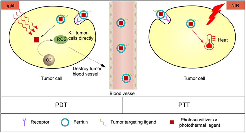 |
Figure 4 Ferritin for photodynamic PDT and PTT. PDT: Ferritin delivers photosensitizers to tumor cells. Under irradiation with a specific wavelength of laser light, the photosensitizers generate reactive oxygen species in the presence of oxygen, directly killing tumor cells and destroying tumor blood vessels; PTT: Ferritin transports photothermal agents into tumor cells. By absorbing NIR light and converting it into heat, the localized high temperature generated thereby destroys tumor cells or tissues. Reproduced with permission from ref,69 CC BY 4.0. Copyright © 2023 by the authors. |
Currently, research on ferritin nanocarriers in lung cancer therapy is still dominated by preclinical studies, with some strategies entering clinical translation evaluation. However, no large-scale human clinical trial data have been publicly reported. Nevertheless, ferritin possesses dual functions as a drug carrier and an imaging agent, holding promise for achieving integrated diagnosis and treatment of lung cancer.
Antibody-Drug Conjugates (ADCs) are among the most successful nanoformulations in clinical cancer therapy. These drugs target tumor-specific antigens via antibodies and deliver highly potent chemotherapeutic agents (drug payloads) into tumor cells in a nanoscale structure (10–15 nm), enabling precise tumor eradication and reduced systemic toxicity, thus providing crucial support for the precise treatment of lung cancer.
HER2 is a transmembrane glycoprotein receptor with intracellular tyrosine kinase activity.70 Its activation triggers a series of subcellular signaling pathways that regulate the growth, differentiation, and migration of epithelial cells across various cell lineages.71,72 Trastuzumab-emtansine (T-DM1) is the first ADC tested in advanced HER2-mutant NSCLC. The nanoparticle-based formulation, NPs-T-DM1, developed based on T-DM1, can passively target tumor blood vessels via the EPR effect while retaining the HER2-specific binding capability of trastuzumab.73,74 The DM1 encapsulated within these nanoparticles is slowly released in the tumor microenvironment, capable of penetrating the tumor stroma and killing adjacent HER2-negative cells, thereby addressing the issue of tumor heterogeneity.75,76 However, long-term use may induce downregulation of HER2 expression in tumor cells, leading to drug resistance, and some patients still experience toxic side effects.75 Therefore, drug resistance and cumulative toxicity of antibody-conjugated nanocarriers remain key challenges to be addressed in future research.
Synthetic Polymeric Nanoparticles
Synthetic polymeric nanocarriers are constructed through specific fabrication processes using artificially chemically synthesized macromolecules (ie, synthetic polymers) as the core matrix. Owing to their chemical versatility, they are also promising nanotherapeutic tools, and a variety of synthetic polymeric nanocarriers have entered clinical trial stages.
Matrix-Type Polymeric Nanoparticles
Studies on the preparation of disulfiram‑loaded poly (lactic‑co‑glycolic acid) (PLGA) nanoparticles have demonstrated that modulating parameters such as PLGA polymer type, loading and molecular weight of stabilizers (eg, polyvinyl alcohol), and sonication time enables precise control over particle size, encapsulation efficiency, and release kinetics. These adjustments further significantly influence the stability of the drug in serum, its copper‑ion‑mediated activation efficiency, and in vitro cytotoxicity against non‑small cell lung cancer cells.77 On the other hand, to address the challenge of delivering hydrophilic peptide drugs, a non‑aqueous nanoprecipitation technique has been employed to prepare poly (sebacic anhydride) (PSA) nanoparticles under strictly anhydrous conditions. This process effectively prevents polymer hydrolysis and premature drug release, achieves high drug‑loading encapsulation of neuropeptides, and leverages the rapid degradation characteristics of PSA to yield release profiles with either burst or sustained patterns, offering a novel strategy for local delivery systems such as nasal administration.78
Polymeric Micelles
Polymer micelles, as extensively studied self-assembled core-shell nanostructures, typically range in size from 10 to 100 nm. Their hydrophilic shell, composed of polyethylene glycol, reduces uptake by the reticuloendothelial system and significantly prolongs systemic circulation time, thereby promoting selective drug accumulation in tumor tissues through the enhanced permeability and retention effect. The hydrophobic core efficiently encapsulates poorly water-soluble anticancer drugs, substantially reducing the need for organic solvents and exhibiting prolonged circulation, tumor-targeted accumulation, and controlled release capabilities. By delivering insoluble drugs, polymer micelles can markedly enhance the safety and efficacy of chemotherapy regimens for lung cancer79–81 (Figure 5).
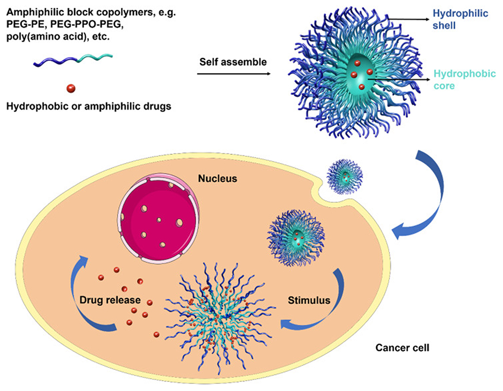 |
Figure 5 Structural and Functional Properties of Polymer micelles (PMs). PMs are self-assembly products of block copolymers with the core–shell structure consisting of a hydrophobic core surrounded by a hydrophilic shell. The core of PMs permits to entrap hydrophobic and amphiphilic drugs, allowing controlled drug release, while the shell can prevent the intake of PMs by the RES, prolonging the blood circulation time of PMs, which will further increase the drug accumulation in the tumor sites. When encountering intracellular or external stimuli such as pH, enzyme, redox, laser, etc, PMs will disassemble to release the drug. PEG, poly(ethylene glycol); PE, phosphatidylethanolamine; PPO, polyphenylene oxygen. Reproduced with permission from ref,81 CC BY 4.0. Copyright© The Royal Society of Chemistry. |
Studies have shown that micelles prepared from the specific amphiphilic block copolymer palmitoyl‑poly (methacryloyloxyethyl phosphorylcholine), owing to their distinct polarity contrast and stable amphiphilic structure, outperform traditional polyethylene glycol‑based micellar systems in terms of drug loading capacity, cellular uptake efficiency, and inhibitory effects against multidrug‑resistant tumor cells. This offers a new strategy for addressing the challenge of clinical drug resistance.82
Due to their ultra-small size, polymeric micelles can enhance the water solubility of chemotherapeutic drugs, prolong blood half-life, and retain the EPR effect, thereby passively accumulating at tumor sites through the leaky vascular system.83,84 This is crucial for the treatment of solid tumors via passive targeting, especially for tumors with poorly differentiated blood vessels.85
Furthermore, by modifying their surface with targeting ligands (eg, antibodies, peptides) or integrating imaging probes (eg, fluorophores, radionuclides), micelles can achieve active targeting toward tumor cells and enable real‑time imaging tracking. Simultaneously, through the incorporation of pH‑sensitive, thermosensitive, redox‑responsive, or light‑sensitive polymers, micelles can undergo structural dissociation in response to specific tumor microenvironmental cues or external stimuli, thereby achieving site‑specific and time‑controlled drug release. This approach facilitates the construction of an integrated “diagnosis‑therapy‑monitoring” intelligent nanoplatform. The convergence and advancement of these strategies are driving polymer micelle systems toward more precise, efficient, and safe directions in tumor therapy.
The hydrophilic segment PEG is the most commonly used material for drug delivery micelles. PEG forms a hydrophilic corona on the micelle surface, which can minimize non-specific interactions with blood components and prolong circulation time. Besides PEG, poly(N-vinylpyrrolidone) (PVP)86 and poly(N-isopropylacrylamide) (pNIPAM)87,88 are also used as hydrophilic components of micelles.
Blank polymeric micelles exhibit excellent biocompatibility. In lung cancer treatment, monomethoxy poly(ethylene glycol)-poly(caprolactone)-D-α-tocopheryl polyethylene glycol 1000 succinate (MPEG-PCL-TPGS) is used as a nanocarrier for delivering paclitaxel (PTX). Studies have shown that PTX-loaded MPEG-PCL-TPGS (PTX/MPT) micelles exhibit a sustained-release profile for 168 hours, with accelerated drug release under acidic conditions. They also show higher cellular uptake through endocytic pathways. Thus, the biocompatible polymeric nanosystem loaded with PTX holds potential as a delivery system for lung cancer treatment.89 Furthermore, by modifying their surface with targeting ligands (eg, antibodies, peptides) or integrating imaging probes (eg, fluorophores, radionuclides), micelles can achieve active targeting toward tumor cells and enable real-time imaging tracking. Simultaneously, through the incorporation of pH-sensitive, thermosensitive, redox-responsive, or light-sensitive polymers, micelles can undergo structural dissociation in response to specific tumor microenvironmental cues or external stimuli, thereby achieving site-specific and time-controlled drug release. This approach facilitates the construction of an integrated “diagnosis-therapy-monitoring” intelligent nanoplatform.90 The convergence and advancement of these strategies are driving polymer micelle systems toward more precise, efficient, and safe directions in tumor therapy. However, it still has limitations including poor formulation stability, low drug loading capacity and remains in the clinical trial stage.88
Dendrimers
Dendrimer nanocarriers are a class of highly symmetric spherical compounds composed of repetitive branched molecules ranging from 1 to 100 nm, with highly branched three-dimensional structures, and have emerged as important platforms for tumor-targeted therapy and drug co-delivery. Their properties are mainly determined by functional surface groups, enabling them to serve as core components of biomaterials for targeted therapy and diagnosis, as well as to efficiently deliver drugs and genes both in vitro and in vivo. In applications for tumor therapy and diagnosis, dendrimers can load hydrophobic drugs and nucleic acids (DNA/RNA) via entrapment or conjugation, achieve tumor-targeted delivery with the aid of targeting moieties such as folic acid and antibodies as well as the EPR effect, and simultaneously improve the solubility and bioavailability of drugs. Among them, polyamidoamine (PAMAM), polypropyleneimine (PPI), and polylysine (PLL) are the most extensively studied types.91 Lee et al confirmed that the formulation of doxorubicin conjugated with biodegradable dendrimers exhibits superior anti-tumor efficacy to free doxorubicin, with activity comparable to that of doxorubicin liposomes (Doxil®).92
As the most widely used dendrimers in the field of drug delivery, polyamidoamine (PAMAM) features three drug encapsulation sites, namely molecular voids, branching points, and surface groups. The construction of tumor-targeted PAMAM-albumin nanoparticle assemblies and co-controlled release PAMAM-PAMAM assemblies enables PAMAM to comprehensively optimize the performance of the entire drug delivery process, improve the water solubility and stability of hydrophobic drugs, enhance dissolution rate, and achieve controlled release. Consequently, PAMAM is expected to serve as the core carrier of next-generation intelligent drug delivery systems, and its potential needs to be explored through interdisciplinary collaboration in the future.93
The construction of novel dendrimers using biocompatible components, as well as the surface modification of dendrimers via PEGylation, acetylation, glycosylation, and amino acid functionalization, have improved the safety of dendrimer-based nanotherapeutics and enabled the integration of imaging agents to achieve integrated tumor-targeted diagnosis and therapy.94,95 Based on the characteristic that the hydrophobic inner cavities of dendrimers can load various chemotherapeutic agents and imaging agents, researchers have also developed a variety of dendrimer-based immunotherapeutic formulations, which significantly improve the therapeutic index of current cancer immunotherapies and ultimately enhance tumor treatment outcomes. Although dendrimer-based nanomedicines have not yet been translated into clinical applications, they are expected to overcome the current challenges associated with cancer immunotherapy and transform the existing paradigm of cancer immunotherapy.96
Polymer-Drug Conjugates
Polymer-drug conjugates are covalently attached to biocompatible, water-soluble polymer backbones—such as poly(N-(2-hydroxypropyl)methacrylamide (HPMA)—via cleavable linkages, forming nano-sized constructs. A representative example, the HPMA-doxorubicin conjugate (HPMA-Dox), along with its further developed intelligent nanoassemblies (PMD), primarily relies on the EPR effect for tumor accumulation. Its design innovation lies in the incorporation of microenvironment-responsive linkers, such as peptides cleavable by tumor-overexpressed matrix metalloproteinases (MMPs), enabling precise and controlled drug release at the tumor site and significantly reducing systemic toxicity. These conjugates offer multiple therapeutic advantages: they improve pharmacokinetics by extending drug circulation and enhancing tumor accumulation;97 demonstrably reduce cardiotoxicity compared to free doxorubicin.98; and show potential in overcoming multidrug resistance by mechanisms such as ATP depletion to inhibit P-glycoprotein-mediated drug efflux.99 However, high-molecular-weight polymer conjugates may face limited penetration into dense tumor tissues due to their size, and most promising systems remain in preclinical stages, with clinical translation efficiency and long-term safety requiring further validation.100,101
Viral Nanoparticles
Viral nanoparticles, including natural viral vectors, virus-like particles (VLPs), and artificially engineered viruses, possess inherent targeting ability, high-efficiency delivery capacity, and immunomodulatory properties. Certain viruses (eg, adenoviruses) naturally tend to infect epithelial cells and bind to receptors on the surface of lung cancer cells (eg, coxsackie-adenovirus receptor (CAR), epidermal growth factor receptor (EGFR)), thereby achieving tumor-specific delivery.For example, ONCOS-102, an oncolytic adenovirus, selectively infects lung cancer cells by targeting the EphA2 receptor.102 However, viral nanoparticles still carry the risk of inducing excessive immune responses in the host. Consequently, their research in lung cancer treatment is currently in the transitional stage of clinical translation, though they may become ideal materials for lung cancer diagnosis and treatment in the future.
Inorganic Nano-Delivery Systems
Inorganic Nano-delivery systems refer to nanoscale carrier platforms constructed on the basis of inorganic materials, typically with a particle size ranging from 1 to 1000 nm. Compared with organic nanomaterials, inorganic Nano-delivery systems are emerging stars in the biomedical field, possessing advantages such as simple preparation, excellent controllability of shape and size, and easy surface modification. They can realize drug protection, controlled release and precise targeted delivery, thereby significantly improving therapeutic efficacy and reducing systemic toxic side effects.103,104 Moreover, the unique optical, electrical and magnetic properties of inorganic nanomaterials endow them with potential functions including imaging development, targeted delivery and synergistic drug therapy. Common inorganic nanomaterials include: metal-based nanoparticles, non-metallic inorganic materials and composite inorganic materials.
Metal Nano-Delivery Systems
Gold Nanoparticles
Gold Nanoparticles (AuNPs) exhibit various physicochemical properties such as unique optical properties,105 good biocompatibility106 and controllable synthesis capability,107 thus demonstrating multi-dimensional application potential in the field of lung cancer treatment. AuNPs can be effectively applied in multiple biomedical fields, including targeted drug delivery,108,109 biosensors,110,111 molecular imaging,112,113 ultrasound ablation,114 and photothermal therapy.115,116 They are particularly utilized in ultra-sensitive detection and imaging-based therapeutic technologies, which are essential for the treatment of life-threatening diseases such as cancer.117 With the continuous improvement of the preparation methods for AuNPs, a variety of AuNPs with different sizes and morphologies have been successfully prepared at present, namely gold nanospheres, gold nanorods, gold nanocages, gold nanoshells, and gold nanostars. These AuNPs of different sizes and shapes possess distinct therapeutic functions118,119 (Figure 6).
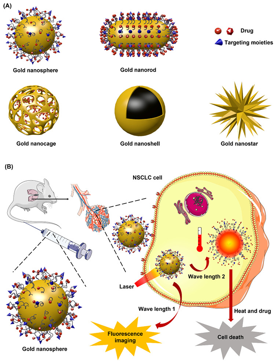 |
Figure 6 Nanostructures and Applications of AuNPs. AuNPs are stable colloid solutions of Au atom clusters that are remotely controlled by NIR. (A) AuNPs have diverse shapes containing gold nanospheres, nanorods, nanocages, nanoshells, and nanostars. (B) AuNPs can be employed as drug delivery systems to transport imaging agents and therapeutic drugs for both diagnosis and treatment, as bio-sensors for fluorescence imaging triggered by NIR, and as photothermal therapeutic agents which produce cell-killing heat by laser irradiation. Reproduced with permission from ref,81 CC BY 4.0. Copyright© The Royal Society of Chemistry. |
Among gold nanoparticles, gold nanorods (AuNRs) stand out as one of the most crucial theranostic nanomaterials due to their unique optical and electronic properties. AuNRs exhibit a rod-like morphology, and their aspect ratio (AR) is a key factor determining the surface plasmon resonance (SPR) properties. During the synthesis process, the SPR peak can be precisely regulated;120 thus, AuNRs can be synthesized as ideal SPR materials, contrast agents in optical imaging121 and nanoscale heat sources in therapeutic applications.122–124 Since both photoacoustic imaging (PAI) and plasmonic photothermal therapy (PPTT) utilize AuNRs as optical absorbers, the integration of diagnosis and treatment can be achieved using AuNRs.125 Meanwhile, the photothermal property of AuNRs enables them to absorb light emitted by light sources and convert it into heat, thereby inducing tumor cell destruction. A study employed AuNRs of different sizes as light absorbers for pulsed wave photothermal therapy (PW-PPTT) on the lung cancer cell line (A549). The results showed that after lung cancer cells were co-incubated with gold nanorods (AuNRs) of different sizes and subsequently exposed to pulsed wave (PW) laser irradiation, the induced cell mortality rate was significantly higher than that of cells without AuNR co-incubation.126 However, gold nanoparticles exhibit local pulmonary toxicity and insufficient targeting efficacy, and they are currently in the clinical trial phase.
Iron Oxide Nanoparticles
Magnetic iron oxide nanoparticles are widely used in biomedical applications.127–130 However, bare iron oxide nanoparticles exhibit poor stability and dispersibility when directly exposed to the environment, and their magnetic properties fail to support high-sensitivity magnetic resonance imaging (MRI). In 2012, Hou et al proposed a bromide-induced wet chemical synthesis method to prepare high-purity and morphology-controllable iron carbide (Fe5C2) nanoparticles (Fe5C2-NPs).131 As an emerging magnetic nanomaterial, Fe5C2-NPs contain both carbon and iron elements, possessing multiple excellent and unique properties such as high saturation magnetization, strong corrosion resistance, and superior photothermal conversion performance. Consequently, they can be applied in MRI and magnetic targeting in the field of cancer theranostics.132,133 Meanwhile, relying on endogenous stimuli in the tumor microenvironment (TME) such as hypoxia, weak acidity, high hydrogen peroxide (H2O2) levels, and high glutathione (GSH) levels,134,135 Fe5C2-NPs can undergo specific reactions at the tumor site, improving treatment precision.
In the field of nanomaterials, gold-iron carbide Janus nanoparticles (Au–Fe2C Janus NPs) are composed of gold (Au) and iron carbide (Fe2C). These two components integrate the photothermal conversion and CT imaging functions of the Au component, as well as the magnetic response and MRI imaging functions of the Fe2C component, enabling the composite performance of “theranostic integration”.136–138 Studies have shown that Au–Fe2C Janus NPs exhibit an in vitro photothermal conversion efficiency of up to 30.2%, and after accumulating at the tumor site, the central temperature of the tumor exceeds 42°C under laser irradiation. This indicates that they are excellent PTT agents and exert a strong inhibitory effect on tumor growth.127,136 By modifying targeting molecules (eg, anti-EGFR antibodies, Affibody ZHER2:342), Au–Fe2C Janus NPs can specifically bind to receptors on the surface of lung cancer cells (eg, EGFR, HER2), thereby improving tumor accumulation efficiency.136,139,140 However, the clinical translation of Au–Fe2C Janus NPs needs to overcome multiple challenges including material specificity, safety, and manufacturing processes. Currently, they are still in the laboratory research stage. If progress can be made in areas such as precision targeting and combination therapy in the future, they are expected to provide a new option for lung cancer treatment.
Silica Nanoparticles
Silica Nanoparticles (SNPs) are widely used in the biomedical field, especially in lung cancer diagnosis and treatment, due to their high design flexibility, good biocompatibility, and ability to improve the bioavailability of drug molecules at specific sites. Among them, SNPs have attracted extensive attention as one of the most biocompatible drug carriers.
Mesoporous Silica Nanoparticles
Among various types of nanoparticles, mesoporous silica nanoparticles (MSNPs) have emerged as a cutting-edge and promising multifunctional platform in the field of cancer therapy, thanks to numerous advantages such as large specific surface area, controllable pore size and morphology, easy surface modification, high drug-loading capacity, and biodegradability.23
With highly ordered mesoporous structures (pore size: 2–50 nm), ultra-large specific surface area, and modifiable surfaces,23,141,142 MSNPs exert effects in lung cancer through multi-dimensional mechanisms:143 their mesoporous channels can efficiently load chemotherapeutic drugs, targeted drugs, immunomodulators, etc, and achieve tumor microenvironment-triggered release via pH/enzyme-sensitive responsive gating molecules;144 they can enhance tumor accumulation and lung retention by conjugating targeting molecules (such as anti-EGFR antibodies and RGD peptides) on the surface or designing aerosol inhalation formulations; meanwhile, they can integrate photothermal/photodynamic agents to achieve chemotherapy-physical therapy synergy, or load immune adjuvants to induce immunogenic cell death and reverse the immunosuppressive microenvironment. Additionally, they can realize multimodal imaging by loading superparamagnetic iron oxide nanoparticles (SPIONs), doping Gd3⁺/18F, or modifying fluorescent dyes, providing integrated support for lung cancer diagnosis, treatment, and monitoring.
Compared with carriers such as liposomes and polymeric nanoparticles, MSNs exhibit significant advantages in lung cancer applications: they show higher stability in resisting degradation and external pressure;145 they can recognize specific cells and receptors to achieve the expected efficacy at the target site while avoiding side effects on non-target tissues;146 embedding iron oxide nanocrystals into MSNs can serve as targeted contrast agents, enabling magnetic manipulation and MRI to become excellent nano-diagnostic tools;147 they have strong structural adjustability, and the mesoporous pore size, particle size, and surface charge can be customized by regulating the synthesis process to adapt to different lung cancer subtypes.148,149 However, MSNs still face multiple bottlenecks in clinical translation for lung cancer: potential biosafety hazards, as large-sized MSNs degrade slowly in the lungs and may induce chronic inflammation or even increase cytotoxicity; insufficient targeting efficiency and tumor penetration, with low passive targeting efficiency for lung cancer subtypes with sparse blood vessels; pending optimization of production and stability, with high synthesis costs; and difficulty in solving drug resistance and recurrence issues.150,151
Porous Silicon Nanoparticles
As a new type of inorganic nanomaterial, porous silicon nanoparticles (PSiNPs) have demonstrated multi-dimensional research potential in lung cancer treatment. PSiNPs enable controlled release of chemotherapeutic drugs and are involved in gene immunotherapy. The mesoporous structure of PSiNPs can efficiently load chemotherapeutic drugs (such as doxorubicin and cisplatin), and achieve tumor microenvironment-triggered release through surface-modified stimulus-responsive linkers (such as pH-sensitive hydrazone bonds and light-responsive o-nitrobenzyl groups). For example, the PSiNPs designed by the Wiley team covalently conjugate drug-linker complexes via click chemistry, with a drug-loading capacity of up to 250 µg/mg, and the drug release rate is significantly increased under acidic conditions. In a mouse model of lung cancer, this delivery system can significantly inhibit tumor growth with lower systemic toxicity than traditional chemotherapy.152 PSiNPs can simultaneously deliver small interfering RNA (siRNA, such as siRNA targeting survivin) and chemotherapeutic drugs, reversing tumor drug resistance through synergistic effects. For instance, PEG-modified PSiNPs loaded with survivin siRNA can effectively silence drug-resistant genes in lung cancer cells and enhance the killing effect of chemotherapeutic drugs.153 With the advancement of research on lung cancer nano-delivery systems, researchers are attempting to innovate combined treatment modes based on PSiNPs.
After modifying photothermal agents (such as gold nanoshells and porphyrins) on the surface of PSiNPs, they generate heat (42–45°C) under near-infrared (NIR) light irradiation, which not only directly damages the tumor cell membrane but also promotes the release of chemotherapeutic drugs in the mesopores. Studies have shown that PSiNPs combined with NIR therapy can reduce the survival rate of lung cancer cells to 6.7%, and no recurrence was observed after complete tumor regression.154 In addition, PSiNPs can also act as sonosensitizers, inducing reactive oxygen species production in combination with ultrasound to enhance the tumor-killing effect.154 However, PSiNPs also face shortcomings such as large pore size, poor permeability, and high cost, and are currently still in the preclinical trial stage.
Silica-Based Composite Materials Nanoparticles
Although silica nanomaterials are still in the preclinical stage of lung cancer clinical trials, researchers have been continuously exploring their potential. A study constructed an AuNR@S-MCM-41 composite nanosystem, in which AuNRs were anchored on the surface of thiol-functionalized mesoporous silica (S-MCM-41) via gold-thiol bonds, enabling efficient loading of the chemotherapeutic drug doxorubicin (DOX).In the A549 lung cancer cell model, the cell-killing efficiency of AuNR@S-MCM-41-DOX was significantly higher than that of chemotherapy alone or photothermal therapy alone (the cell survival rate in the combined therapy group was only 18%). The mechanisms involved lysosomal escape, mitochondrial damage, and reactive oxygen species (ROS) burst. Furthermore, the thiol-functionalized mesoporous silica loaded with AuNRs (AuNR@S-MCM-41) combines photothermal therapy and chemotherapeutic drug delivery, achieving pH- and near-infrared (NIR)-dual responsive release, which enhances the killing effect on lung cancer cells.149
Transition Metal Dichalcogenides
Transition metal dichalcogenides(TMDs) (such as MoS2 and WS2) are a class of two-dimensional layered nanomaterials with the general formula MX2.155 Their core characteristics lie in their atomically thin exfoliability, high specific surface area, and the transition from an indirect bandgap in bulk form to a direct bandgap in monolayer form, which endows them with exceptional optoelectronic properties and strong photoluminescence. Moreover, their surfaces are easily functionalized, exhibit catalytic-like activity, possess good biocompatibility and degradability, and can respond to stimuli such as light and pH, forming the physicochemical foundation for their applications in the biomedical field.156
Integrated cancer theranostic applications based on two-dimensional transition metal dichalcogenides, such as chemotherapy sensitization, have been explored.157 Zhang et al157 successfully constructed a multifunctional nano-drug delivery system (MoS2-SS-HA-Er) based on molybdenum disulfide (MoS2) by chemically exfoliating MoS2 into nanosheets, synthesizing HA-SS-NH2 via EDC/NHS reaction, subsequently functionalizing MoS2 nanosheets with HA at defect sites, and loading erlotinib (Er) (Figure 7). This system not only significantly enhances the stability and biocompatibility of MoS2 nanosheets in physiological environments but also endows them with tumor-targeting capability mediated by CD44 receptors. Moreover, under near-infrared laser irradiation, it can simultaneously generate photothermal effects and trigger controlled drug release, achieving spatiotemporal synergy between chemotherapy and photothermal therapy. In vitro experiments of this study demonstrated that the system can be specifically internalized by drug-resistant lung cancer cells, exhibiting significant synergistic cytotoxicity and the ability to induce cell apoptosis. In vivo experiments further confirmed its excellent tumor-targeted accumulation, photothermal conversion efficiency, and remarkable tumor-suppressive effect with low systemic toxicity. Although this study has not systematically evaluated the long-term in vivo metabolic pathways and potential immunogenicity of the material, it provides an effective novel nanoplatform for developing targeted synergistic therapeutic strategies against drug-resistant lung cancer.
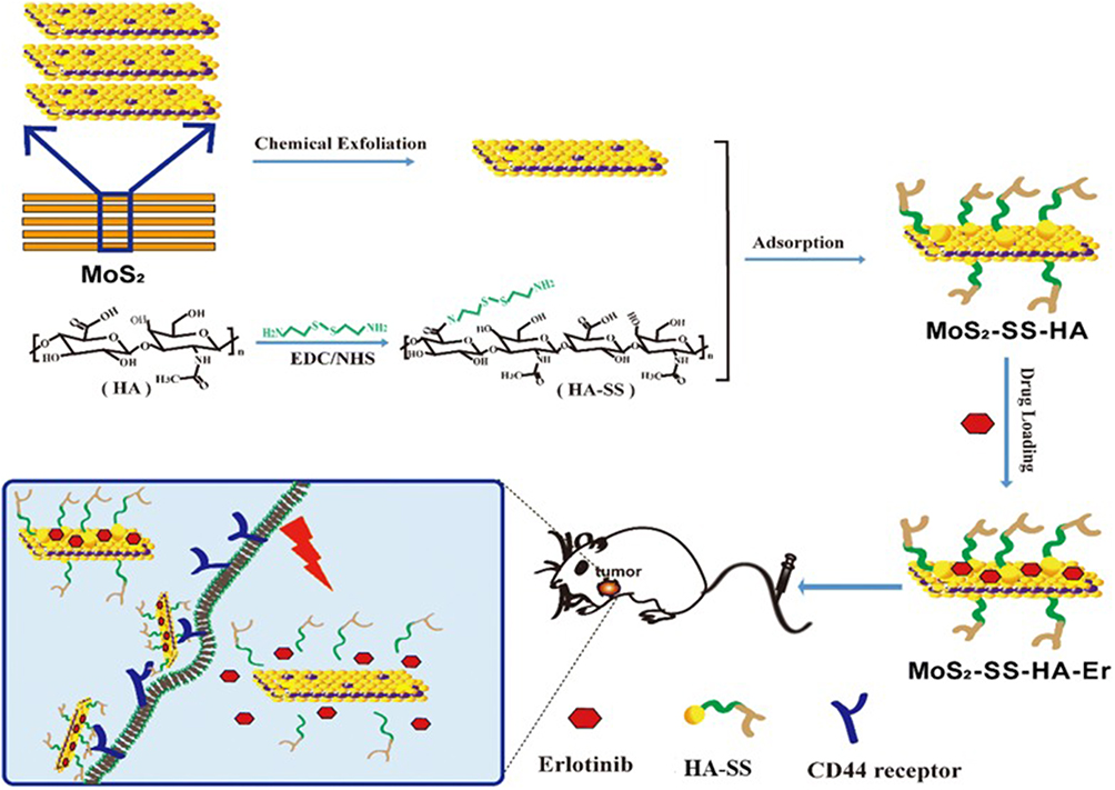 |
Figure 7 Schematic illustration depicting the construction of MoS2-SS-HA-based nanocomposites for synergistic chemo-photothermal therapy. Chemical exfoliation of MoS2 flakes to nanosheets and synthesis of HA-SS-NH2 via EDC/NHS reaction. Subsequently, the MoS2 nanosheets are functionalized with HA and loading Er at the defect sites to obtain MoS2-SS-HA-Er nanosheets, which could target tumor site mediated by CD44 receptor, and produce the hyperthermia upon NIR irradiation. Reproduced with permission from ref,157 CC BY 4.0. Copyright© The Author(s) 2019. |
Jin et al158 focused on two-dimensional transition metal dichalcogenides WS2 and WSe2, which possess excellent physicochemical properties and have been utilized as chemical sensitizers for biomedical applications. This study found that both WS2 and WSe2 nanosheets can adhere to the surface of A549 cells and internalize into the cells, increasing the chances of interacting with cell surface proteins and cytoplasmic proteins, thereby inducing cellular autophagy. Leveraging this mechanism, pretreatment with WS2 and WSe2 significantly enhanced the sensitivity of A549 cells to the chemotherapeutic drug doxorubicin (DOX). However, further in vivo studies are required to validate this effect.
Graphene Nanomaterials
Graphene-based nanomaterials demonstrate immense potential in lung cancer theranostics due to their ultra-high specific surface area (approximately 2600 m2/g), excellent electrical conductivity, and versatile functionalizability.159 In diagnostics, electrochemical sensors based on reduced graphene oxide and fluorescent probes utilizing graphene quantum dots enable highly sensitive and specific detection of lung cancer biomarkers, facilitating early non-invasive diagnosis.160,161 In the therapeutic domain, graphene oxide and its derivatives serve as intelligent drug delivery platforms, enabling targeted drug delivery for lung cancer and achieving synergistic photothermal/photodynamic therapy under near-infrared light irradiation, which enhances therapeutic efficacy while overcoming tumor drug resistance.162–164 Furthermore, multifunctional graphene-based systems that integrate diagnostic imaging with synergistic therapy, along with dual-targeting designs for modulating the tumor microenvironment, are advancing the development of integrated lung cancer theranostics. Although clinical translation still faces challenges such as biosafety concerns and scalable manufacturing, the combination of artificial intelligence and genomic technologies holds promise for graphene-based nanomaterials to provide innovative solutions for precision medicine in lung cancer.
Black Phosphorus Nanoparticles
Long-term use of chemotherapeutic drugs in lung cancer leads to drug resistance, reduced efficacy, and adverse side effects. To circumvent these drawbacks, targeted therapy and photothermal therapy have emerged as effective solutions. As an emerging two-dimensional material, black phosphorus (BP) has become an ideal carrier for synergistic PTT and chemotherapy in NSCLC treatment, owing to its unique anisotropic optical properties, high specific surface area (up to 1000 m2/g), and biodegradability.
Black phosphorus exhibits excellent photothermal properties, making it suitable for PTT.165 Chen et al166 developed PEG- and polyethylenimine (PEI)-modified black phosphorus nanosheets (PPBP) for the targeted delivery of small interfering RNA (siRNA) against human telomerase reverse transcriptase (hTERT). Under NIR laser irradiation, PPBP displayed efficient PTT/PDT activity and achieved controlled siRNA release through pH- and reactive oxygen species (ROS)-responsive degradation. In in vivo experiments, PPBP-siRNA significantly inhibited tumor growth and metastasis, verifying the synergistic effect of gene therapy combined with PTT/PDT.
Black phosphorus has a broad absorption band in the NIR region (700–1000 nm) with a photothermal conversion efficiency of up to 30.84%, which is significantly higher than that of traditional gold nanoparticles (22–23%). Under 808 nm laser irradiation, black phosphorus nanosheets (BPNSs) can increase the central temperature of tumors to over 45°C, inducing cancer cell apoptosis.167 Upon light irradiation, black phosphorus can generate ROS, which damages tumor cell DNA via PDT; meanwhile, PTT-induced immunogenic cell death (ICD) can release tumor antigens and activate systemic anti-tumor immunity.168 Black phosphorus also possesses high drug-loading capacity and targeted release properties, which can enhance chemotherapeutic efficacy. Lin et al169 first combined black phosphorus nanoparticles (BPNPs) with gefitinib, an epidermal growth factor receptor tyrosine kinase inhibitor (EGFR-TKI), to construct a targeted nanocarrier (BPGM). This system enhanced targeting ability through tumor cell membrane camouflage and achieved synergy between PTT and chemotherapy under NIR laser irradiation. Additionally, researchers encapsulated black phosphorus quantum dots (BPQDs) with exosomes (hEX) to develop a tumor vaccine (hEX@BP). In in vivo experiments, this vaccine exhibited superior PTT efficacy and stronger tumor-targeting capability, providing an effective immunophotothermal therapy for cancer.170
However, the phosphorus atoms in BPNPs have high reactivity, resulting in poor in vivo stability, susceptibility to oxidative degradation, and toxic degradation products.171,172 Currently, the targeted modification of BPNPs mostly relies on single ligands (eg, anti-EGFR antibodies). Nevertheless, NSCLC exhibits heterogeneity (some tumor cells have low or no EGFR expression), leading to the failure of precise targeting in some tumor cells.171,172 Therefore, research on BP NPs in NSCLC treatment is currently in the preclinical development stage. Although some technologies have entered translational medicine research, they have not yet advanced to human clinical trials.
Inorganic-Organic Hybrid Nanoparticles
Traditional inorganic materials (eg, metal oxides) exhibit high stability and unique optical/electrical properties, but they suffer from poor biocompatibility and proneness to agglomeration. Organic materials (eg, polymers), on the other hand, possess flexibility and processability, yet they are limited by insufficient thermal stability and weak mechanical properties. Inorganic-organic hybrid nanoparticles can integrate the advantages of both materials, meet the requirements for multiple functions, and exhibit numerous advantages compared with other nanocarriers (eg, liposomes and polymeric nanoparticles), making them a viable option for cancer treatment.
Metal-Organic Framework Nanoparticles
Metal-organic frameworks (MOFs) are a novel class of porous hybrid nanomaterials formed through the self-assembly of metal ions and organic ligands via coordination bonds. Their structure offers high designability and functional versatility: the metal-organic framework serves as the core, providing an ultra-high specific surface area, porosity, and drug-loading capacity; functional molecules such as chemotherapeutic drugs, genes, or imaging agents can be loaded within the pores; and the surface can be further modified with PEG, targeting ligands, or biomimetic membranes to enhance stability, enable active targeting, and improve biocompatibility.173 This multi-layered architecture makes MOFs an ideal platform for intelligent nanoscale drug delivery.Du et al174 prepared a copper-based MOF (Cu-MOF) core Nano-delivery system via a hydrothermal method, loaded it with the anticancer drug doxorubicin (DOX), and then coated keratin on the Cu-MOF core through a lactamization reaction to construct a Cu-MOFs@Keratin nanodrug delivery system. The construction of this system improves biocompatibility. After entering tumor cells via endocytosis, the system disintegrates under the stimulation of the intracellular microenvironment, enabling sustained release of the loaded DOX. It is adaptable to the low pH value and high concentration of glutathione (GSH) in the tumor microenvironment (TME). In addition, this drug delivery system can also generate reactive oxygen species (·OH) through a cascade redox reaction, realizing glutathione-triggered chemodynamic therapy.
MOFs can serve not only as drug carriers but also as highly sensitive biosensors. For example, they can be used to construct enzyme-free electrochemical sensors for detecting cancer-related biomarkers such as hydrogen peroxide (H2O2), offering new tools for early diagnosis.175,176 In terms of therapy, MOFs can achieve controllable drug release in response to the tumor microenvironment, such as pH changes, high concentrations of glutathione, or specific enzymes.177 Moreover, they can integrate chemotherapy, photothermal therapy, and photodynamic therapy to launch synergistic.
Yang et al178 developed the biomimetic “biological bomb” system, the study utilized zinc-based MOF (ZIF-8) as the core, loaded with the suicide gene pHSVtk and the drug ganciclovir (GCV), achieving a gene loading efficiency as high as 85.2% and a drug loading capacity of 7.54%.179,180 This carrier has a particle size of approximately 154 nm, a specific surface area of 1025 m2/g, and exhibits significant pH-responsive release characteristics in acidic environments (release rate > 80% within 48 hours). To further enhance performance, the research coated the ZIF-8 surface with a human bone marrow mesenchymal stem cell membrane (hBMSCM), forming biomimetic nanoparticles (BEFORE), which significantly improved the system’s stability, biocompatibility, and cellular uptake efficiency. Both in vitro and in vivo experiments demonstrated that BEFORE can be efficiently internalized by lung cancer cells via endocytosis, induce tumor cell apoptosis, and cause no significant organ damage, highlighting its promising therapeutic potential and biosafety.
Although MOF nanoparticles show certain potential as DDS for lung cancer treatment, their clinical application still faces multiple challenges including their biocompatibility, complex preparation processes, high costs,181 cytotoxicity, organ damage, structural stability182 and metabolic disorders.178
Quantum Dot Hybrid Nano-Delivery Systems
Quantum dots are spherical semiconductor nanoparticles with versatile surface chemical properties. Compared with other drug carriers, quantum dots possess larger specific surface area, smaller size, and stronger adsorption capacity, and their surface chemical properties can be modulated. They hold great potential in various biological applications, including bioimaging and drug delivery systems.183,184 Pilch et al185 constructed a nanoparticle-based drug delivery system by connecting novel anticancer compounds (asymmetric diacridine derivatives, UAs) to non-toxic quantum dots (silver-indium-zinc-sulfur nanocrystals) via non-covalent bonds. The conjugation of asymmetric diacridine derivatives (UAs) with quantum dots (QDs) could induce cellular senescence in human lung cancer H460 cells, and the QD-UA hybrids exhibited a significant senescence-inducing effect.
Limitations and Prospects
Significant breakthroughs have been achieved in the research of Nano-delivery systems in the field of lung cancer diagnosis and treatment; however, their translation from the laboratory to clinical application still faces numerous challenges. The primary issues to be addressed are the physicochemical properties and biosafety of nanocarrier materials.186 On one hand, in lung cancer treatment, Nano-delivery systems may introduce cationic groups to improve antigen-loading efficiency and enhance anti-tumor activity, which could increase cytotoxicity.187,188 On the other hand, non-biogenic materials such as inorganic nanoparticles exhibit stable loading and release properties, but they have problems including long in vivo retention time and unclear metabolic pathways, which may pose long-term biosafety risks, such as potential immunogenic reactions and toxicity triggered in the body.189,190 More critically, the interaction mechanism between nanocarriers and biological systems has not been fully elucidated.187 In addition, the lung has a dense capillary network and a mucociliary clearance mechanism, which directly affect the effective deposition and retention of nanocarriers. Meanwhile, a large number of alveolar macrophages rapidly phagocytose inhaled nanoparticles, leading to a decrease in the therapeutic concentration at the tumor site.191,192 These challenges complicate the application of nanoparticle drug delivery systems in lung cancer.
The clinical translation of Nano-delivery systems in lung cancer treatment is in a critical stage of accelerating the transition from laboratory breakthroughs to clinical application. First, for lung treatment, inhalation administration can simulate the natural infection pathway, directly activate lung mucosal immunity, and significantly improve the retention of nanoparticles in lung tissue.193 Future delivery systems will further optimize particle size properties and adopt bionic design combined with pulmonary surfactants to ensure efficient delivery of particles to deep lung tissue and specific uptake by tumor cells.194 Meanwhile, multi-target synergy will replace single targeting to recognize multiple antigens on the surface of tumor cells, improving targeting accuracy.195 Second, future nanocarriers will focus more on multi-functionalization and intelligent upgrading, evolving from a single delivery function to a “theranostic integration” multi-functional platform. This platform will integrate drug delivery, imaging diagnosis, efficacy monitoring, and immune vaccine functions. By loading chemotherapeutic drugs and contrast agents, it can realize visualized tumor treatment and real-time efficacy evaluation.196,197 In addition, the involvement of computer technology will completely change the research and development paradigm of nanocarriers. In the future, the physicochemical properties, in vivo behavior, and interactions with biomolecules of nanomaterials can be predicted in a virtual environment, which will significantly shorten the carrier optimization cycle and accelerate the translation of nanodrugs from the laboratory to the clinic.198
Conclusions
This review provides a comprehensive synthesis of the latest advancements in NDDS for LC therapy, systematically analyzing their classification, structural features, underlying mechanisms, and translational status. Organic NDDSs—exemplified by liposomes, polymeric nanoparticles, and viral nanoparticles—exhibit superior biocompatibility and versatile drug-loading capabilities, with several formulations (eg, nab-paclitaxel) already integrated into clinical practice. Inorganic NDDSs, such as gold nanoparticles and mesoporous silica nanoparticles, harness distinctive optical, magnetic, and structural properties to enable synergistic therapeutic outcomes and precision imaging, thereby offering transformative potential for targeted treatment modalities. Inorganic-organic hybrid systems, including LPHNPs and MOFs, synergistically combine the strengths of both material classes, resulting in enhanced stability, targeting specificity, and therapeutic performance.
Despite these promising developments, the clinical translation of NDDSs for LC confronts significant obstacles. Critical biosafety concerns—such as prolonged in vivo retention of non-biogenic materials, potential immunogenicity, and poorly defined metabolic pathways—demand rigorous and systematic investigation. Furthermore, the unique pulmonary physiological microenvironment, characterized by the mucociliary clearance mechanism and alveolar macrophage phagocytosis, imposes substantial barriers to the effective deposition and sustained retention of nanocarriers at tumor sites. Additionally, the fundamental interaction mechanisms between nanomaterials and biological systems remain inadequately understood, limiting the rational design and predictable behavior of NDDSs in vivo.
Moving forward, several strategic directions are pivotal to advancing the field: optimizing inhalation-based delivery to achieve deep lung penetration, employing biomimetic designs and multi-targeting strategies to enhance tumor specificity, and developing intelligent “theranostic” platforms that integrate diagnosis, therapy, and monitoring in a single system. Equally important is the integration of computational modeling and artificial intelligence, which holds the potential to revolutionize NDDS development by predicting the physicochemical and biological behaviors of nanocarriers, thereby significantly shortening the R&D timeline and accelerating translational progress.
In conclusion, through continued interdisciplinary innovation spanning materials science, pharmaceutical engineering, and biomedical research, NDDSs are poised to fundamentally redefine the therapeutic landscape of lung cancer. By enabling more precise, effective, and patient-tailored treatment strategies, NDDSs not only promise to elevate therapeutic efficacy and safety but also to improve overall patient quality of life—ushering in a new paradigm of intelligent, minimally invasive, and personalized oncology care.
Acknowledgments
The authors would like to thank Dr. Qunhua Zhou (Department of Medical Chemistry, Medical School, Zhengzhou Health College, Zhengzhou 450000, China) for her assistance with the revision. Zhengjun Li, Chang Liu, and Zexi Di share first authorship.
Disclosure
The authors declare no conflict of interest.
References
1. Bray F, Laversanne M, Sung H, et al. Global cancer statistics 2022: GLOBOCAN estimates of incidence and mortality worldwide for 36 cancers in 185 countries. CA Cancer J Clin. 2024;74(3):229–25. doi:10.3322/caac.21834
2. Li Y, Yan B, He S. Advances and challenges in the treatment of lung cancer. Biomed Pharmacother. 2023;169:115891. doi:10.1016/j.biopha.2023.115891
3. Meyer ML, Fitzgerald BG, Paz-Ares L, et al. New promises and challenges in the treatment of advanced non-small-cell lung cancer. Lancet. 2024;404(10454):803–822. doi:10.1016/S0140-6736(24)01029-8
4. Cryer AM, Thorley AJ. Nanotechnology in the diagnosis and treatment of lung cancer. Pharmacol Ther. 2019;198:189–205. doi:10.1016/j.pharmthera.2019.02.010
5. Lopez-Davila V, Seifalian AM, Loizidou M. Organic nanocarriers for cancer drug delivery. Curr Opin Pharmacol. 2012;12(4):414–419. doi:10.1016/j.coph.2012.02.011
6. Zingg R, Fischer M. The consolidation of nanomedicine. Wiley Interdiscip Rev Nanomed Nanobiotechnol. 2019;11(6):e1569. doi:10.1002/wnan.1569
7. Jia Y, Jiang Y, He Y, et al. Approved nanomedicine against diseases. Pharmaceutics. 2023;15(3):774. doi:10.3390/pharmaceutics15030774
8. Greish K, Mathur A, Bakhiet M, Taurin S. Nanomedicine: is it lost in translation? Ther Deliv. 2018;9(4):269–285. doi:10.4155/tde-2017-0118
9. Liang T, Zhang B, Xing Z, et al. Adapting and remolding: orchestrating tumor microenvironment normalization with photodynamic therapy by size transformable nanoframeworks. Angew Chem Int Ed Engl. 2021;60(20):11464–11473. doi:10.1002/anie.202102180
10. Yang L, Tang J, Yin H, et al. Self-assembled nanoparticles for tumor-triggered targeting dual-mode NIRF/MR imaging and photodynamic therapy applications. ACS Biomater Sci Eng. 2022;8(2):880–892. doi:10.1021/acsbiomaterials.1c01418
11. Han X, Gong C, Yang Q, et al. Biomimetic nano-drug delivery system: an emerging platform for promoting tumor treatment. Int J Nanomed. 2024;19:571–608. doi:10.2147/IJN.S442877
12. Cardoso VMO, Bistaffa MJ, Sterman RG, et al. Nanomedicine innovations for lung cancer diagnosis and therapy. ACS Appl Mater Interfaces. 2025;17(9):13197–13220. doi:10.1021/acsami.4c16840
13. Adrianzen Herrera D, Ashai N, Perez-Soler R, Cheng H. Nanoparticle albumin bound-paclitaxel for treatment of advanced non-small cell lung cancer: an evaluation of the clinical evidence. Expert Opin Pharmacother. 2019;20(1):95–102. doi:10.1080/14656566.2018.1546290
14. Gupta N, Hatoum H, Dy GK. First line treatment of advanced non-small-cell lung cancer - specific focus on albumin bound paclitaxel. Int J Nanomed. 2014;9:209–221. doi:10.2147/IJN.S41770
15. Su H, Jia J, Mao Y, Zhu R, Li Z. A real-world analysis of FDA Adverse Event Reporting System (FAERS) events for liposomal and conventional doxorubicins. Sci Rep. 2024;14(1):5095. doi:10.1038/s41598-024-55185-4
16. Wei QY, Xu YM, Lau ATY. Recent progress of nanocarrier-based therapy for solid malignancies. Cancers. 2020;12(10):2783. doi:10.3390/cancers12102783
17. Zhong Q, da Rocha SR. Poly(amidoamine) dendrimer–doxorubicin conjugates: in vitro characteristics and pseudosolution formulation in pressurized metered-dose inhalers. Mol Pharm. 2016;13(3):1058–1072. doi:10.1021/acs.molpharmaceut.5b00876
18. Kaminskas LM, Kelly BD, McLeod VM, et al. Characterisation and tumour targeting of PEGylated polylysine dendrimers bearing doxorubicin via a pH labile linker. J Control Release. 2011;152(2):241–248. doi:10.1016/j.jconrel.2011.02.005
19. Sharma AK, Gothwal A, Kesharwani P, et al. Dendrimer nanoarchitectures for cancer diagnosis and anticancer drug delivery. Drug Discov Today. 2017;22(2):314–326. doi:10.1016/j.drudis.2016.09.013
20. Guo S, Vieweger M, Zhang K, et al. Ultra-thermostable RNA nanoparticles for solubilizing and high-yield loading of paclitaxel for breast cancer therapy. Nat Commun. 2020;11(1):972. doi:10.1038/s41467-020-14780-5
21. Park K, Lee GY, Kim Y-S, et al. Heparin-deoxycholic acid chemical conjugate as an anticancer drug carrier and its antitumor activity. J Control Release. 2006;114(3):300–306. doi:10.1016/j.jconrel.2006.05.017
22. Choudhary S, Gupta L, Rani S, Dave K, Gupta U. Impact of Dendrimers on Solubility of Hydrophobic Drug Molecules. Front Pharmacol. 2017;8:261. doi:10.3389/fphar.2017.00261
23. Dhingra S, Goyal S, Thirumal D, et al. Mesoporous silica nanoparticles: a versatile carrier platform in lung cancer management. Nanomedicine. 2024;19(15):1331–1346. doi:10.1080/17435889.2024.2348438
24. Zi Y, Yang K, He J, et al. Strategies to enhance drug delivery to solid tumors by harnessing the EPR effects and alternative targeting mechanisms. Adv Drug Deliv Rev. 2022;188:114449. doi:10.1016/j.addr.2022.114449
25. Danhier F. To exploit the tumor microenvironment: since the EPR effect fails in the clinic, what is the future of nanomedicine? J Control Release. 2016;244:108–121. doi:10.1016/j.jconrel.2016.11.015
26. De Jong WH, Borm PJ. Drug delivery and nanoparticles:applications and hazards. Int J Nanomed. 2008;3:133–149. doi:10.2147/ijn.s596
27. Ailuno G, Iacobazzi RM, Lopalco A, et al. The pharmaceutical technology approach on imaging innovations from Italian research. Pharmaceutics. 2021;13(8):1214. doi:10.3390/pharmaceutics13081214
28. Lobo G, Paiva KLR, Silva ALG, et al. Nanocarriers used in drug delivery to enhance immune system in cancer therapy. Pharmaceutics. 2021;13(8):1167. doi:10.3390/pharmaceutics13081167
29. Hussein Kamareddine M, Ghosn Y, Tawk A, et al. Organic nanoparticles as drug delivery systems and their potential role in the treatment of chronic myeloid leukemia. Technol Cancer Res Treat. 2019;18:1533033819879902. doi:10.1177/1533033819879902
30. Yeo S, Kim MJ, Shim YK, Yoon I, Lee WK. Solid lipid nanoparticles of curcumin designed for enhanced bioavailability and anticancer efficiency. ACS Omega. 2022;7(40):35875–35884. doi:10.1021/acsomega.2c04407
31. Obeid MA, Alsaadi M, Aljabali AA. Recent updates in curcumin delivery. J Liposome Res. 2023;33(1):53–64. doi:10.1080/08982104.2022.2086567
32. Saffarionpour S, Diosady LL. Curcumin, a potent therapeutic nutraceutical and its enhanced delivery and bioaccessibility by pickering emulsions. Drug Deliv Transl Res. 2022;12(1):124–157. doi:10.1007/s13346-021-00936-3
33. Kanwal Q, Ahmed M, Hamza M, et al. Curcumin nanoparticles: physicochemical fabrication, characterization, antioxidant, enzyme inhibition, molecular docking and simulation studies. RSC Adv. 2023;13(32):22268–22280. doi:10.1039/d3ra01432k
34. Oh JY, Choi E, Jana B, et al. Protein-precoated surface of metal-organic framework nanoparticles for targeted delivery. Small. 2023;19:e2300218. doi:10.1002/smll.202300218
35. Conte C, Longobardi G, Barbieri A, et al. Non-covalent strategies to functionalize polymeric nanoparticles with NGR peptides for targeting breast cancer. Int J Pharm. 2023;633:122618. doi:10.1016/j.ijpharm.2023.122618
36. Ilies BD, Yildiz I, Abbas M. Peptide-conjugated nanoparticle platforms for targeted delivery, imaging, and biosensing applications. Chembiochem. 2024;25(10):e202300867. doi:10.1002/cbic.202300867
37. Su YC, Chen YC, Lo YH, Chang CH, Chen YF. Hydrophobic interactions enhance doxorubicin delivery from hyaluronic acid nanogels. Eur J Pharm Biopharm. 2025;210:114676. doi:10.1016/j.ejpb.2025.114676
38. Arvapalli DM, Sheardy AT, Allado K, et al. Design of curcumin loaded carbon nanodots delivery system: enhanced bioavailability, release kinetics, and anticancer activity. ACS Appl Bio Mater. 2020;3(12):8776–8785. doi:10.1021/acsabm.0c01144
39. Zhang J, Deng M, Xu C, et al. Dual-prodrug-based hyaluronic acid nanoplatform provides cascade-boosted drug delivery for oxidative stress-enhanced chemotherapy. ACS Appl Mater Interfaces. 2024;16(38):50459–50473. doi:10.1021/acsami.4c11662
40. Ruan G, Ye L, Ke J, et al. All-in-one gadolinium-doxorubicin nanoassemblies for spatial delivery and chemoresistance reversal in tumor microenvironments. ACS Appl Mater Interfaces. 2025;17(13):19348–19366. doi:10.1021/acsami.4c21725
41. Jeong M, Shin S, Lee G, et al. Engineered lipid nanoparticles enable therapeutic gene silencing of GTSE1 for the treatment of liver fibrosis. J Control Release. 2024;374:337–348. doi:10.1016/j.jconrel.2024.08.012
42. Kim SJ, Puranik N, Yadav D, Jin JO, Lee PCW. Lipid nanocarrier-based drug delivery systems: therapeutic advances in the treatment of lung cancer. Int J Nanomed. 2023;18:2659–2676. doi:10.2147/IJN.S406415
43. Martins S, Sarmento B, Ferreira DC, Souto EB. Lipid-based colloidal carriers for peptide and protein delivery--liposomes versus lipid nanoparticles. Int J Nanomed. 2007;2(4):595–607.
44. Xing H, Hwang K, Lu Y. Recent developments of liposomes as nanocarriers for theranostic applications. Theranostics. 2016;6(9):1336–1352. doi:10.7150/thno.15464
45. Has C, Sunthar P. A comprehensive review on recent preparation techniques of liposomes. J Liposome Res. 2020;30(4):336–365. doi:10.1080/08982104.2019.1668010
46. Mohammad-Jafari K, Naghib SM, Mozafari MR. Liposomal nanoformulation-encapsulated paclitaxel for reducing chemotherapy side effects in lung cancer treatments: recent advances and future outlooks. Curr Med Chem. 2025;32(25):5155–5179. doi:10.2174/0109298673308951240921121345
47. Zhang J, Du Q, Song X, et al. Evaluation of the tumor-targeting efficiency and intratumor heterogeneity of anticancer drugs using quantitative mass spectrometry imaging. Theranostics. 2020;10(6):2621–2630. doi:10.7150/thno.41763
48. Aranguren A, Torres CE, Munoz-Camargo C, Osma JF, Cruz JC. Synthesis of nanoscale liposomes via low-cost microfluidic systems. Micromachines. 2020;11(12):1050. doi:10.3390/mi11121050
49. Fu X, Shi Y, Wu H, et al. Inhalable liposomal delivery of osimertinib and DNA for treating primary and metastasis lung cancer. Nat Commun. 2025;16(1):3336. doi:10.1038/s41467-025-58312-5
50. Badea CT, Clark DP, Holbrook M, Srivastava M, Mowery Y, Ghaghada KB. Functional imaging of tumor vasculature using iodine and gadolinium-based nanoparticle contrast agents: a comparison of spectral micro-CT using energy integrating and photon counting detectors. Phys Med Biol. 2019;64(6):065007. doi:10.1088/1361-6560/ab03e2
51. Abla KK, Zahwi M, El-Lakany SA, Karam M. Advances in siRNA-loaded liposomes for lung cancer: challenges and future perspectives. Int J Biol Macromol. 2025;319:145461. doi:10.1016/j.ijbiomac.2025.145461
52. Sun J, Bi C, Chan HM, et al. Curcumin-loaded solid lipid nanoparticles have prolonged in vitro antitumour activity, cellular uptake and improved in vivo bioavailability. Colloids Surf B Biointerfaces. 2013;111:367–375. doi:10.1016/j.colsurfb.2013.06.032
53. Jana M, Biswas UK, Patro CS, et al. Solid lipid nanoparticles: a review of their biomedical applications and preparation. Pharm Nanotechnol. 2025;13(4):758–774. doi:10.2174/0122117385312175240502100018
54. Bae KH, Lee JY, Lee SH, et al. Optically traceable solid lipid nanoparticles loaded with siRNA and paclitaxel for synergistic chemotherapy with in situ imaging. Adv Healthc Mater. 2013;2(4):576–584. doi:10.1002/adhm.201200338
55. Aggarwal BB, Kumar A, Bharti AC. Anticancer potential of curcumin: preclinical and clinical studies. Anticancer Res. 2003;23(1A):363–398.
56. Liu W, Zhai Y, Heng X, et al. Oral bioavailability of curcumin: problems and advancements. J Drug Target. 2016;24(8):694–702. doi:10.3109/1061186X.2016.1157883
57. Nikwade V, Choudhary N, Solanki R, et al. Fabrication and characterization of ConA-conjugated curcumin-loaded solid lipid nanoparticles for theranostic applications in lung cancer treatment. Nanoscale. 2025;17(6):3203–3210. doi:10.1039/d4nr03157a
58. Li M, Fang G, Zahid F, et al. Co-delivery of paclitaxel and curcumin loaded solid lipid nanoparticles for improved targeting of lung cancer: in vitro and in vivo investigation. Heliyon. 2024;10(9):e30290. doi:10.1016/j.heliyon.2024.e30290
59. Rahman MA, Ali A, Rahamathulla M, et al. Fabrication of sustained release curcumin-loaded solid lipid nanoparticles (Cur-SLNs) as a potential drug delivery system for the treatment of lung cancer: optimization of formulation and in vitro biological evaluation. Polymers. 2023;15(3):542. doi:10.3390/polym15030542
60. Basniwal RK, Khosla R, Jain N. Improving the anticancer activity of curcumin using nanocurcumin dispersion in water. Nutr Cancer. 2014;66(6):1015–1022. doi:10.1080/01635581.2014.936948
61. Gupta T, Singh J, Kaur S, et al. Enhancing bioavailability and stability of Curcumin Using Solid Lipid Nanoparticles (CLEN): a covenant for its effectiveness. Front Bioeng Biotechnol. 2020;8:879. doi:10.3389/fbioe.2020.00879
62. Zhong SJ, Xing YD, Dong LY, et al. Progress in the study of curcumin metabolism in vivo. J Asian Nat Prod Res. 2025;27(5):639–657. doi:10.1080/10286020.2024.2420619
63. Liu Y, Zhao F, Wang Q, et al. Current perspectives on paclitaxel: focus on its production, delivery and combination therapy. Mini Rev Med Chem. 2023;23(18):1780–1796. doi:10.2174/1389557523666230210145150
64. Yardley DA. nab-Paclitaxel mechanisms of action and delivery. J Control Release. 2013;170(3):365–372. doi:10.1016/j.jconrel.2013.05.041
65. Zhou C, Hu Y, Arkania E, et al. A global Phase 3 study of serplulimab plus chemotherapy as first-line treatment for advanced squamous non-small-cell lung cancer (ASTRUM-004). Cancer Cell. 2024;42(2):198–208e193. doi:10.1016/j.ccell.2023.12.004
66. Liu Y, Dong Y, Zhu H, Jing W, Guo H, Yu J. Nanoparticle albumin-bound paclitaxel in elder patients with advanced squamous non-small-cell lung cancer: a retrospective study. Cancer Med. 2020;9(4):1365–1373. doi:10.1002/cam4.2791
67. Fu F, Wang W, Wu L, et al. Inhalable biomineralized liposomes for cyclic Ca(2+)-burst-centered endoplasmic reticulum stress enhanced lung cancer ferroptosis therapy. ACS Nano. 2023;17(6):5486–5502. doi:10.1021/acsnano.2c10830
68. Ma Y, Dong Y, Li X, Wang F, Zhang Y. Tumor-penetrating peptide-functionalized ferritin enhances antitumor activity of paclitaxel. ACS Appl Bio Mater. 2021;4(3):2654–2663. doi:10.1021/acsabm.0c01613
69. Deng G, Li Y, Liang N, et al. Ferritin in cancer therapy: a pleiotropic tumoraffin nanocage-based transport. Cancer Med. 2023;12(10):11570–11588. doi:10.1002/cam4.5778
70. King CR, Kraus MH, Aaronson SA. Amplification of a Novel v-erbB-related gene in a human mammary carcinoma. Science. 1985;229(4717):974–976. doi:10.1126/science.2992089
71. Gassmann M, Casagranda F, Orioli D, et al. Aberrant neural and cardiac development in mice lacking the ErbB4 neuregulin receptor. Nature. 1995;378(6555):390–394. doi:10.1038/378390a0
72. Threadgill DW, Dlugosz AA, Hansen LA, et al. Targeted disruption of mouse EGF receptor: effect of genetic background on mutant phenotype. Science. 1995;269(5221):230–234. doi:10.1126/science.7618084
73. Yu X, Wu H, Hu H, et al. Zein nanoparticles as nontoxic delivery system for maytansine in the treatment of non-small cell lung cancer. Drug Deliv. 2020;27(1):100–109. doi:10.1080/10717544.2019.1704942
74. Gao S, Zhang W, Wang R, et al. Nanoparticles encapsulating nitrosylated maytansine to enhance radiation therapy. ACS Nano. 2020;14(2):1468–1481. doi:10.1021/acsnano.9b05976
75. Liu F, Mao K, Chen H, et al. Enhancing the safety and efficacy of Trastuzumab Emtansine (T-DM1) through nano-delivery system in breast cancer therapy. Small. 2024;20(50):e2400977. doi:10.1002/smll.202400977
76. Yaman S, Ramachandramoorthy H, Iyer P, et al. Targeted chemotherapy via HER2-based chimeric antigen receptor (CAR) engineered T-cell membrane coated polymeric nanoparticles. Bioact Mater. 2024;34:422–435. doi:10.1016/j.bioactmat.2023.12.027
77. Najlah M, Ahmed Z, Iqbal M, et al. Development and characterisation of disulfiram-loaded PLGA nanoparticles for the treatment of non-small cell lung cancer. Eur J Pharm Biopharm. 2017;112:224–233. doi:10.1016/j.ejpb.2016.11.032
78. Haim Zada M, Rottenberg Y, Domb AJ. Peptide loaded polymeric nanoparticles by non-aqueous nanoprecipitation. J Colloid Interface Sci. 2022;622:904–913. doi:10.1016/j.jcis.2022.05.007
79. Gothwal A, Khan I, Gupta U. Polymeric micelles: recent advancements in the delivery of anticancer drugs. Pharm Res. 2016;33(1):18–39. doi:10.1007/s11095-015-1784-1
80. Hanafy NAN, El-Kemary M, Leporatti S. Micelles structure development as a strategy to improve smart cancer therapy. Cancers. 2018;10(7):238. doi:10.3390/cancers10070238
81. Jiang P, Liang B, Zhang Z, et al. New insights into nanosystems for non-small-cell lung cancer: diagnosis and treatment. RSC Adv. 2023;13(28):19540–19564. doi:10.1039/d3ra03099g
82. Zhang M, Zhang Z, Song X, et al. Synthesis and characterization of Palmitoyl-block-poly(methacryloyloxyethyl phosphorylcholine) polymer micelles for anticancer drug delivery. Biomacromolecules. 2022;23(11):4586–4596. doi:10.1021/acs.biomac.2c00838
83. Shen F, Fang Y, Wu Y, et al. Metal ions and nanometallic materials in antitumor immunity: function, application, and perspective. J Nanobiotechnology. 2023;21(1):20. doi:10.1186/s12951-023-01771-z
84. Cinteza LO, Ohulchanskyy TY, Sahoo Y, et al. Diacyllipid micelle-based nanocarrier for magnetically guided delivery of drugs in photodynamic therapy. Mol Pharm. 2006;3(4):415–423. doi:10.1021/mp060015p
85. Davis ME, Chen ZG, Shin DM. Nanoparticle therapeutics: an emerging treatment modality for cancer. Nat Rev Drug Discov. 2008;7(9):771–782. doi:10.1038/nrd2614
86. Benahmed A, Ranger M, Leroux JC. Novel polymeric micelles based on the amphiphilic diblock copolymer poly(N-vinyl-2-pyrrolidone)-block-poly(D,L-lactide). Pharm Res. 2001;18(3):323–328. doi:10.1023/a:1011054930439
87. Chung JE, Yokoyama M, Aoyagi T, Sakurai Y, Okano T. Effect of molecular architecture of hydrophobically modified poly(N-isopropylacrylamide) on the formation of thermoresponsive core-shell micellar drug carriers. J Control Release. 1998;53(1–3):119–130. doi:10.1016/s0168-3659(97)00244-7
88. Kim DW, Kim S-Y, Kim H-K, et al. Multicenter Phase II trial of Genexol-PM, a novel Cremophor-free, polymeric micelle formulation of paclitaxel, with cisplatin in patients with advanced non-small-cell lung cancer. Ann Oncol. 2007;18(12):2009–2014. doi:10.1093/annonc/mdm374
89. Zhang XY, Zhang YD. Enhanced antiproliferative and apoptosis effect of paclitaxel-loaded polymeric micelles against non-small cell lung cancers. Tumour Biol. 2015;36(7):4949–4959. doi:10.1007/s13277-015-3142-7
90. Oerlemans C, Bult W, Bos M, et al. Polymeric micelles in anticancer therapy: targeting, imaging and triggered release. Pharm Res. 2010;27(12):2569–2589. doi:10.1007/s11095-010-0233-4
91. Palmerston Mendes L, Pan J, Torchilin VP. Dendrimers as nanocarriers for nucleic acid and drug delivery in cancer therapy. Molecules. 2017;22(9):1401. doi:10.3390/molecules22091401
92. Lee CC, Gillies ER, Fox ME, et al. A single dose of doxorubicin-functionalized bow-tie dendrimer cures mice bearing C-26 colon carcinomas. Proc Natl Acad Sci U S A. 2006;103(45):16649–16654. doi:10.1073/pnas.0607705103
93. Chauhan AS. Dendrimers for Drug Delivery. Molecules. 2018;23(4):938. doi:10.3390/molecules23040938
94. Cheng Y, Zhao L, Li Y, Xu T. Design of biocompatible dendrimers for cancer diagnosis and therapy: current status and future perspectives. Chem Soc Rev. 2011;40(5):2673–2703. doi:10.1039/c0cs00097c
95. Tarach P, Janaszewska A. Recent advances in preclinical research using PAMAM dendrimers for cancer gene therapy. Int J Mol Sci. 2021;22(6):2912. doi:10.3390/ijms22062912
96. Rawding PA, Bu J, Wang J, et al. Dendrimers for cancer immunotherapy: avidity-based drug delivery vehicles for effective anti-tumor immune response. Wiley Interdiscip Rev Nanomed Nanobiotechnol. 2022;14(2):e1752. doi:10.1002/wnan.1752
97. Zhou M, Tang M, Zhang H, Luo K, Huang Y. HPMA polymeric nanocarriers for anticancer drugs with tumor microenvironment-responsive extracellular biodegradation and intracellular drug release. J Biomed Nanotechnol. 2019;15(8):1688–1700. doi:10.1166/jbn.2019.2727
98. de Agustin de Oro JJ, Soria LM, Fernandez AP, Segarra VT. Ultrasound in the evaluation of rheumatoid arthritis. Eur J Rheumatol. 2022;11(3):S277–S282. doi:10.5152/eurjrheum.2022.20224
99. Peng Y, Liu D, Diao Z, et al. Corrigendum to “Down-regulation of B2R contributes to preeclampsia by inhibiting human trophoblast cell invasion and angiogenesis” [Pregnancy Hypertens. 21 (2020) 14-22]. Pregnancy Hypertens. 2021;26:1. doi:10.1016/j.preghy.2021.07.247
100. Seymour LW, Ferry DR, Kerr DJ, et al. Phase II studies of polymer-doxorubicin (PK1, FCE28068) in the treatment of breast, lung and colorectal cancer. Int J Oncol. 2009;34(6):1629–1636. doi:10.3892/ijo_00000293
101. Li J. Predictors of family caregiver burden in Shanghai. J Appl Gerontol. 2021;40(7):703–712. doi:10.1177/0733464820951029
102. Behr M, Kaufmann JK, Ketzer P, et al. Adenoviruses using the cancer marker EphA2 as a receptor in vitro and in vivo by genetic ligand insertion into different capsid scaffolds. PLoS One. 2014;9(4):e95723. doi:10.1371/journal.pone.0095723
103. Saifullah B, Hussein MZ. Inorganic nanolayers: structure, preparation, and biomedical applications. Int J Nanomed. 2015;10:5609–5633. doi:10.2147/IJN.S72330
104. Sreeharsha N, Chitrapriya N, Jang YJ, Kenchappa V. Evaluation of nanoparticle drug-delivery systems used in preclinical studies. Ther Deliv. 2021;12(4):325–336. doi:10.4155/tde-2020-0116
105. Dreaden EC, Alkilany AM, Huang X, Murphy CJ, El-Sayed MA. The golden age: gold nanoparticles for biomedicine. Chem Soc Rev. 2012;41(7):2740–2779. doi:10.1039/c1cs15237h
106. Vigderman L, Zubarev ER. Therapeutic platforms based on gold nanoparticles and their covalent conjugates with drug molecules. Adv Drug Deliv Rev. 2013;65(5):663–676. doi:10.1016/j.addr.2012.05.004
107. Singhana B, Slattery P, Chen A, Wallace M, Melancon MP. Light-activatable gold nanoshells for drug delivery applications. AAPS Pharm Sci Tech. 2014;15(3):741–752. doi:10.1208/s12249-014-0097-8
108. Saha K, Agasti SS, Kim C, Li X, Rotello VM. Gold nanoparticles in chemical and biological sensing. Chem Rev. 2012;112(5):2739–2779. doi:10.1021/cr2001178
109. Chen Y, Feng X. Gold nanoparticles for skin drug delivery. Int J Pharm. 2022;625:122122. doi:10.1016/j.ijpharm.2022.122122
110. Springer T, Chadtova Song X, Ermini ML, Lamacova J, Homola J. Functional gold nanoparticles for optical affinity biosensing. Anal Bioanal Chem. 2017;409(16):4087–4097. doi:10.1007/s00216-017-0355-1
111. Boisselier E, Astruc D. Gold nanoparticles in nanomedicine: preparations, imaging, diagnostics, therapies and toxicity. Chem Soc Rev. 2009;38(6):1759–1782. doi:10.1039/b806051g
112. Savla R, Minko T. Nanoparticle design considerations for molecular imaging of apoptosis: diagnostic, prognostic, and therapeutic value. Adv Drug Deliv Rev. 2017;113:122–140. doi:10.1016/j.addr.2016.06.016
113. McLaughlan JR, Cowell DMJ, Freear S. Gold nanoparticle nucleated cavitation for enhanced high intensity focused ultrasound therapy. Phys Med Biol. 2017;63(1):015004. doi:10.1088/1361-6560/aa97e9
114. Huang X, El-Sayed IH, Qian W, El-Sayed MA. Cancer cell imaging and photothermal therapy in the near-infrared region by using gold nanorods. J Am Chem Soc. 2006;128(6):2115–2120. doi:10.1021/ja057254a
115. Al-Thani AN, Jan AG, Abbas M, Geetha M, Sadasivuni KK. Nanoparticles in cancer theragnostic and drug delivery: a comprehensive review. Life Sci. 2024;352:122899. doi:10.1016/j.lfs.2024.122899
116. Cheng X, Sun R, Yin L, et al. Light-triggered assembly of gold nanoparticles for photothermal therapy and photoacoustic imaging of tumors in vivo. Adv Mater. 2017;29(6). doi:10.1002/adma.201604894
117. Ghosh P, Han G, De M, Kim CK, Rotello VM. Gold nanoparticles in delivery applications. Adv Drug Deliv Rev. 2008;60(11):1307–1315. doi:10.1016/j.addr.2008.03.016
118. Rai A, Ferreira L. Biomedical applications of the peptide decorated gold nanoparticles. Crit Rev Biotechnol. 2021;41(2):186–215. doi:10.1080/07388551.2020.1853031
119. Guo J, Rahme K, He Y, et al. Gold nanoparticles enlighten the future of cancer theranostics. Int J Nanomed. 2017;12:6131–6152. doi:10.2147/IJN.S140772
120. Zhang Q, Zhou Y, Villarreal E, et al. Faceted gold nanorods: nanocuboids, convex nanocuboids, and concave nanocuboids. Nano Lett. 2015;15(6):4161–4169. doi:10.1021/acs.nanolett.5b01286
121. Wang Y, Chen Y, Zhao Y, Liu S. Compressed sensing for biomedical photoacoustic imaging: a review. Sensors. 2024;24. doi:10.3390/s24092670
122. Huang X, Jain PK, El-Sayed IH, El-Sayed MA. Plasmonic photothermal therapy (PPTT) using gold nanoparticles. Lasers Med Sci. 2008;23(3):217–228. doi:10.1007/s10103-007-0470-x
123. Annesi F, Pane A, Losso MA, et al. Thermo-plasmonic killing of Escherichia coli TG1 bacteria. Materials. 2019;12(9):1530. doi:10.3390/ma12091530
124. Choi WI, Sahu A, Kim YH, Tae G. Photothermal cancer therapy and imaging based on gold nanorods. Ann Biomed Eng. 2012;40(2):534–546. doi:10.1007/s10439-011-0388-0
125. Qin J, Peng Z, Li B, et al. Gold nanorods as a theranostic platform for in vitro and in vivo imaging and photothermal therapy of inflammatory macrophages. Nanoscale. 2015;7(33):13991–14001. doi:10.1039/c5nr02521d
126. Knights OB, McLaughlan JR. Gold nanorods for light-based lung cancer theranostics. Int J Mol Sci. 2018;19(11):3318. doi:10.3390/ijms19113318
127. Wang S, Hou Y. New types of magnetic nanoparticles for stimuli-responsive theranostic nanoplatforms. Adv Sci. 2024;11(8):e2305459. doi:10.1002/advs.202305459
128. Li M, Li Y, Zheng J, et al. Ultrasound-responsive nanocarriers with siRNA and Fe(3)O(4) regulate macrophage polarization and phagocytosis for augmented non-small cell lung cancer immunotherapy. J Nanobiotechnology. 2024;22(1):605. doi:10.1186/s12951-024-02883-w
129. Liang X, Chen M, Bhattarai P, et al. Complementing cancer photodynamic therapy with ferroptosis through iron oxide loaded porphyrin-grafted lipid nanoparticles. ACS Nano. 2021;15(12):20164–20180. doi:10.1021/acsnano.1c08108
130. Zhang H, Deng L, Liu H, et al. Enhanced fluorescence/magnetic resonance dual imaging and gene therapy of liver cancer using cationized amylose nanoprobe. Mater Today Bio. 2022;13:100220. doi:10.1016/j.mtbio.2022.100220
131. Yang C, Zhao H, Hou Y, Ma D. Fe5C2 nanoparticles: a facile bromide-induced synthesis and as an active phase for fischer-tropsch synthesis. J Am Chem Soc. 2012;134(38):15814–15821. doi:10.1021/ja305048p
132. Tang W, Zhen Z, Yang C, et al. Fe5C2 nanoparticles with high mri contrast enhancement for tumor imaging. Small. 2014;10(7):1245–1249. doi:10.1002/smll.201303263
133. Huang G, Hu J, Zhang H, et al. Highly magnetic iron carbide nanoparticles as effective T(2) contrast agents. Nanoscale. 2014;6(2):726–730. doi:10.1039/c3nr04691e
134. Wang Y, Chen Y, Fang J. Post-transcriptional and post-translational regulation of central carbon metabolic enzymes in cancer. Anticancer Agents Med Chem. 2017;17(11):1456–1465. doi:10.2174/1871520617666170327110712
135. Hou W, Xie Y, Song X, et al. Autophagy promotes ferroptosis by degradation of ferritin. Autophagy. 2016;12(8):1425–1428. doi:10.1080/15548627.2016.1187366
136. Ju Y, Zhang H, Yu J, et al. Monodisperse Au-Fe(2)C janus nanoparticles: an attractive multifunctional material for triple-modal imaging-guided tumor photothermal therapy. ACS Nano. 2017;11(9):9239–9248. doi:10.1021/acsnano.7b04461
137. Efremova MV, Spasova M, Heidelmann M, et al. Room temperature synthesized solid solution AuFe nanoparticles and their transformation into Au/Fe Janus nanocrystals. Nanoscale. 2021;13(23):10402–10413. doi:10.1039/d1nr00383f
138. Su H, Hurd Price C-A, Jing L, et al. Janus particles: design, preparation, and biomedical applications. Mater Today Bio. 2019;4:100033. doi:10.1016/j.mtbio.2019.100033
139. Yu J, Yang C, Li J, et al. Multifunctional Fe5 C2 nanoparticles: a targeted theranostic platform for magnetic resonance imaging and photoacoustic tomography-guided photothermal therapy. Adv Mater. 2014;26(24):4114–4120. doi:10.1002/adma.201305811
140. Yang Z, Zhao T, Huang X, et al. Modulating the phases of iron carbide nanoparticles: from a perspective of interfering with the carbon penetration of Fe@Fe(3)O(4) by selectively adsorbed halide ions. Chem Sci. 2017;8(1):473–481. doi:10.1039/c6sc01819j
141. Kesse S, Boakye-Yiadom KO, Ochete BO, et al. Mesoporous silica nanomaterials: versatile nanocarriers for cancer theranostics and drug and gene delivery. Pharmaceutics. 2019;11(2):77. doi:10.3390/pharmaceutics11020077
142. Lilhore UK, Poongodi M, Kaur A, et al. Hybrid model for detection of cervical cancer using causal analysis and machine learning techniques. Comput Math Methods Med. 2022;2022:4688327. doi:10.1155/2022/4688327
143. Song Y, Zhou B, Du X, et al. Folic acid (FA)-conjugated mesoporous silica nanoparticles combined with MRP-1 siRNA improves the suppressive effects of myricetin on non-small cell lung cancer (NSCLC). Biomed Pharmacother. 2020;125:109561. doi:10.1016/j.biopha.2019.109561
144. Yu A, Dai X, Wang Z, et al. Recent Advances of Mesoporous Silica as a Platform for Cancer Immunotherapy. Biosensors. 2022;12(2):109. doi:10.3390/bios12020109
145. Trzeciak K, Chotera-Ouda A, Bak S II, Potrzebowski MJ. Mesoporous silica particles as drug delivery systems-the state of the art in loading methods and the recent progress in analytical techniques for monitoring these processes. Pharmaceutics. 2021;13(7):950. doi:10.3390/pharmaceutics13070950
146. Olivieri F, Castaldo R, Cocca M, Gentile G, Lavorgna M. Mesoporous silica nanoparticles as carriers of active agents for smart anticorrosive organic coatings: a critical review. Nanoscale. 2021;13(20):9091–9111. doi:10.1039/d1nr01899j
147. Yuan D, Ellis CM, Davis JJ. Mesoporous silica nanoparticles in bioimaging. Materials. 2020;13(17):3795. doi:10.3390/ma13173795
148. Pavan C, Delle Piane M, Gullo M, et al. The puzzling issue of silica toxicity: are silanols bridging the gaps between surface states and pathogenicity? Part Fibre Toxicol. 2019;16(1):32. doi:10.1186/s12989-019-0315-3
149. Deinavizadeh M, Kiasat AR, Shafiei M, et al. Synergistic chemo-photothermal therapy using gold nanorods supported on thiol-functionalized mesoporous silica for lung cancer treatment. Sci Rep. 2024;14(1):4373. doi:10.1038/s41598-024-54778-3
150. Munaweera I, Shi Y, Koneru B, et al. Nitric oxide- and cisplatin-releasing silica nanoparticles for use against non-small cell lung cancer. J Inorg Biochem. 2015;153:23–31. doi:10.1016/j.jinorgbio.2015.09.002
151. Guo X, Guo N, Zhao J, Cai Y. Active targeting co-delivery system based on hollow mesoporous silica nanoparticles for antitumor therapy in ovarian cancer stem-like cells. Oncol Rep. 2017;38(3):1442–1450. doi:10.3892/or.2017.5829
152. Xue Y, Bai H, Peng B, et al. Porous silicon nanocarriers with stimulus-cleavable linkers for effective cancer therapy. Adv Healthc Mater. 2022;11(12):e2200076. doi:10.1002/adhm.202200076
153. Wan Y, Apostolou S, Dronov R, Kuss B, Voelcker NH. Cancer-targeting siRNA delivery from porous silicon nanoparticles. Nanomedicine. 2014;9(15):2309–2321. doi:10.2217/nnm.14.12
154. Hong C, Lee J, Zheng H, Hong SS, Lee C. Porous silicon nanoparticles for cancer photothermotherapy. Nanoscale Res Lett. 2011;6(1):321. doi:10.1186/1556-276X-6-321
155. Meivita MP, Chan SSY, Go SX, et al. WS(2)/Polyethylene glycol nanostructures for ultra-efficient MCF-7 cancer cell ablation and electrothermal therapy. ACS Omega. 2022;7(27):23075–23082. doi:10.1021/acsomega.2c00284
156. Saravanan N, Bajhal S, Thinakaran J, Sundaramurthy A. Multifunctional nanozymes based on MoS(2) for synergistic catalytic activity and cancer photothermal therapy. Nanomedicine. 2025;20(13):1621–1635. doi:10.1080/17435889.2025.2510891
157. Zhang C, Zhang D, Liu J, et al. Functionalized MoS(2)-erlotinib produces hyperthermia under NIR. J Nanobiotechnology. 2019;17(1):76. doi:10.1186/s12951-019-0508-9
158. Jin W, Yang T, Jia J, Jia J, Zhou X. Enhanced sensitivity of A549 cells to doxorubicin with WS(2) and WSe(2) nanosheets via the induction of autophagy. Int J Mol Sci. 2024;25(2). doi:10.3390/ijms25021164
159. Kiani MN, Khaliq H, Abubakar M, et al. Advancing the potential of nanoparticles for cancer detection and precision therapeutics. Med Oncol. 2025;42(7):239. doi:10.1007/s12032-025-02782-6
160. Mukherjee S, Mukherjee A, Bytesnikova Z, et al. 2D graphene-based advanced nanoarchitectonics for electrochemical biosensors: applications in cancer biomarker detection. Biosens Bioelectron. 2024;250:116050. doi:10.1016/j.bios.2024.116050
161. Khan R, Qureshi A, Azhar M, et al. Recent progress of fluorescent carbon dots and graphene quantum dots for biosensors: synthesis of solution methods and their medical applications. J Fluoresc. 2025;35(5):2623–2640. doi:10.1007/s10895-024-03809-3
162. Andoh V, Ocansey D, Naveed H, et al. The advancing role of nanocomposites in cancer diagnosis and treatment. Int J Nanomed. 2024;19:6099–6126. doi:10.2147/IJN.S471360
163. Tiwari S, Rudani BA, Tiwari P, Bahadur P, Flora SJS. Photodynamic therapy of cancer using graphene nanomaterials. Expert Opin Drug Deliv. 2024;21(9):1331–1348. doi:10.1080/17425247.2024.2398604
164. Zhang P, Zheng L, Zhang X, et al. Mesoporous graphene oxide nanocomposite effective for combined chemo/photo therapy against non-small cell lung cancer. Int J Nanomed. 2024;19:7493–7508. doi:10.2147/IJN.S460767
165. Kassaee SN, Richard D, Ayoko GA, Islam N. Lipid polymer hybrid nanoparticles against lung cancer and their application as inhalable formulation. Nanomedicine. 2024;19(25):2113–2133. doi:10.1080/17435889.2024.2387530
166. Chen L, Chen C, Chen W, et al. Biodegradable black phosphorus nanosheets mediate specific delivery of hTERT siRNA for synergistic cancer therapy. ACS Appl Mater Interfaces. 2018;10(25):21137–21148. doi:10.1021/acsami.8b04807
167. Xu D, Liu J, Wang Y, et al. Black phosphorus nanosheet with high thermal conversion efficiency for photodynamic/photothermal/immunotherapy. ACS Biomater Sci Eng. 2020;6(9):4940–4948. doi:10.1021/acsbiomaterials.0c00984
168. Zhong G, Miao Y, Zhou J, et al. Near-infrared light-induced photothermal and immunotherapy system for lung cancer bone metastasis treatment with simultaneous bone repair. Bioact Mater. 2025;52:182–199. doi:10.1016/j.bioactmat.2025.06.008
169. Lin Z, Deng Q, Fang Q, et al. Black phosphorus nanoparticles for dual therapy of non-small cell lung cancer. J Drug Target. 2022;30(6):614–622. doi:10.1080/1061186X.2022.2032093
170. Liu Q, Fan T, Zheng Y, et al. Immunogenic exosome-encapsulated black phosphorus nanoparticles as an effective anticancer photo-nanovaccine. Nanoscale. 2020;12(38):19939–19952. doi:10.1039/d0nr05953f
171. Hong C, Liu Y, Shi D, et al. Radiofrequency-responsive black phosphorus nanogel crosslinked with cisplatin for precise synergy in multi-modal tumor therapies. J Control Release. 2024;373:853–866. doi:10.1016/j.jconrel.2024.07.075
172. Gao N, Nie J, Wang H, et al. A versatile platform based on black phosphorus nanosheets with enhanced stability for cancer synergistic therapy. J Biomed Nanotechnol. 2018;14(11):1883–1897. doi:10.1166/jbn.2018.2632
173. Yang J, Dai D, Zhang X, et al. Multifunctional metal-organic framework (MOF)-based nanoplatforms for cancer therapy: from single to combination therapy. Theranostics. 2023;13(1):295–323. doi:10.7150/thno.80687
174. Du J, Chen G, Yuan X, Yuan J, Li L. Multi-stimuli responsive Cu-MOFs@Keratin drug delivery system for chemodynamic therapy. Front Bioeng Biotechnol. 2023;11:1125348. doi:10.3389/fbioe.2023.1125348
175. Geraskevich AV, Solomonenko AN, Dorozhko EV, Korotkova EI, Barek J. Electrochemical sensors for the detection of reactive oxygen species in biological systems: a critical review. Crit Rev Anal Chem. 2024;54(4):742–774. doi:10.1080/10408347.2022.2098669
176. Mohan B, Kumar S, Xi H, et al. Fabricated Metal-Organic Frameworks (MOFs) as luminescent and electrochemical biosensors for cancer biomarkers detection. Biosens Bioelectron. 2022;197:113738. doi:10.1016/j.bios.2021.113738
177. Pantwalawalkar J, Mhettar P, Nangare S, et al. Stimuli-responsive design of metal-organic frameworks for cancer theranostics: current challenges and future perspective. ACS Biomater Sci Eng. 2023;9(8):4497–4526. doi:10.1021/acsbiomaterials.3c00507
178. Yang L, Chen H, Kaziem AE, et al. Effects of exposure to different types of metal-organic framework nanoparticles on the gut microbiota and liver metabolism of adult zebrafish. ACS Nano. 2024;18(37):25425–25445. doi:10.1021/acsnano.4c03451
179. Yang L, Lin Y, Zhang J, et al. Biomimetic metal-organic frameworks navigated biological bombs for efficient lung cancer therapy. J Colloid Interface Sci. 2022;625:532–543. doi:10.1016/j.jcis.2022.06.008
180. Sun Y, Zheng L, Yang Y, et al. Metal-organic framework nanocarriers for drug delivery in biomedical applications. Nanomicro Lett. 2020;12(1):103. doi:10.1007/s40820-020-00423-3
181. Azbell TJ, Pitt TA, Jerozal RT, Mandel RM, Milner PJ. Simplifying the synthesis of metal-organic frameworks. Acc Mater Res. 2023;4(10):867–878. doi:10.1021/accountsmr.3c00121
182. Lin Y, Min K, Ma W, et al. Probing the stability of metal-organic frameworks by structure-responsive mass spectrometry imaging. Chem Sci. 2024;15(10):3698–3706. doi:10.1039/d4sc00021h
183. Zhao MX, Zhu BJ. The research and applications of quantum dots as nano-carriers for targeted drug delivery and cancer therapy. Nanoscale Res Lett. 2016;11(1):207. doi:10.1186/s11671-016-1394-9
184. Matea CT, Mocan T, Tabaran F, et al. Quantum dots in imaging, drug delivery and sensor applications. Int J Nanomed. 2017;12:5421–5431. doi:10.2147/IJN.S138624
185. Pilch J, Kowalik P, Bujak P, Nowicka AM, Augustin E. Quantum dots as a good carriers of unsymmetrical bisacridines for modulating cellular uptake and the biological response in lung and colon cancer cells. Nanomaterials. 2021;11(2):462. doi:10.3390/nano11020462
186. Yi M, Li Y, Jie H, Deng S. Nanotechnology-driven drug delivery systems for lung cancer: computational advances and clinical perspectives. Thorac Cancer. 2025;16(14):e70134. doi:10.1111/1759-7714.70134
187. Qian Y, Liang X, Yang J, et al. Hyaluronan reduces cationic liposome-induced toxicity and enhances the antitumor effect of targeted gene delivery in mice. ACS Appl Mater Interfaces. 2018;10(38):32006–32016. doi:10.1021/acsami.8b12393
188. Gandhapudi SK, Ward M, Bush JPC, et al. Antigen priming with enantiospecific cationic lipid nanoparticles induces potent antitumor CTL responses through novel induction of a type I IFN response. J Immunol. 2019;202(12):3524–3536. doi:10.4049/jimmunol.1801634
189. Hadrup N, Vogel U, Jacobsen NR. Biokinetics of inhaled silver, gold, copper oxide, and zinc oxide nanoparticles: a review. Nanotoxicology. 2025;19(3):259–289. doi:10.1080/17435390.2025.2476994
190. Lin W, Huang YW, Zhou XD, Ma Y. In vitro toxicity of silica nanoparticles in human lung cancer cells. Toxicol Appl Pharmacol. 2006;217(3):252–259. doi:10.1016/j.taap.2006.10.004
191. Asgharian B, Price OT. Deposition of ultrafine (nano) particles in the human lung. Inhal Toxicol. 2007;19(13):1045–1054. doi:10.1080/08958370701626501
192. Kirch J, Guenther M, Doshi N, et al. Mucociliary clearance of micro- and nanoparticles is independent of size, shape and charge--an ex vivo and in silico approach. J Control Release. 2012;159(1):128–134. doi:10.1016/j.jconrel.2011.12.015
193. Kurosaki T, Katafuchi Y, Hashizume J, et al. Induction of mucosal immunity by pulmonary administration of a cell-targeting nanoparticle. Drug Deliv. 2021;28(1):1585–1593. doi:10.1080/10717544.2021.1955040
194. Gonsalves A, Sorkhdini P, Bazinet J, et al. Development and characterization of lung surfactant-coated polymer nanoparticles for pulmonary drug delivery. Biomater Adv. 2023;150:213430. doi:10.1016/j.bioadv.2023.213430
195. Singh D, Singh S, Tandon N. Multi-epitope ligand-conjugated nanoparticles for tumor neoantigen targeting: advancing molecular precision in cancer immunotherapy. Med Oncol. 2025;42(9):424. doi:10.1007/s12032-025-02986-w
196. Chen J, Ran P, Xu Y, et al. Biomimetic multifunctional nanoparticles for improved radiotherapy and immunotherapy in cancer treatment. Mater Today Bio. 2025;32:101698. doi:10.1016/j.mtbio.2025.101698
197. Xu Q, Li S, Qiu M, et al. A bimodal MRI-traceable nanozyme with immune-activated capability for immunotherapy of lung metastases. ACS Appl Bio Mater. 2025;8(7):5818–5829. doi:10.1021/acsabm.5c00565
198. Lin Y, Lin P, Xu R, et al. Nanovaccines empowering CD8(+) T cells: a precision strategy to enhance cancer immunotherapy. Theranostics. 2025;15(7):3098–3121. doi:10.7150/thno.107856
 © 2026 The Author(s). This work is published and licensed by Dove Medical Press Limited. The
full terms of this license are available at https://www.dovepress.com/terms
and incorporate the Creative Commons Attribution
- Non Commercial (unported, 4.0) License.
By accessing the work you hereby accept the Terms. Non-commercial uses of the work are permitted
without any further permission from Dove Medical Press Limited, provided the work is properly
attributed. For permission for commercial use of this work, please see paragraphs 4.2 and 5 of our Terms.
© 2026 The Author(s). This work is published and licensed by Dove Medical Press Limited. The
full terms of this license are available at https://www.dovepress.com/terms
and incorporate the Creative Commons Attribution
- Non Commercial (unported, 4.0) License.
By accessing the work you hereby accept the Terms. Non-commercial uses of the work are permitted
without any further permission from Dove Medical Press Limited, provided the work is properly
attributed. For permission for commercial use of this work, please see paragraphs 4.2 and 5 of our Terms.