
Back to Journals » Clinical, Cosmetic and Investigational Dermatology » Volume 19
Rapidly Progressive Perianal Giant Condyloma in an Immunocompetent Adolescent: An Unusual Abuse-Associated Case
Authors Achdiat PA  , Handayani Y , Avriyanti E , Firdaus CP , Fakhrena F, Maharani RH
Received 6 March 2026
Accepted for publication 7 May 2026
Published 20 May 2026 Volume 2026:19 603469
DOI https://doi.org/10.2147/CCID.S603469
Checked for plagiarism Yes
Review by Single anonymous peer review
Peer reviewer comments 2
Editor who approved publication: Dr Michela Starace
Pati Aji Achdiat, Yulisa Handayani, Erda Avriyanti, Chaerani Pratiwi Firdaus, Firda Fakhrena, Retno Hesty Maharani
Department of Dermatology and Venereology, Faculty of Medicine, Universitas Padjadjaran - Dr. Hasan Sadikin Hospital, Bandung, West Java, Indonesia
Correspondence: Pati Aji Achdiat, Department of Dermatology and Venereology, Faculty of Medicine, Universitas Padjadjaran - Dr. Hasan Sadikin Hospital, Jl. Pasteur 38, Bandung, West Java, 40161, Indonesia, Tel +6281225955478 ext. 3449, Fax +62222032426, Email [email protected]
Abstract: Buschke–Löwenstein tumor (BLT) is a rare, locally aggressive variant of condyloma acuminata associated predominantly with HPV genotypes 6 and 11. It presents as a progressively enlarging, exophytic, verrucous mass in the anogenital region with infiltrative growth and potential for significant local destruction. Predisposing factors include diabetes mellitus, tobacco and alcohol use, pregnancy, chemotherapy, and immunosuppression; however, cases in immunocompetent individuals have been reported. A 15-year-old female presented with an 8.5 cm perianal verrucous mass. She had no comorbidities or conventional risk factors but reported sexual assault 1.5 years earlier. Histopathology confirmed giant condyloma acuminata, and PCR genotyping identified HPV type 11. Laboratory findings, including HIV serology, were unremarkable. Complete surgical excision was performed without complications. At the one-month follow-up after the excision procedure, all lesions had disappeared, and no new lesions had appeared. This case highlights the unusual occurrence of BLT in an immunocompetent adolescent after sexual assault and emphasizes the importance of early recognition, consideration of underlying abuse, and timely management to prevent disease progression.
Keywords: adolescent, Buschke–Löwenstein tumor, giant condyloma acuminata, HPV, sexual transmission
Introduction
Giant condyloma acuminata, also known as Buschke–Löwenstein tumor (BLT), was initially documented by Abraham Buschke and Ludwig Löwenstein in 1925 on the penis of a male patient, representing a rare sexually transmitted disease (STD).1 It manifests as a slowly enlarging, cauliflower-like lesion in the genital or anorectal region, with gradual infiltration into adjacent tissues, and typically presents in the anal, perianal region, vulva, vagina, scrotum, perineum, and bladder. The condition originates from a chronic condyloma acuminata, which may expand to diameters exceeding 10 cm.2
In pediatric populations, anogenital warts are uncommon, and their presence particularly in older children, raises concern for possible sexual abuse.3 While most HPV infections are effectively cleared by the immune system, a minority of patients develop persistent infection, which may lead to extensive lesions such as BLT. Epidemiological data about BLT in children are limited, and the prevalence within this age group remains undetermined. According to multiple studies, condyloma acuminata typically manifests in children between the ages of 2.8 and 5.6, with the majority of cases occurring in females.3,4
The immune system effectively prevents 90% of HPV infections and is associated with significant localized cell-mediated immune responses. However, approximately 10% of individuals develop a persistent infection, which carries a risk of developing benign proliferative lesions, high-grade precursors, and eventually invasive carcinomas.5,6 Patients exhibiting a high degree of susceptibility to localized formation, fast progression, and elevated recurrence rates of BLT often demonstrate different types of immunodeficiency.7 Diabetes mellitus, alcoholism, tobacco consumption, poor local hygiene, chemotherapy, pregnancy, immunosuppression, and immunosuppressive medication are among the risk factors associated with BLT.7,8
Although rare, some immunocompetent individuals with severe HPV infections have been reported.9 However, the prevalence of such cases in patients without acquired immunodeficiencies may suggests an underlying congenital immune deficiency.10 Certain gene mutations are believed to be associated with increased vulnerability to various HPV induced lesions, including BLT. It may result in diminished quantities of both antigen presenting cells (APCs) and T lymphocytes.8
Despite being a benign lesion, BLT exhibits local aggressiveness and carries the potential for malignant development. BLT has a recurrence rate of up to 67% and is locally invasive.8 In addition, the lesions have been observed to occasionally develop into large exophytic masses that have been documented to cause complications with vaginal delivery, urinary regulation, and bowel movements. Furthermore, patients often present with various symptoms, including hemorrhoids, constipation, difficulty in defecating and urinating, dysuria, abdominal bloating, and fatigue.11
In this report, we present a case of BLT in a 15-year-old immunocompetent female with no underlying illnesses or known risk factors. A history of sexual violence approximately one year prior to presentation raises the possibility of abuse-related HPV transmission, which is a recognized concern in pediatric cases of anogenital warts and BLT. Although BLT is typically associated with immunocompromised states, its occurrence in immunocompetent individuals, particularly adolescents, is uncommon. This case highlights the potential for condyloma acuminata to progress to BLT even in otherwise healthy individuals, emphasizing the importance of early recognition, comprehensive evaluation, and consideration of additional contributing factors such as congenital immune defects and hormonal influences.
Case Report
A 15-year-old female was referred from the pediatric department with multiple, painful, skin-colored lumps in the perianal area. The initial lesion was a pea-sized skin-colored lump on the perianal area for one year prior to consultation. Ten months later, the lumps significantly increased in size and number, giving a cauliflower-like appearance with an unpleasant odor. She additionally noted persistent vaginal discharge for the past year. The discharge was white, watery, and malodorous; however, it was not accompanied by dysuria or lower abdominal pain. For these complaints, the patient was assessed by the pediatric department, diagnosed with anogenital warts, and referred to the pediatric surgery department for the excision of the warts. The patient also reported the presence of multiple ulcers on the vagina that had self-healed 1.5 years prior to consultation. The patient is unmarried, but she had been the victim of a sexual violation by four unidentified males 1.5 years prior to the consultation. The patient exhibited a lack of memory regarding the sexual intercourse, whether it was anal or vaginal, and whether a condom was utilized or not. This was because she claimed to have been administered sleeping pills. However, she denied a history of other STI symptoms, unexplained weight loss, chronic diarrhea, or a persistent cough. The patient has not exhibited any history of smoking or consumption of narcotics or alcohol. There is no family history of similar cases.
A physical examination revealed that the vital signs were within normal limits, and the patient’s overall health status was good, with a normal body mass index. Venereological examination revealed cauliflower-like lesions on the perianal regions ranging in size from 1×0.5x0.3 cm to 8.5x3x2 cm (Figure 1A and B) and verrucous and hyperkeratotic papules on the vaginal introitus ranging from 0.1×0.1x0.1 cm to 0.3×0.1x0.1 (Figure 1C). A white discharge was also observed at the vaginal introitus and ostium cervix, characterized by its substantial quantity and viscosity. No cutaneous lesions were detected in any other anatomical site, and lymphadenopathy was not observed.
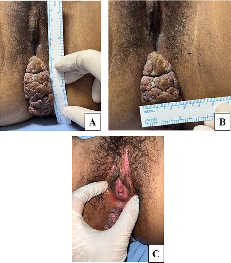 |
Figure 1 At the initial visit, the patient exhibited clinical manifestations of solitary, cauliflower-like lesions on the perianal region (A and B). Verrucous and hyperkeratotic papules were also observed on the vaginal introitus (C). |
The acetowhite findings were positive; however, laboratory tests for human immunodeficiency virus (HIV), hepatitis B surface antigen (HBsAg), Treponema pallidum hemagglutination assay (TPHA), and the venereal disease research laboratory (VDRL) titer were non-reactive. The thorax radiography of the patient revealed no evidence of tuberculosis. Blood glucose level and other laboratory findings were within normal limits. The pregnancy test shows a negative result. Gram staining revealed co-existing findings of bacterial vaginosis (Figure 2A), fulfilling Amsel’s criteria, and non-gonococcal cervicitis (Figure 2B). The dermoscopic evaluation of the lesion revealed a knob-like appearance (Figure 3).
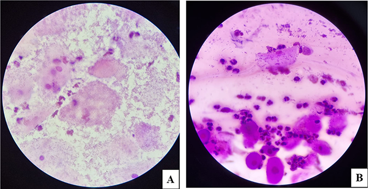 |
Figure 2 Gram staining of the vaginal specimen revealed clue cells (A) and Gram staining of the cervical specimen showed PMN over 30 cells per field, indicating non-gonococcal cervicitis (B). |
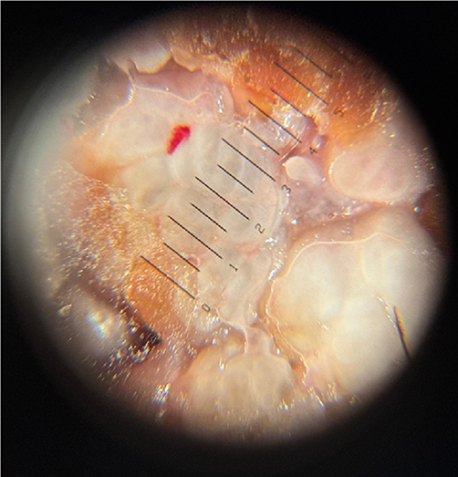 |
Figure 3 The dermoscopy examination revealed knob-like appearance. |
A shave biopsy procedure was performed on the perianal lesion of the patient, and the histopathological examination revealed keratinized stratified squamous epithelium with acanthosis, papillomatosis, hyperkeratosis, and parakeratosis (Figure 4A and B). Koilocytosis (Figure 4C) was observed in some nuclei, but there were no signs of malignancy. Genotyping with polymerase chain reaction (PCR) yielded positive results for HPV types 11. These findings are consistent with AGW on the vaginal introitus, BLT on region perianal, non-gonococcal cervicitis, and bacterial vaginosis. The temporal relationship between the sexual assault and the subsequent rapid progression of perianal lesions raises a strong suspicion of HPV transmission related to the assault, despite the patient’s otherwise immunocompetent status.
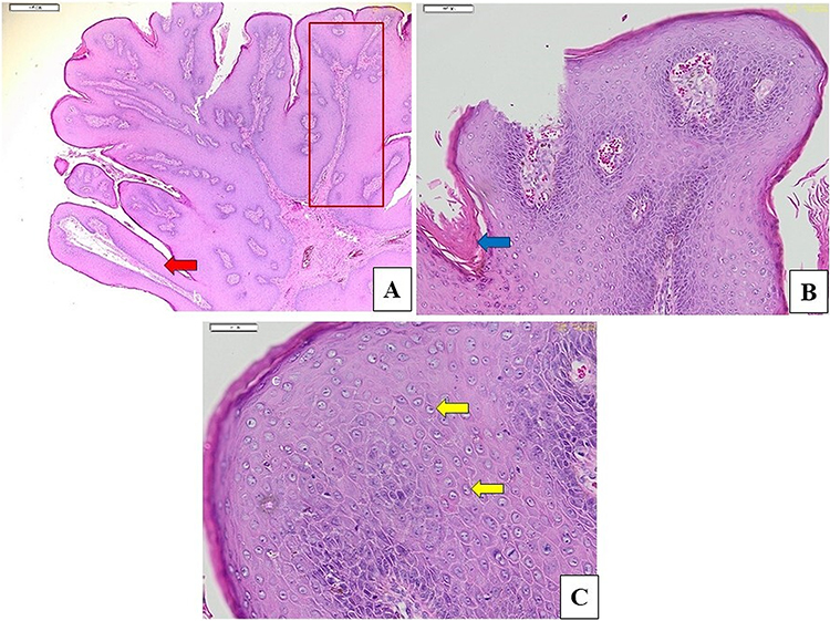 |
Figure 4 (A–C) Histopathological examination showed (A) papillomatosis (red arrow), akantosis (red box); (B) hyperkeratosis (blue arrow); (C) koilocytosis (yellow arrow) (Hematoxylin-Eosin stain; original magnification, 20x (A), 100x (B), 200x (C) respectively). |
The patient subsequently underwent surgical excision by the pediatric surgery department for her BLT (Figure 5). The patient was also given a single dose of 2 grams of metronidazole and 1 gram of azithromycin to treat bacterial vaginosis and non-gonococcal cervicitis, respectively. Notably, the AGW on the vaginal introitus exhibited spontaneous healing prior to the initiation of treatment (Figure 6A). No complications were identified postoperatively (Figure 6B).
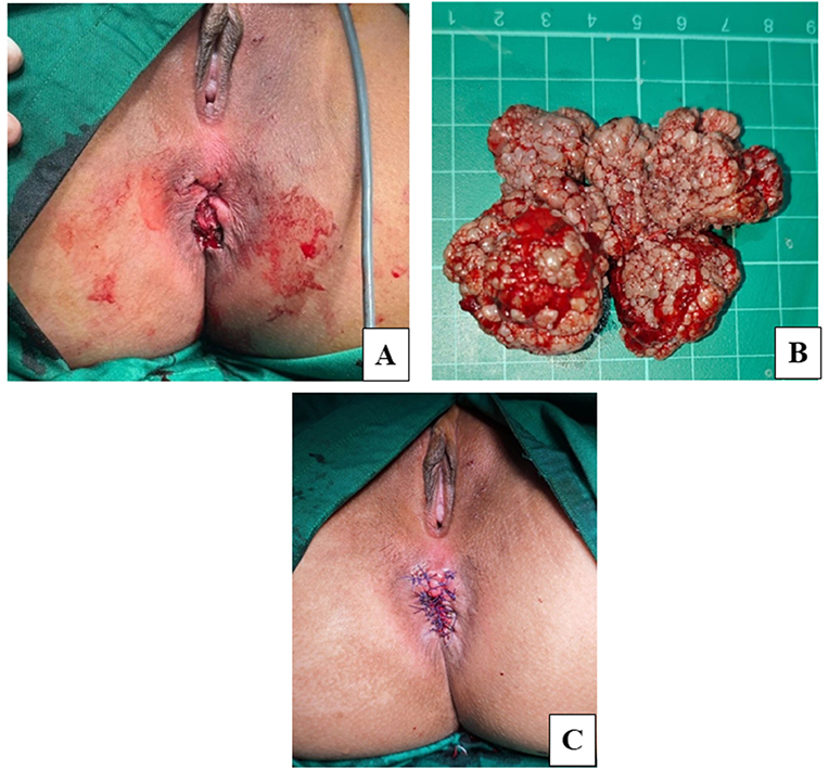 |
Figure 5 (A–C) The patient’s postoperative condition (A), the lesion excised during the procedure (B), and the surgical wound after suturing (C) are presented. |
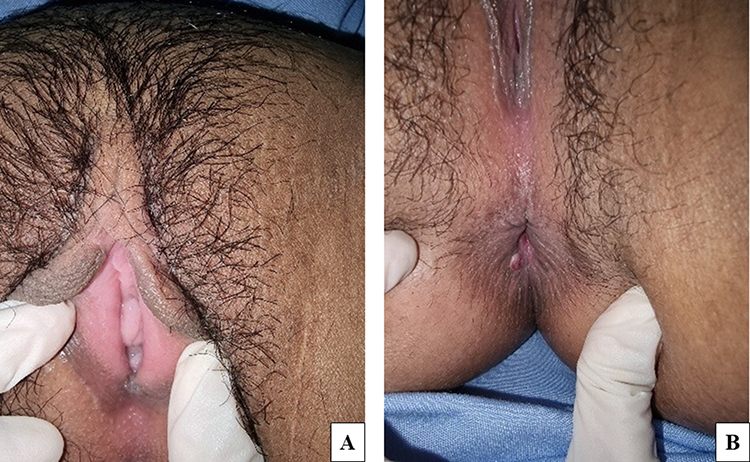 |
Figure 6 (A and B) The patient’s condition 38th days after surgical excision revealed spontaneous remission of condyloma acuminata on vaginal introitus (A), the postoperative wound on the perianal area has closed and there is no new growth of condyloma acuminata (B). |
Discussion
Giant condyloma acuminata, or BLT, is a rare type of condyloma acuminata that impacts 0.1% of the population and is caused by HPV.12 HPV is classified into low-risk and highrisk varieties based on its carcinogenic potential. Infection with low-risk HPV strains typically remains asymptomatic or benign, with lesions resolving spontaneously after several weeks or months. Low-risk HPV types, such as HPV-6 and HPV-11, are associated with condyloma and also contribute to the development of BLT, characterized by destructive local growth, while high-risk subtypes 16, 18, 31, 33, 35, and 45 can lead to high-grade dysplasia and squamous cell carcinoma.2,13
A higher prevalence of BLT is observed after puberty, although it can occur at any age. Men are more affected than women, usually between 30 and 50 years old, with a male/female sex ratio of 2.7:1.14 It predominantly occurs in men on the penis, urethra, and scrotal regions in 94% of cases, and in women on the vulva in 90% of cases, while merely 10% to 17% involve the anoperineal region, occuring in anal intercourse and oral sex. Despite its rising prevalence in recent years, BLT continues to be an uncommon issue among children. Whenever it is found in children, sexual abuse must be investigated.15 The majority of children infected with HPV exhibit symptoms within two years of their initial sexual intercourse. In female adolescents, perianal condyloma may arise via autoinoculation of secretions due to primary cervicovaginal infection or by anal intercourse.2 While there are no case studies directly blaming sexual abuse as the exclusive cause of BLT in children, the evidence indicates sexual abuse is a recognized and concerning transmission route for HPV in children, particularly for anogenital lesions like condyloma acuminata and potentially BLT. Most pediatric HPV-related lesions in older children carry a high index of suspicion for sexual abuse, especially when occurring at genital sites and in children older than 4 years.4 The patient in this case reported that the sexual assault occurred approximately 18 months prior to presentation, with a pea-sized lesion first noted about 12 months before consultation, indicating a 6-month interval between exposure and lesion onset. The patient is unable to recall details regarding the sexual intercourse, including anal or vaginal, attributing her memory loss to the administration of sedative medications. In the present case, the temporal association between the assault and lesion onset supports the consideration of suspected sexual transmission as a contributing factor.
The clinical manifestation of BLT is distinctive. Typically, it manifests as a single, cauliflower-like tumoral lesion that develops in the external genitalia or the perianal area. The tumor gradually increases in size, and in cases where medical intervention is delayed, it may reach or exceed a diameter of 10–15 cm. Due to inadequate hygiene and maceration associated with bacterial superinfection, the lesions produce a malodorous scent.16 Constipation, hemorrhoids, difficulty defecating and urinating, dysuria, bloating, and fatigue usually accompanied the symptom.17 This case report describes a patient with a solitary, cauliflower-like lesion in the perianal region, measuring 8.5 cm in length, and accompanied by a foul odor, in accordance with the clinical manifestations of BLT. In comparison, Ambriz GonzalesIn comparison, Ambriz González et al18 reported a similar case in a 12-year-old girl presenting with a 15×20 cm perianal BLT. The tumor was linked to HPV infection, although the subtype could not be identified. There was no evidence of sexual abuse, and the child had poor hygiene. The patient underwent two-stage surgical excision and topical 5-fluorouracil, with no recurrence and excellent outcomes at one year.
People who take immunosuppressant drugs or who have HIV are more likely than the general population to get a severe HPV infection. Other factors that make this risk higher are diabetes mellitus, heavy drinking, smoking, bad local hygiene, chemotherapy, and pregnancy.7–9 The study found that CD4+ T cells and monocytes/macrophages become predominant in regressing condylomas. HIV infection induces an immunosuppressive state by reducing CD4+ lymphocyte numbers, limiting dendritic cell activation, and diminishing CD8+ lymphocyte function. Notably, CD8+ lymphocyte activation is essential for the eradication of HPV-infected epithelial cells.2 CD4+ T lymphocytes play a critical role in the immune response to HPV, as evidenced by the increased incidence of extensive dysplastic lesions in patients other than those with HIV infection, such as those receiving immunosuppressive therapy or those with congenital immune system defects.8
Congenital immune system defects result from mutations in specific genes involved in immune regulation, including serine peptidase inhibitor Kazal type 5 (SPINK5), GATA2, CXC chemokine receptor 4 (CXCR4), and dedicator of cytokinesis 8 (DOCK8). These genetic alterations have been linked to an increased susceptibility to HPV-related lesions compared to healthy individuals, such as large anogenital warts, by impairing APC and reducing T lymphocyte levels.2 A case documented by Li et al19 reported a 26-year-old female BLT patient with Netherton syndrome, caused by mutations in the SPINK5 gene.
This mutation leads to dysfunction of lympho-epithelial Kazal-type related inhibitor (LEKT1), a serine protease essential for skin and hair morphogenesis, as well as mucosal epithelial defense against inflammation and microbial infections. Similarly, GATA2 deficiency has been associated with HPV persistence in over 75% of affected individuals, with approximately 50% at increased risk of recurrent infections and warts due to the depletion of critical immune cell lineages, including B cells, dendritic cells, monocytes, and natural killer cells.20
In addition, CXCR4, a transmembrane receptor found on leukocytes, endothelial cells, and stem cells, is essential for immune signalling in HPV regulation. Its interaction with Janus Kinase (JAK) 2 and JAK3 activates the JAK/Signal Transducer and Activator of Transcription (STAT) pathway, while binding to stromal-derived factor-1 (SDF-1) mediates chemotaxis, adhesion, and immune cell accumulation.21 Defective CXCR4 impairs SDF-1 signalling, hinders leukocyte migration, and facilitates HPV replication and disease progression. DOCK8 is involved in facilitating leukocyte migration; its deficiency leads to impaired immunoregulation and subsequent dissemination of HPV. Lymphopenia develops with age, impacting CD8+ and CD4+ T cell populations. In mice, the deficiency of DOCK8 limited the suppression of thymic T regulatory cells.20
Laboratory investigations, immunoglobulin levels, and genetic sequencing are necessary to confirm an immune defect.22,23 Achdiat et al19 reported a 17-year-old male with condyloma acuminata on the penis, exhibiting no symptoms of immunodeficiency, and laboratory test results confirming the absence of immunodeficiency. This patient did not respond to treatment with the measles, mumps, and rubella (MMR) vaccine—a stark contrast from prior studies that have shown successful resolution of condyloma acuminata with MMR vaccination. Considering these findings, patients with severe or treatment-resistant HPV-related warts should be assessed for possible underlying immunological deficiencies. The patient in this case report was considered immunocompetent, with no signs of HIV, TB, or any underlying conditions. While a congenital immunological deficiency cannot be excluded, there is yet no clinical evidence to substantiate this hypothesis. Therefore, targeted genetic evaluation may be considered in future investigations.
Several sources of evidence suggest that HPV infection may be influenced by hormonal factors. When estrogen is present, the HPV 16 E6 and E7 genes work more efficiently in SiHa cervical cancer cells.23 A study by Cui et al24 identified estrogen receptors in condyloma tissue using immunohistochemistry in the majority of lesions; research by Fakheri et al25 also showed that 76% of lesions in pathologically confirmed condyloma acuminata had progesterone receptors. These findings underscore the possible influence of hormones on HPV-related lesions, while evidence specifically addressing hormonal influences in adolescent patients remains limited, highlighting the need for further studies in this population.
Buschke–Löwenstein tumors are frequently detected via a comprehensive clinical history and visual assessment of the lesions. DNA detection techniques, such as polymerase chain reaction (PCR), can offer supplementary diagnostics by confirming the diagnosis and facilitating genotyping.8 PCR analysis of the patient’s lesion confirmed the presence of HPV type 11, supporting the diagnosis of BLT.
The clinical and histological characteristics of BLT and condyloma acuminata exhibit similarities, including epithelial proliferation, thickening, infrequent mitotic figures in the basal and spiny layers, parakeratosis, and hyperkeratosis. However, in BLT, papillomatosis, acanthosis, and ridge elongation are more prominent; mitotic activity is increased, and keratinization of individual cells along with keratin beads is observable.17,23 On a histopathologic examination of this patient, epithelium was found to have characteristics such as hyperplasia, acanthosis, prominent papillomatosis, hyperkeratosis, and parakeratosis. Cell nuclei are within normal limits without signs of malignancy, and some nuclei are koilocytes, in concordance with BLT.
Due to the absence of a consensus on the optimal treatment approach for BLT, treatment algorithms are predominantly informed by case reports and small case series.26 Therapeutic options for BLT encompass imiquimod and podophyllin for topical chemotherapy, intralesional 5-fluorouracil injection, cryotherapy, curettage, radical resection, either as separate therapies or in conjunction with neoadjuvant or adjuvant chemotherapy, chemotherapy alone, and radiation therapy.27 A wide radical excision, followed by reconstructive surgery, seems to be the optimal therapeutic strategy for BLT management because of the risk of malignancy and local destruction.2,28 It is necessary to surgically excise the perianal BLT with well-defined secure margins, maintaining a minimum distance of 1 cm, while meticulously dissecting the subcutaneous fatty tissues from the external anal sphincter.29 In this case report, the patient underwent wide excision and reconstructive surgery by the pediatric surgery department to prevent malignancy and any further destruction. The lesion was entirely removed with sufficient margins to guarantee total removal while safeguarding the adjacent healthy tissue. During the operation, the mass was observed to be well-defined, with no deep penetration into surrounding tissues. Following excision, the resulting defect was assessed and deemed suitable for primary closure. However, the lesions on the vaginal introitus were spontaneously resolved as expected in 90% of cases of HPV infection.
The prognosis of BLT presumably depends on tumor size, malignancy transformation, recurrent infections, secondary infections, and immunodeficiencies.2 The predominant complications of BLT include necrosis, fistula formation, and superinfection. Soft tissue infiltration and various sequelae, including fistula, abscess, surgical incision hemorrhage, soft tissue infection, flap failure, urinary tract infection, urethral blockage, anal stenosis, and fecal incontinence, are linked to morbidity in BLT patients.12 The local recurrence rate of BLT has been documented to be as high as 67% with a 10-month mean recurrence interval.18 Sandoval et al1 recommend a follow-up schedule of every six months after the complete healing of the wound for the first two years. Following this initial period, the long-term follow-up should be conducted on an annual basis.11 The patient in this report is at present under monitoring. In the most recent followup, no new lesions have been observed, no complication have occurred after surgery, and the patient remains under observation to date. The patient was informed about the significance of the HPV vaccine to prevent more HPV-related problems, and periodic follow-up visits were arranged to monitor for any new lesions or recurrences.
This case underscores the potential for severe HPV-related disease to develop in immunocompetent adolescents following suspected sexual assault, highlighting the importance of thorough evaluation, early intervention, and long-term monitoring.
Conclusion
The Buschke–Löwenstein tumor is an uncommon variant of condyloma acuminata that usually occurs in individuals with immunodeficiency, underlying diseases, alcohol use, smoking, poor hygiene, or chemotherapy and during pregnancy. This case underscores the necessity of acknowledging the advancement of condyloma acuminata to BLT, even in the absence of acquired immunodeficiency and other recognized risk factors. In instances of suspected sexual abuse, prompt multidisciplinary evaluation by pediatric, gynecological, or pediatric surgery and psychosocial teams is crucial for ensuring proper screening, protection, and comprehensive patient care.
Ethics Statement
This study was conducted in compliance with the Declaration of Helsinki, Good Clinical Practice, and local regulatory requirements, and was approved by the Medical Ethics Committee of Dr. Hasan Sadikin General Hospital Bandung (approval number DP.04.03/D.XIV.6.5/419/2025). In cases with a history suggestive of abuse, particular attention was given to maintaining patient confidentiality and adhering to institutional safeguarding protocols, including appropriate multidisciplinary involvement. The potential history of abuse was handled with sensitivity, prioritizing patient safety and psychological well-being.
Consent Statement
Written informed consent for publication of the case details and images was obtained from the patient and her mother, who signed the consent form as her legal guardian.
Acknowledgment
The authors would like to thank the staff of Dermatology and Venereology Department, Faculty of Medicine, Universitas Padjadjaran–Dr. Hasan Sadikin General Hospital. The authors acknowledge Universitas Padjadjaran for the support through the Hibah Riset Internal Universitas Padjadjaran – Faculty Assignment Grant 2025 in the preparation of this review, and the financial support for publication costs provided through a reimbursement scheme via the Indonesian Endowment Fund for Education (LPDP), on behalf of the Indonesian Ministry of Higher Education, Science and Technology, under the EQUITY Program (Contract No. 4303/B3/DT.03.08/2025 and 3927/UN6.RKT/HK.07.00/2025).
Disclosure
The authors report no conflicts of interest in this study.
References
1. Sandoval I, Hernández R, Torres E, Yanque O. Giant condylomata acuminata of Buschke–Lowenstein. J Obstet Gynaecol. 2020;40(4):582–10. PMID: 31373237. doi:10.1080/01443615.2019.1607834
2. Purzycka-Bohdan D, Nowicki RJ, Herms F, Casanova JL, Fouéré S, Béziat V. The pathogenesis of giant condyloma acuminatum (Buschke–Lowenstein Tumor): an overview. Int J Mol Sci. 2022;23(9):4547. PMID: 35562936; PMCID: PMC9100137. doi:10.3390/ijms23094547
3. Drumond DG, Castelo BB, Esperança SD, Dominiquini FB, de Góis Speck NM. Therapeutic approaches to condyloma acuminatum in children and adolescents. Medicina. 2022;55:1–10.
4. Sinclair KA, Woods CR, Sinal SH. Venereal warts in children. Pediatr Rev. 2011;32(3):115–21; quiz 121. PMID: 21364015. doi:10.1542/pir.32-3-115
5. Niazy F, Rostami K, Motabar AR. Giant condyloma acuminatum of vulva frustrating treatment challenge. World J Plast Surg. 2015;4(2):159–162. PMID: 26284185; PMCID: PMC4537608.
6. Evrüke İM, Usta Korkut İ. Giant condyloma acuminatum in a female renal transplant recipient with rapid development: a case report. Cukurova Med J. 2023;48(1):297–300. doi:10.17826/cumj.1227717
7. Atkinson AL, Pursell N, Sisay A. The giant condyloma (buschke-löwenstein tumor) in the immunocompromised patient. Case Rep Obstet Gynecol. 2014;2014:793534. PMID: 25328732; PMCID: PMC4190693. doi:10.1155/2014/793534
8. Grosu-Bularda A, Hariga CS, Dumitru CS, et al. Clinicopathological findings and comprehensive review of Buschke–Löwenstein tumors based on a case study. J Pers Med. 2024;14(8):1–15. doi:10.3390/jpm14080887
9. Béziat V, Casanova JL, Jouanguy E. Human genetic and immunological dissection of papillomavirus-driven diseases: new insights into their pathogenesis. Curr Opin Virol. 2021;51:9–15. PMID: 34555675; PMCID: PMC8743045. doi:10.1016/j.coviro.2021.09.002
10. Nanda S, Mohapatra J, Mahapatra M, Parija J, Nayak B. An exasperating and unusual presentation of Buschke–Löwenstein tumor in an adolescent girl: a case report with review of literature. Indian J Med Paediatr Oncol. 2024;45:1–5.
11. Loo GH, Lim LY, Zainuddin ZM, Fam XI. Staged resection in the management of HIV-related anogenital giant condyloma acuminatum. A case report. Ann Med Surg. 2019;48:73–76. PMID: 31737262; PMCID: PMC6849151. doi:10.1016/j.amsu.2019.10.024
12. Suárez-Ibarrola R, Heinze A, Sánchez-Sagástegui F, et al. Giant condyloma acuminatum in the genital, perineal and perianal region in a pediatric patient. Literature review and case report. Urol Case Rep. 2016;7:14–16. PMID: 27335781; PMCID: PMC4909496. doi:10.1016/j.eucr.2016.02.003
13. Boda D, Cutoiu A, Bratu D, Bejinariu N, Crutescu R. Buschke–Löwenstein tumors: a series of 7 case reports. Exp Ther Med. 2022;23(6):393. PMID: 35495587; PMCID: PMC9047027. doi:10.3892/etm.2022.11320
14. Bouhout T, Ramdani A, Kharkhach A, Serji B. A report of two rare cases of Buschke–Löwenstein tumor. Cureus. 2024;16(1):e52700. PMID: 38384644; PMCID: PMC10879842. doi:10.7759/cureus.52700
15. Tampa M, Malin-Benea A, Sarbu MI, Benea V, Georgescu SR. A case of giant rapidly evolving Buschke–Löwenstein tumor in an immunocompetent patient. Revista Românå de Boli Infectioase. 2013;5:83–87.
16. Nieves-Condoy JF, Acuña-Pinzón CL, Chavarría-Chavira JL, Hinojosa-Ugarte D, Zúñiga-Vázquez LA. Giant condyloma acuminata (Buschke–Lowenstein Tumor): review of an unusual disease and difficult to manage. Infect Dis Obstet Gynecol. 2021;2021:9919446. PMID: 34305393; PMCID: PMC8266468. doi:10.1155/2021/9919446
17. Pérez-González A, Cachay E, Ocampo A, Poveda E. Update on the epidemiological features and clinical implications of Human Papillomavirus Infection (HPV) and Human Immunodeficiency Virus (HIV) Coinfection. Microorganisms. 2022;10(5):1047. PMID: 35630489; PMCID: PMC9147826. doi:10.3390/microorganisms10051047
18. Ambriz-González G, Escobedo-Zavala LC, Carrillo de la Mora F, et al. Buschke–Löwenstein tumor in childhood: a case report. J Pediatr Surg. 2005;40(9):e25–7. PMID: 16150329. doi:10.1016/j.jpedsurg.2005.05.070
19. Achdiat PA, Ismiranty D, Hindritiani R, Rizqandaru T, Usman HA, Maharani RH. The oncogenic human papillomavirus involvement as a risk factor of measles, mumps, and rubella vaccine immunotherapy failure in anogenital warts. Int Med Case Rep J. 2025;18:83–90. PMID: 39830042; PMCID: PMC11742370. doi:10.2147/IMCRJ.S498892
20. Hewavisenti RV, Arena J, Ahlenstiel CL, Sasson SC. Human papillomavirus in the setting of immunodeficiency: pathogenesis and the emergence of next-generation therapies to reduce the high associated cancer risk. Front Immunol. 2023;14:1112513. PMID: 36960048; PMCID: PMC10027931. doi:10.3389/fimmu.2023.1112513
21. Leiding JW, Holland SM. Warts and all: human papillomavirus in primary immunodeficiencies. J Allergy Clin Immunol. 2012;130(5):1030–1048. PMID: 23036745; PMCID: PMC3517887. doi:10.1016/j.jaci.2012.07.049
22. Chu EY, Freeman AF, Jing H, et al. Cutaneous manifestations of DOCK8 deficiency syndrome. Arch Dermatol. 2012;148(1):79–84. PMID: 21931011; PMCID: PMC4103903. doi:10.1001/archdermatol.2011.262
23. Winer RL, Koutsky LA. Genital human papillomavirus infection. In: Holmes KK, Sparling PF, Stamm WE, Piot P, Wasserheit JN, Corey L, editors. Sexually Transmitted Diseases.
24. Cui MH, Liu YQ, Li HL, Li SR. Human papillomavirus in condyloma acuminata and other benign lesions of the female genital tract. Chin Med J. 1994;107(9):703–708. PMID: 7805465.
25. Fakheri T, Afsharian M, Malekianzadeh E, Khazaei S, Izadi B, Kanani M. Estrogen and progesterone receptor expression in vulvar condyloma acuminata. Int J Collab Res Intern Med Public Health. 2012;4(4):380–387.
26. Balogh N, Kolozsi P, Tóth D. A rare malignancy: a case report of early progression of anal Buschke–Löwenstein tumor into squamous cell carcinoma in an immunocompetent patient. Int J Surg Case Rep. 2024;119:109715. PMID: 38704971; PMCID: PMC11087946. doi:10.1016/j.ijscr.2024.109715
27. Müdüroğlu M, Güllüoğlu YB, Taşlıpınar M, et al. An extraordinary case of Buschke–Löwenstein tumor: multiple localization, malignant transformation, and clinical insights. Afr J Urol. 2024;30(56):1–7. doi:10.1186/s12301-024-00459-6
28. Hum M, Chow E, Schuurmans N, Dytoc M. Case of giant vulvar condyloma acuminata successfully treated with imiquimod 3.75% cream: a case report. SAGE Open Med Case Rep. 2018;6:2050313X18802143. PMID: 30345054; PMCID: PMC6180360. doi:10.1177/2050313X18802143
29. Mihailov R, Tatu AL, Niculet E, et al. Surgical management of perianal giant condyloma acuminatum of Buschke and Löwenstein: case Presentation. Life. 2023;13(9):1916. PMID: 37763319; PMCID: PMC10532963. doi:10.3390/life13091916
 © 2026 The Author(s). This work is published and licensed by Dove Medical Press Limited. The
full terms of this license are available at https://www.dovepress.com/terms
and incorporate the Creative Commons Attribution
- Non Commercial (unported, 4.0) License.
By accessing the work you hereby accept the Terms. Non-commercial uses of the work are permitted
without any further permission from Dove Medical Press Limited, provided the work is properly
attributed. For permission for commercial use of this work, please see paragraphs 4.2 and 5 of our Terms.
© 2026 The Author(s). This work is published and licensed by Dove Medical Press Limited. The
full terms of this license are available at https://www.dovepress.com/terms
and incorporate the Creative Commons Attribution
- Non Commercial (unported, 4.0) License.
By accessing the work you hereby accept the Terms. Non-commercial uses of the work are permitted
without any further permission from Dove Medical Press Limited, provided the work is properly
attributed. For permission for commercial use of this work, please see paragraphs 4.2 and 5 of our Terms.