
Back to Journals » International Journal of Nanomedicine » Volume 21
Radiolabeled Iron-Based Nanomaterials for Cancer Diagnosis and Therapy
Authors Ma YX, Dai KL, Sun N, Zhang S
Received 9 December 2025
Accepted for publication 4 March 2026
Published 20 March 2026 Volume 2026:21 587365
DOI https://doi.org/10.2147/IJN.S587365
Checked for plagiarism Yes
Review by Single anonymous peer review
Peer reviewer comments 2
Editor who approved publication: Dr RDK Misra
Yan-Xi Ma,* Kun-Long Dai,* Na Sun, Song Zhang
Department of Nuclear Medicine, Xin Qiao Hospital, Army Medical University, Chong Qing, 400037, People’s Republic of China
*These authors contributed equally to this work
Correspondence: Kun-Long Dai; Song Zhang, Email [email protected]; [email protected]
Abstract: Cancer remains one of the leading causes of death worldwide, with diagnosis and treatment continuing to pose significant challenges. In recent years, iron-based nanomaterials (iron oxide-based nanomaterials, iron-based complex conjugates) have garnered unprecedented attention due to their excellent functionality, superior biocompatibility and potential for multifunctional integration. Due to the non-specific nature of radionuclides. Their integration with nanomaterials offers promising opportunities for precise cancer diagnosis and effective treatment by enabling targeted delivery, controlled release, and synergistic combination therapies. This review systematically elucidates the latest advancements in radiolabeled iron-based nanomaterials for cancer diagnosis and therapy, with a focus on radiolabeling methods, multimodal imaging, and the combination of radiotherapy with various treatment modalities, including photothermal therapy (PTT), magnetic hyperthermia therapy (MHT), photodynamic therapy (PDT), immunotherapy (IT), and chemodynamic therapy (CDT). Finally, the prospects for the development of radiolabeled iron-based nanomaterials are discussed, along with key research priorities for the future.
Keywords: radionuclides, iron-based nanomaterials, multimodal imaging, combination therapy
Introduction
Cancer ranks as the second most common cause of mortality globally. According to World Health Organization (WHO) projections, global cancer incidence may increase by 60% within the next 20 years.1 The growing burden of cancer presents substantial challenges to both public health and economic systems, making early detection and effective treatment universally recognized priorities.2 While modern oncology has made significant advances, conventional diagnostic and therapeutic approaches face limitations due to tumor heterogeneity, drug resistance, and adverse effects. Current screening methods primarily rely on biomarkers and imaging techniques (eg, X-ray, ultrasound, MRI); however, definitive diagnosis often requires invasive biopsy or surgical sampling.
The current options for cancer therapy modalities include surgery, chemotherapy, radiotherapy, immunotherapy, monoclonal antibodies, targeted therapy, photodynamic therapy, and laser ablation. However, each approach has inherent drawbacks.3 Conventional cancer treatment often entails surgical intervention, which serves as the first-line approach for most malignancies by enabling direct removal of the tumor. Despite its efficacy, surgery is associated with significant risks and considerable physical trauma. Chemotherapy induces severe systemic toxicity. External beam radiotherapy, though widely used, delivers suboptimal radiation doses to tumors due to energy absorption by surrounding healthy tissues, resulting in collateral damage.4,5 In contrast, internal radiotherapy (radionuclide therapy) offers a targeted approach by delivering cytotoxic radiation directly to tumors or their microenvironment. Unlike biological therapies that modulate signaling pathways, radionuclides induce tumor cell death via localized ionizing radiation, which induces DNA strand breaks.6,7 This method improves therapeutic precision while minimizing off-target effects.
Radionuclides are categorized into diagnostic and therapeutic nuclides. Diagnostic radionuclides are essential for patient stratification planning, leading to enhanced outcomes in targeted radionuclide therapy. Additionally, radionuclides can be utilized in conjunction with positron emission tomography (PET) and single photon emission computed tomography (SPECT) techniques to precisely monitor systemic disease lesions and patient stratification (Table 1). 8–10
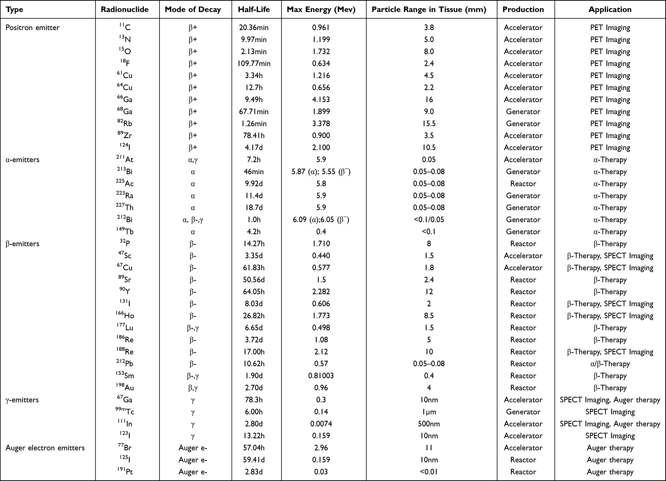 |
Table 1 Radionuclide properties11–15 |
Molecular delivery systems play a crucial role in enhancing the tumor-targeting capabilities of various radionuclide constructs, encompassing small molecules, polypeptides, antibodies, nanomaterials, and microspheres.11,16–20 Despite advancements, radionuclide-based therapies encounter significant challenges in both diagnosis and treatment. Some cancers exhibit resistance to radionuclide therapy, while issues such as non-specific distribution, limited bioavailability, and ionization-induced damage to non-target organs further complicate their efficacy.12,13,21 To address these challenges, the integration of nanomedicine offers a promising avenue to broaden the diagnostic and therapeutic landscape. Radionuclide nano systems are emerging as powerful tools for tumor diagnosis and treatment Specifically, leveraging nanomaterials for targeted delivery of radionuclides to tumor tissues holds potential to enhance bioavailability and reduce toxicity to normal tissues.7,14
Recent advancements in radiopharmaceuticals have demonstrated the utilization of nanomaterials in diagnosing and treating various cancers, leveraging their distinctive physical and chemical properties.22–24 Nanomaterials have emerged as pivotal players in diverse nuclear medicine domains, notably in the creation of radionuclide nanoparticle carriers for deployment as contrast agents in computed tomography (CT) imaging.25 These carriers enable the loading, transport, and delivery of a multitude of radionuclides within a single nanomaterial. In contrast to numerous small molecule radiopharmaceuticals, nanomaterials possess a superior specific surface area-to-volume ratio, rendering them optimal candidates for targeted radionuclide delivery. Furthermore, many nanoparticles offer supplementary optical, magnetic, and other imaging and therapeutic functionalities.26,27
Transition metal nanomaterials are one of the most representative nanomaterials in the field of nanomedicine. Iron-based nanomaterials have been widely used in tumor diagnosis and treatment due to their unique magnetism, good biocompatibility, chemical stability, and various morphologies.28–30 Among various candidates, iron-based nanomaterials present distinct advantages over other materials like gold, which is also notable for imaging and radiosensitization. (1) Biocompatibility and Clinical Translation: Iron oxide-based nanomaterials (eg, Ferumoxytol) have received approval from the U.S. Food and Drug Administration (FDA) as MRI contrast agents and drug carriers, providing a clear regulatory pathway and established safety profile.31 (2) Multifunctionality: They possess unique magnetism enabling MRI contrast, magnetic targeting, and magnetic hyperthermia (MHT).32,33 (3) Fenton Activity: Iron ions can catalyze the production of reactive oxygen species (ROS) via Fenton reactions, facilitating chemodynamic therapy (CDT). (4) Cost-Effectiveness and Abundance: Iron is more abundant and less expensive than gold, facilitating scalable production. While gold nanoparticles exhibit excellent surface plasmon resonance for photothermal therapy, iron-based materials offer a broader spectrum of inherent therapeutic mechanisms and easier clinical integration, making them a compelling platform for radiolabeled theranostics.34
This review aims to provide a comprehensive and critical update specifically on radiolabeled iron-based platforms. It also serves as a valuable supplement to existing earlier reviews. An area that has evolved rapidly but lacks a recent dedicated synthesis comparing labeling strategies, multimodal applications, and translational challenges. It summarizes the latest research progress and key radiolabeling technologies of radioisotope iron-based nanomaterials, focusing on their breakthrough applications and innovative strategies in multimodal imaging-guided precision diagnosis and targeted radiotherapy. It also discusses the key challenges and future plans for clinical translation.
Loading Radionuclides in Nanomaterials
A key factor in designing radio nanomedicines is radio stability the firm attachment of radionuclides to the material during storage, administration, and delivery to the target site. Otherwise, free radionuclides may cause systemic toxicity to normal tissues through undesired radiation exposure. To date, various strategies have been developed for labeling iron-based nanomaterials. Based on the location of radionuclides within the material, these methods can be categorized as surface conjugation, internal incorporation and interface engineering.28,35,36 Specific techniques include adsorption (physical binding), chelation (for metallic radionuclides, eg, 64Cu, 68Ga, 89Zr), covalent conjugation (for non-metallic radionuclides, eg, 124I, 131I) and encapsulation (core-shell entrapment).
Surface Conjugation
Surface conjugation is a method for immobilizing radionuclides onto material surfaces, comprising two categories: direct surface labeling and indirect surface labeling. In the direct method, radionuclides form chemical bonds directly with the material surface. Most examples rely on coordination bonds between chemical groups on the nanomaterial surface—such as Fe2O3, -PO3H, -SH, or -OH—and the radionuclides. The indirect method involves introducing chelating agents or prosthetic groups onto the material surface to enable binding with both metallic and non-metallic radionuclides.
The direct surface labeling of radionuclides, sometimes referred to as chemisorption, is a highly efficient and straightforward technique that relies on the unique chemical properties of nanomaterial surfaces to achieve robust attachment of radionuclides.37 This method has been employed in catalysis and analytical chemistry to elucidate interaction mechanisms between metals and materials.38 In 2013, Cai et al reported the first application of this technique to label iron-based nanomaterials, attributing the radiolabeling mechanism to the formation of stable As-Fe3O4 complexes, where AsIIIO3 trigonal pyramids or AsVO4 tetrahedra can occupy vacant tetrahedral sites on the octahedrally terminated (111) surface of Fe3O4.39 For non-metallic radionuclides, their strong affinity for iron-based materials is often exploited for labeling. For example, Zhu et al used 125I to label Fe3O4-Ag heterodimers, achieving MRI/SPECT dual-modality imaging probes through rapid binding of 125I with Ag in the nanoparticles.40
In addition, radiolabeling methods based on surface adsorption have been developed for clinically approved iron oxide drugs (eg, Feraheme, Ferumoxytol, and Fercarbotran).41–43 By heating the reaction mixture at 120°C for 1 hour, 64Cu tightly binds to the iron oxide core, achieving a radiochemical yield of 66 ± 6%. The same reaction conditions are applicable to other metallic radionuclides in different oxidation states, such as 89Zr and 111In, with labeling yields of 93 ± 3% and 91 ± 2%, respectively.44 The variation in yields can be attributed to differences in the coordination chemistry, ionic radius, and optimal oxidation state for surface binding for each metal ion (Cu2⁺, Zr4⁺, In3⁺). Both in vitro stability assays and in vivo biodistribution studies demonstrated that the resulting 89Zr radiolabeled nanoparticles exhibit favorable pharmacokinetic profiles, with negligible impact on their physical and biological properties.
Recently, Patrick et al introduced the concept of surface radio-mineralization (SRM) to describe the heat-induced radiolabeling (HIR) method for iron oxide and ferrite nanoparticles. By applying HIR to label bare polymer-coated multinuclear and surface-functionalized iron oxide nanoparticles with 111In and 89Zr, they found that the radio-mineralized metal oxides on the nanoparticle surface did not alter the nanoparticles’ original physical properties.45
Compared to indirect labeling methods requiring chelators or prosthetic groups, direct surface radiolabeling offers distinct advantages: 1) Operational Simplicity: Most protocols can be completed via a straightforward one-step procedure. This approach also enables labeling of radionuclides difficult to chelate conventionally (eg, 72As, 69Ge). 2) Reduced Interference: Avoids potential side effects associated with chelators/prosthetic groups. Enhanced Functionalization Capacity: Without surface-bound chelators/prosthetic groups, more space remains for multifunctional engineering (eg, conjugation of fluorophores, targeting ligands, or therapeutic agents to create versatile nanoplatforms). 3) Improved Distribution Accuracy: Prevents inconsistencies in biodistribution caused by ligand dissociation, enabling more precise tracking of nanomaterial metabolism in vivo. However, the primary technical challenge lies in the high-temperature requirements (typically 100–120°C), which may compromise the integrity of thermolabile nanoparticle formulations, thereby limiting applicability.
Indirect Surface Labeling Using Chelating Agents
Radionuclides with metallic characteristics typically require chelating agents to connect them to nanomaterials through coordination chemistry methods. The choice of chelating agent is crucial for achieving stable radiolabeling. Factors such as the coordination number, coordination geometry, ionic radius, and charge of the radionuclide must generally be considered to ensure proper matching with the radionuclide. Additionally, according to the hard/soft acid-base theory, the hardness of different metallic radionuclides determines whether they coordinate more readily with hard or soft donor ligands.46 Therefore, the “hardness” of the metal ion must be evaluated, and the selected ligand should have appropriate hard/soft donor atoms and correct electronic properties to improve the kinetic inertness of the complex.47
Chelating agents are mainly divided into two categories: acyclic/linear chelators and macrocyclic chelators (Figure 1). Acyclic or linear chelators can rapidly form complexes with metal ions, making them suitable for applications requiring fast reactions. Due to their structural flexibility, complexes formed by linear chelators may be less stable than those formed by macrocyclic chelators.48 In contrast, macrocyclic chelators have rigid and predetermined structures, providing a more stable coordination environment.49 Although the complexation process is slower, the final products often exhibit improved stability, making them more suitable for long-term in vivo applications. The ideal chelating agent should be capable of functioning under mild conditions (eg, neutral pH and room temperature) while demonstrating sufficient kinetic inertness and thermodynamic stability in biological systems to ensure the stability and efficacy of radiolabeled compounds during imaging and therapy.48
 |
Figure 1 Common linear and macrocyclic chelators and matched radionuclides. |
A critical consideration for chelator design is functionality within the tumor microenvironment (TME), which is often slightly acidic (pH ~6.5–6.8). Some chelators may exhibit reduced binding affinity or kinetic stability at lower pH, potentially leading to premature release of the radionuclide. Therefore, developing pH-resistant chelators or using macrocyclic chelators known for high kinetic inertness (eg, DOTA derivatives for trivalent metals) is crucial for applications in cancer theranostics.
Chelating agents can load metallic radionuclides through coordination. However, bifunctional chelators (BFCs) are typically required to successfully load metallic radionuclides onto nanoparticles.50 BFCs can simultaneously bind to nanoparticles and chelate metallic radionuclides, improving labeling efficiency. During the conjugation process of bifunctional chelators, the formation of chemical bonds between functional groups on the nanoparticle surface and the BFC is essential.51 These chemical bonds can be formed through various reactions, including amine coupling, carboxyl coupling, and thiol coupling. The choice of reaction depends on the functional groups present on the nanoparticle surface and the structure of the chelator. Click chemistry, noted for its rapid and efficient reaction characteristics, has been widely applied in radiolabeling of nanomaterials.52
Gong et al successfully labeled cyclic arginine-glycine-aspartic acid (cRGD)-conjugated superparamagnetic iron oxide (SPIO) nanocarriers with 64Cu using NOTA chelator for dual-modality PET/MRI imaging of tumors. The NOTA chelator was conjugated to SPIO nanoparticles through a reaction between maleimide groups on the SPIO’s polyethylene glycol (PEG) surface and thiol-functionalized NOTA, followed by efficient 64Cu radiolabeling. This successful surface conjugation enabled quantitative comparison of tumor accumulation between targeted cRGD-SPIO and non-targeted SPIO formulations.53 In another study, the Blower group reported a novel bifunctional chelator (dtcbp) featuring a dithiocarbamate group for binding 64Cu and a bisphosphonate group with strong affinity for SPIO. The stability of [64Cu(dtcbp)2]-SPIO nanoparticles was tested in high concentrations of ethylenediaminetetraacetic acid (EDTA) and human serum, showing strong binding of 64Cu to SPIO nanoparticles with minimal ligand dissociation.54 Most recently, Grimm’s research group achieved significant progress by constructing 89Zr-labeled ferumoxytol (an FDA-approved iron oxide nanoparticle formulation) using desferrioxamine (DFO) chelator, significantly enhancing PET/MRI detection sensitivity for tumor-draining lymph nodes. This approach demonstrated remarkable radiolabeling efficiency exceeding 90% while maintaining the nanoparticles’ critical physical properties including size distribution, surface charge, and magnetic relaxivity.55
Indirect Surface Labeling Using Prosthetic Groups
Non-metallic radionuclides, such as 11C/14C and halogens (18F, 76Br, and iodine isotopes [123I, 124I, 125I, 131I]), are typically covalently conjugated to nanomaterials through reactive functional groups on surface ligands. The extremely short half-life of 11C (20.4 minutes) necessitates rapid radiolabeling procedures. To label superparamagnetic iron oxide nanoparticles (SPIONs) with 11C, [11C]methyl iodide is used as a methylation agent, reacting with hydroxyl or amine groups on the nanoparticles via O-methylation or N-methylation. However, this approach suffers from low radiochemical yields (<3%), primarily due to particle agglomeration and low ligand surface density.56 In contrast, 14C has a much longer half-life (5.70×10³ years) and relatively simpler labeling chemistry. Retterer et al successfully incorporated 14C directly into the carbon backbone of organic molecules on Fe-Si-(COO−)3 nanoparticle surfaces, demonstrating that the 14C-labeled nanoparticles exhibited identical chemical properties to their non-radioactive counterparts.57 Furthermore, 18F labeling of iron-based nanomaterials is commonly achieved using click chemistry approaches.58
Halogen radiolabeling is a strategy that integrates radioactive halogens (primarily radioiodine) into nanomaterials through small-molecule radiochemical reactions. This method typically employs iodination reagents such as Iodogen,59 Iodo-Beads,60 Chloramine-T,61 and Bolton-Hunter reagents.62 These traditional iodination agents - Iodogen, Iodo-beads, and Chloramine-T - play a crucial role in radiolabeling biomolecules, particularly tyrosine residues and their derivatives. These reagents act as oxidants that rapidly react with iodide ions to generate highly reactive electrophilic species. The resulting electrophiles can efficiently substitute the ortho-position of phenolic hydroxyl groups on tyrosine residues, enabling effective radiolabeling of target molecules. This process is not only rapid (typically completed within seconds to minutes) but also achieves high radiolabeling efficiency, ensuring both the effectiveness and stability of the labeling. For instance, Gao et al developed a dual-modality molecular probe (Fe3O4-3H11-125I) by using Iodogen method.63
Internal Doping
Internal doping is commonly used to fabricate nanomaterials containing encapsulated radionuclides. This method effectively prevents radionuclide leaching, thereby avoiding in vivo labeling issues caused by ion migration, protein chelation, or ligand detachment. For iron-based nanomaterials, this approach includes techniques such as radiochemical doping and isotope exchange.
Radiochemical doping involves reacting a mixture of radioactive nuclide-containing precursors with non-radioactive (“cold”) nanomaterial precursors in a one-step synthesis to produce radiolabeled nanomaterials. From a chemical perspective, trace amounts of radioactive nuclides (“hot” precursors) are introduced into the nanomaterial precursors, initiating co-precipitation and resulting in the incorporation of radionuclides into the nanomaterial’s crystal lattice. As a widely used non-chelator-based radiolabeling techniques, this approach ensures high radiochemical stability while preserving the structural integrity of the material. The incorporation of radionuclides into the crystal lattice addresses the issue of signal loss associated with surface labels by physically shielding the radionuclide and integrating it into a stable matrix. These doped isotopes remain “inactive” in terms of unintended chemical interactions because they are substitutionally or interstitially placed within the lattice points, sharing the local chemical environment of the host atoms.
For instance, Moya et al employed a dual radiolabeling strategy: during nanoparticle synthesis, they doped Iron oxide nanoparticles (IONPs) with 111In in the aqueous phase, encapsulated them in poly(lactide-co-glycolide) nanoparticles, and then attached 125I to the surface-coating bovine serum albumin (BSA) via electrophilic substitution.64 While aqueous-phase synthesis offers advantages such as high efficiency and easy post-processing, it often leads to undesirable nanomaterial morphology, low phase purity, and broad size distribution. These limitations can be mitigated by using organic solvents. For example, Gao et al synthesized 111In-doped IONPs by thermally decomposing iron acetylacetonate in dibenzyl ether, yielding uniformly sized, highly crystalline nanoparticles.65
Isotope exchange involves replacing (or exchanging) stable elements present on nanomaterials with radionuclides and can be categorized into heteroexchange and homoexchange. The distinction lies in whether the exchange occurs between different elements (heteroexchange) or between different isotopes of the same element (homoexchange). The advantage of this method is its operational simplicity. However, only a few examples in the literature have employed this technique for radiolabeling. For instance, Pospisilova et al reported the preparation of 59Fe-IONP by incubating non-radioactive IONPs with 59FeCl3 for 24 hours, achieving a radiolabeling yield of 83% and demonstrating high radiochemical stability (less than 2% of 59Fe was released after 35 days of incubation in 0.1 M PBS at pH 7.4 and in rat serum).66 However, the fate and long-term stability of such compounds within the complex tumor microenvironment (TME), characterized by hypoxia, acidity, and high protease activity, require further investigation. The safety of these radionuclides hinges on their retention within the nanocarrier. Potential release over very long periods (months to years) raises concerns about chronic radiation exposure. Therefore, comprehensive studies tracking isotope release kinetics, biodistribution over extended periods, and thorough toxicological evaluations are essential before clinical translation.
Interface Engineering
In addition to labeling radionuclides on the exterior or interior of nanomaterials, they can also be anchored at the interface between nanomaterials and ligands. Gao et al introduced an anchoring group-mediated radiolabeling strategy, in which bisphosphonate-functionalized PEG derivatives a dual function: acting as chelating groups for radionuclides and anchoring groups for surface PEGylation. This enables radionuclides to be immobilized between phosphate groups of adjacent PEG ligands. The SPECT tracer 99mTc was labeled onto Fe3O4 nanoparticles through the strong affinity of bisphosphonate-PEG toward both 99mTc and Fe3O4 nanoparticles. The 99mTc ions bridge phosphate groups on adjacent PEG ligands, further enhancing their binding stability to Fe3O4 particles. The reaction completes within 30 minutes at room temperature with a radiolabeling yield of approximately 50%.67
Comparative Advantages and Challenges of Labeling Strategies
Direct surface labeling (eg, HIR) offers simplicity and avoids exogenous chelators but may require harsh conditions. Indirect labeling via chelators provides high stability for metallic radionuclides through well-optimized coordination chemistry but adds molecular complexity and potential immunogenicity. Internal doping boasts the highest stability by locking radionuclides within the lattice, making it ideal for long-term tracking or therapy, but synthesis is more complex and post-modification flexibility is reduced. Interface engineering cleverly integrates labeling with surface modification, offering stability and functionality at mild conditions, though radiolabeling yields can be variable. The optimal choice depends on the specific radionuclide, intended application (imaging or therapy), required stability, and the need for additional surface functionalization.
Nuclear Medicine Imaging-Related Multimodality Imaging
Accurate tumor imaging is of great significance for the early diagnosis of tumors and intraoperative imaging guidance. Currently, the mainstream imaging technologies used in hospitals include MRI, X-ray CT, PET, SPECT, and US imaging. However, each single imaging modality has its inherent limitations, resulting in poor detection sensitivity or low diagnostic accuracy.68 Multimodal imaging combines the strengths of individual technologies, offering new opportunities for improved diagnostic strategies. For instance, SPECT/MRI or PET/MRI imaging technologies, which combine high-resolution MRI with SPECT or PET, not only provide high-resolution anatomical images but also offer physiological information, thereby enhancing the overall system’s detection sensitivity and image resolution. Due to their excellent biocompatibility and magnetic properties, numerous radiolabeled iron-based nanomaterials have been designed and developed as multimodal imaging probes to improve imaging performance, enabling more accurate and sensitive tumor diagnosis.
SPECT or PET/MRI Dual-Modality Imaging
MRI is a powerful non-invasive imaging technique that provides extensive information about the body, including anatomical, physiological, and molecular data. The principle of MRI technology is primarily based on the fact that different biological tissues produce distinct imaging signals due to variations in water content and hydrogen atom relaxation times under different external magnetic field conditions. When a tissue undergoes pathological changes, the relaxation time of hydrogen atoms in the diseased tissue differs from that in normal tissue. Therefore, this method can effectively detect pathological tissues. Owing to its excellent spatial resolution and deep tissue penetration, MRI is widely used in tumor diagnosis, treatment, and therapeutic monitoring.69 However, overlapping relaxation times between healthy and lesioned tissues may compromise imaging sensitivity.70 Integrating MRI with nuclear medicine imaging (SPECT or PET) overcomes the limitations of single-modal approaches, delivering synergistic information for accurate tumor diagnosis. By radiolabeling paramagnetic materials used as MRI contrast agents, a bimodal imaging platform combining high sensitivity and high spatial resolution can be established. Among these, iron oxide nanomaterials are extensively applied due to their high relaxivity (a measure of their efficiency in shortening the relaxation times of water protons, thus enhancing contrast), superior contrast enhancement, and low toxicity.71 Moreover, particles within specific size ranges exhibit paramagnetic properties at room temperature, preventing aggregation, significantly reducing the risk of capillary embolism and enhancing biosafety. (Table 2) summarizes representative examples of radiolabeled iron-based nanomaterials in nuclear medicine-related dual-modality imaging.
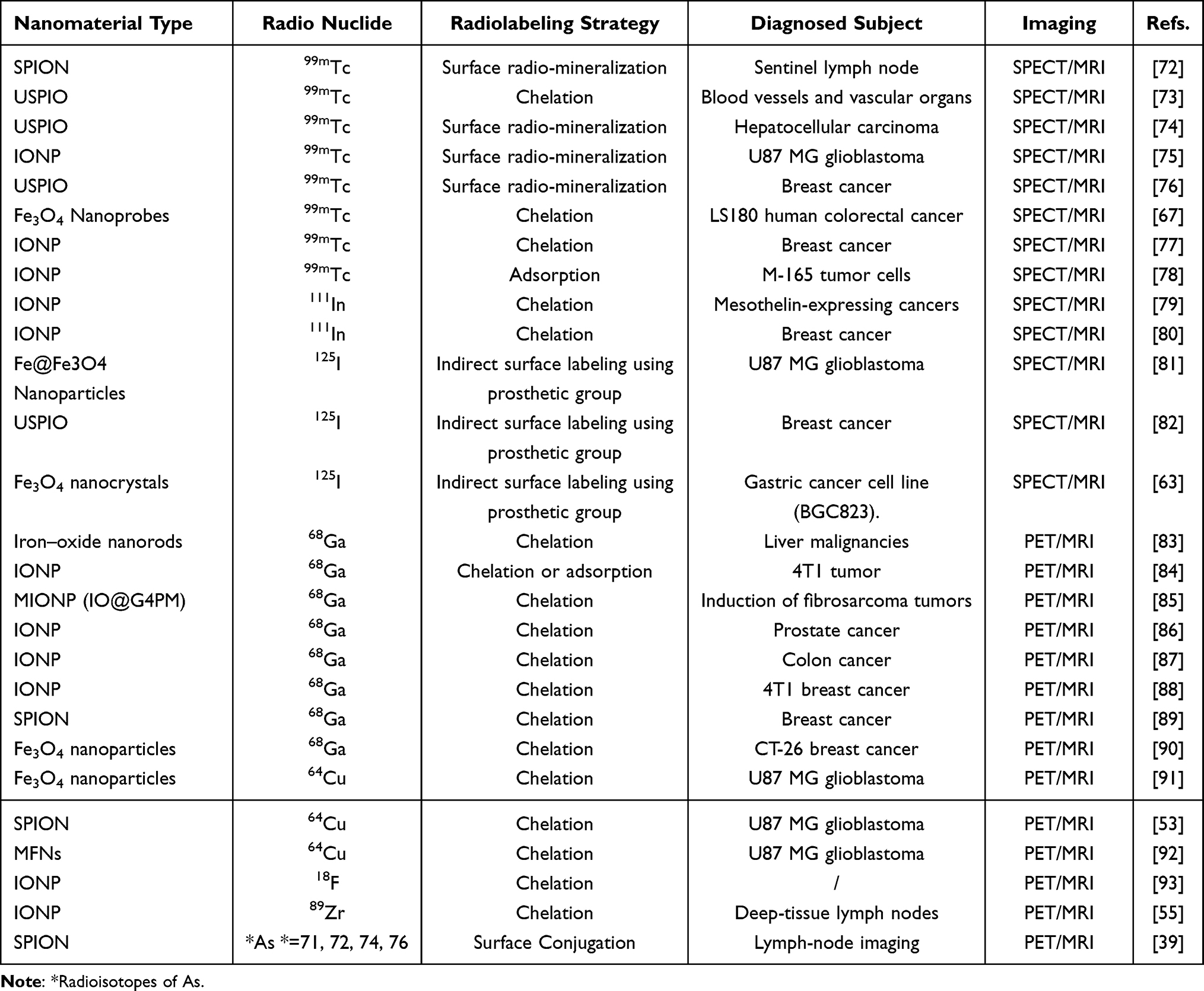 |
Table 2 Table Summarizes the Application of Iron-Based Nanomaterials in Dual-Modal Imaging |
99mTc (t1/2 = 6 h) is the most commonly used radionuclide in SPECT, accounting for over 80% of diagnostic radiopharmaceuticals in current nuclear imaging. 99mTc-based radioactive iron-based nanoparticles play a significant role in tumor diagnosis. For example, Rosales et al used radiolabeled bisphosphonate (BP) conjugated to the surface of ultrasmall superparamagnetic iron oxide (USPIO) nanoparticles for T1-weighted MRI-SPECT multimodal imaging.73 The resulting nanoparticles demonstrated good stability in water or saline, with a near-zero potential at neutral pH. Longitudinal (r1) and transverse (r2) relaxivity values measured at a clinically relevant magnetic field (3 T) showed an r1 of 9.5 mM−1s−1 and an r2/r1 ratio of 2.97, making these USPIOs attractive as T1-weighted MRI contrast agents under high magnetic fields. Compared to non-functionalized USPIOs, these nanoparticles achieved comparable signal enhancement using only a quarter of the dose. Furthermore, the nanoparticles exhibited a long circulation time (t1/2 = 2.97 h), allowing high-spatial-resolution visualization of vasculature and vascular organs (Figure 2).
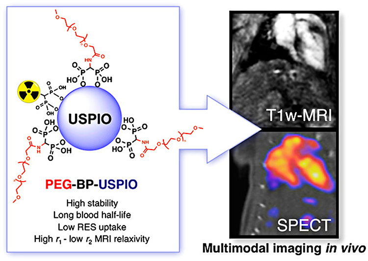 |
Figure 2 Showing a new stealth PEG polymer conjugate containing a terminal 1,1-bisphosphonate (BP) group for strong and stable binding to the surface of ultrasmall-superparamagnetic oxide nanomaterials (USPIOs). This polymer, PEG(5)-BP-USPIO Can be used for in vivo Multimodal (T1 MRI-SPECT) imaging through radioactive labeling technology. Reproduced with permission from Ref.,73 © 2012 American Chemical Society. |
Gao et al developed a tumor-microenvironment-responsive SPECT/MRI dual-modal probe for enhanced tumor imaging67 (Figure 3). In this work, a tumor-specific Arg-Gly-Asp (RGD) peptide for tumor targeting and a “self-marking” self-peptide were linked via a disulfide bond. The self-peptide helps the nanoprobes evade uptake by the reticuloendothelial system (RES) before reaching the tumor site via blood circulation. Upon reaching the tumor site, the disulfide bond is cleaved by the high concentration of glutathione (GSH) in the tumor microenvironment. Subsequently, the RGD anchors the nanoprobes to the surface of cancer cells highly expressing the RGD receptor αvβ3. The exposed thiol groups then react with maleimide residues on another adjacent particle to crosslink the particles, thereby significantly improving the contrast enhancement capability of the nanoprobes. Both in vitro and in vivo experiments demonstrated that the aggregates significantly improved the MRI contrast enhancement performance of the Fe3O4 particles. These results demonstrate the potential of radiolabeled iron-based nanoparticles as targeted multimodal imaging agents for molecular imaging.
 |
Figure 3 Schematic drawing of the antiphagocytosis 99mTc-labeled Fe3O4 nanoparticles and their responsiveness to GSH-triggering within tumor microenvironment for forming particle aggregates through interparticle crosslinking reaction. Reproduced with permission from Ref.,67 © 2017 WILEY-VCH Verlag GmbH & Co. KGaA, Weinheim. |
In addition to 99mTc, other commonly used gamma-emitting radionuclides such as 111In, 123I, 125I, 67Ga and 177Lu have been explored for SPECT imaging. Among these, radioiodine (123I and 131I) can be used for diagnosing thyroid cancer, 67Ga for imaging lymphoma, and 111In has been used for diagnosing neuroendocrine tumors. Furthermore, Lutetium-177 (177Lu) is not only suitable as a radionuclide for SPECT imaging, but also applicable for therapeutic applications due to its low-energy (0.49 MeV) beta-minus (β−) emission and gamma emissions (208 keV (11%), 113 keV (6.6%)).94
Compared to SPECT, PET imaging offers higher accuracy, sensitivity, and resolution. Hybrid imaging combining high-resolution MRI and PET may provide a better solution for future early cancer diagnosis. Thanks to the multifunctional surface properties of the materials, iron-based nanomaterials can be used to prepare imaging probes by radiolabeling with PET imaging nuclides.
68Ga (t1/2 = 68 min) is an ideal PET imaging radionuclide. Its short half-life ensures reduced radiation exposure for patients. Furthermore, due to its excellent chemical properties, it can form stable complexes with various nanoparticles.95 For example, Jeong et al developed a prostate-specific membrane antigen (PSMA)-targeted iron-based nanoparticle. Iron oxide nanoparticles were encapsulated with three amphiphiles containing PEG, DOTA, and a PSMA-targeting ligand in an aqueous medium. PET and MRI imaging results showed that, in a dual-tumor xenograft mouse model study, the contrast agent was selectively taken up only by 22Rv1 (PSMA-positive) tumors, not by PC-3 (PSMA-negative) tumors. The dual-modal imaging not only exhibited high resolution but also provided quantitative information. Therefore, 68Ga-DOTA-IO-GUL is a promising dual-mode agent for imaging prostate cancer (Figure 4).86
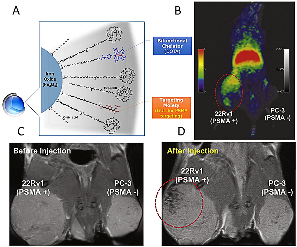 |
Figure 4 (A) showing the diagram of encapsulated DOTA-IO-GUL. (B) In vivo PET result. PSMA-selective uptake result in micro PET imaging. (SUVmean = 0.668) 68Ga-DOTA-IO-GUL (10.2 MBq, 0.1 mL) tail vein injection after 1 h imaging. Left tumor is PSMA-positive (22Rv1) and right tumor is PSMA-negative (PC-3). Red circle demonstrates the selective uptake of DOTA-IO-GUL to the left positive tumor. (C) In vivo micro MRI image results. Before injection, control image; (D) after injection of DOTA-IO-GUL (0.1 mL, 0.2 mM) through the tail vein (1 h). Left tumor is PSMA positive (22Rv1) and right tumor is PSMA-negative (PC-3). Red circle demonstrates the selective uptake of DOTA-IO-GUL to the positive tumor (49.1 Fe ng/g). Reproduced with permission from Ref.,86 © 2015 The Authors Published by Elsevier Inc. |
64Cu (t1/2 = 12.7 h) is a commonly used positron emitter that can be attached to nanoparticles via coordination with chelators. Shen et al developed a PET/MRI dual-modal imaging agent based on SPIONs.91 They prepared the PET/MRI dual-modal imaging probe by attaching 64Cu to the surface of DOTA-modified SPIONs. α(v)β (3)-targeting RGD (Arg-Gly-Asp) peptides were further conjugated to the SPIONs along with 64Cu to achieve highly specific tumor targeting. Subsequently, using human glioblastoma tumor-bearing mice expressing αvβ3 as the animal model, MRI and MicroPET were applied to observe the in vivo behavior of the probe. These results indicated that this probe possesses good tumor targeting capability and is a potential PET/MR imaging tool.
18F (t1/2 = 110 min) is the most commonly used PET radionuclide in clinical practice, typically labeled onto organic molecules. Due to its strong affinity for Al3⁺ ions, it offers convenience for labeling nanomaterials. Cheng et al successfully prepared an 18F-AlF-labeled dual-modal probe by reacting Al3⁺ ions and radioactive 18F− ions with NOTA-conjugated IONPs.93 Thorek et al prepared 89Zr-radiolabeled iron oxide nanoparticles, namely 89Zr-ferumoxytol, for lymphatic system imaging.85 Cai’s group developed a chelator-free method for radiolabeling SPIONs with radioactive arsenic (*AsIII).39 Owing to the strong affinity of arsenic for magnetic materials, *AsIII is firmly adsorbed onto the SPION surface. Following intravenous injection of free *As and *As-IONP, PET imaging was performed to investigate the biodistribution of *As-IONP in Balb/C mice. The results revealed that free *As exhibited high bladder uptake at 0.5 h and 3 h post-injection. *As-IONP underwent hepatic and splenic clearance, with the significant bladder signal likely corresponding to *As desorption from nanoparticles in vivo.
PET/Fluorescence Dual-Modality Imaging
Fluorescence imaging is clinically used to differentiate pathological tissues from normal tissues and to guide surgical resection, and it has been widely applied in tumor diagnosis and treatment.96 It is categorized into visible light imaging and near-infrared (NIR) imaging, with NIR imaging being more commonly used due to its deeper tissue penetration. Based on wavelength, NIR imaging can be further divided into NIR-I (650–950 nm) and NIR-II (1000–1700 nm) fluorescence imaging.97 Compared to visible light and NIR-I fluorescence imaging, NIR-II fluorescence imaging utilizes longer excitation and emission wavelengths. This reduces scattering of both incident and emitted light by biological tissues, resulting in higher spatial resolution and greater penetration depth.98,99 Furthermore, significantly lower autofluorescence occurs in NIR-II imaging due to reduced tissue absorption of NIR-II photons.100 Consequently, NIR fluorescence imaging has become a prominent research focus in in vivo fluorescence imaging in recent years.
Radiolabeled iron-based nanomaterials conjugated with fluorescent dyes can be engineered into PET/fluorescence dual-modality imaging probes.101 For instance, Weissleder et al functionalized aminated dextran-coated iron oxide nanoparticles by “clicking” fluorescent dyes, the PET radionuclide 18F, and targeting ligands onto the material. This probe was used for PET/FMT dual-modality imaging of macrophages in BALB/c mice bearing CT26 colon carcinoma tumors on their flanks. In vitro and in vivo results demonstrated excellent correlation between FMT and PET in terms of probe concentration (r2 > 0.99) and spatial signal distribution (r2 > 0.85), indicating strong quantitative capabilities. These findings suggest that FMT can serve as a valuable imaging modality complementary to radionuclide-based techniques.58
Tri-Modality or Multimodality Imaging
Building on the successful application of dual-modality imaging probes in tumor diagnosis, researchers have developed tri-modality or multimodality imaging probes to integrate their respective advantages and obtain complementary diagnostic information. For instance, tri-modality imaging materials have been reported, such as PET/NIRF/MRI,102 PET/PAI/MRI,103,104 SPECT/PAI/MRI,105 Optical/PET/MRI,60 and SPECT/OFI/MRI.106 Quad-modality imaging materials, such as PET/NIRF/PAI/CT,107 have also been documented.
Chen et al modified the surface of IONPs with dopamine to generate nanoconjugates that can be readily encapsulated within a human serum albumin (HSA) matrix-a clinically utilized drug carrier.102 The HSA-coated IONPs (HSA-IONPs), dual-labeled with 64Cu-DOTA and Cy5.5, were tested in a subcutaneous U87 MG xenograft mouse model. In vivo PET/NIRF/MRI tri-modality imaging demonstrated that HSA-IONPs exhibited high retention, favorable extravasation efficiency, and low macrophage uptake in tumor regions.
Liu et al functionalized IONPs and self-assembled them onto the surface of two-dimensional molybdenum disulfide (MoS2) nanosheets via thiol chemistry, followed by 64Cu adsorption onto the nanoparticles.103 PET/PAI/MRI imaging in 4T1 tumor-bearing mice revealed that the hybrid nanoparticles possessed excellent serum stability and effective tumor retention. This combined imaging approach not only provided physiological information about the tumor but also enabled the non-invasive acquisition of molecular and anatomical data at varying depths.
Yoo et al developed an Optical/PET/MRI tri-modality imaging probe leveraging the Cherenkov radiation (CR) phenomenon.60 Cherenkov radiation is light emitted when charged particles traverse a dielectric medium at a speed greater than that of light in the same medium. While Cherenkov luminescence (CL) imaging is limited by low penetration depth due to its short wavelength, its integration with nuclear imaging effectively overcomes this limitation. The study combined a PET radionuclide (124I) with an MRI probe to fabricate a tri-modality nanoprobe. This probe enabled accurate imaging of sentinel lymph nodes (SLNs), and by eliminating the need for an external light source, it effectively avoided background interference from external fluorescence.
A representative example of quad-modality imaging probes was reported by Liu et al, who modified Bi2Se3 nanosheets with FeSe2 nanoparticles (FeSe2/Bi2Se3) and radiolabeled them with 64Cu for PET/MRI/CT/PAI multimodal imaging in tumor-bearing mice.107 The probe exhibited a high *r*2 relaxivity, strong X-ray attenuation capability, and intense NIR absorption. At 24 h post-injection, the tumor site showed: darkening effects on MRI, significantly increased CT Hounsfield unit values, enhanced photoacoustic signals, and pronounced tumor contrast in PET. These concordant imaging results demonstrated pronounced tumor accumulation of FeSe2/Bi2Se3-PEG via the EPR effect.
Applications of Nanomaterials for Cancer Treatment
In recent years, iron-based nanomaterials have played a pivotal role in cancer therapy owing to their exceptional functional versatility and biosafety.16 Their magnetic properties, biocompatibility, and ease of functionalization enable diverse therapeutic applications—from targeted drug delivery to hyperthermia- and immunotherapy-based strategies. Inherent magnetism ensures precise tumor-site localization, conferring superior targeting capabilities. Furthermore, their catalytic activity (eg, in Fenton reactions) allows efficient generation of reactive oxygen species (ROS) for oxidative stress-induced tumor cell death.108 Radiolabeled iron-based nanomaterials can also incorporate organic molecules or other ions to create multifunctional nanoplatforms for multimodal theranostics, enhancing radiotherapeutic efficacy while reducing radiation dosage. This section highlights recent advances in radiolabeled iron-based nanoplatforms for cancer treatment, focusing on TRT and its synergistic applications in MHT, PDT, CDT, and immunotherapy, with emphasis on underlying mechanistic principles.
Targeted Radionuclide Therapy (TRT)
TRT is an emerging therapeutic approach that delivers radionuclides specifically to tumor-associated targets. Due to its efficacy and safety, TRT has evolved into a promising cancer treatment.109 The demand for TRT in oncology is growing rapidly, and the FDA has approved more than 50 radiopharmaceuticals.110 MRI, a widely used clinical tool, offers excellent tissue penetration, high sensitivity, and precise imaging capabilities. It plays a crucial role not only in tumor diagnosis but also in monitoring the accumulation of therapeutic agents within tumors, thereby guiding radionuclide therapy in real time.111
In 2024, Gao et al reported a multifunctional iron oxide nanomedicine-based TRT system. Beyond enhancing MRI contrast for imaging, this nanodrug also exhibits mucoadhesive properties and bioorthogonal functionality. Specifically, magnetic iron oxide nanoparticles were coated with hyaluronic acid co-labeled with Dibenzocyclooctyne (DBCO) and 177Lu. Upon intravesical instillation, DBCO groups on the nanoparticle surface underwent click reactions with azide groups on engineered cancer cells, enabling bioorthogonal targeting and efficient internalization into tumor cells. MRI visualized bladder cancer lesions, and significant tumor growth inhibition was observed. The results demonstrated the nanoprobes’ exceptional ability to reduce tumor size and downstage bladder tumors. This study provides a valuable strategy for integrating MRI with therapeutic radionuclides to achieve theranostics—a unified diagnostic and therapeutic approach112 (Figure 5).
 |
Figure 5 A scheme showing the iron oxide nanoparticle was used as a carrier that was coated with hyaluronic acid (HA) for facilitating mucosa penetration. Dibenzocyclooctyne (DBCO) was introduced into the HA coating layer to react through bioorthogonal reaction with azide as an artificial receptor of bladder cancer cells, to improve the cellular internalization of the nanoprobe labeled with 177Lu. Through magnetic resonance imaging, the targeted imaging of both nonmuscle-invasive bladder cancer (NMIBC) and muscle-invasive bladder cancer (MIBC) was realized after intravesical instillation of the multifunctional probe, Reproduced with permission from Ref.,112 © 2024 American Chemical Society. |
Combination of RT and PTT
PTT is an effective non-invasive cancer treatment113 that utilizes heat generated from photothermal agents (PTAs) irradiated by specific wavelengths of light to eliminate tumor cells while sparing healthy tissues. Traditional radiotherapy relies on oxygen within the tumor microenvironment; however, hypoxia and insufficient blood supply in solid tumors limit RT efficacy. Combining PTT with RT substantially enhances therapeutic outcomes. A novel cancer treatment technology has emerged that leverages metallic nanostructures to absorb NIR light through surface plasmon resonance (SPR) and convert it into heat via PTT. An intriguing example of this strategy was reported by Liu et al in 2016107 (Figure 6). The study developed a unique and simple method to construct novel two-dimensional FeSe2-decorated Bi2Se3 nanosheets. These nanomaterials were then labeled with the radioisotope 64Cu using a chelator-free approach, demonstrating excellent biocompatibility and efficient tumor accumulation. The further combination of near-infrared laser and X-ray irradiation enabled a synergistic effect between photothermal therapy and radiotherapy in vivo. The combined treatment group demonstrated significant cooperative tumor destruction and highly effective inhibition of tumor growth, fully highlighting the substantial application value of the composite material. After systemic administration, the material was rapidly excreted with minimal retention within 30 days, and no obvious toxic or side effects were observed. These results indicate promising clinical translation potential for this material.
 |
Figure 6 A scheme showing the structure of FeSe2/Bi2Se3 composite-nanostructure for multimodal imaging and photothermal-radiation combined tumor therapy. Reproduced with permission from Ref.,107 © 2016 WILEY-VCH Verlag GmbH & Co. KGaA, Weinheim. |
Combination of RT and MHT
Magnetic hyperthermia therapy involves exposing magnetic nanomaterials (such as iron oxide) to an appropriate alternating magnetic field, utilizing the generated heat to kill tumor cells. When the materials are concentrated in the tumor region, the thermal effect is localized to that area without harming normal tissues. Thus, this method offers advantages such as excellent selectivity, biocompatibility, and deep tissue penetration. In 2010, it was officially approved for clinical use.114 When combined with radiotherapy, the two modalities integrate their strengths and overcome the limitations of individual therapies, opening new avenues for cancer treatment.
Zhang et al constructed a novel combined therapeutic system for liver cancer using PEI-Mn0.5Zn0.5Fe2O4 nanoparticles (PEI-MZF-NPs) as both a magnetic medium and gene delivery vector.115 They evaluated the therapeutic efficacy and safety of the pHRE-Egr1-HSV-TK/131I-antiAFP McAb-GCV/MFH system against liver cancer in vitro and in vivo. Due to the excellent magnetic properties of the nanoparticles, they generate heat under an alternating magnetic field, enabling hyperthermia therapy. Meanwhile, 131I not only kills liver cancer cells but also activates the Egr1 promoter, initiating gene therapy. The anti-AFP monoclonal antibody ensures targeted treatment, thereby achieving multi-modal targeted killing of liver cancer through gene therapy, radionuclides, and hyperthermia.
Stanković et al conjugated 131I-radiolabeled CC49 antibodies to SPIONs via the reactive groups of 3-aminopropyltriethoxysilane (APTES).116 After intratumoral administration in NOD-SCID mice bearing LS174T human colon adenocarcinoma xenografts, the nanoparticles exhibited specific and prolonged local retention. Compared to the untreated group, the combined therapy induced significant tumor growth inhibition. Histological analysis confirmed necrosis and apoptosis in tumor cells without systemic toxicity.
In a recent study, Bilewicz et al synthesized SPIONs coated with a radioactive gold (198Au) layer, designed for multimodal hepatocellular carcinoma (HCC) treatment combining radionuclide therapy and magnetic hyperthermia.117 The synthesized nanoparticles exhibited superparamagnetic properties with a saturation magnetization of 50 emu/g, reaching a temperature of 43°C under a magnetic field frequency of 386 kHz. In vitro results demonstrated significant cytotoxicity, with cell viability dropping below 8% after 72 h at a radioactive concentration of 2.5 MBq/mL.
Radionuclide Therapy-Enhanced Cherenkov Photodynamic Therapy
PDT is a minimally invasive medical technology with broad application prospects, already utilized in both fundamental research and clinical settings for treating various diseases, including fungal infections, skin conditions, and multiple cancers.118,119 However, conventional external light irradiation in PDT often suffers from rapid tissue attenuation, limiting its clinical use primarily to superficial lesions. To overcome this limitation, various nano-photosensitizers have been engineered to enable PDT activation using different excitation sources, including NIR light,120,121 X-ray radiation,122 and self-illumination.123 Among these, Cherenkov radiation -induced PDT-where Cherenkov luminescence (CL) generated by radionuclides activates the photosensitizer to produce ROS-represents a novel PDT approach that eliminates the need for external light excitation.124 Combining CR with nanoparticles, particularly metal-based nanomaterials, enhances both cancer diagnosis and therapy.
Cai et al designed a magnetically targeted nanostructure based on high-performance magnetic (Zn0.4Mn0.6)Fe2O4 nanoparticles (MNPs), surface-conjugated with meso-tetra(4-carboxyphenyl)porphyrin (TCPP) and chelator-free-labeled 89Zr (89Zr-MNP/TCPP), for magnetically enhanced CR-induced PDT.125 The 89Zr-labeled MNP surface enables PET imaging to track in vivo distribution, while the excited TCPP generates singlet oxygen to destroy tumor cells. Results demonstrate that 89Zr-MNP/TCPP nanostructures serve as an efficient magnetic carrier, achieving high tumor accumulation under an external magnetic field, thereby significantly enhancing the therapeutic efficacy of CR-induced PDT by overcoming the depth and external light dependency of conventional PDT. This magnetically targeted CR-induced PDT achieves rapid and substantial suppression of tumor growth in vivo. Moreover, this strategy integrates multiple imaging modalities, providing a highly precise tool for evaluating tumor treatment progression.
Combination of RT and Immunotherapy
The combination of PD-1/PD-L1 inhibitors with radiolabeled nanomaterials not only enables tumor radiotherapy but also enhances positive immunomodulation while suppressing negative immune resistance, achieving dual anti-tumor effects and improving survival outcomes.
For example, Tian et al synthesized FeTA nanoparticles via coordination between ferric iron (Fe3⁺) and tannic acid (TA), followed by chelation-free loading of radionuclides (131I, 90Y, 177Lu, and 225Ac) onto the nanomaterial.126 These nanoparticles were further functionalized with tetra-arm PEG-SH via a Michael addition reaction, yielding a biocompatible brachytherapy hydrogel. Finally, imiquimod (a TLR7 agonist) was loaded to synergize with anti-PD-L1 antibody therapy, triggering a potent anti-tumor immune response and inhibiting metastatic tumor growth. This combined radionuclide-immunotherapy was tested in a CT26 colon cancer metastasis model. Results demonstrated that the combination therapy effectively inhibited the growth of metastatic tumors by activating CD8⁺ T cell-mediated anti-tumor immune responses. In addition, the treatment had no significant adverse effects on healthy tissues (including the heart, liver, spleen, lungs, and kidneys). In conclusion, this imageable brachytherapy hydrogel provides a novel strategy for developing clinically translatable radiopharmaceuticals (Figure 7).
 |
Figure 7 The schematic illustration of imageable brachytherapy with chelator-free radiolabeled FeTA-PEG hydrogel combining immune checkpoint-blockade therapy to activate antitumor immune responses for inhibiting cancer metastases. Reproduced with permission from Ref.,126 © 2024 Wiley-VCH GmbH. |
Ferroptosis, a non-apoptotic form of regulated cell death (RCD) discovered by Stockwell in 2012, is characterized by the accumulation of labile iron in the cytoplasm and excessive lipid peroxidation.127 The regulation of ferroptosis offers a novel approach for cancer treatment, with advantages including the ability to combat therapy-resistant tumor cells, effectively inhibit tumor growth, and generate mild immunogenicity.128 Several small-molecule drugs have been developed as ferroptosis-inducing compounds (FINs) to trigger tumor ferroptosis. However, organic molecules face limitations such as poor solubility, nonspecific distribution, systemic toxicity, and low bioavailability.129 Consequently, researchers have designed various ferroptosis inducers based on metal complexes and nanomaterials. Among these, iron-based nanomaterials have recently shown promise for efficient ferroptosis therapy and immunotherapy.130
Recently, Sun et al developed 131I-labeled single-crystal ultrasmall iron nanoparticles (USINPs) to explore the relationship among radiotherapy, ferroptosis, and immunotherapy131 (Figure 8). The study involved the preparation of renal-clearable USINPs and 131I-aPD-L1 nanoassemblies through the affinity between fluorophenylboronic acid-modified USINPs and 131I-aPD-L1. This assembly demonstrated stable circulation in the bloodstream and excellent tumor-targeting capability. Both in vitro and in vivo experiments revealed that USINPs exhibit excellent tumor-targeting capability and effectively inhibit tumor growth. USINPs-induced ferroptosis enhanced tumor radiosensitization to 131I, while 131I-mediated RT further potentiated ferroptosis. Simultaneously, immunogenic cell death (ICD) triggered by radiotherapy and ferroptosis, when combined with PD-L1 immune checkpoint blockade therapy, exhibited robust antitumor immunity. This study provides a novel strategy to improve the accumulation of ferroptosis inducers and radiopharmaceuticals in tumors.
 |
Figure 8 USINAs(131I-aPD-L1) accumulate in the tumor sites due to the EPR effect and PD-L1 targeting and disassemble into USINPs and 131I-aPD-L1 upon extracellular ATP in the tumor microenvironment.131I-aPD-L1 bind to PD-L1 receptors on the surface of tumor cells for 131I-mediated radiopharmaceutical therapy (RPT) as well as immune checkpoint blockade (ICB) therapy. On one hand,131I promote ferroptosis via ROS generation and SLC7A11 down-regulation; on the other hand, USINPs initiate ferroptosis with decreased GPX4 to sensitize cancer cells to 131I with enhanced RPT efficacy. Combining ICB with ferroptosis and RPT further enhance antitumor immunotherapy. “↑” indicates a relative increase or accumulation of the indicator (eg, LPO, ROS), while “↓” denotes a relative decrease or reduction in the indicator (eg, GSH, cystine, cysteine). Reproduced with permission from Ref.,131 © 2024 Elsevier Ltd. |
Radionuclide Therapy-Enhanced CDT
CDT is an emerging cancer treatment strategy based on the Fenton or Fenton-like reactions of transition metals.132 It converts the abundant H2O2 in the tumor microenvironment (TME) into highly oxidizing hydroxyl radicals (·OH), which cause irreversible damage to cellular components such as lipids, proteins, and nucleic acids, ultimately leading to tumor cell death.133 Typically, the efficacy of CDT depends on the reaction rate of the introduced Fenton/Fenton-like reactions, which is influenced by factors such as the metal catalyst,134 H2O2 concentration,135 pH,136 and external energy fields. Consequently, various strategies have been designed and reported to enhance intratumoral Fenton/Fenton-like reactions by manipulating these limiting factors to improve CDT outcomes.137 However, these approaches face significant constraints due to the rate-limiting nature of the Fe3+/Fe2+ cycle, which severely restricts Fenton reaction efficiency.
Radionuclides emit various types of radiation (eg, α, β, γ rays) during decay, interacting with surrounding matter to generate low-energy electrons, such as hydrated electrons in aqueous solutions. Therefore, incorporating radionuclides into CDT nanomaterials can accelerate the reduction of transition metals from higher to lower oxidation states, thereby enhancing CDT efficacy.
In the study by Bu et al138 iron-based metal-organic framework (MOF) nanoparticles were synthesized using FeCl3 and 2-hydroxyterephthalic acid (2-HTP), followed by modification with PEG and radiolabeling with 125I. The porous structure facilitated H2O2 enrichment, boosting the Fenton reaction. Simultaneously, the presence of 125I induced continuous generation of hydrated electrons, promoting the conversion of Fe3+ to Fe2+. This mechanism led to potent antitumor effects in both in vitro and in vivo pancreatic cancer models (Figure 9). Compared to the control group, 25I-MIL-88B(Fe) NPs exhibited significant antitumor efficacy. Moreover, 125I-MIL-88B(Fe) nanoparticles did not induce acute or chronic toxicity in mice and demonstrated satisfactory in vivo biocompatibility.
 |
Figure 9 Radioiodine-induced eaq−generation promotes the conversion of Fe3+/Fe2+ in 125I-MIL-88B(Fe) NPs-mediated Fenton reaction, resulting in accelerated OH production and enhanced CDT through the ROS/MAPK/p53 pathway-mediated cell apoptosis. Reproduced with permission from Ref.,138 © 2024 Wiley-VCH GmbH. |
Summary and Outlook
Radiolabeled iron-based nanomaterials have shown great promise in tumor diagnosis and treatment. Their unique magnetism, excellent biocompatibility, and capability to integrate diagnostic and therapeutic modalities make them invaluable tools for overcoming the limitations of conventional cancer approaches. From enhancing imaging sensitivity and specificity in MRI applications to enabling precise and minimally invasive therapies—such as magnetic hyperthermia, photodynamic therapy, chemodynamic therapy, and immunotherapy—these materials contribute significantly to precision diagnosis and treatment of cancer. Moreover, the integration of nanomaterials with advanced technologies like radiomics offers direction for next-generation cancer treatment strategies. Despite these encouraging achievements, radiolabeled iron-based nanomaterials face challenges in transitioning from basic research to clinical trials.
Biosafety of Nanomaterials
The potential for large-scale clinical application of nanomaterials predominantly hinges on their biosafety,139 as the sustainable advancement of nanomedicine relies on resolving nanotoxicological and biosecurity concerns. Iron-based nanomaterials, as inorganic metallic compounds, may induce chemical toxicity mediated by iron ions during practical use. These materials can accumulate in normal organs (eg, kidneys, liver, spleen), leading to prolonged in vivo retention in healthy tissues and consequent systemic side effects. Additionally, the retention of radioactive components may pose risks of radiation-induced damage. To address these challenges, we propose two strategic approaches: 1) Surface Engineering: Modifying material surfaces through techniques such as PEGylation to alter bio-interactions, thereby minimizing protein adsorption and extending circulation time to reduce in vivo toxicity. 2) Biodegradable Design: Developing intelligently engineered iron-based nanoprobes with controlled in vivo degradability. These materials should efficiently accumulate in target tissues while undergoing programmed degradation after completing imaging or therapeutic functions—requiring careful reconciliation of the trade-off between degradability and stability.
Scalable Production of Materials
Successful clinical integration of radiolabeled nanomaterials necessitates robust validation protocols to verify material stability, therapeutic efficacy, and safety profiles under clinically relevant conditions. Concurrently, synthesis and radiolabeling procedures must be simple, reproducible, and scalable with relatively low production costs. Pharmacological and toxicological parameters must demonstrate strict consistency across different batches of radiolabeled products. Therefore, the transition from preclinical studies to clinical application has been slow but this can be addressed by adopting standardized preclinical validation systems and Good Manufacturing Practice (GMP) to ensure safety and efficacy.140,141 A critical approach to addressing these challenges involves developing kit-based production systems, enabling repeatable, large-scale manufacturing to ensure safe clinical deployment.
Development of Novel Nanomaterials
As research advances, greater emphasis should be placed on constructing novel nanomaterials with innovative functionalities to meet personalized theranostic demands. For instance, integrating multifunctional modules into materials enables diagnostic-therapeutic integration.142 Furthermore, selecting endogenous components as surface modifiers presents a superior approach to reducing adverse reaction risks—such as utilizing biomimetic membrane camouflage to enhance nanomaterial targeting and biocompatibility.143 Ultimately, integrating artificial intelligence (AI) and big data into cancer diagnosis and treatment processes offers compelling advantages.144 These technologies will transform drug discovery, optimize personalized therapeutic strategies, and significantly enhance safety-efficacy profiles. By leveraging AI algorithms like Artificial Neural Networks (ANN), researchers can simulate critical parameters including particle size, zeta potential, and encapsulation efficiency to synthesize nanoparticles with optimized therapeutic performance.145–147
Conclusions
This review has systematically explored the burgeoning field of radiolabeled iron-based nanomaterials for cancer theranostics. The most compelling benefits of these platforms include: (1) Multimodal Capabilities: The intrinsic properties of iron enable high-quality MRI, while radiolabeling provides sensitive PET/SPECT, allowing for anatomical, functional, and molecular imaging in a single agent. (2) Synergistic Therapeutics: Iron-based cores serve as hubs for combining radiotherapy with PTT, MHT, CDT, and immunotherapy, often leading to supra-additive antitumor effects. (3) Clinical Translation Potential: The established safety profile of iron oxide nanoparticles, coupled with FDA approval for some formulations, provides a significant head start for clinical adoption compared to many other nanoplatforms.
However, significant challenges remain to be addressed: (1) Long-Term Fate and Safety: The chronic biodistribution, degradation, and clearance pathways of these complex nanomaterials, especially when combined with radioactive components, require more comprehensive long-term studies. (2) Manufacturing and Quality Control: Reproducible, large-scale, and cost-effective synthesis of radiolabeled nanomaterials with stringent quality control for clinical use is non-trivial. (3) Targeting Efficiency: While passive targeting via the EPR effect is valuable, active and specific tumor targeting needs improvement to reduce off-target accumulation and enhance therapeutic indices. (4) Personalization: Developing strategies to select the right nanoplatform (material type, radionuclide, combination therapy) for the right patient based on tumor biology is a key future direction.
Future research should prioritize the development of “smart” nanomaterials with biodegradable backbones, stimuli-responsive activation, and enhanced tumor-specific targeting. Furthermore, the integration of artificial intelligence for nanomaterial design, treatment planning, and outcome prediction holds immense promise. With continued interdisciplinary efforts focusing on these challenges, radiolabeled iron-based nanomaterials are poised to make the critical leap from promising laboratory constructs to transformative clinical cancer therapies.
Abbreviations
SPECT, Single photon emission computed tomography; PET, Positron emission tomography; MRI, Magnetic resonance imaging; TRT, Targeted radiotherapy; RT, Radionuclide therapy; PTT, Photothermal therapy; MHT, Magnetic hyperthermia therapy; PDT, Photodynamic therapy; IT, Immunotherapy; CDT, Chemodynamic therapy; WHO, World Health Organization; CT, Computed tomography; FDA, U.S. Food and Drug Administration; SRM, Surface radio-mineralization; HIR, Heat-induced radiolabeling; BFCs, Bifunctional chelators; Crgd, cyclic arginine-glycine-aspartic acid; SPIO, Superparamagnetic iron oxide; PEG, Polyethylene glycol; EDTA, Ethylenediaminetetraacetic acid; DFO, Desferrioxamine; SPIONs, Superparamagnetic iron oxide nanoparticles; IONPs Iron oxide nanoparticles; BSA, Bovine serum albumin; US, Ultrasound; BP, Bisphosphonate; USPIO, Ultrasmall-superparamagnetic oxide nanomaterials; MIONP, Multifunctional magnetic iron oxide nanoparticles; MFNs, Manganese ferrite nanoparticles; RES, Reticuloendothelial system; GSH, Glutathione; PSMA, Prostate-specific membrane antigen; NIR, Near-infrared; FMT, Fluorescence mediated tomography; NIFR, Near-infrared fluorescence; PAI, Photoacoustic Imaging; OFI, Optical fluorescence Imaging; HAS, Human serum albumin; CR, Cherenkov radiation; CL, Cherenkov luminescence; SLNs, Sentinel lymph nodes; ROS, Reactive oxygen species; DBCO, Dibenzocyclooctyne; NMIBC, Nonmuscle-invasive bladder cancer; MIBC, Muscle-invasive bladder cancer; PTAs, Photothermal agents; SPR, Surface plasmon resonance; APTES, 3-aminopropyltriethoxysilane; HCC, Hepatocellular carcinoma; TCPP, Meso-tetra(4-carboxyphenyl)porphyrin; TA, Tannic acid; RCD, Regulated cell death; FINs, Ferroptosis-inducing compounds; USINPs, Ultrasmall iron nanoparticles; ICD, Immunogenic cell death; RPT, Radiopharmaceutical therapy; ICB, Immune checkpoint blockade; TME, Tumor microenvironment; MOF, Metal-organic framework; 2-HTP, 2-hydroxyterephthalic acid; AI, Artificial intelligence; ANN, Artificial Neural Networks.
Data Sharing Statement
No data were used for the research described in the article.
Consent for Publication
We confirm that the details of any images can be published. Copyright permission is granted.
Acknowledgments
This work was supported by Young PhD Talents Cultivation Project of of Xinqiao Hospital of Army Medical University (No: 2025YQB003).
Author Contributions
All authors made a significant contribution to the work reported, whether that is in the conception, study design, execution, acquisition of data, analysis and interpretation, or in all these areas; took part in drafting, revising or critically reviewing the article; gave final approval of the version to be published; have agreed on the journal to which the article has been submitted; and agree to be accountable for all aspects of the work.
Funding
This work was supported by Young PhD Talents Cultivation Project of Xinqiao Hospital of Army Medical University (No: 2025YQB003).
Disclosure
The authors declare that they have no competing interests in this work.
References
1. Sgouros G, Bodei L, McDevitt MR, Nedrow JR. Radiopharmaceutical therapy in cancer: clinical advances and challenges. Nat Rev Drug Disc. 2020;19(9):589–26. doi:10.1038/s41573-020-0073-9
2. Bray F, Ferlay J, Soerjomataram I, Siegel RL, Torre LA, Jemal A. Global cancer statistics 2018: globocan estimates of incidence and mortality worldwide for 36 cancers in 185 countries. Ca A Cancer J Clin. 2018;68(6):394–424. doi:10.3322/caac.21492
3. Yassin MT, Al-Otibi FO, Al-Sahli SA, El-Wetidy MS, Mohamed S. Metal oxide nanoparticles as efficient nanocarriers for targeted cancer therapy: addressing chemotherapy-induced disabilities. Cancers. 2024;16(24):4234. doi:10.3390/cancers16244234
4. Hagiwara Y, Oike T, Niimi A, et al. Clustered DNA double-strand break formation and the repair pathway following heavy-ion irradiation. J Radiat Res. 2019;60(1):69–79. doi:10.1093/jrr/rry096
5. Karpuz M, Silindir-Gunay M, Ozer AY. Current and future approaches for effective cancer imaging and treatment. Cancer Biother Radiopharm. 2018;33(2):39–51. doi:10.1089/cbr.2017.2378
6. Bentzen SM, Constine LS, Deasy JO, et al. Quantitative analyses of normal tissue effects in the clinic (quantec): an introduction to the scientific issues. Int J Radiat Oncol Biol Phys. 2010;76(3):S3–S9. doi:10.1016/j.ijrobp.2009.09.040
7. Pei P, Liu T, Shen W, Liu Z, Yang K. Biomaterial-mediated internal radioisotope therapy. Mater Horizons. 2021;8(5):1348–1366. doi:10.1039/d0mh01761b
8. Aghanejad A, Jalilian AR, Fazaeli Y, Beiki D, Fateh B, Khalaj A. Radiosynthesis and biodistribution studies of [62Zn/62Cu]–plerixafor complex as a novel in vivo PET generator for chemokine receptor imaging. J Radioanal Nucl Chem. 2013;299(3):1635–1644. doi:10.1007/s10967-013-2822-2
9. Mirzaei A, Jalilian AR, Aghanejad A, et al. Preparation and evaluation of 68ga-ecc as a pet renal imaging agent. Nuclear Med Mol Imag. 2015;49(3):208–216. doi:10.1007/s13139-015-0323-7
10. Ritt P. Recent developments in SPECT/CT. Seminars Nuclear Med. 2022;52(3):276–285. doi:10.1053/j.semnuclmed.2022.01.004
11. Zhang S, Wang X, Gao X, et al. Radiopharmaceuticals and their applications in medicine. Signal Transduction Targeted Ther. 2025;10(1):1. doi:10.1038/s41392-024-02041-6
12. Pellico J, Gawne PJ, de Rosales R TM. Radiolabelling of nanomaterials for medical imaging and therapy. Chem Soc Rev. 2021;50(5):3355–3423. doi:10.1039/d0cs00384k
13. Sun N, Wang T, Zhang S. Radionuclide-labelled nanoparticles for cancer combination therapy: a review. J Nanobiotechnol. 2024;22:728. doi:10.1186/s12951-024-03020-3
14. Roy I, Krishnan S, Kabashin AV, Zavestovskaya IN, Prasad PN. Transforming nuclear medicine with nanoradiopharmaceuticals. ACS Nano. 2022;16(4):5036–5061. doi:10.1021/acsnano.1c10550
15. Papadimitroulas P, Loudos G, Nikiforidis GC, Kagadis GC. A dose point kernel database using GATE Monte Carlo simulation toolkit for nuclear medicine applications: comparison with other monte carlo codes. Med Phys. 2012;39(8):5238–5247. doi:10.1118/1.4737096
16. Bodei L, Herrmann K, Schöder H, Scott AM, Lewis JS. Radiotheranostics in oncology: current challenges and emerging opportunities. Nat Rev Clin Oncol. 2022;19(8):534–550. doi:10.1038/s41571-022-00652-y
17. Bollineni VR, Collette S, Liu Y. Functional and molecular imaging in cancer drug development. Chin Clin Oncol. 2014;3(2):17. doi:10.3978/j.issn.2304-3865.2014.05.05
18. Ramadoss K, Vadivel V, A V, L K. Magnetic nanoparticle-based approaches in cancer therapy–a critical review. Int J Appl Pharm. 2022;14(6):21–27. doi:10.22159/ijap.2022v14i6.45064
19. Viswanath D, Won -Y-Y. Combining radiotherapy (RT) and photodynamic therapy (PDT): clinical studies on conventional RT-PDT approaches and novel nanoparticle-based RT-PDT approaches under preclinical evaluation. ACS Biomat Sci Eng. 2022;8(9):3644–3658. doi:10.1021/acsbiomaterials.2c00287
20. Stéen EJL, Edem PE, Nørregaard K, et al. Pretargeting in nuclear imaging and radionuclide therapy: improving efficacy of theranostics and nanomedicines. Biomaterials. 2018;179:209–245. doi:10.1016/j.biomaterials.2018.06.021
21. Nabi PN, Vahidfar N, Tohidkia MR, Hamidi AA, Omidi Y, Aghanejad A. Mucin-1 conjugated polyamidoamine-based nanoparticles for image-guided delivery of gefitinib to breast cancer. Int J Biol Macromol. 2021;174:185–197. doi:10.1016/j.ijbiomac.2021.01.170
22. Chen G, Roy I, Yang C, Prasad PN. Nanochemistry and nanomedicine for nanoparticle-based diagnostics and therapy. Chem Rev. 2016;116(5):2826–2885. doi:10.1021/acs.chemrev.5b00148
23. Farokhzad OC, Langer R. Impact of nanotechnology on drug delivery. ACS Nano. 2009;3(1):16–20. doi:10.1021/nn900002m
24. Nie S, Xing Y, Kim GJ, Simons JW. Nanotechnology applications in cancer. Annual Rev Biomed Eng. 2007;9(1):257–288. doi:10.1146/annurev.bioeng.9.060906.152025
25. Smith BR, Gambhir SS. Nanomaterials for in vivo imaging. Chem Rev. 2017;117(3):901–986. doi:10.1021/acs.chemrev.6b00073
26. Silindir-Gunay M, Ozer AY. Nanosized drug delivery systems as radiopharmaceuticals. In: Nanostructures for Cancer Therapy. 2017:563–592.
27. Suman SK, Subramanian S, Mukherjee A. Combination radionuclide therapy: a new paradigm. Nuclear Med Biol. 2021;98-99:40–58. doi:10.1016/j.nucmedbio.2021.05.001
28. Cui M, Zhu M, Tang D, Cao Z, Xiao H, Shang K. Nanotheranostics with radionuclides for cancer diagnosis and therapy. Adv Funct Mater. 2025;35(23):2423445. doi:10.1002/adfm.202423445
29. Dadfar SM, Roemhild K, Drude NI, et al. Iron oxide nanoparticles: diagnostic, therapeutic and theranostic applications. Adv Drug Delivery Rev. 2019;138:302–325. doi:10.1016/j.addr.2019.01.005
30. Ogbezode JE, Ezealigo US, Bello A, Anye VC, Onwualu AP. A narrative review of the synthesis, characterization, and applications of iron oxide nanoparticles. Discover Nano. 2023;18:125. doi:10.1186/s11671-023-03898-2
31. Wu X, Zhang H. Therapeutic strategies of iron-based nanomaterials for cancer therapy. Biomed Mater. 2021;16:032003. doi:10.1088/1748-605X/abd0c4
32. Zhou H, Guo M, Li J, et al. Hypoxia-Triggered self-assembly of ultrasmall iron oxide nanoparticles to amplify the imaging signal of a tumor. J Am Chem Soc. 2021;143(4):1846–1853. doi:10.1021/jacs.0c10245
33. Wang L, Zhang X, He L, et al. Iron-based nanomaterials for modulating tumor microenvironment. WIREs Nanomed Nanobiotechnol. 2025;17(1):e70001. doi:10.1002/wnan.70001
34. Ren X, Zheng R, Fang X, et al. Red blood cell membrane camouflaged magnetic nanoclusters for imaging-guided photothermal therapy. Biomaterials. 2016;92:13–24. doi:10.1016/j.biomaterials.2016.03.026
35. Ge J, Zhang Q, Zeng J, Gu Z, Gao M. Radiolabeling nanomaterials for multimodality imaging: new insights into nuclear medicine and cancer diagnosis. Biomaterials. 2020;228:119553. doi:10.1016/j.biomaterials.2019.119553
36. Lemaître TA, Burgoyne AR, Ooms M, Parac-Vogt TN, Cardinaels T. Inorganic radiolabeled nanomaterials in cancer therapy: a review. ACS Appl Nano Mater. 2022;5(7):8680–8709. doi:10.1021/acsanm.2c01204
37. Roldan Cuenya B, Croy JR, Mostafa S, Behafarid F, et al. Solving the structure of size-selected Pt nanocatalysts synthesized by inverse micelle encapsulation. J Am Chem Soc. 2010;132(25):8747–8756. doi:10.1021/ja101997z
38. Roldan Cuenya B, Behafarid F. Nanocatalysis: size- and shape-dependent chemisorption and catalytic reactivity. Surf Sci Rep. 2015;70(2):135–187. doi:10.1016/j.surfrep.2015.01.001
39. Chen F, Ellison PA, Lewis CM, et al. Chelator-free synthesis of a dual-modality PET/MRI agent. Angew Chem Int Ed. 2013;52(50):13319–13323. doi:10.1002/anie.201306306
40. Zhu J, Zhang B, Tian J, et al. Synthesis of heterodimer radionuclide nanoparticles for magnetic resonance and single-photon emission computed tomography dual-modality imaging. Nanoscale. 2015;7(8):3392–3395. doi:10.1039/c4nr07255c
41. Abdollah MRA, Carter TJ, Jones C, et al. Fucoidan prolongs the circulation time of dextran-coated Iron oxide nanoparticles. ACS Nano. 2018;12(2):1156–1169. doi:10.1021/acsnano.7b06734
42. Yuan H, Wilks MQ, Normandin MD, El Fakhri G, Kaittanis C, Josephson L. Heat-induced radiolabeling and fluorescence labeling of Feraheme nanoparticles for PET/SPECT imaging and flow cytometry. Nat Protocols. 2018;13(2):392–412. doi:10.1038/nprot.2017.133
43. Normandin MD, Yuan H, Wilks MQ, et al. Heat-induced radiolabeling of nanoparticles for monocyte tracking by PET. Angew Chem Int Ed. 2015;54(44):13002–13006. doi:10.1002/anie.201505525
44. Boros E, Bowen AM, Josephson L, Vasdev N, Holland JP. Chelate-free metal ion binding and heat-induced radiolabeling of iron oxide nanoparticles. Chem Sci. 2015;6(1):225–236. doi:10.1039/c4sc02778g
45. Patrick PS, Bogart LK, Macdonald TJ, et al. Surface radio-mineralisation mediates chelate-free radiolabelling of iron oxide nanoparticles. Chem Sci. 2019;10(9):2592–2597. doi:10.1039/c8sc04895a
46. Cui W-R, Xu W, Chen Y-R, et al. Olefin-linked cationic covalent organic frameworks for efficient extraction of ReO4−/99TcO4−. J Hazard Mater. 2023;446:130603. doi:10.1016/j.jhazmat.2022.130603
47. Hu A, Wilson JJ. Advancing chelation strategies for large metal ions for nuclear medicine applications. Acc Chem Res. 2022;55(6):904–915. doi:10.1021/acs.accounts.2c00003
48. Thiele NA, Brown V, Kelly JM, et al. An eighteen-membered macrocyclic ligand for Actinium-225 targeted alpha therapy. Angew Chem Int Ed. 2017;56(46):14712–14717. doi:10.1002/anie.201709532
49. Mewis RE, Archibald SJ. Biomedical applications of macrocyclic ligand complexes. Coord Chem Rev. 2010;254(15):1686–1712. doi:10.1016/j.ccr.2010.02.025
50. Desai P, Rimal R, Sahnoun SEM, et al. Radiolabeled nanocarriers as theranostics-advancement from peptides to nanocarriers. Small. 2022;18(25):2200673. doi:10.1002/smll.202200673
51. Li Z, Jin Q, Huang C, Dasa S, et al. Trackable and targeted phage as positron emission tomography (PET) agent for cancer imaging. Theranostics. 2011;1:371–380. doi:10.7150/thno/v01p0371
52. Wang C-F, Sarparanta MP, Mäkilä EM, et al. Multifunctional porous silicon nanoparticles for cancer theranostics. Biomaterials. 2015;48:108–118. doi:10.1016/j.biomaterials.2015.01.008
53. Yang X, Hong H, Grailer JJ, et al. cRGD-functionalized, DOX-conjugated, and 64Cu-labeled superparamagnetic iron oxide nanoparticles for targeted anticancer drug delivery and PET/MR imaging. Biomaterials. 2011;32(17):4151–4160. doi:10.1016/j.biomaterials.2011.02.006
54. Rafael torres Martin de Rosales RT, Paul RL, Jauregui-Osoro M, et al. Synthesis of 64CuII-bis(dithiocarbamatebisphosphonate) and its conjugation with superparamagnetic iron oxide nanoparticles: in vivo evaluation as dual-modality PET-MRI agent. Angew Chem Int Ed. 2011;50(24):5509–5513. doi:10.1002/anie.201007894
55. Thorek DLJ, Ulmert D, Diop N-FM, et al. Non-invasive mapping of deep-tissue lymph nodes in live animals using a multimodal PET/MRI nanoparticle. Nat Commun. 2014;5:3097. doi:10.1038/ncomms4097
56. Sharma R, Xu Y, Kim SW, et al. Carbon-11 radiolabeling of iron-oxide nanoparticles for dual-modality PET/MR imaging. Nanoscale. 2013;5(16):7476–7483. doi:10.1039/c3nr02519e
57. Nallathamby PD, Mortensen NP, Palko HA, et al. New surface radiolabeling schemes of super paramagnetic iron oxide nanoparticles (SPIONs) for biodistribution studies. Nanoscale. 2015;7(15):6545–6555. doi:10.1039/c4nr06441k
58. Nahrendorf M, Keliher E, Marinelli B, et al. Hybrid PET-optical imaging using targeted probes. Proc Natl Acad Sci. 2010;107(17):7910–7915. doi:10.1073/pnas.0915163107
59. Tang Y, Zhang C, Wang J, et al. MRI/SPECT/Fluorescent tri-modal probe for evaluating the homing and therapeutic efficacy of transplanted mesenchymal stem cells in a rat ischemic stroke model. Adv Funct Mater. 2014;25(7):1024–1034. doi:10.1002/adfm.201402930
60. Park JC, Yu MK, An GI, et al. Facile preparation of a hybrid nanoprobe for triple-modality Optical/PET/MR imaging. Small. 2010;6(24):2863–2868. doi:10.1002/smll.201001418
61. Sun H, Zhang B, Jiang X, et al. Radiolabeled ultra-small fe3o4 nanoprobes for tumor-targeted multimodal imaging. Nanomedicine. 2018;14(1):5–17. doi:10.2217/nnm-2018-0219
62. Kumar R, Roy I, Ohulchanskky TY, et al. In vivo biodistribution and clearance studies using multimodal organically modified silica nanoparticles. ACS Nano. 2010;4(2):699–708. doi:10.1021/nn901146y
63. Liu S, Jia B, Qiao R, et al. A Novel Type of Dual- modality molecular probe for MR and nuclear imaging of tumor: preparation, characterization and in vivo application. Mol Pharmaceut. 2009;6(4):1074–1082. doi:10.1021/mp900143a
64. Llop J, Jiang P, Marradi M, et al. Visualisation of dual radiolabelled poly(lactide-co-glycolide) nanoparticle degradation in vivo using energy-discriminant SPECT. J Mat Chem B. 2015;3(30):6293–6300. doi:10.1039/c5tb01157d
65. Zeng J, Jia B, Qiao R, et al. In situ 111In-doping for achieving biocompatible and non-leachable 111In-labeled Fe3O4 nanoparticles. Chem Commun. 2014;50(17):2170–2172. doi:10.1039/c3cc48948e
66. Pospisilova M, Zapotocky V, Nesporova K, et al. Preparation and biodistribution of 59Fe-radiolabelled iron oxide nanoparticles. J Nanopart Res. 2017;19:80. doi:10.1007/s11051-016-3719-0
67. Gao Z, Hou Y, Zeng J, et al. Tumor microenvironment-triggered aggregation of antiphagocytosis 99mTc-Labeled Fe3O4 nanoprobes for enhanced tumor imaging in vivo. Adv Mater. 2017;29(24):1701095. doi:10.1002/adma.201701095
68. Siddique S, Chow JCL. Application of nanomaterials in biomedical imaging and cancer therapy. Nanomaterials. 2020;10(9):1700. doi:10.3390/nano10091700
69. Heinzmann K, Carter LM, Lewis JS, Aboagye EO. Multiplexed imaging for diagnosis and therapy. Nat Biomed Eng. 2017;1(9):697–713. doi:10.1038/s41551-017-0131-8
70. Goel S, England CG, Chen F, Cai W. Positron emission tomography and nanotechnology: a dynamic duo for cancer theranostics. Adv Drug Delivery Rev. 2017;113:157–176. doi:10.1016/j.addr.2016.08.001
71. Li J, Zheng L, Cai H, et al. Polyethyleneimine-mediated synthesis of folic acid-targeted iron oxide nanoparticles for in vivo tumor MR imaging. Biomaterials. 2013;34(33):8382–8392. doi:10.1016/j.biomaterials.2013.07.070
72. Madru R, Kjellman P, Olsson F, et al. 99mTc-Labeled superparamagnetic iron oxide nanoparticles for multimodality SPECT/MRI of sentinel lymph nodes. J Nucl Med. 2012;53(3):459–463. doi:10.2967/jnumed.111.092437
73. Sandiford L, Phinikaridou A, Protti A, et al. Bisphosphonate-anchored pegylation and radiolabeling of superparamagnetic iron oxide: long-circulating nanoparticles for in vivo multimodal (T1 MRI-SPECT) imaging. ACS Nano. 2012;7(1):500–512. doi:10.1021/nn3046055
74. Zhao Y, Yao Q, Tan H, et al. Design and preliminary assessment of 99mTc-labeled ultrasmall superparamagnetic iron oxide-conjugated bevacizumab for single photon emission computed tomography/magnetic resonance imaging of hepatocellular carcinoma. J Radioanal Nucl Chem. 2013;299(3):1273–1280. doi:10.1007/s10967-013-2846-7
75. Tsiapa I, Efthimiadou EK, Fragogeorgi E, et al. 99mTc-labeled aminosilane-coated iron oxide nanoparticles for molecular imaging of ανβ3 -mediated tumor expression and feasibility for hyperthermia treatment. J Colloid Interface Sci. 2014;433:163–175. doi:10.1016/j.jcis.2014.07.032
76. Shanehsazzadeh S, Gruettner C, Lahooti A, et al. Monoclonal antibody conjugated magnetic nanoparticles could target MUC-1-positive cells in vitro but not in vivo. Contrast Media Mol Imag. 2014;10(3):225–236. doi:10.1002/cmmi.1627
77. de Souza Albernaz M, Toma SH, Clanton J, Araki K, Santos-Oliveira R. Decorated superparamagnetic iron oxide nanoparticles with monoclonal antibody and diethylene-triamine-pentaacetic acid labeled with Thechnetium-99m and Galium-68 for breast cancer imaging. Pharm Res. 2018;35:24. doi:10.1007/s11095-017-2320-2
78. Tsoukalas C, Psimadas D, Kastis GA, et al. A novel metal-based imaging probe for targeted dual-modality SPECT/MR imaging of angiogenesis. Front Chem. 2018;6:224. doi:10.3389/fchem.2018.00224
79. Misri R, Meier D, Yung AC, Kozlowski P, Häfeli UO. Development and evaluation of a dual-modality (MRI/SPECT) molecular imaging bioprobe. Nanomed Nanotechnol Biol Med. 2012;8(6):1007–1016. doi:10.1016/j.nano.2011.10.013
80. Zolata H, Abbasi Davani F, Afarideh H. Synthesis, characterization and theranostic evaluation of Indium-111 labeled multifunctional superparamagnetic iron oxide nanoparticles. Nuclear Med Biol. 2015;42(2):164–170. doi:10.1016/j.nucmedbio.2014.09.007
81. Wang J, Zhao H, Zhou Z, et al. MR/SPECT imaging guided photothermal therapy of tumor-targeting Fe@Fe3O4 nanoparticles in vivo with low mononuclear phagocyte uptake. ACS Appl Mater Interfaces. 2016;8(31):19872–19882. doi:10.1021/acsami.6b04639
82. Deng S, Zhang W, Zhang B, et al. Radiolabeled cyclic arginine-glycine-aspartic (RGD)-conjugated iron oxide nanoparticles as single-photon emission computed tomography (SPECT) and magnetic resonance imaging (MRI) dual-modality agents for imaging of breast cancer. J Nanopart Res. 2015;17:19. doi:10.1007/s11051-014-2845-9
83. Burke BP, Baghdadi N, Kownacka AE, et al. Chelator free gallium-68 radiolabelling of silica coated iron oxide nanorods via surface interactions. Nanoscale. 2015;7(36):14889–14896. doi:10.1039/c5nr02753e
84. Papadopoulou S, Kolokithas-Ntoukas A, Salvanou E-A, et al. Chelator-free/chelator-mediated radiolabeling of colloidally stabilized iron oxide nanoparticles for biomedical imaging. Nanomaterials. 2021;11(7):1677. doi:10.3390/nano11071677
85. Almasi T, Gholipour N, Akhlaghi M, et al. Development of Ga-68 radiolabeled DOTA functionalized and acetylated PAMAM dendrimer-coated iron oxide nanoparticles as PET/MR dual-modal imaging agent. Int J Polym Mater Polym Biomater. 2020;70(15):1077–1089. doi:10.1080/00914037.2020.1785451
86. Moon S-H, Yang BY, Kim YJ, et al. Development of a complementary PET/MR dual-modal imaging probe for targeting prostate-specific membrane antigen (PSMA). Nanomed Nanotechnol Biol Med. 2016;12(4):871–879. doi:10.1016/j.nano.2015.12.368
87. S-m K, Chae MK, Yim MS, et al. Hybrid PET/MR imaging of tumors using an oleanolic acid-conjugated nanoparticle. Biomaterials. 2013;34(33):8114–8121. doi:10.1016/j.biomaterials.2013.07.078
88. Gholipour N, Akhlaghi M, Mokhtari Kheirabadi A, Geramifar P, Beiki D. Development of Ga-68 labeled, biotinylated thiosemicarbazone dextran-coated iron oxide nanoparticles as multimodal PET/MRI probe. Int J Biol Macromol. 2020;148:932–941. doi:10.1016/j.ijbiomac.2020.01.208
89. Hajiramezanali M, Atyabi F, Mosayebnia M, et al. 68Ga-radiolabeled bombesin-conjugated to trimethyl chitosan-coated superparamagnetic nanoparticles for molecular imaging: preparation, characterization and biological evaluation. Int J Nanomed. 2019;14:2591–2605. doi:10.2147/ijn.S195223
90. Cho -B-B, Park JH, Jung SJ, et al. Synthesis and characterization of 68Ga labeled Fe3O4 nanoparticles for positron emission tomography (PET) and magnetic resonance imaging (MRI). J Radioanal Nucl Chem. 2015;305(1):169–178. doi:10.1007/s10967-015-4026-4
91. Lee H-Y, Li Z, Chen K, et al. PET/MRI dual-modality tumor imaging using arginine-glycine-aspartic (RGD)-conjugated radiolabeled iron oxide nanoparticles. J Nucl Med. 2008;49(8):1371–1379. doi:10.2967/jnumed.108.051243
92. Shi X, Shen L. Integrin αvβ3 receptor targeting PET/MRI dual-modal imaging probe based on the 64 Cu labeled manganese ferrite nanoparticles. J Inorganic Biochem. 2018;186:257–263. doi:10.1016/j.jinorgbio.2018.06.004
93. Sun Z, Cheng K, Wu F, et al. Robust surface coating for a fast, facile fluorine-18 labeling of iron oxide nanoparticles for PET/MR dual-modality imaging. Nanoscale. 2016;8(47):19644–19653. doi:10.1039/c6nr07298d
94. Cutler CS, Hennkens HM, Sisay N, Huclier-Markai S, Jurisson SS. Radiometals for combined imaging and therapy. Chem Rev. 2012;113(2):858–883. doi:10.1021/cr3003104
95. Hicks RJ, Hofman MS. Is there still a role for SPECT–CT in oncology in the PET-CT era? Nat Rev Clin Oncol. 2012;9(12):712–720. doi:10.1038/nrclinonc.2012.188
96. Liberale G, Bohlok A, Bormans A, et al. Indocyanine green fluorescence imaging for sentinel lymph node detection in colorectal cancer: a systematic review. Eur J Surg Oncol. 2018;44(9):1301–1306. doi:10.1016/j.ejso.2018.05.034
97. Discovery and technology for human health. Nat Biomed Eng. 2017;1:0001. doi:10.1038/s41551-016-0001
98. Chen G, Zhang Y, Li C, Huang D, Wang Q, Wang Q. Recent advances in tracking the transplanted stem cells using near-infrared fluorescent nanoprobes: turning from the first to the second near-infrared window. Adv Healthcare Mater. 2018;7(20):1800497. doi:10.1002/adhm.201800497
99. Wang H, Mu X, Yang J, Liang Y, Zhang X-D, Ming D. Brain imaging with near-infrared fluorophores. Coord Chem Rev. 2019;380:550–571. doi:10.1016/j.ccr.2018.11.003
100. Liu Y, Bhattarai P, Dai Z, Chen X. Photothermal therapy and photoacoustic imaging via nanotheranostics in fighting cancer. Chem Soc Rev. 2019;48(7):2053–2108. doi:10.1039/c8cs00618k
101. Liu TW, MacDonald TD, Shi J, Wilson BC, Zheng G. Intrinsically Copper-64-labeled organic nanoparticles as radiotracers. Angew Chem Int Ed. 2012;51(52):13128–13131. doi:10.1002/anie.201206939
102. Xie J, Chen K, Huang J, et al. PET/NIRF/MRI triple functional iron oxide nanoparticles. Biomaterials. 2010;31(11):3016–3022. doi:10.1016/j.biomaterials.2010.01.010
103. Liu T, Shi S, Liang C, et al. Iron Oxide decorated MoS2 nanosheets with double pegylation for chelator-free radiolabeling and multimodal imaging guided photothermal therapy. ACS Nano. 2015;9(1):950–960. doi:10.1021/nn506757x
104. Jin Q, Zhu W, Jiang D, et al. Ultra-small iron-gallic acid coordination polymer nanoparticles for chelator-free labeling of 64Cu and multimodal imaging-guided photothermal therapy. Nanoscale. 2017;9(34):12609–12617. doi:10.1039/c7nr03086j
105. Chen L, Chen J, Qiu S, et al. Biodegradable nanoagents with short biological halflife for SPECT/PAI/MRI multimodality imaging and PTT therapy of tumors. Small. 2017;14(4):1702700. doi:10.1002/smll.201702700
106. Madru R, Svenmarker P, Ingvar C, et al. Development of a hybrid nanoprobe for triple-modality MR/SPECT/Optical fluorescence imaging. Diagnostics. 2014;4(1):13–26. doi:10.3390/diagnostics4010013
107. Cheng L, Shen S, Shi S, et al. FeSe2-decorated Bi2Se3 nanosheets fabricated via cation exchange for chelator-free 64Cu-labeling and multimodal image-guided photothermal-radiation therapy. Adv Funct Mater. 2016;26(13):2185–2197. doi:10.1002/adfm.201504810
108. Wei R, Fu G, Li Z, Liu Y, Xue M. Engineering iron-based nanomaterials for breast cancer therapy associated with ferroptosis. Nanomedicine. 2024;19(6):537–555. doi:10.2217/nnm-2023-0270
109. Kostelnik TI, Orvig C. Radioactive main group and rare earth metals for imaging and therapy. Chem Rev. 2018;119(2):902–956. doi:10.1021/acs.chemrev.8b00294
110. Sun J, Huangfu Z, Yang J, et al. Imaging-guided targeted radionuclide tumor therapy: from concept to clinical translation. Adv Drug Delivery Rev. 2022;190:114538. doi:10.1016/j.addr.2022.114538
111. Valerio M, Donaldson I, Emberton M, et al. Detection of clinically significant prostate cancer using magnetic resonance imaging–ultrasound fusion targeted biopsy: a systematic review. Europ Urol. 2015;68(1):8–19. doi:10.1016/j.eururo.2014.10.026
112. Li Y, Shan S, Zhang R, et al. Imaging and downstaging bladder cancer with the 177Lu-labeled bioorthogonal nanoprobe. ACS Nano. 2024;18(26):17209–17217. doi:10.1021/acsnano.4c04303
113. Han HS, Choi KY. Advances in nanomaterial-mediated photothermal cancer therapies: toward clinical applications. Biomedicines. 2021;9(3):305. doi:10.3390/biomedicines9030305
114. Asín L, Ibarra MR, Tres A, Goya GF. Controlled cell death by magnetic hyperthermia: effects of exposure time, field amplitude, and nanoparticle concentration. Pharm Res. 2012;29(5):1319–1327. doi:10.1007/s11095-012-0710-z
115. Lin M, Huang J, Jiang X, et al. A combination hepatoma-targeted therapy based on nanotechnology: pHRE-Egr1-HSV-TK/131I-antiAFPMcAb-GCV/MFH. Sci Rep. 2016;6:33524. doi:10.1038/srep33524
116. Stanković A, Mihailović J, Mirković M, et al. Aminosilanized flower-structured superparamagnetic iron oxide nanoparticles coupled to 131I-labeled CC49 antibody for combined radionuclide and hyperthermia therapy of cancer. Int J Pharm. 2020;587:119628. doi:10.1016/j.ijpharm.2020.119628
117. Gharibkandi NA, Żuk M, Muftuler FZB, Wawrowicz K, Żelechowska-matysiak K, Bilewicz A. 198Au-coated superparamagnetic iron oxide nanoparticles for dual magnetic hyperthermia and radionuclide therapy of hepatocellular carcinoma. Int J Mol Sci. 2023;24(6):5282. doi:10.3390/ijms24065282
118. Lucky SS, Soo KC, Zhang Y. Nanoparticles in photodynamic therapy. Chem Rev. 2015;115(4):1990–2042. doi:10.1021/cr5004198
119. Fan W, Huang P, Chen X. Overcoming the Achilles’ heel of photodynamic therapy. Chem Soc Rev. 2016;45(23):6488–6519. doi:10.1039/c6cs00616g
120. Liu Y, Liu Y, Bu W, et al. Hypoxia induced by upconversion-based photodynamic therapy: towards highly effective synergistic bioreductive therapy in tumors. Angew Chem Int Ed. 2015;54(28):8105–8109. doi:10.1002/anie.201500478
121. Huang L, Li Z, Zhao Y, et al. Ultralow-power near infrared lamp light operable targeted organic nanoparticle photodynamic therapy. J Am Chem Soc. 2016;138(44):14586–14591. doi:10.1021/jacs.6b05390
122. Zhang C, Zhao K, Bu W, et al. Marriage of scintillator and semiconductor for synchronous radiotherapy and deep photodynamic therapy with diminished oxygen dependence. Angew Chem Int Ed. 2014;54(6):1770–1774. doi:10.1002/anie.201408472
123. Goel S, Ferreira CA, Chen F, et al. Activatable hybrid nanotheranostics for tetramodal imaging and synergistic photothermal/photodynamic therapy. Adv Mater. 2017;30(6):1704367. doi:10.1002/adma.201704367
124. Kotagiri N, Sudlow GP, Akers WJ, Achilefu S. Breaking the depth dependency of phototherapy with Cerenkov radiation and low-radiance-responsive nanophotosensitizers. Nature Nanotechnol. 2015;10(4):370–379. doi:10.1038/nnano.2015.17
125. Ni D, Ferreira CA, Barnhart TE, et al. Magnetic targeting of nanotheranostics enhances cerenkov radiation-induced photodynamic therapy. J Am Chem Soc. 2018;140(44):14971–14979. doi:10.1021/jacs.8b09374
126. Cui Z, Wang L, Liu W, et al. Imageable brachytherapy with chelator-free radiolabeling hydrogel. Adv Healthcare Mater. 2024;13(26):2401438. doi:10.1002/adhm.202401438
127. Dixon Scott J, Lemberg Kathryn M, Lamprecht Michael R, et al. Ferroptosis: an iron-dependent form of nonapoptotic cell death. Cell. 2012, 149(5): 1060–1072. doi: 10.1016/j.cell.2012.03.042
128. Liu J, Zhan J, Zhang Y, et al. Ultrathin clay nanoparticles-mediated mutual reinforcement of ferroptosis and cancer immunotherapy. Adv Mater. 2023;36(9):2309562. doi:10.1002/adma.202309562
129. Xia Y, Zhang J, Liu G. A prospective strategy leveraging nanomedicine for cancer therapy: pouring ferroptosis on apoptosis. Nano Today. 2023;48:101740. doi:10.1016/j.nantod.2022.101740
130. Liang H, Wu X, Zhao G, Feng K, Ni K, Sun X. Renal Clearable Ultrasmall single-crystal Fe nanoparticles for highly selective and effective ferroptosis therapy and immunotherapy. J Am Chem Soc. 2021;143(38):15812–15823. doi:10.1021/jacs.1c07471
131. Shen J, Feng K, Yu J, et al. Responsive and traceless assembly of iron nanoparticles and 131I labeled radiopharmaceuticals for ferroptosis enhanced radio-immunotherapy. Biomaterials. 2025;313:122795. doi:10.1016/j.biomaterials.2024.122795
132. Tang Z, Zhao P, Wang H, Liu Y, Bu W. Biomedicine meets Fenton chemistry. Chem Rev. 2021;121(4):1981–2019. doi:10.1021/acs.chemrev.0c00977
133. Tang Z, Liu Y, He M, Bu W. Chemodynamic therapy: tumour microenvironment-mediated Fenton and Fenton-like reactions. Angew Chem Int Ed. 2018;58(4):946–956. doi:10.1002/anie.201805664
134. Zhou H, Li X, Niu D, et al. Ultrasensitive chemodynamic therapy: bimetallic peroxide triggers high ph-activated, synergistic effect/H2O2 self-supply-mediated cascade Fenton chemistry. Adv Healthcare Mater. 2021;10(9):2002126. doi:10.1002/adhm.202002126
135. Luo L, Wang H, Tian W, et al. Targeting ferroptosis-based cancer therapy using nanomaterials: strategies and applications. Theranostics. 2021;11(20):9937–9952. doi:10.7150/thno.65480
136. Hu T, Yan L, Wang Z, et al. A pH-responsive ultrathin Cu-based nanoplatform for specific photothermal and chemodynamic synergistic therapy. Chem Sci. 2021;12(7):2594–2603. doi:10.1039/d0sc06742c
137. Xin J, Deng C, Aras O, Zhou M, Wu C, An F. Chemodynamic nanomaterials for cancer theranostics. J Nanobiotechnol. 2021;19:192. doi:10.1186/s12951-021-00936-y
138. Wang T, Wang C, Wang Y, et al. Radioiodine-mediated transition metal valence conversion for enhanced chemodynamic therapy. Adv Funct Mater. 2024;34(34):2400791. doi:10.1002/adfm.202400791
139. Zhang N, Xiong G, Liu Z. Toxicity of metal-based nanoparticles: challenges in the nano era. Front Bioeng Biotechnol. 2022;10:1001572. doi:10.3389/fbioe.2022.1001572
140. Valencia PM, Farokhzad OC, Karnik R, Langer R. Microfluidic technologies for accelerating the clinical translation of nanoparticles. Nat Nanotechnol. 2012;7(10):623–629. doi:10.1038/nnano.2012.168
141. Matsuura N, Rowlands JA. Towards new functional nanostructures for medical imaging. Med Phys. 2008;35(10):4474–4487. doi:10.1118/1.2966595
142. Gawne PJ, Ferreira M, Papaluca M, Grimm J, Decuzzi P. New opportunities and old challenges in the clinical translation of nanotheranostics. Nat Rev Mater. 2023;8(12):783–798. doi:10.1038/s41578-023-00581-x
143. Wang Z, Guo Y, Fan Y, et al. Metal-phenolic-network-coated dendrimer-drug conjugates for tumor MR imaging and chemo/chemodynamic therapy via amplification of endoplasmic reticulum stress. Adv Mater. 2022;34(7):e2107009. doi:10.1002/adma.202107009
144. Xu Y, Liu X, Cao X, et al. Artificial intelligence: a powerful paradigm for scientific research. Innovation. 2021;2(4):100179. doi:10.1016/j.xinn.2021.100179
145. Lin Z, Chou WC, Cheng YH, He C, Monteiro-Riviere NA, Riviere JE. Predicting nanoparticle delivery to tumors using machine learning and artificial intelligence approaches. Int J Nanomed. 2022;17:1365–1379. doi:10.2147/IJN.S344208
146. Rao L, Yuan Y, Shen X, Yu G, Chen X. Designing nanotheranostics with machine learning. Nat Nanotechnol. 2024;19(12):1769–1781. doi:10.1038/s41565-024-01753-8
147. Dhaliwal A, Ma J, Zheng M, et al. Deep learning for automatic organ and tumor segmentation in nanomedicine pharmacokinetics. Theranostics. 2024;14(3):973–987. doi:10.7150/thno.90246
 © 2026 The Author(s). This work is published and licensed by Dove Medical Press Limited. The
full terms of this license are available at https://www.dovepress.com/terms
and incorporate the Creative Commons Attribution
- Non Commercial (unported, 4.0) License.
By accessing the work you hereby accept the Terms. Non-commercial uses of the work are permitted
without any further permission from Dove Medical Press Limited, provided the work is properly
attributed. For permission for commercial use of this work, please see paragraphs 4.2 and 5 of our Terms.
© 2026 The Author(s). This work is published and licensed by Dove Medical Press Limited. The
full terms of this license are available at https://www.dovepress.com/terms
and incorporate the Creative Commons Attribution
- Non Commercial (unported, 4.0) License.
By accessing the work you hereby accept the Terms. Non-commercial uses of the work are permitted
without any further permission from Dove Medical Press Limited, provided the work is properly
attributed. For permission for commercial use of this work, please see paragraphs 4.2 and 5 of our Terms.
Recommended articles
Recent Advances in Near-Infrared Cyanine Dye-Based Fluorescent Nanoprobes for Tumor Imaging and Therapy
Gao Y, Zhu L, Du Z, Xiong J, Ma R, Aili M, Alifu N, Dong B
International Journal of Nanomedicine 2025, 20:13911-13937
Published Date: 19 November 2025
An Experimental Study into the Photodynamic/Sonodynamic Therapy of Triple-Negative Breast Cancer Mediated by the Novel Nano-Photonic Sensitizer NBDPBr Nanoparticles
Huang Q, Wang L, Zhang Y, Zhao L, Zhang T
International Journal of Nanomedicine 2026, 21:576927
Published Date: 28 March 2026