Back to Journals » International Medical Case Reports Journal » Volume 18
Pyogenic Granuloma of the Larynx: A Case Report
Authors Moallim Hussein AA, Sheikh OA ![]() , Mukhtar AA
, Mukhtar AA ![]() , Abdi IA, Ahmed NM, Mohamed SS
, Abdi IA, Ahmed NM, Mohamed SS ![]() , Kahiye MA
, Kahiye MA ![]() , Nur MA, Asseyr AF
, Nur MA, Asseyr AF ![]()
Received 2 September 2024
Accepted for publication 16 January 2025
Published 27 January 2025 Volume 2025:18 Pages 181—186
DOI https://doi.org/10.2147/IMCRJ.S494200
Checked for plagiarism Yes
Review by Single anonymous peer review
Peer reviewer comments 2
Editor who approved publication: Professor Thomas E Hutson
Abdullahi Ali Moallim Hussein,1 Omar Adam Sheikh,2 Abdiwahab Abdirahman Mukhtar,3 Intisar Ahmed Abdi,4 Nasra Mohamed Ahmed,5 Shukri Said Mohamed,6 Mohamed Ali Kahiye,7 Mohamed Abdikarim Nur,8 Abdullahi Farah Asseyr9
1Department of Otorhinolaryngology, Welcare Specialty Hospital, Mogadishu, Somalia; 2Department of Basic Medical Sciences, Faculty of Medicine, Somali National University, Mogadishu, Somalia; 3Department of Otorhinolaryngology, Port Elizabeth Provincial Hospital, Eastern Cape, South Africa; 4Department of Anesthesia, Welcare Specialty Hospital, Mogadishu, Somalia; 5Department of Clinical Sciences, Faculty of Medicine and Surgery, Somali National University, Mogadishu, Somalia; 6Department of Pediatric Surgery, Mogadishu Somali Turkey Recep Tayyip Erdoğan Training and Research Hospital, Mogadishu, Somalia; 7Department of Pathology, Sahan Pathology Lab, Mogadishu, Somalia; 8Department of Surgery, Faculty of Medicine and Surgery, Somali National University, Mogadishu, Somalia; 9Department of Pediatrics and Neonatology, Welcare Specialty Hospital, Mogadishu, Somalia
Correspondence: Omar Adam Sheikh, Department of Basic Medical Sciences, Faculty of Medicine and Surgery, Somali National University, 21 October Road, Waberi, Mogadishu, Somalia, Tel +252615950095, Email [email protected]
Introduction: Pyogenic granulomas are benign, painless vascular tumors on the skin and mucosal surfaces. They are commonly found in the head and neck region, although their occurrence in the larynx is rare. The term “lobular capillary hemangioma” is now used to describe the histological appearance of these tumors more accurately. Predisposing factors for pyogenic granuloma of the larynx have been linked to prior traumatic injuries, such as intubation, previous surgeries, or laryngopharyngeal reflux.
Case Presentation: A 33-year-old male visited our clinic with complaints of progressive dysphonia and choking for two months. The patient did not experience dysphagia or dyspnea. There was no history of previous intubation, gastroesophageal reflux disease, or other chronic illnesses. Upon laryngoscopic examination, a polypoid lesion measuring 25× 8 mm was found on the posterior commissure of the right vocal cord. No abnormalities were observed during the ear and nose examinations.
Discussion: Pyogenic granuloma, or lobular capillary hemangioma, is a hyperplastic lesion that develops on the skin and mucous membranes. It often occurs in response to trauma or hormonal factors. Histologically, pyogenic granulomas differ from granulation tissues due to their lobular arrangement of capillaries in the deeper portions of the lesion. The term “lobular capillary hemangioma” has been recently introduced to emphasize this phenomenon. Some of the conditions considered in the differential diagnosis include hemangioma, hemangioendothelioma, angiofibroma, angiosarcoma, Kaposi sarcoma, Wegener’s granuloma, Crohn’s disease, granulomatous infections such as tuberculosis and histoplasmosis, traumatic granuloma, carcinosarcoma, as well as verrucous and squamous cell carcinoma.
Conclusion: We present a case report of lobular capillary hemangioma of the larynx, including its histopathological diagnosis and management course.
Keywords: vocal cord, laryngoscope, hemangioma, bartonella
Introduction
Pyogenic granuloma is a benign vascular tumor typically occurring on the skin and mucous membranes. It is generally painless and can affect people of all ages, with the highest occurrence in the second and third decades of life.1 Pyogenic granuloma is a misnomer because it is not a granuloma, which is histologically produced by bacterial infection. To more precisely characterize the distinctive appearance of these lesions, the term “lobular capillary hemangioma” was established.1 Although the exact cause of pyogenic granuloma is still unknown, infections, hormonal changes, and mild repeated trauma have all been reported as triggering factors. It affects more women of childbearing age than men and typically manifests during pregnancy.2 It has been hypothesized that pyogenic organisms, mostly Bartonella species, are the cause, although this has not been proven.3 In 1897, Poncet and Dor introduced the first description of this condition.4 Predisposing factors of laryngeal pyogenic granuloma have been linked to prior traumatic injuries, such as intubation, previous surgeries, or laryngopharyngeal reflux.1
Some authors believe that vocal cord granulomas caused by phonotrauma, intubation, or laryngopharyngeal reflux are often misdiagnosed as pyogenic granulomas (PG), when the correct pathological diagnosis is more likely simple granulation tissue. Some authors assert that PG does not occur in the larynx or trachea.2 It is important to mention that our patient did not have previous intubation or trauma to the larynx.
Therefore, we present a case study of laryngeal pyogenic granuloma, along with its histological diagnosis and management course.
Case Presentation
A 33-year-old male was referred to our Otolaryngology Department at Welcare Specialty Hospital with complaints of a foreign body sensation in the throat, progressive dysphonia, and choking for two months. The patient has no dysphagia or dyspnea. There is no history of previous intubation, gastroesophageal reflux disease, or other chronic illnesses. There is no history of voice overuse or abuse. He reported no history of smoking and does not consume alcohol.
During the ENT examination, the larynx was examined using a 70-degree rigid laryngoscope, and a polypoid lesion measuring 25×8 mm was found on the posterior commissure of the right vocal cord (Figure 1). The epiglottis, arytenoid cartilage, and postcricoid area were normal. No palpable cervical lymph nodes were detected. No abnormalities were noted in the ear and nose examinations.
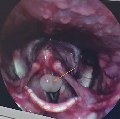 |
Figure 1 A rigid 70-degree endoscopic picture shows a left-sided pedunculated mass on the posterior commissure of the left Vocal cord. |
The patient underwent surgery to remove the lesion using endoscopy while under general anesthesia (Figures 2 and 3). The histopathological examination revealed a polypoid tissue with proliferating blood vessels and inflammatory cells, diagnosed as lobular capillary hemangiomas (Figures 4 and 5). The patient returned for a follow-up three months post-operation and showed no signs of recurrence.
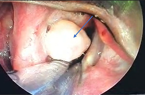 |
Figure 2 The intraoperative endoscopic picture shows a left-sided pedunculated mass on the posterior commissure of the left Vocal cord. |
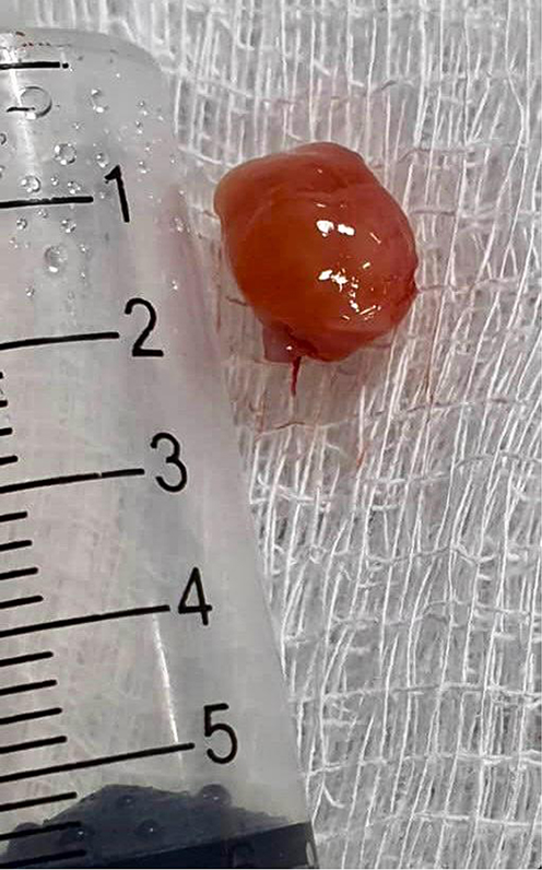 |
Figure 3 Postoperative excision of the Mass. |
 |
Figure 4 Photomicrograph showing pedunculated tissue with surface epithelial ulceration lined fibrinopurulent material under H&E stain (x40). |
 |
Figure 5 Photomicrograph showing engorged blood vessels and hemosiderin-laden macrophages under H&E stain (x100). |
Discussion
Pyogenic granuloma, or lobular capillary hemangioma, is a benign vascular lesion affecting the skin and mucous membranes. It is characterized by a lobular capillary architecture.5 Factors associated with the development of this pathological lesion include trauma, periodontal and gingival diseases, infections, and poor dental hygiene.4 Some researchers suggest that this is a hyperproliferative vascular response to a low-virulence infectious organism.6 Poncet and Dor proposed that these organisms were fungi when they first described this lesion in 1897.4
Pyogenic granuloma is a red, lobulated mass on a pedunculated or sessile base.7 Between 1970 and 2008, a review performed at the Federal University of Rio Grande do Norte analyzed 9300 vascular lesions of the oral cavity.8 Among these, 293 cases of pyogenic granulomas were diagnosed based on biopsy results. The majority of these cases (83%) were found in the gingiva, followed by the lip (5.3%), tongue (5.3%), palate (4.5%), and buccal mucosa (0.8%). The larynx showed no lesions diagnosed as pyogenic granuloma.8
Although pyogenic granuloma of the larynx is considered rare, some studies have been reported in the literature. One study reported a case of pyogenic granuloma in the left vocal fold of a 60-year-old male.1 Another study also indicated that an 80-year-old female with a lesion on the right false vocal cord was histopathologically confirmed to have a pyogenic granuloma.8
Hormonal changes, particularly during pregnancy, are believed to be the cause of pyogenic granuloma. According to Hanick et al, a female patient in her third trimester presented to the emergency room with frank hemoptysis.2 Differential diagnosis is crucial for identifying the pathology and determining the appropriate treatment and prognosis of the disease. Several benign and malignant lesions, such as hemangioma, hemangioendothelioma, angiofibroma, angiosarcoma, Kaposi’s sarcoma, Wegener’s granuloma, and Crohn’s disease, along with granulomatous infectious diseases like tuberculosis and histoplasmosis, traumatic granuloma, carcinosarcoma, and verrucous and squamous cell carcinoma, should be considered as potential differential diagnoses for pyogenic granuloma of the larynx.4
A 20-year-old woman visited the clinic complaining of hoarseness, shortness of breath, and a cough following a traumatic injury to her throat. An examination performed under general anesthesia revealed a lesion that completely obstructed the supraglottic area and was tethered to the superior aspect of the right arytenoid vocal process. Egilmez et al report a case involving a male patient diagnosed with a pyogenic granuloma that originated from the posterior aspect of the left vocal fold, obstructing the rima glottidis. This patient had a history of intubation prior to his diagnosis.1 In contrast, our patient did not have any history of intubation or trauma to the larynx.
Conclusion
Pyogenic granulomas are rarely found in the larynx. Symptoms can vary in severity, from mild to profound, depending on the size of the lesion. The differential diagnoses should consider mass lesions originating from the posterior glottis and pyogenic granuloma.
Several hypotheses have been proposed to explain the causes of pyogenic granuloma; however, the exact etiology remains unknown. Pyogenic granuloma is a non-neoplastic growth, so proper diagnosis, prevention, and management of these lesions are essential to prevent complications.
Ethics Statement
Based on the regulations of the review board of the Welcare Specialty Hospital, institutional review board approval is not required for case reports.
Informed Consent Statement
Written informed consent was obtained from the patient for the publication of any potentially identifiable images or data included in this article.
Acknowledgments
We would like to thank all the participants and Welcare Specialty Hospital for their valuable contributions to the study; also, we would to thank Professor Dr Abdullahi Farah Asseir, MD for all contributions, corrections, and critical reviews.
Disclosure
The authors report no conflicts of interest associated with this publication.
References
1. Hijazi LO, Asiri M, Al Mahdi MJ, Pharaon M. Pyogenic granuloma of the larynx. J Surg Case Report. 2022;2022(7):rjac299. doi:10.1093/jscr/rjac299
2. Hanick AL, Meleca JB, Billings SD, Bryson PC. Pyogenic granuloma of the larynx: a rare cause of hemoptysis. Am J Otolaryngol. 2019;40(2):331–333. doi:10.1016/j.amjoto.2018.10.014
3. Levy I, Rolain JM, Lepidi H, et al. Is pyogenic granuloma associated with bartonella infection? J Am Acad Dermatol. 2005;53(6):1065–1066. doi:10.1016/j.jaad.2005.08.046
4. Egilmez OK, Uzun L, Ozkanli SS, Kalcioglu MT. Pyogenic granuloma of the larynx: a case report. Online J Otolaryngol. 2016;6(1).
5. Karadağli T, Yalçin Ş, Yildiz M, Akpolat N. Pyogenic granuloma of the epiglottis: a case report. Türk Otolarengoloji Arsivi. 2007;45(1):41–44.
6. Jafarzadeh H, Sanatkhani M, Mohtasham N. Oral pyogenic granuloma: a review. J Oral Sci. 2006;48(4):167–175. doi:10.2334/josnusd.48.167
7. Gordón-Núñez MA, de Vasconcelos Carvalho M, Benevenuto TG, Lopes MF, Silva LM, Galvão HC. Oral pyogenic granuloma: a retrospective analysis of 293 cases in a Brazilian population. J Oral Maxillofacial Surg. 2010;68(9):2185–2188. doi:10.1016/j.joms.2009.07.070
8. Lai S, Kelleher K, Sataloff RT. Vocal fold granuloma: the “ball-valve” phenomenon. Ear Nose Throat J. 2000;79(11):836. doi:10.1177/014556130007901103
 © 2025 The Author(s). This work is published and licensed by Dove Medical Press Limited. The
full terms of this license are available at https://www.dovepress.com/terms
and incorporate the Creative Commons Attribution
- Non Commercial (unported, 3.0) License.
By accessing the work you hereby accept the Terms. Non-commercial uses of the work are permitted
without any further permission from Dove Medical Press Limited, provided the work is properly
attributed. For permission for commercial use of this work, please see paragraphs 4.2 and 5 of our Terms.
© 2025 The Author(s). This work is published and licensed by Dove Medical Press Limited. The
full terms of this license are available at https://www.dovepress.com/terms
and incorporate the Creative Commons Attribution
- Non Commercial (unported, 3.0) License.
By accessing the work you hereby accept the Terms. Non-commercial uses of the work are permitted
without any further permission from Dove Medical Press Limited, provided the work is properly
attributed. For permission for commercial use of this work, please see paragraphs 4.2 and 5 of our Terms.