
Back to Journals » Clinical, Cosmetic and Investigational Dermatology » Volume 19
Pseudoxanthoma Elasticum-Like Papillary Dermal Elastolysis: A Case Report
Authors Ye J, Yang X, Lv W, Ma T
Received 8 April 2026
Accepted for publication 11 July 2026
Published 24 July 2026 Volume 2026:19 615254
DOI https://doi.org/10.2147/CCID.S615254
Checked for plagiarism Yes
Review by Single anonymous peer review
Peer reviewer comments 2
Editor who approved publication: Dr Jeffrey Weinberg
Junhong Ye,1 Xianxu Yang,1 Wen Lv,2 Tianming Ma3
1Department of Dermatology, Hainan Fifth People’s Hospital, Haikou, Hainan, People’s Republic of China; 2Department of Allergy, Hainan Fifth People’s Hospital, Haikou, Hainan, People’s Republic of China; 3Department of Dermatology, The Second Affiliated Hospital of Heilongjiang University of Chinese Medicine, Harbin, Heilongjiang, People’s Republic of China
Correspondence: Tianming Ma, Email [email protected]
Abstract: We report a clinical case of pseudoxanthoma elasticum-like papillary dermal elastolysis. A 36-year-old female presented to our institution with a 30-year history of spontaneously erupting papules on her anterior neck. These lesions had gradually increased in both number and size without any identifiable trigger. Physical examination revealed numerous densely distributed, firm, pale-yellow subcutaneous papules localized to the anterior neck, right supraclavicular region, right shoulder, and right posterior neck. Dermoscopy demonstrated pale or milky-white structureless areas with globular or irregular morphologies. Some lesions exhibited slight confluence. Focal linear or atypical vessels were observed between the lesions, but no classic frog-spawn appearance or typical vascular structures were present. Histopathologic examination of serial sections showed mild acanthosis and papillomatosis. The superficial dermis exhibited mild vascular dilatation accompanied by a sparse perivascular inflammatory infiltrate. Crucially, elastic tissue staining revealed fragmented and markedly reduced elastic fibers within the papillary dermis. Consequently, the diagnosis of pseudoxanthoma elasticum-like papillary dermal elastolysis was established.
Keywords: pseudoxanthoma elasticum-like, papillary dermis, elastolysis
Introduction
Pseudoxanthoma elasticum-like papillary dermal elastolysis (PXE-PDE) is a dermatosis with a strong predilection for the neck. It typically presents as multiple pale-yellow or skin-colored, nonfollicular papules that may gradually coalesce into plaques. Patients are generally asymptomatic.1 The exact etiology of the disease remains unclear. Current medical literature suggests potential associations with aging, ultraviolet radiation, abnormal elastogenesis, and genetic factors.2 Histopathologically, elastic tissue staining demonstrates a loss of the elastic plexus in the papillary dermis alongside the presence of melanocytes.3 The condition lacks systemic involvement and is primarily a cosmetic concern. Given the current absence of effective therapeutic options, establishing an accurate and efficient diagnosis is critical. PXE-PDE is an uncommon and relatively underrecognized acquired elastolytic dermatosis. Although more than 50 cases have been reported in the literature, its clinical recognition remains challenging because it may mimic pseudoxanthoma elasticum and other elastolytic disorders. The lesions most commonly involve the neck and supraclavicular areas, but other sites, including the shoulders, upper trunk, axillae, and antecubital regions, may also be affected.4 The present case deserves attention because the patient was relatively young and had a remarkably long 30-year disease course, suggesting possible early onset, whereas PXE-PDE is more commonly described in older women. Previously reported frog-spawn-like structures were not observed in the present case.5
Clinical Data
Medical History
A 36-year-old female patient presented to our outpatient clinic on April 15, 2025. She developed pale-yellow papules on her anterior neck 30 years ago without an identifiable trigger. These lesions gradually increased in both number and size. She remained asymptomatic and had not received any prior treatment. Current presentation showed numerous firm, pale-yellow, miliary subcutaneous papules. They were densely distributed across the anterior neck, right supraclavicular region, right shoulder, and right posterior neck. Throughout the disease course, the patient reported no fever, weight loss, or exposure to known allergens. Her general well-being, appetite, and sleep were normal. She denied any other significant medical history or a family history of similar conditions.
Physical Examination
The patient was in good general health. Systemic examination revealed no remarkable findings. Dermatological evaluation identified multiple miliary, pale-yellow papules localizing to the anterior neck, right supraclavicular region, right shoulder, and right posterior neck (Figure 1). The papules were densely clustered. The lesions were flat, firm, and slightly refractile, with no tenderness elicited upon palpation.
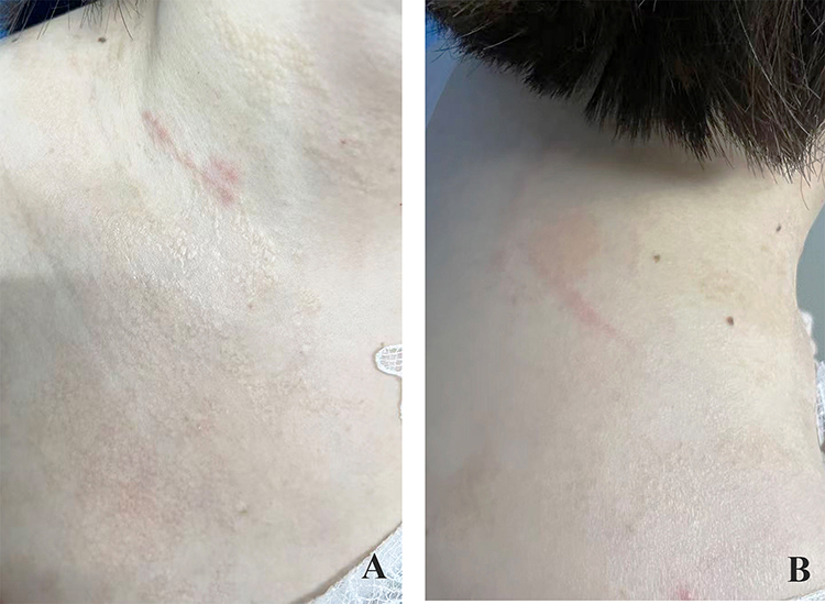 |
Figure 1 Numerous pale-yellow papules are visible on the right anterior neck and supraclavicular region (A). Abundant pale-yellow papules are also distributed across the skin of the right shoulder and posterior neck (B). |
Laboratory and Auxiliary Investigations
Routine blood and urine tests were unremarkable. Hepatic and renal function profiles were within normal limits. Serological testing for Treponema pallidum, hepatitis C virus, human immunodeficiency virus, and hepatitis B surface antigen were all negative. Electrocardiography showed no abnormalities. Classic pseudoxanthoma elasticum was considered in the differential diagnosis. The patient reported no visual impairment, intermittent claudication, chest discomfort, gastrointestinal bleeding, or other symptoms suggestive of ocular, cardiovascular, gastrointestinal, or peripheral arterial involvement. Because the lesions were localized, asymptomatic, and clinically benign, and because the patient declined further examinations due to economic concerns, fundoscopy, formal peripheral arterial assessment, calcification staining, and circulating pyrophosphate measurement were not performed. Therefore, the distinction from classic pseudoxanthoma elasticum was based mainly on the clinical presentation, absence of systemic manifestations, and histopathological findings. Dermoscopic examination of the skin lesions was performed using a digital dermatoscope (Model: BN-PFMF-8001). Under non-polarized light at 20× magnification, the lesions appeared as yellow, structureless areas with globular or irregular shapes, and some showed partial confluence (Figure 2A). Under polarized light at the same magnification, focally distributed linear or atypical vessels were observed between the lesions, while no typical frogspawn-like structures or vascular patterns were identified (Figure 2B). Histopathological examination of a biopsy specimen from the anterior neck demonstrated mild epidermal acanthosis and papillomatosis. The superficial dermis showed mild vascular dilatation accompanied by a sparse perivascular inflammatory infiltrate (Figure 3). Elastic tissue staining revealed that elastic fibers within the papillary dermis were fragmented and markedly reduced (Figure 4). Based on the clinical presentation, absence of subjective symptoms, laboratory findings, dermoscopic features, and histopathological results, a final diagnosis of pseudoxanthoma elasticum-like papillary dermal elastolysis was established.
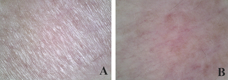 |
Figure 2 Under non-polarized light at 20× magnification, the lesions appear as yellow, structureless areas with globular or irregular shapes, and some show partial confluence (A). Under polarized light at 20× magnification, focally distributed linear or atypical vessels are visible between the lesions, while no typical frogspawn-like structures or vascular patterns are observed (B). |
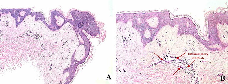 |
Figure 3 Histopathological findings. Hematoxylin and eosin staining shows mild epidermal acanthosis and papillomatosis at low magnification ((A) ×40). At higher magnification, mild vascular dilatation with sparse perivascular inflammatory infiltrate is observed in the superficial dermis ((B) ×100). |
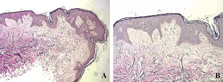 |
Figure 4 Elastic tissue staining. Elastic fibers within the papillary dermis are fragmented and markedly reduced at low magnification ((A) ×40) and higher magnification ((B) ×100). |
Treatment
Because no established curative treatment is available for PXE-PDE, the patient was mainly advised to undergo clinical observation and strict photoprotection with physical barriers. An empirical topical regimen, consisting of desonide cream alternating with 0.1% tacrolimus ointment, was discussed as a symptomatic anti-inflammatory option, considering the mild superficial perivascular inflammatory infiltrate observed histopathologically. However, the patient had no pain, pruritus, or functional impairment, and she declined pharmacological treatment because of the cosmetic-only nature of the lesions and economic concerns. At the one-year follow-up, the lesions remained stable without notable progression or spontaneous regression.
Discussion
Pseudoxanthoma elasticum-like papillary dermal elastolysis is a dermatosis defined by the dissolution of the elastic fiber network within the papillary dermis.1 Clinically, it manifests as asymptomatic, pale-yellow, non-cystic papules. These lesions often display a cobblestone or gooseflesh-like morphology and may coalesce into distinct plaques. Distribution is typically symmetrical, heavily favoring the neck and supraclavicular regions.2 The condition progresses indolently. Affected sites lack any history of prior inflammation or trauma, and the disease is completely devoid of systemic manifestations. Though primarily observed in the elderly population, the definitive etiology remains elusive. Current perspectives suggest a complex interplay of intrinsic aging, ultraviolet radiation exposure, abnormal elastogenesis, and genetic susceptibility.3 Establishing a diagnosis depends heavily on clinical presentation and specialized histological evaluation. Routine hematoxylin and eosin (HE) staining generally yields non-specific findings. Conversely, elastic tissue stains readily demonstrate the hallmark diagnostic features: a complete loss of elastic fibers in the upper papillary dermis accompanied by a depletion of the elastic network in the deeper reticular dermis.6
Upon presentation at our institution, the patient’s diagnosis was definitively confirmed through clinical evaluation, dermoscopy, histopathology, and elastic tissue staining. In clinical practice, this condition must be primarily differentiated from pseudoxanthoma elasticum (PXE), mid-dermal elastolysis, white fibrous papulosis of the neck, and linear focal elastosis.
Pseudoxanthoma elasticum (PXE):7,8 Pseudoxanthoma elasticum was the most important differential diagnosis in this case. PXE is an inherited disorder of elastic tissue mineralization and may involve the skin, eyes, cardiovascular system, and gastrointestinal tract. Its onset often occurs during adolescence or early adulthood, although the clinical presentation and severity are highly heterogeneous. The onset of PXE most commonly occurs during the second decade of life, although earlier or later presentations have been reported. Its clinical presentation and disease severity are highly heterogeneous among affected patients. Histopathologically, PXE is characterized by fragmented and mineralized elastic fibers, mainly in the mid-to-deep reticular dermis, with calcium-phosphate mineralization that can be demonstrated by special stains such as von Kossa or Alizarin Red. In the present case, classic PXE was considered but was less favored because the patient had localized cutaneous lesions, no clinical evidence of ocular, cardiovascular, gastrointestinal, or peripheral arterial involvement, and elastic tissue staining showed fragmented and markedly reduced elastic fibers predominantly within the papillary dermis. Nevertheless, fundoscopy, formal peripheral arterial assessment, calcification staining, and circulating pyrophosphate measurement were not performed, which represents a limitation of this case.
Other resembling dermatoses were considered but were less compatible with the clinical and histopathological findings. Mid-dermal elastolysis usually presents with well-demarcated wrinkled patches, perifollicular papules, or persistent reticular erythema, with elastic fiber loss mainly located in the mid-dermis.9,10 These features were absent in our patient, whose elastic fiber changes were predominantly confined to the papillary dermis. White fibrous papulosis of the neck was also unlikely because the lesions were yellowish rather than white, and histopathology did not show prominent papillary dermal fibrosis or thickened collagen bundles.11,12 Linear focal elastosis was excluded because the patient did not present with linear yellow striae on the trunk or back, and histology showed reduced and fragmented elastic fibers rather than an accumulation of elongated elastic fibers.13,14
There is currently no established effective treatment for this condition. Disease management relies primarily on clinical observation. Because the patient was completely asymptomatic aside from the aesthetic impact of the papules, our intervention was limited to advising strict photoprotection. We strongly emphasized the use of physical barriers to prevent ultraviolet radiation from potentially extending the affected area. The patient declined experimental therapies. After one year of follow-up, her lesions have remained entirely stable, exhibiting neither expansion nor signs of spontaneous regression.
Conclusion
This case describes PXE-PDE in a relatively young patient with a 30-year disease duration, suggesting a possible early-onset and long-standing clinical course. Although PXE-PDE has been reported previously, this presentation may broaden its recognized clinical spectrum and reminds clinicians that PXE-PDE-like lesions are not restricted to elderly patients. Diagnosis should rely on clinicopathological correlation, especially elastic tissue staining showing fragmentation and marked reduction of elastic fibers in the papillary dermis. Since no established curative treatment is available, observation and photoprotection remain reasonable management options for asymptomatic cases. The empirical topical regimen was discussed only as an anti-inflammatory option, and the patient ultimately declined pharmacological treatment.
Ethical Informed Consent Statement
Written informed consent has been provided by the patient to have the case details and any accompanying images published. This case has been approved by the Fifth People’s Hospital of Hainan Province and can be made public.
Funding
Project supported by Hainan Province Clinical Medical Center.
Disclosure
The authors report no conflicts of interest in this work.
References
1. Murshidi R, Bani Hamad S, Al-Tarawneh A. Pseudoxanthoma elasticum-like papillary dermal elastolysis; A report of two cases and a literature review. Dermatol Online J. 2024;30(5). doi:10.5070/D330564429
2. Panagou E, Ratynska M, Heelan K. Pseudoxanthoma elasticum-like papillary dermal elastolysis: a case report and review of literature. Int J Dermatol. 2019;58(1):93–6. PMID: 29907963. doi:10.1111/ijd.14093
3. Setó-Torrent N, Iglesias-Sancho M, Arandes-Marcocci J, Fernández-Figueras MT. Pseudoxanthoma elasticum-like papillary dermal elastolysis in non-exposed skin. An Bras Dermatol. 2020;95(2):247–249. doi:10.1016/j.abd.2019.08.024
4. Vázquez-Osorio I, Rosón E, Suárez-Peñaranda JM, Vázquez-Veiga H. Pseudoxanthoma elasticum-like papillary dermal elastolysis. Actas Dermosifiliogr. 2015;106(4):333–336. English, Spanish. doi:10.1016/j.ad.2014.10.002
5. Mercuri SR, Cestone E, Bearzi P, Rizzo N, Paolino G. Dermoscopical and pathological findings in pseudoxanthoma elasticum-like papillary dermal elastolysis. G Ital Dermatol Venereol. 2020;155(5):692–693. doi:10.23736/S0392-0488.18.06090-X
6. Revelles JM, Machan S, Pielasinski U, et al. Pseudoxanthoma elasticum-like papillary dermal elastolysis: immunohistochemical study using elastic fiber cross-reactivity with an antibody against amyloid P component. Am J Dermatopathol. 2012;34(6):637–643. doi:10.1097/DAD.0b013e318257be63
7. Uitto J, Jiang Q, Váradi A, Bercovitch LG, Terry SF. Pseudoxanthoma elasticum: diagnostic features, classification, and treatment options. Expert Opin Orphan Drugs. 2014;2(6):567–577. doi:10.1517/21678707.2014.908702
8. Ladewig MS, Götting C, Szliska C, et al. Pseudoxanthoma elasticum: genetische Grundlagen, Manifestationen und therapeutische Ansätze [Pseudoxanthoma elasticum]. Ophthalmologe. 2006;103(6):537–551; quiz 552–553. German. doi:10.1007/s00347-006-1353-4
9. Aryal S, Li Y, Bashyal S, Qiang LY, Rehman A. A case report on mid-dermal elastolysis: a distinctive presentation on the neck. Cureus. 2023;15(9):e45724. doi:10.7759/cureus.45724
10. Zhang LH, Zhang Y, Li YL. Mid-dermal elastolysis: a case report and literature review. Indian J Dermatol. 2023;68(2):217–219. doi:10.4103/ijd.ijd_957_22
11. Calvão J, Ferreira BR, Cardoso JC. Multiple whitish papules on the posterior neck of an elderly woman. An Bras Dermatol. 2020;95(1):102–104. doi:10.1016/j.abd.2019.05.003
12. Schaffer PR, Tran K, Kaplan J, et al. White fibrous papulosis of the neck. Dermatol Online J. 2014;20(12):
13. Ahmad A, Al-Sughayer F, Al-Sabah H, Al-Lafi A. Linear Focal Elastosis: a Peculiar Dermatosis. Clin Case Rep. 2025;13(9):e70860. doi:10.1002/ccr3.70860
14. Aung T, Noakes R, Kasem K. Linear focal elastosis: a case report with a brief literature review. Clin Exp Dermatol. 2025;50(9):1873–1875. doi:10.1093/ced/llaf197
 © 2026 The Author(s). This work is published and licensed by Dove Medical Press Limited. The
full terms of this license are available at https://www.dovepress.com/terms
and incorporate the Creative Commons Attribution
- Non Commercial (unported, 4.0) License.
By accessing the work you hereby accept the Terms. Non-commercial uses of the work are permitted
without any further permission from Dove Medical Press Limited, provided the work is properly
attributed. For permission for commercial use of this work, please see paragraphs 4.2 and 5 of our Terms.
© 2026 The Author(s). This work is published and licensed by Dove Medical Press Limited. The
full terms of this license are available at https://www.dovepress.com/terms
and incorporate the Creative Commons Attribution
- Non Commercial (unported, 4.0) License.
By accessing the work you hereby accept the Terms. Non-commercial uses of the work are permitted
without any further permission from Dove Medical Press Limited, provided the work is properly
attributed. For permission for commercial use of this work, please see paragraphs 4.2 and 5 of our Terms.