
Back to Journals » Journal of Experimental Pharmacology » Volume 17
Protective Effects of Tamarillo (Solanum betaceum) Extract Against Apoptosis in Lead Acetate-Induced Mice Testicular Damage
Authors I’tishom R  , Agustinus A, Khaerunnisa S , Abutari CA, Undaryati YM, Rezano A , Margiana R, Putra WMI
Received 29 April 2024
Accepted for publication 23 June 2025
Published 22 July 2025 Volume 2025:17 Pages 485—496
DOI https://doi.org/10.2147/JEP.S476046
Checked for plagiarism Yes
Review by Single anonymous peer review
Peer reviewer comments 2
Editor who approved publication: Prof. Dr. Abdelwahab Omri
Reny I’tishom,1– 4 Agustinus Agustinus,1,2,4 Siti Khaerunnisa,4,5 Cinta Ayu Abutari,2 Yeti Mareta Undaryati,3 Andri Rezano,2,6 Ria Margiana,2,7 Wildan Maulana Ishom Putra4
1Department of Biomedical Sciences, Faculty of Medicine, Universitas Airlangga, Surabaya, East Java, Indonesia; 2Andrology Study Program, Faculty of Medicine, Universitas Airlangga, Surabaya, East Java, Indonesia; 3Doctoral Program of Medical Science, Faculty of Medicine, Universitas Airlangga, Surabaya, East Java, Indonesia; 4Medical Study Program, Faculty of Medicine, Universitas Airlangga, Surabaya, East Java, Indonesia; 5Department of Biochemistry and Physiology, Faculty of Medicine, Universitas Airlangga, Surabaya, East Java, Indonesia; 6Department of Biomedical Sciences, Faculty of Medicine, Universitas Padjadjaran, Bandung, West Java, Indonesia; 7Department of Anatomy, Faculty of Medicine, Universitas Indonesia, Jakarta, Jakarta, Indonesia
Correspondence: Reny I’tishom, Department of Biomedical Sciences Faculty of Medicine, Universitas Airlangga Campus A, Jl. Mayjen Prof. Dr. Moestopo 47, Surabaya, East Java, 60131, Indonesia, Tel +62 31 5020251, Email [email protected]
Purpose: To evaluate the protective effects of Tamarillo (Solanum betaceum Cav) extract on lead acetate-induced testicular germ cell apoptosis in male mice.
Methods: A post-test-only control group experimental study was conducted on Balb/c mice. Thirty-five male mice 12 weeks old were randomly divided into 5 groups: two control groups (K-, K+) and three treatment groups (P1, P2, P3). The K- received distilled water, and the K+ received 75 mg/kg lead acetate. The P1, P2, and P3 received 100, 200, and 400 mg/kg of Tamarillo crude extracts (TCE), respectively for 35 days, and on the fourth day, were given lead acetate 75 mg/kg one hour after the TCE administration by gavage tube. The effect of TCE against lead-induced oxidative stress in mice was determined by the expression of mouse testicular apoptosis using terminal deoxynucleotidyl Transferase-mediated dUTP nick end labeling (TUNEL) assay, the expression of Caspase-3, and SOD.
Results: TCE at the dose of 400 mg/kg showed comparable apoptosis and SOD expression to the negative control group (K-). Notably, the level of Caspase-3 among treatment groups showed lower expression than the K- group despite the injection of lead acetate.
Conclusion: This study demonstrated that TCE exhibits antioxidant activity and protects the reproductive system by inhibiting lead acetate to induce oxidative damage and testicular damage.
Keywords: antioxidant, testicular germ cell, oxidative damage, tamarillo, testicular apoptosis
Introduction
Tamarillo (Solanum betaceum Cav.) is a fruit species native to the Andean regions of South America, Southeast Asia, and the North Island of New Zealand.1–3 Due to its high resemblance in its flesh texture to a tomato, it is also known as a “tree tomato”.2 Ripened fruit broadly distinguished by the colors of yellow-orange, red, and purple exhibiting a slightly bitter, sour, and astringent taste with a characteristic aroma.4 These flavors originate from the fruit’s 70 volatile compounds and organic acids.5–10 Among these compounds, phenolics, carotenoids, and anthocyanins are considered to be the main bioactive components for health-promoting benefits.11 Furthermore, Solanum betaceum is also known for its high nutritional contents such as vitamins A, B6, and C, dietary fiber, and potassium, higher than lemon, banana, pomegranate, and blueberry.12 The health benefits of Solanum betaceum consumption include antioxidative, antiproliferative, antinociceptive, anti-inflammatory, allergenicity, anti-obesity, and antimicrobial properties.13 Despite possessing numerous beneficial properties, Solanum betaceum has yet to be fully exploited due to its unique flavor and color, which remain unpopular.2
Flavonoid represents a remarkable group of plant secondary metabolites and has been widely studied for their potential to counteract the harmful effects of lead toxicity, which induces apoptosis and oxidative damage in various human tissues.14 In total, 800 variants of flavonoids have been explored since their first discovery in the 1930s.15 The pharmaceutical roles of flavonoids include cytotoxic, anticancer, anti-inflammatory, antiviral, antibacterial, cardioprotective, hepatoprotective, neuroprotective, antimalarial, antileishmanial, antitrypanosomal, and antiamoebic.15–18 This is due to their free radical scavenging mechanism, metal chelation capabilities, and highly accurate protein-binding activity.19 Among these flavonoids, anthocyanin dominates the phenolic composition in Solanum betaceum.20 Anthocyanins such as cyanidin, delphinidin, and pelargonidin rutinosides have been discovered in Solanum betaceum from Brazil, Colombia, Ecuador, and New Zealand.4,21–23 Anthocyanins are water-soluble flavonoids generally utilized as a coloring pigment widely present in fruits and vegetables.24 Anthocyanins extinguish reactive radical species by hydrogen atom transfer.25–29
Concerning the anthocyanin levels in other natural sources, some studies have presented evidence of such compounds initiating protective mechanisms towards various diseases. One is anthocyanin extracted from purple yam potentially preventing lead-caused reproductive toxicity due to its antioxidant, anti-apoptotic properties, and JNK signaling pathway.30 Furthermore, a study on anthocyanin (sourced from black beans) active role against varicocele-induced model presented the prevention of oxidative damage, which induces active spermatogenesis and production of high-quality sperm cells.31
This study aimed to analyze the histological profile of apoptosis, Caspase-3, and the superoxide dismutase (SOD) expression in the testis of mice exposed to lead acetate. Hence, deepening the understanding of the Solanum betaceum in testicular protective effect and exploring the possibility of its use in lowering infertility rates.
Materials and Methods
Materials and Equipments
The material used in this study were Balb/C male mice (Mus musculus), Solanum betaceum fruit, ethanol, filter paper, CMC powder, lead acetate, phosphate buffer saline (PBS), methanol, ether, aquabidest, xylol, paraffin, entellan®, ketamine, xylazine, NaCl, Bouin’s fixative solution, hematoxylin counterstain, Terminal deoxynucleotidyl Transferase-mediated dUTP Nick End Labeling (TUNEL; 12156792910, Roche), proteinase-K, hydrogen peroxide, SOD mouse monoclonal antibody (Sigma-Aldrich, SAB4200807) and caspase-3 mouse monoclonal antibody (Sigma-Aldrich, C5737), diaminobenzidine (DAB), Trekkie Avidin-HRP, and Trekkie Universal Link. All substances used were of analytical grade. Necessary instruments for gaining data were a refrigerator, rotary microtome, micro-scale, syringe, sonde, scalpel, pipette, micropipette, tweezers, object glass, pencil, light microscope, hand counter, timer, micrometer, aluminium foil, computer with statistical program.
Plant Materials
Solanum betaceum fruit was collected from farmland in Wonosobo, Central Java, Indonesia (GPS coordinates: 7.3633°S, 109.9031°E). Identification was based on morphological characteristics using standard botanical references and confirmed by consensus among trained staff at the Research Centre for Biology of the Indonesian Institute of Science (LIPI). A voucher specimen (No. 724/IPH.1.01/If.07/III/2017) is deposited at the LIPI herbarium.
Preparation of Ethanolic Extract of S. betaceum
S. betaceum fruit was dried by a fresh air dryer. Dry powder was extracted by maceration using ethanol for three days at room temperature with solvent replacement every 24 hours. The liquid extract was filtered through filter paper to separate filtrate and residue. The liquid extract of S. betaceum was evaporated with a rotary vacuum evaporator to obtain a viscous extract. The ethanol extract of S. betaceum was added to the treatment diet as a suspension using 1% CMC with dose of 2 mL/200 g. According to previous protocols, S. betaceum extract was simultaneously administered to the treatment group three days before lead acetate exposure for 35 days according to previous protocols.32
Preparation of Lead Acetate
Lead acetate was used at a dose of 75 mg/kg, dissolved in distilled water, and saved at room temperature.32
Experimental Design
A post-test control group experimental study was conducted using 12-week-old male Balb/C mice (25–30 g) to evaluate the gonadoprotective effect of the extract of Solanum betaceum (tamarillo) crude extract (TCE). Mice showing signs of illness, mortality, or more than 10% body weight loss during the study were excluded. A total of 35 mice were randomly assigned into five groups (n = 7 per group): a negative control group (K-), a positive control (K+), and three treatment groups (P1, P2, P3). The K-group received distilled water orally throughout the study. In contrast, the K+ group received lead acetate at 75 mg/kg dissolved in distilled water, by oral gavage once daily for 32 consecutive days (starting day 4 to day 35). The treatment groups (P1, P2, P3) received TCE at doses of 100, 200, and 400 mg/kg, respectively, by oral gavage once daily for 35 days. In these groups, lead acetate (75 mg/kg) was administered one hour after the TCE dose, starting from day 4 through day 35. The pretreatment design was adapted from a previously published study.33 Mice were acclimated for 7 days before the experiment, maintained under a 12-hour light/dark cycle at room temperature.34 On day 36, mice were euthanized under intraperitoneal anesthesia using ketamine (70 mg/kg) and xylazine (20 mg/kg), and both testes were excised for further analysis.
Immunohistochemical Analysis
Median laparotomy was conducted, and the testes were collected, then washed in 0.9% physiological NaCl solution and placed in Bouin’s fixative solution. Testicular tissue was processed using the standard method of embedding with paraffin. Then, the tissue block was sliced using a rotary microtome with a thickness of 4–5 μm and stored in an incubator at 65°C for 3 minutes to complete tissue attachment.35 Tissue sections were washed for 15 minutes with three changes of PBS between each step. After deparaffinization and rehydration, tissue sections were exposed to 3% H2O2 for 10 minutes and methanol for 3 minutes to inactivate endogenous peroxidase activity and then to 10% normal serum for 45–60 minutes to block non-specific proteins. Following rinsing with PBS, the tissue section was then incubated with a primary antibody of SOD or Caspase-3 for 48 hours at 4°C.36 The result of the antigen-antibody reaction was visualized using DAB for 2 minutes, followed by washing and counterstaining with hematoxylin for 10 second and rinsing with aquabidest. The tissue sections were then dehydrated with a series of alcohol and cleared with xylol. The last step was mounting using entelan. As a control of staining, tissue sections were incubated with PBS instead of primary antibody. The control staining slide showed a negative reaction with minimal background staining. Positive SOD or Caspase-3 staining was observed as brown stains in the tissue under a light microscope. The qualitative observation of positive tissue reaction was based on the brown color intensity and distribution of the immunoreaction product in the testicular tissues. The quantitative observation was done by counting the number of cell nuclei that give different levels of brown color intensity in 10 random fields from each testicular tissue section with 400x magnification. A blue color in the cell nuclei indicates a negative reaction. ImageJ software was used to count various brown-stained cell nuclei. The percentage of SOD and caspase-3 expression were compared between groups.37
TUNEL Analysis
Testicular cell apoptosis was evaluated using a TUNEL assay based on manufacture instructions. The TUNEL method detects fragmented DNA in the nucleus during apoptotic cell death in situ. Following the de-paraffinized and rehydrated of 5–6 μm thick tissue sections in xylene and graded ethanol series, the slides were treated for 30 min with 15 ug/mL proteinase-K and washed with PBS. Endogenous peroxidase activity was inhibited by 3% hydrogen peroxidase. Subsequently, the slides were incubated with 25 μL TUNEL solution (50 μL of enzyme solution and 450 μL label solution) for 60 minutes at 37°C. They were put into pre-warmed working strength wash buffer at room temperature for 10 minutes and incubated with blocking buffer for 30 minutes. Thorough washes in PBS separated each step. In one observation field, propidium iodide was added to compare apoptotic and non-apoptotic cells. Labelling was visualized using DAB chromogen, followed by hematoxylin counterstain, and sections were dehydrated, cleared, and mounted. Quantitative analysis of testicular apoptosis was estimated according to Hu et al38 by direct observation through a fluorescence microscope (Olympus, Tokyo, Japan). TUNEL will only detect apoptotic cells and give green fluorescence, while propidium iodide will detect non-apoptotic cells and give red fluorescence.38 Examination was carried out by counting positive cells at 400x magnification and counted in 10 random fields from each testicular tissue section.
Data Processing and Analysis
All statistical analyses were performed using the Statistical Package for the Social Sciences (SPSS) 26.0. Data were initially tested for normality using the Shapiro–Wilk test. Variables with p>0.05 were considered normally distributed and analyzed using one-way ANOVA. For variables that did not meet the normal distribution (p<0.05), non-parametric tests were applied, specifically the Kruskal–Wallis, followed by Mann–Whitney U-tests. All data were presented as mean (±) standard deviation (SD), and statistical significance was accepted at p<0.05.
Results
Tamarillo Extract Decreased Apoptosis of Testicular Damage
Apoptotic cells in the testis of the control and treatment groups were identified by TUNEL assay, as shown in Figure 1. The highest increase in the percentage of apoptotic cells in relation to negative control was observed after lead acetate exposure of 8.257 ± 1.350. Only a few TUNEL-positive cells were observed in negative control animals of 1.286 ± 0.414. However, the number and signal density of TUNEL-positive germinal cells significantly decreased in TCE at the dose of 100 mg/kg and 400 mg/kg (Figure 2 and Table 1).
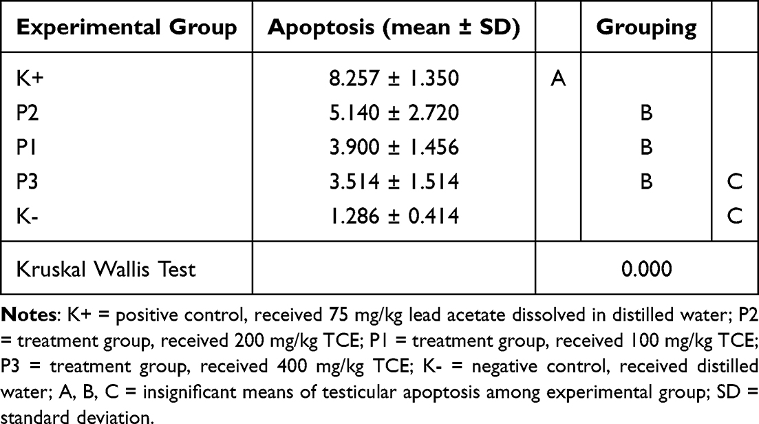 |
Table 1 Means Distribution of Apoptosis Expression Within Experimental Groups |
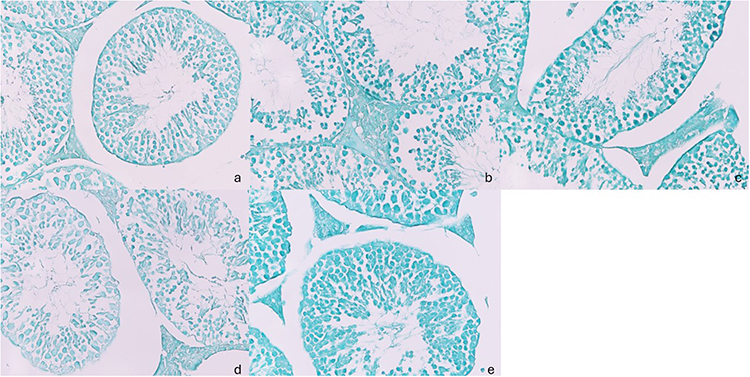 |
Figure 1 Representative of photomicrograph of TUNEL staining in mice testis of negative control (a), positive control (b), TCE at the dose of 100 mg/kg (c); 200 mg/kg (d), and 400 mg/kg (e). |
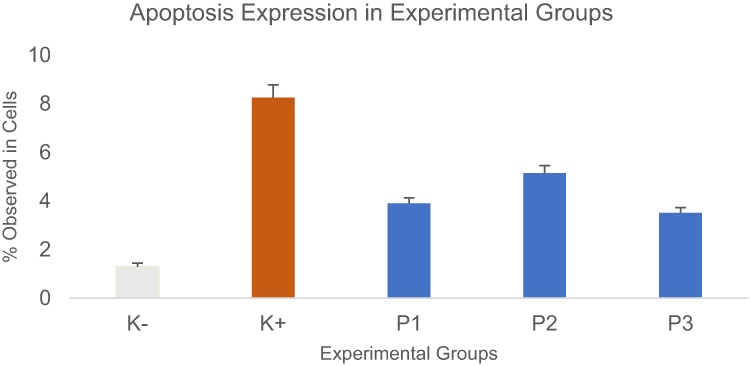 |
Figure 2 Means of testicular apoptosis expression by TUNEL assay. Quantitative analysis of apoptosis in the testis. The percentage of mouse testicular apoptosis was calculated as the ratio of apoptosis-positive seminiferous tubules to the total number of seminiferous tubules. |
The effectiveness of TCE in protecting male reproductive organs was assessed by comparing the means of the control and treatment groups. Table 1 is grouped into similar means of testicular apoptosis-level distribution, A, B, and C, respectively. The treatment groups tended to have a lower apoptosis level than the positive control group suggesting an effective dose of TCE in this range.
The statistically significant differences among the means groups were tested with the Kruskal–Wallis test (Figure 3). The treatment group P3 was found to be the most similar to the negative control group, with no significant difference in means (p=0.115) (Figure 3). This suggests that TCE at the dose of 400 mg/kg reduces similar apoptosis levels as a negative control group.
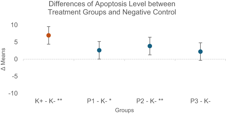 |
Figure 3 Effect of tamarillo extract on apoptosis was measured by comparing differences between treatment groups with the negative control group. Level of significance of p<0.05 and p<0.01 is represented with * and ** respectively. |
Tamarillo Extract Decreased Caspase-3 Expression in Testicular Damage
Various degrees of caspase-3 expression in the testes of all groups were detected by immunohistochemical staining (counterstained with hematoxylin). Caspase-3 facilitates the process of apoptosis in response to testicular damage causing infertility. Histological observation on the control group showed testicular cells were observable and normal testicular architecture. However, staining dramatically increased testicular damage in the mouse exposed to lead acetate. In the mouse, treated with tamarillo extract, the number and morphological integrity of testicular cells were preserved. Observations indicated that the testicular toxic effect of lead acetate was reduced by tamarillo extract (Figure 4).
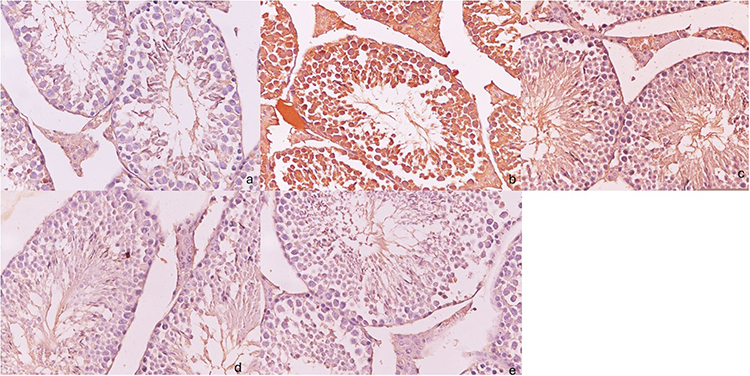 |
Figure 4 Representative of photomicrograph of Caspase-3 immunohistochemical staining in mice testis of negative control (a), positive control (b), TCE at the dose of 100 mg/kg (c); 200 mg/kg (d), and 400 mg/kg (e). |
Almost no specific Caspase-3 immunoreactivity was found in the TCE-treated at the dose of 200 mg/kg. However, heavily labeled Caspase-3-positive cells were seen in mice exposed to lead acetate compared to other experimental groups (Figure 5).
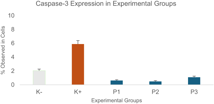 |
Figure 5 Mean of mouse testicular caspase-3 expression by immunohistochemical staining Quantitative analysis of apoptosis in the testis. The percentage of mouse testicular caspase-3 was calculated as the ratio of caspase-3-positive nuclei to the total number of cell nuclei. |
The mean intensity of caspase-3 staining was compared among experimental groups to analyze the amelioration of testicular damage of the tamarillo extract-treated group upon lead acetate exposure and the insignificant means distribution was grouped into Table 2. Lead acetate enhances the highest level of Caspase-3 expression of 5.886b ± 1.472. However, the treatment groups expressed lower testicular Caspase-3 than negative control, suggesting the protective effect of tamarillo extract in male mice exposed to testicular damage.
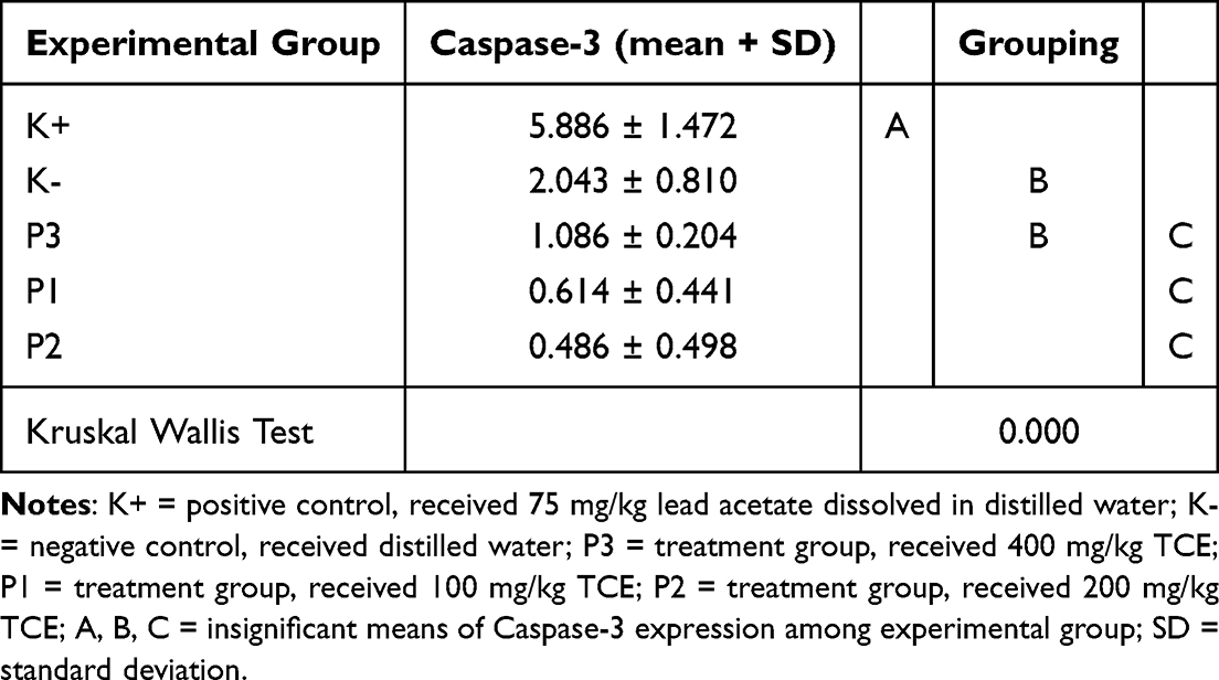 |
Table 2 Means Distribution of Caspase-3 Expression Within Experimental Groups |
The statistically significant differences among the means groups were tested with the Kruskal–Wallis test. This study showed that the TCE-treated group at the dose of 200 mg/kg significantly lower Caspase-3 expression than the negative control group (p=0.05) (Figure 6).
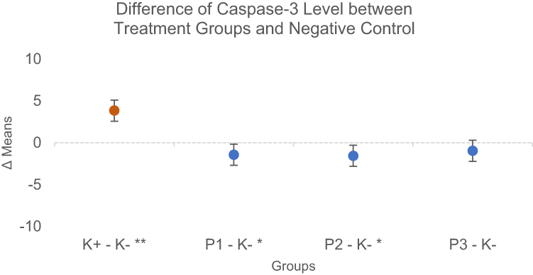 |
Figure 6 Effect of tamarillo extract towards caspase-3 expression measured by comparing differences between treatment groups with the negative control group. Level of significance of p<0.05 and p<0.01 is represented with * and ** respectively. |
Tamarillo Extract Increased SOD Expression in Testicular Damage
Tissue levels of antioxidant enzyme, SOD in each group are observed histologically in Figure 7. Morphological analysis of the mice seminiferous tubules in experimental groups showed mainly in all germ cells, especially in the adluminal compartment.
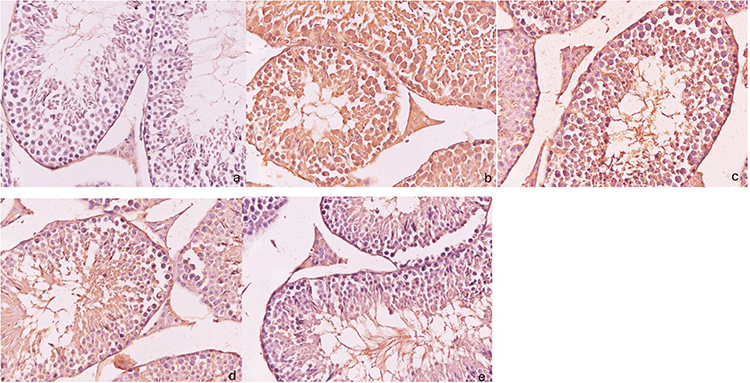 |
Figure 7 Representative of photomicrograph of SOD immunohistochemical staining in mice testis of negative control (a), positive control (b), TCE at the dose of 100 mg/kg (c); 200 mg/kg (d), and 400 mg/kg (e). |
The highest increase in the percentage of SOD expression was in the negative control group at 8.571 ± 1.694, while the lowest increase was in the TCE-treated group at the dose of 100 mg/kg at 2.514 ± 1.014 (Figure 8, Table 3).
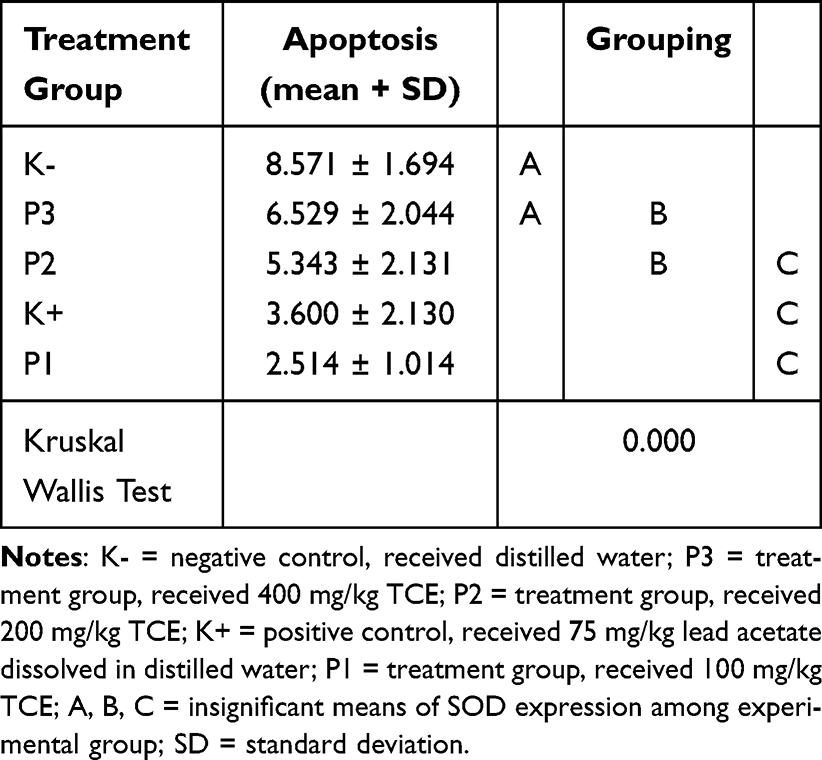 |
Table 3 Means Distribution of SOD Expression Within Experimental Groups |
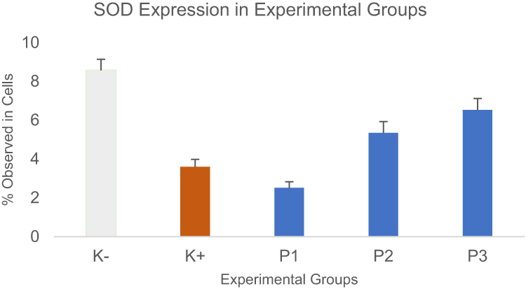 |
Figure 8 Means of mouse testicular SOD expression by immunohistochemical staining Quantitative analysis of apoptosis in the testis. The percentage of mouse testicular SOD was calculated as the ratio of SOD-positive nuclei to the total number of cell nuclei. |
Table 3 is grouped based on the similar means of mouse testicular SOD intensity to show the identical antioxidant activity with the negative control group. The higher dose of TCE treatment showed increasing SOD expression in response to the same damage of testicular tissue induced by lead acetate.
TCE treatment at the dose of 400 mg/kg and negative control demonstrated comparable SOD results (p=0.262) (Figure 9), suggesting the effective dose of TCE to exert antioxidant activity to counter oxidative damage induced by lead acetate.
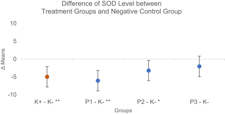 |
Figure 9 Effect of tamarillo extract towards SOD expression measured by comparing differences between treatment groups with the negative control group. Level of significance of p<0.05 and p<0.01 is represented with * and ** respectively. |
Discussion
This study was intended to investigate the effectiveness of tamarillo extract in protecting male fertility following lead acetate exposure. Findings indicate that oral administration of lead acetate significantly increased biomarkers of apoptosis in mice testicular tissue compared to the untreated group. Lead is a major human health hazard present in the environments and biological systems which can affect the gonadal structure and functions and can cause fertility alteration.39,40 Our results agreed that mice exposed to the lead acetate showed irregularity in spermatogenesis, reduced plasma testosterone concentration, and degeneration of seminiferous tubules.41,42
Recent biological agents have been investigated for gonadoprotective effects on testicular function such as dragon fruit,42 moringa,43 chrysin,44 and melatonin.45 In the present study, the addition of tamarillo improved testicular histopathological changes induced by lead. Peroxidative injury and cellular macromolecules cause abnormalities in testicular tissue and the male reproductive system.46 The mechanism of lead-induced testicular toxicity is due to the imbalance between the production of reactive oxygen species (ROS) and the scavenging capacity of antioxidants in the testes, therefore testicles are dependent on antioxidant agents to fight oxidative stress-induced damage.39,46
The results of the present study clearly showed that the levels of apoptosis and Caspase-3 are remarkably increased in mice treated with lead acetate. Lead promotes free radicals and lowers the effects of antioxidants within the cells.46 The imbalance of free radical species produced by lead exposure beyond cellular protective capacity leads to oxidative stress, ultimately resulting in cell apoptosis.47 Lipid membranes are targeted for oxidative damage produced by xenobiotics including heavy metals.48 Furthermore, lead exposure causes several antioxidant enzyme alterations by inhibiting functional SH groups, such as d-aminolevulinic dehydrogenase, superoxide dismutase (SOD), glutathione peroxidase (GPx), and catalase (CAT).46,49 Pretreatment of TCE administration in P1, P2, and P3 provides a strategy to mitigate the antioxidant capacity of TCE to ameliorate lead-induced free radicals.
Apoptosis is a physiological process of programmed cell deletion, which contributes to maintaining the cell number in testicular tissue and removing damaged cells. However, excessive apoptosis could cause male reproductive dysfunction.50 A study reported that a distinct increase of Caspase-3 in time- and dose-dependent manner induced apoptosis of germ cells, suggesting the degree of differences was correlated with the time of lead loading and exposure time to induce damage.51 Our results showed TCE 400 mg/kg is not only lower Caspase-3 to normal levels but also improve the degraded DNA in testicular cells caused by lead acetate.
Several studies used antioxidant evaluation as a marker of high levels of free radicals.52–56 Administration of antioxidants results in increasing the activity of spermatogenesis and steroidogenesis in the testis, thereby preserving fertility. Antioxidants from tamarillo extract provide protective effects in maintaining testicular structure and function.
Free radical damage due to lead exposure depletes antioxidant reserves.57 Cytosolic enzyme, catalase, and glutathione peroxidase enzymes break down H2O2, H2O, and O2. Meanwhile, the superoxide dismutase (SOD) enzyme catalyzes the dismutase reaction and ultimately converts anion radicals into H2O2.58 In the present study, a combination of administration of TCE 400 mg/kg and lead acetate, the level of SOD was increased compared to its level in rats treated only with lead. Decreased SOD expression in testicular tissue may reflect cellular oxidative stress or compensatory mechanism in the response to cellular apoptosis. The activities of testicular SOD were significantly reduced in the lead-exposed mice, while the addition of Artemisia annua extract to lead acetate significantly improved the level of SOD.44 Exogenous antioxidants such as vitamins C and E were also reported to have capabilities in preventing damage to germ cells and testicular tissue caused by excessive free radicals after extended lead acetate exposure.58 A study reported the activity of antioxidants collected from tamarillo extracts with a dosage of IC50 was 1162,608 ppm, which can decelerate lipid peroxidation reaction which is caused by the increase of ROS induced by lead acetate exposure.59 The capability of tamarillo extract to maintain testicular SOD expression seems to be dependent on the dosage. The highest dosage of tamarillo extract 400 mg/kg was comparable to the untreated group, suggesting the critical dose for TCE exerting antioxidant properties against oxidative stress caused by lead acetate. This effect could be due to the presence of anthocyanin and flavonoids, which can fend off various free radicals.60
Conclusion
This study showed that the administration of Solanum betaceum had a significant effect on maintaining testicular structure. Tamarillo exhibited a protective effect on the reproductive system by mitigating lead acetate-induced oxidative stress and excessive cell apoptosis. This might be due to the improved superoxide dismutase level and decreased caspase-3 expression in the testis of male mice.
Data Sharing Statement
The data presented in this study are available on request from the corresponding author. The data are not publicly available due to clarity and usability concerns of the data.
Ethics Approval and Informed Consent
This study was conducted after obtaining a letter of ethical clearance from the Health Research Ethics Committee, Faculty of Medicine Universitas Airlangga with reference number 232/EC/KEPK/FKUA/2020, dated 31 September 2020. All experimental activities were conducted following the ethical declaration of national and international standards for experimental animals to minimize the risk of suffering and provide good animal welfare.
Informed Consent Statement: Not Applicable.
Acknowledgments
The authors would like to thank Universitas Airlangga for their support in funding this project and the staff of the Department of Medical Biology Faculty of Medicine at Universitas Airlangga for their administrative and technical help.
Funding
This research was supported by Universitas Airlangga in compliance with research contract Number: 346/UN3/2020, which was issued on March 27, 2020.
Disclosure
The authors report that there are no conflicts of interest with the work done in this study.
References
1. Wang S, Zhu F. Tamarillo (Solanum betaceum): chemical composition, biological properties, and product innovation. Trends Food Sci Technol. 2019;95:45–58. doi:10.1016/j.tifs.2019.11.004
2. Nallakurumban P, Suja N, Vijayakumar A, Geetha G. Estimation of phytochemicals and antioxidant property of tamarillo (Solanum betaceum) and a value added product tamarillo. IJSPR. 2015;9(2):61–65.
3. Lister C, Morrison S, Kerkhofs N, Wright K. The nutritional composition and health benefits of New Zealand tamarillos. Crop Food Res. 2005;29.
4. Espin S, Gonzalez-Manzano S, Taco V, et al. Phenolic composition and antioxidant capacity of yellow and purple-red Ecuadorian cultivars of tree tomato (Solanum betaceum Cav.). Food Chem. 2016;194:1073–1080. doi:10.1016/j.foodchem.2015.07.131
5. Torrado A, Suárez M, Duque C, Krajewski D, Neugebauer W, Schreier P. Volatile constituents from tamarillo (cyphomandra betacea sendtn.) fruit. Flavour Fragr J. 1995;10:349–354. doi:10.1002/ffj.2730100603
6. Buttery RG, Teranishi R, Ling LC, Turnbaugh JG. Quantitative and sensory studies on tomato paste volatiles. J Agric Food Chem. 1990;38:336–340. doi:10.1021/jf00091a074
7. Suárez M, Duque C, Bicchi C, Wintoch H, Full G, Schreier P. Volatile constituents from the peelings of lulo (Solanum vestissimum d.) fruit. Flavour Fragr J. 1993;8:215–220. doi:10.1002/ffj.2730080409
8. Wong KC, Wong SN. volatile constituents of cyphomandra betacea sendtn. FruiT J Essent Oil Res. 1997;9:357–359. doi:10.1080/10412905.1997.10554261
9. Durant AA, Rodríguez C, Ai S, Herrero C, Jc R, Gupta MP. Analysis of volatile compounds from Solanum betaceum cav. fruits from panama by head-space micro extraction. Rec Nat Prod. 2013;7:15.
10. Garcia JM, Prieto LJ, Guevara A, Malagon D, Osorio C. Chemical studies of yellow tamarillo (Solanum betaceum cav.) fruit flavor by using a molecular sensory approach. Molecules. 2016;21:1729. doi:10.3390/molecules21121729
11. Kou MC, Yen JH, Hong JT, Wang CL, Lin CW, Wu MJCBS. Cyphomandra Betacea Sendt. Phenolics Protect LDL from Oxidation and PC12 Cells from Oxidative Stress. LWT-Food Sci Technol. 2009;42:458–463. doi:10.1016/j.lwt.2008.09.010
12. Diep TT, Rush EC, Yoo MJ. Tamarillo (Solanum betaceum cav.): a review of physicochemical and bioactive properties and potential applications. Food Rev Int. 2020;38:1343–1367. doi:10.1080/87559129.2020.1804931
13. Isla MI, Orqueda ME, Moreno MA, Torres S, Zampini IC. Solanum betaceum fruit waste: a valuable source of bioactive compounds to be used in food and non-foods applications. Foods. 2022;11(21):3363. doi:10.3390/foods11213363
14. Mustafa HN. Ameliorative potential of the quercetin on lead-induced testicular damage: morphohistometric and biochemical analysis. Afr J Urol. 2023;29:36. doi:10.1186/s12301-023-00369-z
15. Kumar S, Pandey AK. Chemistry and biological activities of flavonoids: an overview. Sci World J. 2013;2013:162750. doi:10.1155/2013/162750
16. Khan J, Deb PK, Priya S, et al. Dietary flavonoids: cardioprotective potential with antioxidant effects and their pharmacokinetic, toxicological and therapeutic concerns. Molecules. 2021;27:26. doi:10.3390/molecules26134021
17. Hussain N, Kakoti BB, Rudrapal M, et al. Bioactive antidiabetic flavonoids from the stem bark of Cordia dichotoma forst.: identification, docking and ADMET studies. Molbank. 2021;2.
18. Rudrapal M, Cheti D. Plant flavonoids as potential source of future antimalarial leads. Syst Rev Pharm. 2016;8(1):13–18. doi:10.5530/srp.2017.1.4
19. Rudrapal M, Khairnar SJ, Khan J, et al. Dietary polyphenols and their role in oxidative stress-induced human diseases: insights into protective effects, antioxidant potentials and mechanism(s) of action. Front Pharmacol. 2022;13:806470. doi:10.3389/fphar.2022.806470
20. De Rosso M. HPLC–PDA–MS/MS of anthocyanins and carotenoids from dovyalis and tamarillo fruits. J Agric Food Chem. 2007;55(2):9135–9141. doi:10.1021/jf071316u
21. Osorio C, Hurtado N, Dawid C, Hofmann T, Heredia-Mira FJ, Morales AL. Chemical characterisation of anthocyanins in tamarillo (Solanum betaceum Cav.) and Andes berry (Rubus glaucus Benth.) fruits. Food Chem. 2012;132:1915–1921. doi:10.1016/j.foodchem.2011.12.026
22. Mertz C, Gancel AL, Gunata Z, et al. Phenolic compounds, carotenoids and antioxidant activity of three tropical fruits. J Food Compos Anal. 2009;22:381–387. doi:10.1016/j.jfca.2008.06.008
23. Li W, Gong P, Ma H, et al. Ultrasound treatment degrades, changes the color, and improves the antioxidant activity of the anthocyanins in Red Radish. LWT. 2022;165:113761. doi:10.1016/j.lwt.2022.113761
24. Budiman MR, Wiraswati HL, Rezano A. Purple sweet potato phytochemicals: potential chemo-preventive and anticancer activities. Open Access Maced J Med Sci. 2021;9(F):288–298. doi:10.3889/oamjms.2021.6784
25. Woodford J. A DFT investigation of anthocyanidins. Chem Phys Let. 2005;410:182–187. doi:10.1016/j.cplett.2005.05.067
26. Deepha V, Praveena R, Sadasivam K. DFT studies on antioxidant mechanisms, electronic properties, spectroscopic (FT-IR and UV) and NBO analysis of C-glycosyl flavone, an isoorientin. J Mol Struct. 2015;1082:131–142. doi:10.1016/j.molstruc.2014.10.078
27. Jing P, Zhao S, Ruan S, et al. Quantitative studies on structure–ORAC relationships of anthocyanins from eggplant and radish using 3D-QSAR. Food Chem. 2014;145:365–371. doi:10.1016/j.foodchem.2013.08.082
28. Huang D, Ou B, Prior RL. The chemistry behind antioxidant capacity assays. J Agric Food Chem. 2005;21:1841–1856. doi:10.1021/jf030723c
29. Leopoldini M, Marino T, Russo N, Toscano M. Antioxidant properties of phenolic compounds h-atom versus electron transfer. J Phys Chem A. 2004;108:4916–4922. doi:10.1021/jp037247d
30. Zhou L, Zhang C, Qiang Y, et al. Anthocyanin from purple sweet potato attenuates lead-induced reproductive toxicity mediated by JNK signaling pathway in male mice. Ecotoxicol Environ Saf. 2021;224:112683. doi:10.1016/j.ecoenv.2021.112683
31. Jang H, Kim SJ, Yuk SM, et al. The changes of testis and the effects of anthocyanin on spermatogenesis in rat-induced varicocele. Korean J Androl. 2011;29(1):33–42. doi:10.5534/kja.2011.29.1.33
32. Wirenviona R, I’tishom R, Khaerunnisa S, et al. Solanum betaceum extract give protective effect on spermatozoa morphology of mice exposed to lead acetate. Qanun Medika. 2021;5(1):87–94. doi:10.30651/jqm.v5i1.4594
33. Sudjarwo SA, Sudjarwo GW, Koerniasari K. Protective effect of curcumin on lead acetate-induced testicular toxicity in Wistar rats. Res in Pharm Sci. 2017;12(5):381–390. doi:10.4103/1735-5362.213983
34. National Research Council (US). Institute for Laboratory Animal Research. Guidance for the description of animal research in scientific publications. ILAR J. 2014;55(3):536–540.
35. Kiernan JA. Histological and Histochemical Methods: Theory and Practice.
36. Wresdiyati T, Astawan M, Muchtadi D, Nurdiana Y. Antioxidant activity of Ginger (Zingiber officinale) Oleoresin on the profile of superoxide dismutase (SOD) in the kidney of rats under stress conditions. Jurnal Teknologi Dan Industri Pangan. 2007;7(2):118–125.
37. Gamchi NS, Razi M, Behfar M. Testicular torsion and reperfusion: evidences for biochemical and molecular alterations. Cell Stress Chaperones. 2018;23(3):429–439. doi:10.1007/s12192-017-0855-0
38. Hu JH, Ma JJ, Zhang MH. Enhancement of germ cell apoptosis induced by ethanol in transgenic mice overexpressing Fas Ligand. Cell Res. 2003;13(5):361–367. doi:10.1038/sj.cr.7290181
39. Assi MA, Hezmee MNM, Haron AW, et al. The detrimental effects of lead on human and animal health. Vet World. 2016;9(6):660–671. doi:10.14202/vetworld.2016.660-671
40. Balachandar R, Bagepally BS, Kalahasthi R, et al. Blood level leads and male reproductive hormones: a systematic review and meta-analysis. Toxicology. 2020;443:152574. doi:10.1016/j.tox.2020.152574
41. Haouas Z, Zidi I, Sallem A, et al. Reproductive toxicity of lead acetate in adult male rats: histopathological and cytotoxic studies. J Cytol Histol. 2015;6(1):1000293.
42. Wulandari E, I’tishom R, Sudjarwo SA. The potency of Hylocereus polyrhizus peel extract as protector on lead acetate-induced testicular toxicity in mice. Indian Veterinary J. 2019;96:49–51.
43. Ragab SM, Almohaimeed HM, Alghriany AA, et al. Protective effect of Moringa oleifera leaf ethanolic extract against uranyl acetate-induced testicular dysfunction in rats. Sci Rep. 2024;14:932. doi:10.1038/s41598-023-50854-2
44. Ileriturk M, Benzer F, Aksu EH, et al. Chrysin protects against testicular toxicity caused by lead acetate in rats with its antioxidant, anti-inflammatory, and antiapoptotic properties. J Food Biochem. 2021;45(2):e13593. doi:10.1111/jfbc.13593
45. Ebrahimi ND, Shoejaei-Zhaghani S, Taherifard E. Protective effects of melatonin against physical injuries to testicular tissue: a systematic review and meta-analysis of animal models. Fron Endocrinol. 2023;12:1123999. doi:10.3389/fendo.2023.1123999
46. Flora G, Gupta TA. Toxicity of lead: a review with recent updates. Interdiscip Toxicol. 2012;5(2):47–58. doi:10.2478/v10102-012-0009-2
47. Sudjarwo SA, Koerniasari, K. Protective effects of ethanol extract of mangosteen (Garcinia mangostana L.) pericarp against lead acetate-induced nephrotoxicity in mice. Global J Pharmacol 2015. 9(4):385–391.
48. Owolabi JO, Ghazal OK, Williams FE, Ayodele EO. Effect of Moringa oleifera (Drumstick) leaf extracts on lead induced testicular toxicity in adult Wistar rat (Rattus norvegicus). Int J Biotech Biomed Res. 2012. 2(12):4003–4009.
49. Halliwell B, Gutteridge JM. Role of free radicals and catalytic metal ions in human disease: an overview. Methods Enzymol. 1990;186:1–85. doi:10.1016/0076-6879(90)86093-b
50. Abdel-Daim MM, Abdel-Rahman HG, Dessouki AA, et al. Impact of Garlic (Allium Sativum) oil on cisplatin-induced hepatorenal biochemical and histopathological alterations in Rats. Sci Total Environ 2020. Sci Total Environ. 2020;710:136338. doi:10.1016/j.scitotenv.2019.136338
51. Richburg JH. The relevance of spontaneous- and chemically-induced alteration in testicular in testicular germ cell apoptosis to toxicology. Toxicology. 2000;112–11379–86.
52. Thogan R, Amin YA, Ali RA, et al. Protective effects of Folic acid against reproductive, hematological, hepatic, and renal toxicity induced by Acetamiprid in male Albino rats. Toxicology. 2022;469:153115. doi:10.1016/j.tox.2022.153115
53. Udefa AL, Amama EA, Ea A, et al. Antioxidant, anti-inflammatory and anti-apoptotic effects of hydro-ethanolic extract of Cyperus esculentus L. (tigernut) on lead acetate-induced testicular dysfunction in Wistar rats. Biomed Pharmacother. 2020;129:110491. doi:10.1016/j.biopha.2020.110491
54. Bentaiba K, Belhocine M, Chougrani F, et al. Effectiveness of Withania frutescens root extract on testicular damage induced by lead acetate in adult albino rats. Reprod Toxicol. 2023;115:102–110. doi:10.1016/j.reprotox.2022.12.006
55. Khafaji SS. Antioxidant, anti-inflammatory, and anti-reprotoxic effects of kaempferol and vitamin E on lead acetate-induced testicular toxicity in male rats. Open Vet J. 2023;13(12):1683–1695. doi:10.5455/OVJ.2023.v13.i12.17
56. Amin YA, Noseer EA, Fouad SS, et al. Changes of reproductive indices of the testis due to Trypanosoma evansi infection in dromedary bulls (Camelus dromedarius): semen picture, hormonal profile, histopathology, oxidative parameters, and hematobiochemical profile. J Adv Vet Anim Res. 2020;7(3):537–545. doi:10.5455/javar.2020.g451
57. Jin L, Feng C. Effects of lead on testicular cells apoptosis and expression of caspase-3, Bc-2 and Bax genes in mouse. J Anhui Normal Univ. 2011;34:559–564.
58. Upasani CD, Khera A, Balaraman R. Effect of lead with vitamin E, C, or Spirulina on malondialdehyde, conjugated dienes and hydroperoxides in rats. Indian. J Exp Biol. 2001;39:70–74.
59. Gevrek F, Erdemir F. Investigation of the effect of curcumin, vitamin e and their combination in cisplatin induced testicular apoptosis using immunohistochemical technique. Turk J Urol. 2018;44(1):16–23. doi:10.5152/tud.2017.95752
60. Dewi N, Puspawati N, Swantara D, Asih A, Rita W. 2014. Aktivitas antioksidan senyawa flavonoid ekstrak etanol biji terong 504 belanda (Solanum betaceum, syn) dalam menghambat reaksi peroksidasi lemak pada plasma darah tikus wistar. Indonesian E-J Applied Chem. 2014;2(1):1–16.
 © 2025 The Author(s). This work is published and licensed by Dove Medical Press Limited. The
full terms of this license are available at https://www.dovepress.com/terms
and incorporate the Creative Commons Attribution
- Non Commercial (unported, 4.0) License.
By accessing the work you hereby accept the Terms. Non-commercial uses of the work are permitted
without any further permission from Dove Medical Press Limited, provided the work is properly
attributed. For permission for commercial use of this work, please see paragraphs 4.2 and 5 of our Terms.
© 2025 The Author(s). This work is published and licensed by Dove Medical Press Limited. The
full terms of this license are available at https://www.dovepress.com/terms
and incorporate the Creative Commons Attribution
- Non Commercial (unported, 4.0) License.
By accessing the work you hereby accept the Terms. Non-commercial uses of the work are permitted
without any further permission from Dove Medical Press Limited, provided the work is properly
attributed. For permission for commercial use of this work, please see paragraphs 4.2 and 5 of our Terms.