Back to Journals » International Medical Case Reports Journal » Volume 17
Primary Umbilical Endometriosis (Villar’s Nodule): A Case Report
Authors Baset GY ![]() , Katawazai S
, Katawazai S
Received 14 October 2024
Accepted for publication 5 December 2024
Published 11 December 2024 Volume 2024:17 Pages 1021—1023
DOI https://doi.org/10.2147/IMCRJ.S500922
Checked for plagiarism Yes
Review by Single anonymous peer review
Peer reviewer comments 3
Editor who approved publication: Dr Vinay Kumar
Ghulam Yahia Baset,1 Soma Katawazai2
1Department of Emergency Surgery and Trauma, Kabul University of Medical Science, Ali Abad University Hospital, Kabul, Afghanistan; 2Gynecology and Obstetric Department, Kabul University of Medical Science, Shahr Ara University Hospital, Kabul, Afghanistan
Correspondence: Ghulam Yahia Baset, Email [email protected]
Abstract: Primary umbilical endometriosis is a rare condition in which there is endometrial glands and stroma in the umbilicus. Primary umbilical endometriosis is also called villar’s nodule. This condition is a diagnostic challenge, the pathophysiology of the disease is not well defined and should be considered in all other pathologies of the umbilicus. Surgery is the treatment of choice. Here we present a case of villar’s nodule in a 33-years-old multiparous woman that was successfully treated with surgery.
Keywords: endometriosis, nodule, menstruation cycle, umbilicus
Introduction
Endometriosis is the presence of endometrial glands and stroma outside the normal uterine cavity, which is more common in the pelvic compartment.1 Endmometriosis can also occur in almost every type of tissue and organ, including urinary tract, ovaries, lungs and umbilicus.2 This condition can affects 6–15% of women of reproductive age and 6% of postmenopausal women.3 The incidence of this condition is 2.37–2.49/1000/year and its prevalence rate is 6–8%.3
When there is endometriosis in the umbilicus, it is called umbilical endometriosis.4 Its frequency is estimated to be 0.5–1% of ectopic endometriosis.5 Umbilical endometriosis is divided into two types; primary and secondary. When umbilical endometriosis occurs spontaneously, it is called primary but when it occurs following a gynecological and abdominal surgery, it is called secondary umbilical endometriosis.4 This case report describes primary umbilical endometriosis.
Case Presentation
A 33-year-old multiparous woman came to our hospital, her complaint was pain and bleeding from the umbilicus during the menstruation cycle, each menstruation cycle was regular and for 5 days. She had this history since two years, she was admitted to the surgery ward with the probability of umbilicus endometriosis. On physical examination, there were multiple immobile nodule type lesions in the umbilicus (Figure 1), they were different in size and were firm in consistency. Laboratory examinations were done, Complete Blood Count, Urea, Creatinine, Liver Function Tests and Urine exam were normal.
 |
Figure 1 Nodule type lesions in the umbilicus. |
She was scheduled for operation and the surgery was done under spinal anesthesia. During the surgery an umbilical nodule with no relation to the intraperitoneal organs was found (Figure 2), the umbilicus was removed and was sent for histopathological study (Figure 3). The histopathalogical report confirmed the umbilical endometriosis. The patient was followed-up for 4 months, there was no complication nor recurrence.
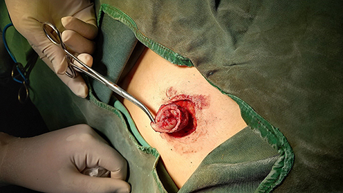 |
Figure 2 Umbilical nodules with no relation to the intraperitoneal organs. |
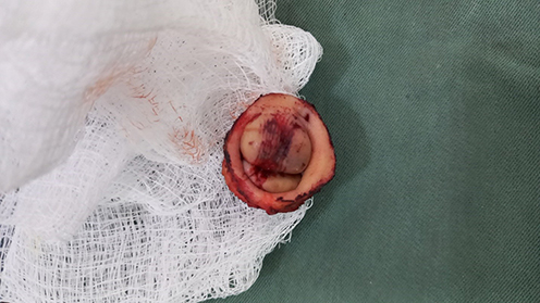 |
Figure 3 Excised umbilicus. |
Discussion
Endometriosis is a benign pathology of women of reproductive age.6 The highest incidence of the disease is at the age of 30–45 years.6 There are several theories about the development of the endometriosis, some authors believe that endometriosis develops from pluripotent cells of the coelom.7 This theory explain that endometriosis can develop in every organ that contains coelomic epithelium including pelvis, umbilicus and hernial sac.7 Embolization by lymphatic vessels is another theory.7 According to this theory blue dye and other materials can migrate retrograde from the pelvis by lymphatic flow to the umbilicus.7
The most important clinical features of the umbilical endometriosis are pain, bleeding, edema and nodule enlargement during menstruation.8 Histopathological examination is the diagnosis gold standard for the umbilical endometriosis.9
Umbilical endometriosis should be differentiated from pyogenic granuloma, umbilical polyps, melanocytic nevus, hemangioma, dismoid and granular cell tumor, melanoma, keloid, umbilical hernia and omphelitis.10 Surgical excision is the only treatment of choice for umbilical endometriosis.4
Conclusion
Primary umbilical endometriosis is a rare condition that should be ruled out from other pathologies of the umbilicus. Diagnosis is made by histopathological study and the treatment of the umbilical endometriosis is surgical excision.
Ethical Approval
This report does not contain any personal information that could lead to the identification of the patient, therefore it is exempt from ethical approval.
Consent for Publication
A written informed consent was obtained from the patient for the publication of this article and the accompanying images. A copy of the written consent is available for review by the editor-in-chief of this journal upon request.
Acknowledgment
The authors would like to express their sincere gratitude to the patient’s guardians for providing consent to include the case details and photographs in this publication.
Funding
No funding was received for this case report.
Disclosure
The authors declare that they have no competing interest in this work.
References
1. Vercellini P, Viganò P, Somigliana E, Fedele L. Endometriosis: pathogenesis and treatment. Nat Rev Endocrinol. 2014;10:261–275. doi:10.1038/nrendo.2013.255
2. Okoye IM, Omiwole O, Adeyoye OM. Primary Umbilical Endometriotic Nodule with an Ultrasound Incidental Finding of Ovarian Endometrioma and a SolitaryIntramural Uterine Myoma: a Case Report. Obs Gynecology Cases-. 2021;8:209. doi:10.23937/2377-9004/1410209
3. BK Daniel BKD, Jens KJ. Primary Umbilical Endometriosis (PUE). Eur J Obstetrics Gynecol Reprod Biol. 2016;9450. doi:10.1016/j.ejogrb.2016.05.030
4. Moniruddin ABM, Raihan HMS, Hasan T, et al. Primary Umbilical Endometriosis (Villar’s Nodule): a RareSymptomatic Umbilical Pathology in An Adult Woman. KYAMC J. 2022;13(1):56–60. doi:10.3329/kyamcj.v13i1.59883
5. Lamoussa Marie Ouedraogo N, Ilboudo S, Ouattara AK, et al. A case report of villa’s nodule in a woman without surgical history. Intern J Surgery Case Rep. 2018;53:186–188. doi:10.1016/j.ijscr.2018.10.066
6. Brown EM, Osswald S, Biediger T. Cutaneous endometriosis of the umbilicus (Villar’s nodule). Int J Women Dermatol. 2020;6:214–215. doi:10.1016/j.ijwd.2020.01.001
7. Dessy LA, Buccheri EM, Chiummariello S, Gagliardi DN, Onesti MG. umbilical endometriosis, our experience. In vivo. 2008;22:811–816.
8. Sengupta M, Naskar A, Gon S, Majumdar B. Villar’s Nodule. Online J Health Allied Scs. 2011;10(1):19.
9. Vega-Castillo JJ, Saenz-Guirado S, Vega-Castillo M, Ruiz-Villaverde R. Umbilical endometriosis: a new dermoscopic pattern. Dermatol Pract Concept. 2022;12(1):e2022023. doi:10.5826/dpc.1201a23
10. Pramanik SR, Mondal S, Paul S, Joycerani D. Primary umbilical endometriosis: a rarity. J Human Reprod Scie. 2014;7(4). doi:10.4103/0974-1208.147495
 © 2024 The Author(s). This work is published and licensed by Dove Medical Press Limited. The
full terms of this license are available at https://www.dovepress.com/terms
and incorporate the Creative Commons Attribution
- Non Commercial (unported, 3.0) License.
By accessing the work you hereby accept the Terms. Non-commercial uses of the work are permitted
without any further permission from Dove Medical Press Limited, provided the work is properly
attributed. For permission for commercial use of this work, please see paragraphs 4.2 and 5 of our Terms.
© 2024 The Author(s). This work is published and licensed by Dove Medical Press Limited. The
full terms of this license are available at https://www.dovepress.com/terms
and incorporate the Creative Commons Attribution
- Non Commercial (unported, 3.0) License.
By accessing the work you hereby accept the Terms. Non-commercial uses of the work are permitted
without any further permission from Dove Medical Press Limited, provided the work is properly
attributed. For permission for commercial use of this work, please see paragraphs 4.2 and 5 of our Terms.