
Back to Journals » Clinical and Experimental Gastroenterology » Volume 19
Primary Colorectal Signet Ring Cell Carcinoma and the Risk of Multiple Primary Gastrointestinal Malignancies: A Retrospective Cohort Based on SEER Database from 2000-2021
Authors Hamed BM, Ellaithy A, Alesawy AF, Alhusban A, Ali N, Al-Shaikh B  , Elshennawy EM, Elbadry SA
Received 13 December 2025
Accepted for publication 17 May 2026
Published 27 May 2026 Volume 2026:19 588726
DOI https://doi.org/10.2147/CEG.S588726
Checked for plagiarism Yes
Review by Single anonymous peer review
Peer reviewer comments 2
Editor who approved publication: Professor Vipul Yagnik
Belal Mohamed Hamed,1 Asmaa Ellaithy,2 Alzahraa Faris Alesawy,3 Alhareth Alhusban,4 Nahla Ali,5 Bushra Al-Shaikh,6 Eslam Mohamed Elshennawy,7 Sally Ali Elbadry8
1Faculty of Medicine, Al-Azhar University, Cairo, Egypt; 2Faculty of Medicine, Suez Canal University, Ismailia, Egypt; 3Faculty of Medicine, Benha University, Qalubiya, Egypt; 4Faculty of Medicine, The University of Jordan, Amman, Jordan; 5Faculty of Medicine, Alexandria University, Alexandria, Egypt; 6Faculty of Medicine, Sana’a University, Sana’a, Yemen; 7Faculty of Medicine, Kafr Alsheikh University, Kafr Alshaikh, Egypt; 8Internal Medicine and Gastroenterology Department, Suez Canal University Hospital, Ismailia, Egypt
Correspondence: Bushra Al-Shaikh, Faculty of Medicine, Sana’a University, Sana’a, Yemen, Email [email protected]
Background: Signet-ring cell carcinoma (SRCC) is a rare subtype of colorectal cancer, constituting 0.5– 2.5% of all adenocarcinomas. It is characterized by signet ring cells as the dominant malignant cell type. Developing gastrointestinal (GI) second primary malignancies (SPMs) after SRCC is a rare event reported in the literature and insufficiently addressed. This study aimed to explore this gap and provide updated evidence about this rare cancer.
Methods: Data were extracted from the Surveillance, Epidemiology, and End Results (SEER) database. Standardized incidence ratio (SIR) analyses with multiple outcome assessment were performed, applying a two-month latency exclusion period to evaluate the risk of GI SPMs in patients diagnosed with primary colorectal SRCC. The SIR was calculated as observed/expected (O/E), with excess absolute risk (EAR) per 10,000. Significance was achieved at 0.05 with a 95% confidence interval (CI).
Results: There was an increased risk for GI SPMs after SRCC in the 2– 11 months interval (O/E=2.49, P< 0.05, EAR=37.31), and over 10 years of follow-up (O/E=3.08, P< 0.05, EAR=59.20). Small intestine SPMs in the 2– 11 months interval had an O/E of 4.17 (P< 0.05, 95% CI: 0.11– 23.26, EAR=1.97), with an overall O/E of 9.62 (P< 0.05, EAR=6.32). No events of GI SPMs were observed among young patients during follow-up (O/E=1.05, P> 0.05, EAR=0.12). Young patients had lack of observed events for GI SPMs (O/E=0.00, EAR=− 0.15), compared to middle-aged (O/E=7.66, P< 0.05) and elderly patients (O/E=2.36, P< 0.05). Patients received chemotherapy showed a slightly higher observed incidence of SPMs (O/E=2.39, P< 0.05, EAR=49.29); however, this finding should be interpreted cautiously given known limitations of chemotherapy data within the SEER database.
Conclusion: Patients diagnosed with colorectal SRCC are at a significantly increased risk of developing GI SPMs. Given the poor overall prognosis of colorectal SRCC, early surveillance protocols must be carefully contextualized at short intervals (6– 12 months) for early detection of GI SPMs. However, a reduced intensity surveillance is recommended beyond 3– 5 years to integrate prevention with long-term follow-up. Recommendations should be tailored according to patients’ risk profiles with individualized patient focused programs.
Keywords: signet-ring cell carcinoma, colorectal cancer, second primary malignancies, gastrointestinal malignancies, SEER database, surveillance
Introduction
Colorectal cancer (CRC) is the third most common cancer worldwide.1 Signet-ring cell carcinoma (SRCC) is a rare subtype of CRC, accounting for less than 1% of all CRC cases and approximately 1.39% of rectal cancers.2 It is characterized histologically by signet ring morphology, where abundant cytoplasmic mucin pushes the nucleus to the peripheral aspect of the cell.3,4 Colorectal SRCC is associated with a poor prognosis due to advanced tumor stage at diagnosis, younger age at onset, and late diagnosis.2,3 The development of second primary malignancies (SPMs) after SRCC has been found to worsen the survival outcomes.5,6 The risk of SPMs in the oesophagus, small intestine, colon, and rectum has been reported to be higher in patients with SRCC than classical adenocarcinoma.5 However, due to the scarcity of SRCC, there is a lack of comprehensive studies examining the association between SPMs and colorectal SRCC.
The underlying mechanism of SPMs is unknown. However, genetic predisposition, environmental factors, and the side effects of primary malignancy treatment may contribute to SPM development.6 Hereditary cancer syndromes may also play a role in the development of multiple gastrointestinal malignancies. Germline mutations in the CDH1 gene, associated with hereditary diffuse gastric cancer (HDGC), have been linked to diffuse gastric carcinoma and occasionally colorectal tumours with signet-ring cell morphology.7 In addition, Lynch syndrome, caused by mutations in DNA mismatch repair genes (MLH1, MSH2, MSH6, and PMS2), predisposes individuals to several gastrointestinal cancers including colorectal, gastric, and small intestinal malignancies.8 These mechanisms may partially explain the clustering of gastrointestinal second primary malignancies observed in patients with colorectal SRCC. Some studies have explored SPM risk in CRC, focusing on the demographics, prognostic factors, clinical aspects, and molecular diagnosis of SRCC.4,6,9 However, risk of GI SPMs after colorectal SRCC is insufficiently addressed in the literature.
This study aimed to bridge this essential literature gap. Understanding the demographics and basic characteristics of this vulnerable population is fundamental to develop effective surveillance and management strategies for early detection of GI SPMs and improving long-term outcomes. The aim of this study was to assess the incidence and risk factors associated with GI SPMs in SRCC patients to provide updated evidence about this rare cancer and reduce the burden of GI SPMs.
Methods
Study Design and Setting
This retrospective cohort study utilized the NCI’s SEER database (17 registries, 2000–2021), which covers approximately 28% of the US population. The database provides comprehensive cancer incidence, survival, and demographic data linked to time-dependent county attributes (eg, income and rurality). Data were linked by state-county and year of diagnosis. The study was reported in line with the STROCSS criteria,10 providing structured recommendations to improve the transparency, clarity, follow-up, and outcome definitions of this cohort SEER-based study.
Participants
Patients diagnosed with primary colorectal SRCC between 2000 and 2021 were included. Demographic characteristics extracted included race (White, Black, and other races: American Indian/Alaska Native and Asian/Pacific Islander), age (pediatrics: <18 years, young adults: 18–39 years, middle-aged: 40–64 years, elderly: ≥65 years, according to the WHO classification,11 marital status (married, unmarried, or unknown), and year of diagnosis. Tumor-related variables included stage (in situ, localized, regional, distant, per SEER staging definitions,12 tumor grade (well-differentiated/grade I, moderately differentiated/grade II, poorly differentiated/grade III, undifferentiated anaplastic/grade IV, and unknown grade), and treatment-related variables (chemotherapy, radiotherapy, and surgery).
Statistical Analysis and Variables
The event was defined as the occurrence of an SPM in a different site from the original cancer at a new GI organ (oesophagus, stomach, small intestine, cecum, appendix, rectum, anus and anal canal, liver, gallbladder, intrahepatic bile duct, pancreas, retro-peritoneum, and colon). The classification follows SEER*Stat definitions, which distinguish SPMs from recurrences or metastases of the primary cancer.12
Two sessions were used in SEER*Stat software version 8.4.3: Frequency session and multiple primary standardized incidence ratio (MP-SIR). The Frequency session provided descriptive statistics for patient demographics (age, gender, race, and marital status), treatment modalities (radiotherapy, chemotherapy, and surgery), tumour stage, SEER grade, and year of diagnosis.
An MP-SIR session calculated the standardized incidence ratio (SIR) as Observed/Expected (O/E) with a 95% confidence interval (CI), with significance achieved at 0.05. Excess Absolute Risk (EAR) was defined as the additional number of SPM cases per 10,000 individuals beyond the expected general population rate, calculated as the difference between observed and expected cases, multiplied by 10,000 (SEER*Stat MP-SIR Session Documentation). The analysis was restricted to primary cancers (sequence number 0 or 1), with exposure based on the recoded date of diagnosis. Latency exclusion period was set to two months, and the study timeframe was from January, 2000 to December, 2021. Patients were followed for +10 years after primary colorectal SRCC diagnosis, with risk assessed at intervals of 2–11 months, 1–5 years, and 10 years. The statistical model applied event site recodes (ICD-O-3/WHO 2008 for SIR). The SIR was stratified by patient characteristics (age, race, marital status, and gender), SPM site (oesophagus, stomach, small intestine, cecum, appendix, rectum, anus and anal canal, liver, gallbladder, intrahepatic bile duct, pancreas, retro-peritoneum, colon), treatment modality, tumour stage, grade, and year of diagnosis to provide insights into incidence variations.
Ethical Considerations
This study was retrospective in design and needed no formal consent, used publicly available, de-identified data from the SEER database. Because the dataset contains no personally identifiable information, institutional review board (IRB) approval was not required for this study.
Results
Baseline Characteristics
Among 8171 patients with colorectal SRCC, about 4275 (52.3%) were males and 47.7% were female. Age distribution showed 54.3% were elderly (n=4434, mean=73.96), followed by middle-aged patients (44.3%). Paediatrics and young adults rarely developed colorectal SRCC (1.4%) as shown in Table 1. Most patients were white (83.1%), while black represented 713 (8.7%), and other races accounted for 7.9% only. The majority had grade III (57.9%), while grade IV represented only 8.3%. Colorectal SRCC was most frequently diagnosed at regional spread stage (42.0%) or after distant spread (41.4%), with fewer cases at localized site (13.2%) or in situ (0.1%). Regarding treatment: 6392 patients had surgical management (78.2%), 4304 received chemotherapy (52.7%), and 943 received radiotherapy (11.7%). Overall, colorectal SRCC was more prevalent among males, elderly patients, and white race, typically diagnosed at an advanced stage with poorly differentiated histological grade. Surgery was the most common management modality, followed by chemotherapy, while radiotherapy played a limited role.
 |
Table 1 Baseline Characteristics for Patients with GI SPMs After the Diagnosis with Colorectal SRCC |
GI SPMs After Primary Colorectal SRCC
This study highlights the demographics and characteristics of patients diagnosed with GI SPMs after primary colorectal SRCC. Males had more risk to develop colorectal SRCC compared to females with observed events of 93 versus 81 along +10 years of follow-up with mean ages of 65.04 and 66.69 years, respectively as shown in Table 2. The highest observed number of events was among the elderly (mean age at event was 73.96), with a rare event of GI SPMs in patients younger than 25 years. The mean age for married patients was 66.22 and was higher compared to single patients or unknown marital status. The white race had the highest risk of GI SPMs (observed=147, mean age at event =66.26 years), with most events diagnosed within 1–5 years after primary SRCC diagnosis. Most colorectal SRCC survivors with GI SPMs were diagnosed between 2011–2020 (Observed=110, mean age=66.14). Regional spread for colorectal SRCC had 74 observed cases with GI SPMs and the mean age was 67.26, making it the most common stage.
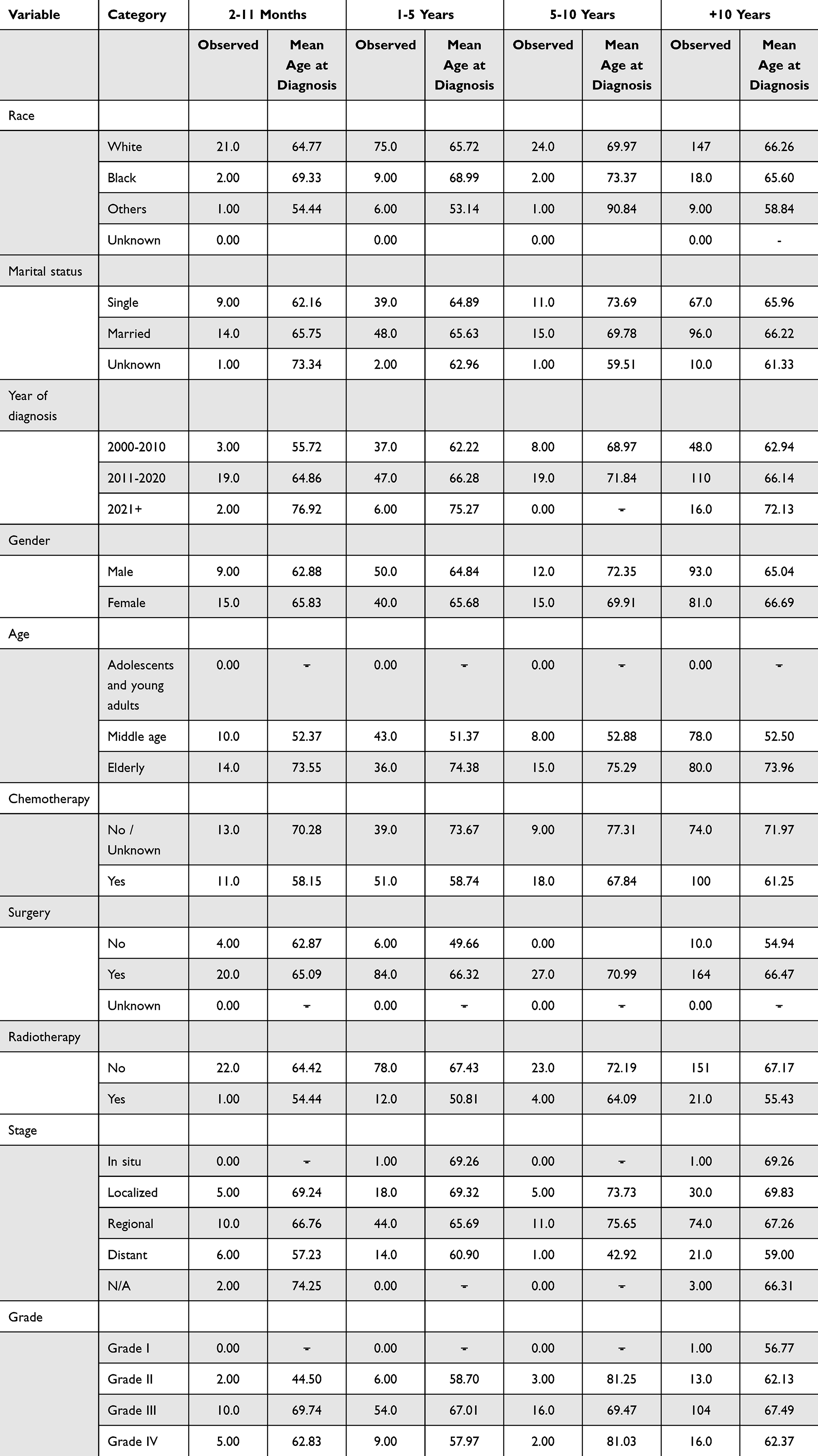 |
Table 2 General Characteristics of Patients with GI SPM After Colorectal SRCC with the Observed Events and Mean Age at Diagnosis Along Different Follow-up Time Intervals |
Colorectal SRCC showed a significant increase in the overall risk of GI SPMs across all sites (O/E=1.57, P<0.05, 95% CI: 1.43–1.72, EAR=85.83) as shown in Table 3 and Figure 1. The risk was significant in the 2–11 months follow-up interval (O/E=1.79, P<0.05, 95% CI: 1.56–2.04, EAR=114.35), with most SPMs arising from the digestive system (O/E=3.00, P<0.05, 95% CI: 2.57–3.47, EAR=58.7). Over 10 years of follow-up, the risk remained high (O/E=3.08, P<0.05, 95% CI: 2.65–3.56, EAR=59.20). Specific GI organs with significantly increased SPMs risk included the rectum (O/E=3.97, P<0.05, 95% CI: 2.49–6.00, EAR=8.19), appendix (O/E=8.44, P<0.05, 95% CI: 2.30–21.61, EAR=1.76), small intestine (O/E=9.45, P<0.05, 95% CI: 5.17–15.86, EAR=6.23), cecum (O/E=3.19, P<0.05, 95% CI: 1.92–4.98, EAR=5.12), and the ascending colon (O/E=3.38, P<0.05, 95% CI: 1.83–5.71, EAR=4.67). Oesophageal SPM risk was increased along +10 years of follow-up (O/E=2.45, P<0.05, 95% CI: 1.06–4.83, EAR=2.36), particularly in the 12–59 months interval (O/E=3.79, P<0.05, 95% CI: 1.23–8.84, EAR=4.32). In contrast, no significant risk was observed for liver and intrahepatic bile duct SPMs (O/E=1.05, P>0.05, 95% CI: 0.45–2.06, EAR=0.12), pancreatic SPMs (O/E=0.93, P>0.05, 95% CI: 0.35–2.02, EAR=−0.15), hepatobiliary system SPMs (including gallbladder and other biliary; O/E=1.15, P>0.05, 95% CI: 0.24–3.37, EAR=0.12). There was an insignificant decrease in SPMs risk for the gum and floor of the mouth (O/E=0.88, P>0.05).
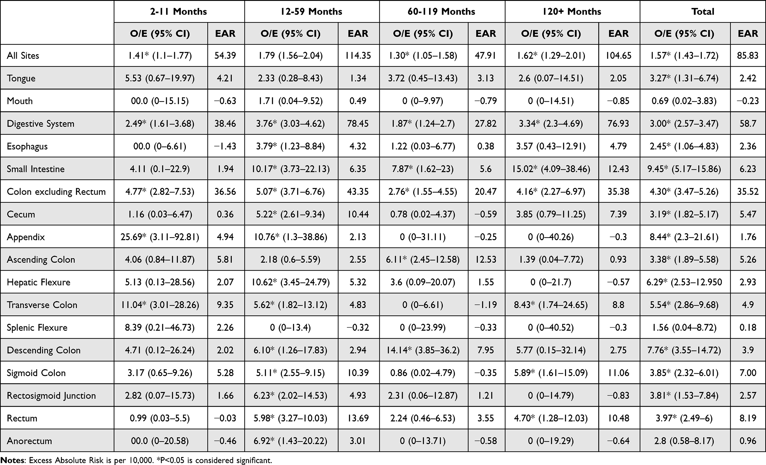 |
Table 3 The Risk of SPMs in Colorectal SRCC Along Different Follow-up Time Intervals in Different GI Sites |
 |
Figure 1 Shows the SPM risk in colorectal SRCC along + 10 years of follow-up in different GI sites relating the SIR to the observed cases. |
Discussion
Colorectal SRCC was more prevalent among males, consistent with global research on colorectal CRC incidence disparities.4,13 However, Hyngstrom et al, using national cancer database data, reported no significant gender differences in SRCC prevalence,14 underscoring the need for further research to resolve these conflicting findings.
Younger patients were less likely to develop colorectal SRCC, aligning with the findings of Jiang et al and Chen et al, who noted a higher incidence in older populations.15,16 Similarly, Nissan et al, in studies from 1986 to 1997, identified a significant correlation between old age and colorectal SRCC incidence.17 These findings support the observed trend of higher SRCC diagnosis rates among the elderly in this study.
Significant racial disparities were observed, with white patients at a markedly higher risk of developing colorectal SRCC, as corroborated by Hyngstrom et al14 Chew et al reported comparable results and highlighted prognostic differences between Asian and white populations.18
Colorectal SRCC is often characterized by advanced disease stage and grade at diagnosis, including large tumor size and high rates of lymph node and peritoneal metastasis,13,16 consistent with the findings of this study that high-grade colorectal SRCC was more likely to develop GI SPMs, although a notable predisposition for GI SPMs was observed in lower-stage disease, such as regional spread.
A pivotal finding of this study was the pronounced susceptibility of colorectal SRCC survivors to secondary luminal GI malignancies. We observed a highly targeted risk profiles, with significant excess risks in the small intestine (O/E=9.45), appendix (O/E=8.44), rectum (O/E=3.97), cecum (O/E=3.19), ascending colon (O/E=3.38), and esophagus (O/E=2.45). Notably, solid accessory organs—including the liver, pancreas, and biliary tract—showed no significant risk, suggesting a vulnerability localized specifically to the GI tract.
This localized risk is likely multifactorial, potentially driven by “field cancerization”, where the shared mucosal lining is sensitized by similar dietary carcinogens and microbiome dysbiosis. Furthermore, shared genetic predispositions and molecular pathways, such as mismatch repair deficiency common in SRCC histologies, may confer a broader vulnerability across the GI epithelium. While late oncogenic effects from primary therapies like pelvic irradiation likely contribute to localized lesions, the widespread GI risk points strongly toward shared environmental and genetic origins, warranting further molecular and genomic profiling.
No specific treatment guidelines exist for colorectal SRCC beyond general CRC protocols. Surgery remains the cornerstone for resectable SRCC, with limited high-quality trials evaluating its efficacy.19 Nearly all patients in this study underwent surgical resection. Fu et al identified an association between poor post-surgical prognosis and factors such as male gender,20 aligning with our results.
The results show a slightly increased risk of GI SPMs following chemotherapy (O/E=2.39, EAR=49.29), but this association should be interpreted cautiously. This incomplete data precludes definitive conclusions about chemotherapy as a direct cause of SPMs in colorectal SRCC survivors. Further research with comprehensive treatment records is recommended for future directions to clarify this association.
Strengths and Weaknesses
This study leverages the comprehensive SEER database, covering 28% of the US population, providing robust and generalizable data on the rare colorectal SRCC. The 10-year follow-up period enabled a thorough assessment of long-term GI SPM risk, emphasizing the need for sustained surveillance. However, the retrospective design introduces potential biases, and small sample sizes in certain subgroups may limit statistical power. Additionally, the lack of detailed treatment data in the SEER database restricts the ability to evaluate specific therapeutic impacts comprehensively (eg: chemotherapy) SEER often underreports systemic therapies and lacks granularity regarding specific agents, dosages, and durations.
Conclusion
This study provides critical insights into the demographic characteristics, therapeutic approaches, and incidence of SPMs in colorectal SRCC. The aggressive nature of SRCC is evident, marked by frequent advanced-stage diagnosis and a significant risk of GI SPMs, particularly in the rectum, appendix, small intestine, cecum, ascending colon, and oesophagus. The results highlight a strong predisposition for GI SPMs, especially within the first-year post-diagnosis, with statistically significant increased risks in the aforementioned sites. Conversely, no notable SPM risk was observed for the gum and floor of the mouth. Given the poor overall prognosis of colorectal SRCC, early surveillance protocols must be carefully contextualized at short intervals (6–12 months) for early detection for any new lesions through imaging, endoscopy, biomarkers follow-up and multidisciplinary team follow-up. However, a reduced intensity surveillance is recommended beyond 3–5 years to integrate prevention with long-term follow-up. Recommendations should be tailored according to patients’ risk profiles with individualized patient focused programs.
Data Sharing Statement
The datasets generated during and/or analysed during the current study are available from the corresponding author on reasonable request.
Ethical Approval
No ethical approval was required as the data used is publicly available and personal data are concealed.
Author Contributions
All authors made a significant contribution to the work reported, whether in the conception, study design, execution, acquisition of data, analysis and interpretation, or in all these areas; took part in drafting, revising or critically reviewing the article; gave final approval of the version to be published; have agreed on the journal to which the article has been submitted; and agree to be accountable for all aspects of the work.
Funding
The authors declare that no funds, grants, or other support were received during the preparation of this manuscript.
Disclosure
The authors have no relevant financial or non-financial interests to disclose. The abstract of this paper was presented at the European Society for Medical oncology (ESMO) GI Conference as a poster presentation with interim findings. The poster’s abstract was published in annals of oncology: (Doi: 10.1016/j.annonc.2024.10.109)
References
1. Korphaisarn K, Morris V, Davis JS, et al. Signet ring cell colorectal cancer: genomic insights into a rare subpopulation of colorectal adenocarcinoma. Br J Cancer. 2019;121(6):505–10. doi:10.1038/s41416-019-0548-9
2. Seog WJ, Dhruv S, Atodaria KP, Polavarapu A. An extremely rare case of rectal signet ring cell carcinoma. Gastroenterol Res. 2022;15(2):106–111. doi:10.14740/gr1516
3. Emile SH, Horesh N, Garoufalia Z, Gefen R, Strassmann V, Wexner SD. Propensity-score matched analysis of survival outcomes of adjuvant therapy in stage II-III signet-ring cell carcinoma of the colon. Clin Colorectal Cancer. 2024;23(1):35–45. doi:10.1016/j.clcc.2023.10.006
4. Weng MT, Chao KH, Tung CC, et al. Characteristics of primary signet ring cell carcinoma of colon and rectum: a case control study. BMC Gastroenterol. 2022;22(1):173. doi:10.1186/s12876-022-02258-1
5. Wu M, Huang M, He C, et al. Risk of second primary malignancies based on the histological subtypes of colorectal cancer. Front Oncol. 2021;11:650937. doi:10.3389/fonc.2021.650937
6. Chen B, Liu B, Yuan Z, et al. Colorectal signet ring cell carcinoma presenting as ulcerating rectosigmoid stricture. ACG Case Rep J. 2023;10(8):e01130. doi:10.14309/crj.0000000000001130
7. Guilford P, Hopkins J, Harraway J, et al. E-cadherin germline mutations in familial gastric cancer. Nature. 1998;392(6674):402–405. doi:10.1038/32918
8. Hampel H, Frankel WL, Martin E, et al. Screening for the lynch syndrome (hereditary nonpolyposis colorectal cancer). N Engl J Med. 2005;352(18):1851–1860. doi:10.1056/NEJMoa043146
9. An Y, Zhou J, Lin G, et al. Clinicopathological and molecular characteristics of colorectal signet ring cell carcinoma: a review. Pathol Oncol Res. 2021;27:1609859. doi:10.3389/pore.2021.1609859
10. Mathew G, Agha R, Albrecht J, et al. STROCSS 2021: strengthening the reporting of cohort, cross-sectional and case-control studies in surgery. Ann Med Surg. 2021;72:103026. doi:10.1016/j.amsu.2021.103026
11. World Health Organization. International Statistical Classification of Diseases and Related Health Problems (ICD-10).
12. SEER program coding and staging manual 2023. 2023.
13. Hugen N, Verhoeven RH, Lemmens VE, et al. Colorectal signet-ring cell carcinoma: benefit from adjuvant chemotherapy but a poor prognostic factor: outcome and adjuvant chemotherapy in signet-ring cell carcinoma. Int J Cancer. 2015;136(2):333–339. doi:10.1002/ijc.28981
14. Hyngstrom JR, Hu CY, Xing Y, et al. Clinicopathology and outcomes for mucinous and signet ring colorectal adenocarcinoma: analysis from the national cancer data base. Ann Surg Oncol. 2012;19(9):2814–2821. doi:10.1245/s10434-012-2321-7
15. Jiang H, Shao D, Zhao P, Wu Y. The predictive and guidance value of signet ring cell histology for stage II/III colon cancer response to chemotherapy. Front Oncol. 2021;11:631995. doi:10.3389/fonc.2021.631995
16. Yeh CY, Wang JY, Chen JS, et al. Clinical significance of signet ring cell rectal carcinoma. Int J Colorectal Dis. 2004;19(2):102–107. doi:10.1007/s00384-003-0515-y
17. Nissan A, Guillem JG, Paty PB, Wong DW, Cohen AM. Signet-ring cell carcinoma of the colon and rectum: a matched control study. Dis Colon Rectum. 1999;42(9):1176–1180. doi:10.1007/BF02238570
18. Chew MH, Yeo SAE, Ng ZP, et al. Critical analysis of mucin and signet ring cell as prognostic factors in an Asian population of 2764 sporadic colorectal cancers. Int J Colorectal Dis. 2010;25(10):1221–1229. doi:10.1007/s00384-010-1033-3
19. Nitsche U, Zimmermann A, Späth C, et al. Mucinous and signet-ring cell colorectal cancers differ from classical adenocarcinomas in tumor biology and prognosis. Ann Surg. 2013;258(5):775–783. doi:10.1097/SLA.0b013e3182a69f7e
20. Fu J, Wu L, Jiang M, et al. Signet ring cell carcinoma of resectable metastatic colorectal cancer has rare surgical value. J Surg Oncol. 2016;114(8):1004–1008. doi:10.1002/jso.24437
 © 2026 The Author(s). This work is published and licensed by Dove Medical Press Limited. The
full terms of this license are available at https://www.dovepress.com/terms
and incorporate the Creative Commons Attribution
- Non Commercial (unported, 4.0) License.
By accessing the work you hereby accept the Terms. Non-commercial uses of the work are permitted
without any further permission from Dove Medical Press Limited, provided the work is properly
attributed. For permission for commercial use of this work, please see paragraphs 4.2 and 5 of our Terms.
© 2026 The Author(s). This work is published and licensed by Dove Medical Press Limited. The
full terms of this license are available at https://www.dovepress.com/terms
and incorporate the Creative Commons Attribution
- Non Commercial (unported, 4.0) License.
By accessing the work you hereby accept the Terms. Non-commercial uses of the work are permitted
without any further permission from Dove Medical Press Limited, provided the work is properly
attributed. For permission for commercial use of this work, please see paragraphs 4.2 and 5 of our Terms.
Recommended articles

Updated Perspectives on the Diagnosis and Management of Familial Adenomatous Polyposis
Kyriakidis F, Kogias D, Venou TM, Karlafti E, Paramythiotis D
The Application of Clinical Genetics 2023, 16:139-153
Published Date: 14 August 2023