
Back to Journals » International Medical Case Reports Journal » Volume 18
Pitfall of Diagnosis Left-Sided Appendicitis Due to Undiagnosed Intestinal Malrotation in Adulthood – a Rare Case Report from Syria and Literature Review
Authors Yousef A, Mohaisen MAB  , Tassabehji O, Atia M, Saleh MM, Saleh MI, Aasi M
Received 15 August 2025
Accepted for publication 25 November 2025
Published 18 December 2025 Volume 2025:18 Pages 1625—1629
DOI https://doi.org/10.2147/IMCRJ.S557979
Checked for plagiarism Yes
Review by Single anonymous peer review
Peer reviewer comments 2
Editor who approved publication: Professor Thomas E Hutson
Ahmad Yousef,1 Mais Alreem Basel Mohaisen,2 Omar Tassabehji,2 Mohammad Atia,3 Mohammad Moaz Saleh,2 Mohammad Ihab Saleh,4 Muataz Aasi1
1Department of General Surgery, Al-Rashid Hospital, Damascus, Syrian Arab Republic; 2Faculty of Medicine, Al-Sham Private University, Damascus, Syrian Arab Republic; 3Department of Gastroenterology, Al-Moujtahed Hospital, Damascus, Syrian Arab Republic; 4Department of Orthopedics, Al-Zabadani Hospital, Damascus, Syrian Arab Republic
Correspondence: Ahmad Yousef, Department of General Surgery, Al-Rashid Hospital, Damascus, Syrian Arab Republic, Tel +963932044578, Email [email protected]
Abstract: Intestinal malrotation, which affects 0.03 to 0.5% of newborns, can induce rare left-sided appendicitis and also go undetected until adulthood because of its unusual clinical signs. Even while it frequently occurs with other congenital defects, it can also manifest in adulthood as a separate abnormality. In this case report, we report on a 33-year-old female patient who has left-sided appendicitis because of intestinal malrotation, an uncommon and challenging illness for which early identification and treatment are essential for a successful outcome.
Keywords: appendectomy, intestinal malrotation, left-sided appendicitis, case report
Introduction
Gastrointestinal malrotations, while frequently associated with other congenital anomalies, often occur as isolated features in adult cases.1 The incidence of reported intestinal malrotation in live births ranges from 0.03% to 0.5%, though the precise incidence in adults remains undetermined.2 Appendicitis in patients with intestinal malrotation represents a relatively rare surgical challenge.1,3 True left-sided acute appendicitis arises in conjunction with congenital anomalies such as situs inversus and intestinal malrotation.4 The latter is a congenital positional anomaly resulting from the non-rotation or incomplete rotation of the primitive intestinal loop around the superior mesenteric artery, occurring in approximately 1 in 500 births.5 While most cases are diagnosed within the first year of life, the incidence in adults is largely unknown. A 2010 review documented 95 cases of left-sided appendicitis, with 23 associated with intestinal malrotation.2,4 When intestinal malrotation is present, acute appendicitis can be difficult to diagnose since it can frequently mimic a wide range of gastrointestinal and genitourinary disorders. In order to enable rapid diagnosis and intervention, emergency doctors, radiologists, and surgeons must have a better understanding of this anatomical variation.2,5 Based on the stage at which the embryologic error occurs, there are three main types of midgut malrotation, a rare fetal anomaly caused by incomplete or failed midgut rotation and fixation during embryonic development: non-rotation (type I), duodenal malrotation (type II), and combined duodenal and cecal malrotation (type III).5 While most cases present early in life, less than 0.5% are identified in adulthood.4,5
Case Presentation
A 33-year-old female presented to the emergency department on November 7, 2024. With severe generalised abdominal pain that began on the day before admission, the pain ultimately localized to the right iliac fossa. In addition, she suffered from anorexia, nausea, vomiting, and a fever of 38°C. In addition to having regular bowel and bladder habits, the patient had no history of chronic disease or surgeries. There were no signs of intestinal obstruction. On Examination, McBurney’s sign was positive. Rovsing’s sign was positive with Rebounding pain. Abdominal and pelvic ultrasound (US) results were normal. No further radiological exams were performed. The patient’s BMI was 27 kg/m2. Laboratory Examination Results: WBC count: 11,000, Neutrophils: 86%, Uric acid: within normal range. Other laboratory parameters were normal: Hemoglobin, serum electrolytes, and renal functions did not reveal any deviations, thus eliminating the absence of systemic disturbances or any impairment due to the acute abdomen. Surgical Procedures:
The patient underwent a laparoscopic appendectomy with general anesthesia, with initial entry via a reversed needle, and pneumoperitoneum was achieved at 15mmHg pressure, with the release of adhesions present in the right iliac fossa.
It is, however, pertinent to point out that malrotation was present, as seen intraoperatively, with the cecum and appendix bases present in the left hypochondrium, and the appendix head, which was swollen due to inflammation, present in the right iliac fossa (Figures 1–5). Appendectomy was achieved by ligature resection, with clipping at the appendix base, with exploration showing malrotation, yet with no evidence or history or symptoms related to the impairment of intestinal or bowel habits, a gutter drain was inserted, and the procedure was completed uneventfully.
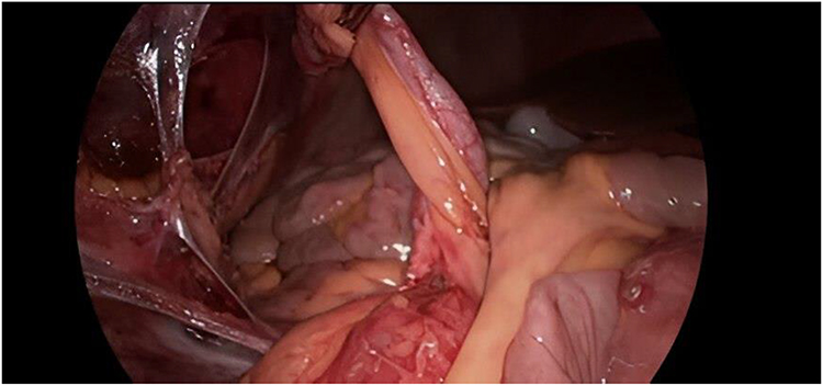 |
Figure 1 The appendix, the liver with the gallbladder, and the ileum. |
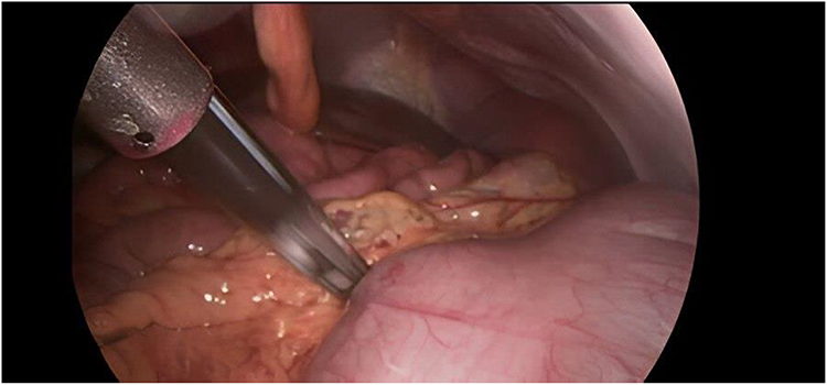 |
Figure 2 The cecum and liver. |
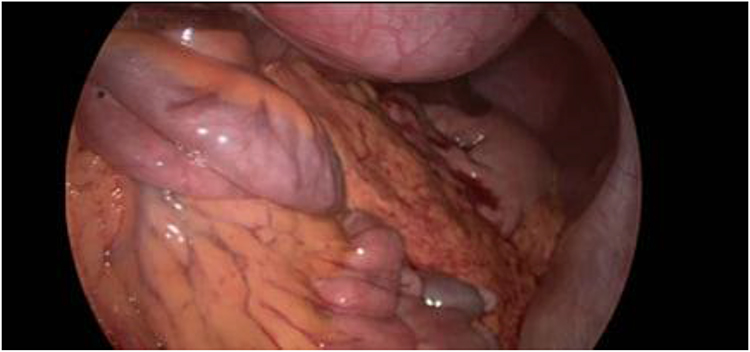 |
Figure 3 The stomach and the cecum. |
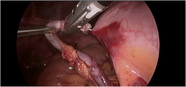 |
Figure 4 The base of the appendix, the cecum, and the small intestines. |
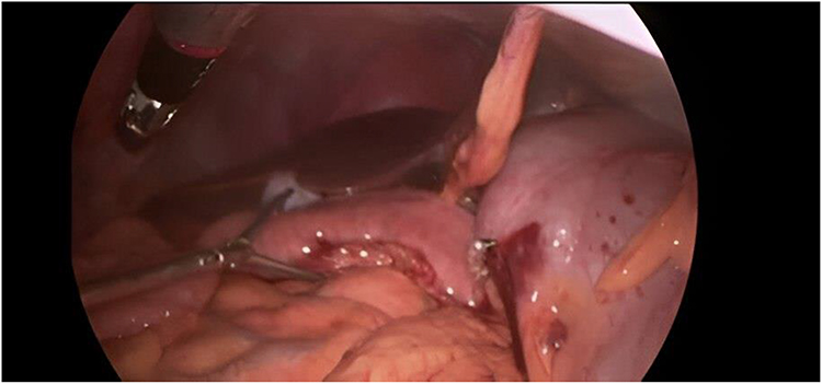 |
Figure 5 Position of the appendix after laparoscopic appendectomy, showing the appendix tip still located in the right iliac fossa, despite malrotation of the cecum to the left upper quadrant. |
Discussion
Recognizing left-sided acute appendicitis early on, especially when paired with intestinal malrotation, is vital due to its rarity and the complexities it brings to diagnosis.6 Intestinal malrotation is estimated to occur in 0.03% to 0.5% of live births, with even rarer instances identified in adulthood, primarily due to the difficulties in clinically diagnosing left-sided acute appendicitis in adults, which often presents with non-specific symptoms and infrequently prompts consideration of malrotation.7 Published literature indicates that left-sided acute appendicitis manifests between the ages of 8 and 63 years, with a prevalence 1.5 times greater in men than in women.8 Our patient was a 33-year-old woman, consistent with the reported age range. The congenital anomaly of intestinal malrotation, which typically occurs due to non-rotation or incomplete rotation of the primitive intestinal loop around the superior mesenteric artery (SMA), complicates the clinical presentation of acute appendicitis.2 Two anatomical anomalies lead to left-sided acute appendicitis: the first is Situs Inversus Totalis (SIT), and the second, a rarer condition, is Midgut Malrotation (MM).9,10 The clinical manifestations of acute appendicitis imitate various gastrointestinal and genitourinary conditions such as gastritis, gastroenteritis, cholecystitis, pyelonephritis, and diverticulitis, among others, resulting in potential cases of misdiagnosis and hence delay in treatment.2,7 In this case, the symptoms were, as they should be, including abdominal pain, vomiting, nausea, and fever, but the uncommon pain areas and anatomy complicated things. The contributory physical findings, including the positivity of McBurney’s sign and Rovsing’s sign, emphasize the importance of a high clinical index of suspicion and specialized imaging studies in similar cases. The efficacy of CT in diagnosing acute appendicitis is well-established, demonstrating an accuracy of 90%-98%. Current literature recommends the utilization of CT scans as the initial diagnostic step following the exclusion of alternative causes using ultrasound.9,11 It cannot be overstated that left-sided appendicitis is extremely rare, especially when intestinal malrotation is present.12 Ladd’s treatment is typically executed when intestinal malrotation manifests as a volvulus, utilizing either an open or laparoscopic technique. However, this procedure is considered optional when malrotation is discovered incidentally or alongside other intra-abdominal conditions such as acute appendicitis.2 Laparoscopic appendectomy is regarded as the gold standard for managing left-sided appendicitis.13,14 In our case, laparoscopic resection led to a quick and complication-free recovery. Only a small number of cases have been reported in previous reviews that highlighting the significance of awareness and early recognition by emergency physicians, radiologists, and surgeons.15
Conclusion
For patients with intestinal malrotation, early diagnosis of left-sided appendicitis is essential for effective therapy and positive results. This case emphasizes the need to examine anatomical abnormalities in the differential diagnosis of abdominal pain, as well as the need for regular education and awareness among healthcare personnel in order to enhance diagnostic accuracy and patient outcomes.
Ethical Approval and Patient Consent
Institutional approval for publication of this case report was obtained from Al-Rashid Hospital, Damascus, Syria. Written informed consent for publication of the clinical details and accompanying images was also obtained from the patient.
Acknowledgment
The authors would like to thank the Scientific Researchers (SRs) Organization, a student-led group based in Damascus, Syria, for their support in this research (contact: [email protected]).
Author Contributions
Mais Alreem Basel Mohaisen, Dr. Mohammad Atia, Omar Tassabehji, Dr. Mohammad Moaz Saleh, and Dr. Mohammad Ihab Saleh contributed to the writing, critical revision, and final approval of the manuscript. Dr. Ahmad Yousef and Dr. Muataz Assi were responsible for data collection and acquisition. Dr. Ahmad Yousef served as the corresponding author. All authors have read and approved the final version of the manuscript and agree to be accountable for all aspects of the work.
Disclosure
The authors report no conflicts of interest in this work.
References
1. Mahat A, Bhusal A, Yadav GK, Mishra U, Duwadi B, Katwal S. Left-sided acute appendicitis in a patient with situs inversus totalis: a case report and a comprehensive review. Radiology Case Reports. 2024;19(3):1020–1025. doi:10.1016/j.radcr.2023.11.073
2. Hoang VT, Van HAT, Hoang TH, Vo TH, Chansomphou V. Left-sided acute appendicitis with congenital gastrointestinal malrotation. J Gastroenterol Hepatol. 2022;6(9):647–648. doi:10.1002/jgh3.12804
3. Kharel H, Pokhrel NB, Kharel Z, Sah D. A case report on left-sided appendicitis with intestinal malrotation. Cureus. 2020;12(1):e6687. doi:10.7759/cureus.6687
4. Chuang PW, Huang BM, Liu CH, Chen CC, Tsai MJ. Left-sided appendicitis in an elderly patient with midgut malrotation. Indian J Surg. 2015;77(Suppl 3):1418–1420. doi:10.1007/s12262-014-1200-9
5. Welte FJ, Grosso M. Left-sided appendicitis in a patient with congenital gastrointestinal malrotation: a case report. J Med Case Rep. 2007;1:92. doi:10.1186/1752-1947-1-92
6. Ahmed W, Ibrahim T, Abbasi MH, Bajwa MA. Left-sided appendicitis in a 14-year-old girl with midgut malrotation. J Coll Physicians Surg Pak. 2018;28(4):317–319. doi:10.29271/jcpsp.2018.04.317
7. Chick JFB, Chauhan NR, Mullen KM, Hanna JW, Bair RJ, Khurana B. Intestinal malrotation and acute left-sided appendicitis. J Emerg Med. 2013;44(4):e333–e334. doi:10.1016/j.jemermed.2012.11.053
8. Perera WR, Hennessy OF. Clinical images. an unusual case of appendicitis. Am J Surg. 2010;199(6):e79–81. doi:10.1016/j.amjsurg.2009.08.047
9. Akbulut S, Ulku A, Senol A, Tas M, Yagmur Y. Left-sided appendicitis: review of 95 published cases and a case report. World J Gastroenterol. 2010;16(44):5598–5602. doi:10.3748/wjg.v16.i44.5598
10. Bhattarai AM, Devkota Y, Bhattarai AM. Left-sided appendicitis with intestinal non-rotation: a case report. JNMA J Nepal Med Assoc. 2022;60(248):396–398. doi:10.31729/jnma.7274
11. Çağlar E, Arıbaş B, Tiken R, Keskin S. Midgut malrotation presenting with left-sided acute appendicitis and CT inversion sign. BMJ Case Rep. 2014;2014. doi:10.1136/bcr-2013-202709.
12. Taslakian B, Issa G, Hourani R, Akel S. Left-sided appendicitis in children with congenital gastrointestinal malrotation: a diagnostic pitfall in the emergency department. BMJ Case Rep. 2013;2013. doi:10.1136/bcr-2013-009474.
13. AlKeileh TB, Elsayed S, Adam RM, Nour M, Bhagchandi T. Left-sided appendicitis managed laparoscopically: a case report. J Med Case Rep. 2024;18(1):21. doi:10.1186/s13256-023-04301-5
14. Kharel H, Pokhrel NB, Kharel Z, Sah D. A case report on left-sided appendicitis with intestinal malrotation case presentation. Cureus. 2020;12(1):1–8. doi:10.7759/cureus.6687
15. Shekhar A, Hendahewa R, Premaratne G. A diagnostic dilemma: left-sided appendicitis in a 10-year-old boy with previously undiagnosed intestinal malrotation. A case report. Int J Surg Case Rep. 2015;14:10–12. doi:10.1016/j.ijscr.2015.06.036
 © 2025 The Author(s). This work is published and licensed by Dove Medical Press Limited. The
full terms of this license are available at https://www.dovepress.com/terms
and incorporate the Creative Commons Attribution
- Non Commercial (unported, 4.0) License.
By accessing the work you hereby accept the Terms. Non-commercial uses of the work are permitted
without any further permission from Dove Medical Press Limited, provided the work is properly
attributed. For permission for commercial use of this work, please see paragraphs 4.2 and 5 of our Terms.
© 2025 The Author(s). This work is published and licensed by Dove Medical Press Limited. The
full terms of this license are available at https://www.dovepress.com/terms
and incorporate the Creative Commons Attribution
- Non Commercial (unported, 4.0) License.
By accessing the work you hereby accept the Terms. Non-commercial uses of the work are permitted
without any further permission from Dove Medical Press Limited, provided the work is properly
attributed. For permission for commercial use of this work, please see paragraphs 4.2 and 5 of our Terms.