Back to Journals » International Journal of Nanomedicine » Volume 20
Phytochemicals in Bone Therapy: Exploring Natural Alternatives for Bone Health
Authors Abdelnabi H, Mohsin S ![]()
Received 24 February 2025
Accepted for publication 14 July 2025
Published 3 September 2025 Volume 2025:20 Pages 10831—10855
DOI https://doi.org/10.2147/IJN.S524695
Checked for plagiarism Yes
Review by Single anonymous peer review
Peer reviewer comments 3
Editor who approved publication: Dr Krishna Nune
Hiba Abdelnabi, Sahar Mohsin
Department of Anatomy, College of Medicine and Health Sciences, United Arab Emirates University, Al Ain, Abu Dhabi, United Arab Emirates
Correspondence: Sahar Mohsin, Department of Anatomy, College of Medicine and Health Sciences, United Arab Emirates University UAEU, P.O. Box 15551, Al Ain, United Arab Emirates, Tel +97137137516, Email [email protected]
Abstract: Bone diseases such as osteoporosis and osteoarthritis are increasingly prevalent, particularly in aging populations. While conventional treatments, including synthetic drugs and mineral supplements, are effective yet often associated with side effects and long-term economic burdens. Active compounds derived from nature, “Phytochemicals” have garnered attention due to their potential to provide safer and more sustainable alternative therapeutic options. However, due to their complex structure and poor pharmacokinetics, their clinical applications are limited. Nano-drug delivery systems address these limitations by developing phytochemical-based nanocarriers, which enable targeted delivery, protect active compounds, and enhance both pharmacokinetics and pharmacodynamics. Given the limitations of synthetic treatments, there is growing interest in exploring phytochemicals and plants and herbal extracts to support bone health. This review focuses on nano-phytochemical approaches for bone therapy, outlining key phytochemicals, their natural sources, nanoformulations, and mechanisms of action. It also evaluates current commercial supplements and highlights the challenges and future directions for clinical translation of nano-phytomedicine in bone health management.
Keywords: phyto-nanomedicine, phytochemicals, bone, natural compounds, plant-based nanoparticles
Graphical Abstract:

Introduction
For centuries, people worldwide have utilized plants and natural ingredients as their primary source of therapy for various diseases and health conditions. Many of the synthetic drugs were derived from a well-known natural source, then their chemical structures were subsequently modified as needed to improve their efficacy, and bioavailability, and reduce their toxicity and side effects.1 However, over time, the use of natural resources for drug discovery has diminished mainly due to the ease in drug chemical structure modifications, difficulties in dosing of herbal medications, time and money involved, and difficulties in issuing patency. As a result of various factors, particularly the toxicity associated with the long-term use of synthetic drugs and their unpredictable side effects, researchers had the urge and interest to go back to nature and discover its active compounds. Additionally, the high cost of drugs and medications, combined with their adverse effects and inconvenient dosages, has led many people to prefer natural products. This refocus on natural ingredients inspired the Japanese to introduce the term “functional food” which identifies the food that has active ingredients.2 Soon after, when the bioactive compounds were formulated into tablets, capsules, liquids, and other pharmaceutical forms the term “nutraceutical” was introduced.2
Phytochemicals are defined as secondary metabolites, bioactive nonessential compounds originating from natural sources.1,3 Furthermore, the advancement in different new medical fields such as genomics, proteomics, transcriptomics, and metabolomics allowed the utilization of these metabolites in drug discovery. Phytochemicals are classified into major groups including phenolics, alkaloids, and terpenes. In plants, they function in reproduction and growth, defense mechanisms against pathogens, and contribute to plants’ color. Most of them exhibit antioxidant, anti-inflammatory, antibacterial, and anticancer activity at various levels.1,3,4 However, despite their functionality and therapeutic potential, they should be thoroughly studied and investigated before their use, due to their concentration-and-structure-dependent side effects, toxicity, drug interaction, and stability concerns.3,5 This highlights the importance of micro/nanocarriers, micro and nano-drug delivery systems (DDS) such as liposomes, micelles, nanotubes, nanofibers, and solid lipid nanoparticles (SLN) along with topically applied hydrogels and devices, and locally implanted scaffolds. These DDS protect the human body from the loaded drugs’ side effects, guard the drug from metabolic degradation, improve its bioavailability (enhances its pharmacokinetics), and allow targeted therapy.6,7 Hence, developing plant-based nanoparticles and DDS facilitates the delivery of huge natural phytochemicals to their site of action.
Bones, the primary component of the skeletal system, are highly metabolically active with a hierarchical structure. Bone tissue undergoes a continuous cycle of formation and resorption, primarily mediated by two key cell types: osteoblasts and osteoclasts. This dynamic process is extremely controlled by complex signaling pathways such as RANK/RANKL pathway, Wnt/β-catenin pathway, bone morphogenetic protein BMP/Smad pathway, nuclear factor kappa-light-chain-enhancer of activated B cells NF-κB pathway. The imbalance in the specific proteins regulating these pathways lead to bone disorders.
Bone-related diseases and problems affect millions of people globally, and their incidence of occurrence is steadily rising. These disorders include osteoporosis, osteoarthritis, rheumatoid arthritis, and bone loss due to injury, fracture, or trauma. Most of these conditions are age-related, and the long-term use of the available effective drugs is associated with many side effects.8 As a result, there is growing attention in alternative medicine that offer comparable therapeutic benefits with improved safety profiles. For example, capsaicin-based topical creams have shown promise in alleviating inflammation associated with rheumatoid arthritis. This review provides an overview of phytochemicals classifications, the application of natural compounds in bone research, and the challenges associated with utilizing natural extracts as drugs.
Bone Disorders and Current Therapeutic Approaches
Bone tissue consists of both organic and inorganic parts. The inorganic fraction is made of water and ions mainly calcium and phosphate in the form of crystalline hydroxyapatite (HA) which is embedded in collagen (COL) fibers. On the other hand, the organic part is composed of different types of cells, proteins, and extracellular matrix (ECM).8 Osteoblasts, bone-forming cells, are located on the lining surfaces of the bone and develop into osteocytes upon mineralization and fixation in mature bone tissues. Conversely, osteoclasts are bone-resorbing cells that function similarly to macrophages.9,10 The bone remodeling process is extremely governed by a highly ordered balanced series of signaling pathways controlling both osteoblasts and osteoclasts.9 One key protein regulating this process is RANKL, a glycoprotein found on the surface of osteoblasts. RANKL acts as a ligand for the RANK glycoprotein receptor on osteoclasts regulating their differentiation, attachment, and activation.9 Any deterioration in bone homeostasis leads to disorders and diseases often due to genetic abnormalities, skeletal injury or defects, developmental problems, chronic inflammation, or bone and joint degeneration.
Osteoporosis is a silent condition that occurs when the bone’s microstructure and mineralization deteriorate, leading to weak, non-load bearing bones that are more susceptible to fractures.11 Osteoporosis is a multifactorial disease related to a number of causes and risk factors such as age, physical inactivity, hormonal change, weight imbalance, underlying diseases like diabetes mellitus type І and II, rheumatoid arthritis, and hyperthyroidism, drug intake, and genetic predisposition.9,12–18 Maintaining an active lifestyle and a proper diet containing sufficient amounts of minerals is the key to the prevention and treatment of osteoporosis.19 Currently available medication for osteoporosis is mainly bisphosphonates like alendronate and zoledronate, calcitonin, and hormonal therapy.19 Bisphosphonates work by inhibiting osteoclast binding and activation, thus suppressing bone resorption and turnover.20 Long-term use of these drugs has many side effects such as chest pain, stomach and esophagus injuries, chronic kidney disease, ulceration, osteonecrosis, and hypocalcemia.11,21
Similarly, osteoarthritis is the most common degenerative disease caused by obesity, aging, or joint injury, where destruction of the articular cartilage, synovial fluid inflammation, and hypertrophy of bones are the main symptoms.11,22 Modifying the lifestyle to a healthier one by weight loss and physical activity is the first line of treatment. For pain control, non-steroidal anti-inflammatory drugs (NSAIDs) are the safest choice after acetaminophen. If the symptoms worsen, stronger analgesics such as Cox-2 inhibitors then opioids could be prescribed. However, these drugs have significant adverse effects, particularly on the kidneys, liver, and gastrointestinal tract.11 Surgical interventions are the last choice for patients who do not improve with pharmacological treatments.
Rheumatoid arthritis (RA) is a chronic, progressive inflammatory disorder caused by immune system dysfunction. RA is characterized by increased bone resorption, decreased bone formation, and joint inflammation.9 Currently used medications for RA are anti-inflammatory drugs (NSAIDs), glucocorticoids, and disease-modifying anti-rheumatic drugs.11 Less pharmacological options and high associated side effects make RA treatment really challenging. As a result, patients usually tend to prefer alternative therapies including minerals, capsaicin (the active compound in chili peppers) as a local analgesic and anti-inflammatory, or glucosamine.23–25 Glucosamine is a naturally occurring amino monosaccharide component of glycosaminoglycans (GAG), located in connective and cartilage tissues.26 Glucosamine is considered a chondroprotective agent due to its function in maintaining cartilage strength, flexibility, and elasticity.26,27 Moreover, glucosamine is the building block for chitosan and chitin, therefore, it is believed that it could stimulate cartilage regeneration.23,27 Yet, deeper investigation should be carried out to confirm its function and any possible side effects.
Critical-size bone defects are bone defects that cannot heal spontaneously. Bone spontaneous repair is limited; injuries exceeding 1–3 cm are classified as critical-size bone defects.28 These defects may result from fractures, traumatic injuries, tumor resection, or congenital diseases.28,29 Critical-size bone defects have been managed by bone grafting with gold standard autogenous grafts,30 or allografts31 and xenografts.31 Nevertheless, all these grafts have drawbacks such as immune rejection, risk of infectivity in both donor and acceptor sites, immunoreaction, donor-site morbidity, and psychological complications.30,31 Therefore, a new alternative solution was introduced using synthetic materials including calcium-based cement, or polymeric scaffolds.32,33 Natural compounds with osteoinductivity are a good promising choice for scaffold functionalization to increase its biocompatibility, bioactivity, and biodegradability.
Main Osteogenesis Signaling Pathways
Mesenchymal stem cells (MSC) in bone tissues proliferate and differentiate into osteoblasts as a response to specific signaling pathways, mainly bone morphogenetic protein 2 (BMP2) and Wnt/β-catenin pathways as shown in Figure 1. Both pathways share some common factors and their activation results in osteogenic differentiation. Bone morphogenetic protein (BMP)s, a subclass of transforming growth factor‐beta (TGF‐β), are essential in skeletal and tissue development acting as growth factors.34,35 Wnt ligands are secreted lipid-modified signaling glycoproteins with 350–400 amino acids long.36
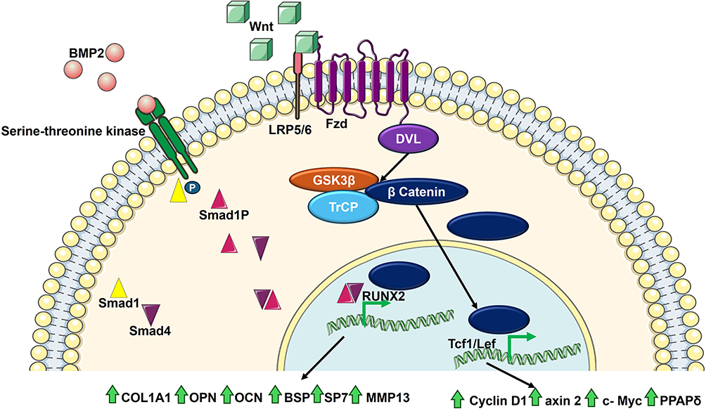 |
Figure 1 Simple schematic representation of BMP2 and Wnt/β-catenin signaling pathways. |
BMP2 binds to its Serine-Threonine kinase receptor, triggering the phosphorylation of Smad1, an intracellular and transcription regulatory protein, which forms a complex with Smad4. This complex then enters the nucleus regulating some gene expression, it recruits runt‐related transcription factors (RUNXs). RUNX2 specifically is a key marker of early osteogenic differentiation. RUNX2 upregulates several osteogenic-related genes mainly secreted phosphoprotein 1 (SPP1) encoding osteopontin (OPN), Bone gamma‐carboxyglutamate (BGLAP) encoding osteocalcin (OCN), collagen type 1 α-1 (COL1A1) encoding type 1 collagen that support and strengthen bone tissues, integrin‐binding sialoprotein (IBSP) encoding bone sialoprotein (BSP) the non-collagen protein in bone matrix, SP7 encoding osterix (OSX), and matrix metalloproteinase 13 (MMP13).36
The canonical Wnt pathway known also as the Wnt/β-catenin signaling pathway is activated by the binding of Wnt ligands to their transmembrane G-protein receptors and low-density lipoprotein (LDL) receptor-related proteins (LRP5/6).36,37 β-catenin has two forms, the phosphorylated-ubiquitinated stable inactive form and the active unphosphorylated one. Glycogen synthase kinase 3β (GSK3β) and β-transducin repeat-containing protein (TrCP) are enzymes causing the phosphorylation and ubiquitination, respectively.36 Wnt 1, 3a, and 8 bind to the frizzled receptor (Fzd), the G-protein, and its coreceptor LRP 5 or 6, resulting in the activation of cytoplasmic phosphoprotein dishevelled (DVL). DVL inhibits GSK3β, resulting in the accumulation of active β-catenin and its internalization to the nucleus, where it binds T cell factor 1 (Tcf1) and lymphoid enhancer factor (Lef) forming a transcription activation complex. This activates the RUNX2 promoter, cyclin D1, axin 2, c-Myc, and peroxisome proliferator‐activated receptor (PPAR‐δ) leading to osteogenic differentiation.36,37 BMP2 itself upregulates Wnt ligands transcription-translation and inhibits TrCP.
Phytochemicals Classes
Phytochemicals are classified into three main classes, phenolics, alkaloids, and terpenes. Figure 2 shows the phytochemical groups and their core chemical structure.
- Phenolics with Aromatic Rings and Hydroxyl Groups. Phenolic acids, flavonoids, tannins, stilbenes, curcuminoids, coumarins, quinones, and lignans are all sub-classes of compounds that contain phenolic functional groups in their molecular structure. According to the structure-activity relationship (SAR), the catechol and hydroxyl (OH) groups are responsible for the antimicrobial, antioxidant, and scavenging activities of these phenolic compounds.38,39 The number and position of OH groups influence bioactivity. Up to four OH groups, especially in an ortho configuration, enhance therapeutic effects.38,40,41
- Phenolic acids including gallic acid, vanillic acid, and caffeic acid enhance immune function and have antiaging, and anti-inflammatory activity.
- Flavonoids are the largest group of phenolics, characterized by their bright colors and sensitivity to environmental factors such as light, temperature, pH, and oxygen. Flavonoids such as luteolin, quercetin, and kaempferol serve as chelating agents, promote cell differentiation, and suppress the activity of nuclear factor kappa-light-chain-enhancer of activated B cells (NF-κB).3 Flavonoids effectively scavenge reactive oxygen species (ROS) and chelate metal ions, reducing oxidative stress and mitigating cellular damage. Their ability to promote cell differentiation is vital for tissue repair and maintaining cellular function. Moreover, by inhibiting the NF-κB activity, a key regulator of immune response to infection, inflammation, and cancer development, flavonoids may contribute to alleviating inflammation and potentially slow cancer progression.3,40,41
- Tannins (Ellagitannins) are water-soluble polyphenols that are hydrolyzed by enzymes or pH change, and they are usually combined with alkaloids, polysaccharides, or proteins forming complexes that can protect tissues and offer healing properties. These compounds are particularly valued for their antibacterial activity, as they can disrupt bacterial cell membranes and inhibit bacterial enzymes. Additionally, their astringent effects on mucosal tissues help protect against ulcer formation by promoting tissue repair and reducing inflammation.42
- Stilbenes have two aromatic rings linked with an ethane bridge. The most famous example resveratrol is extensively studied as an antioxidant and anticancer.3
- Curcuminoids, particularly curcumin found in Curcuma longa (turmeric) and Zingiber officinale (ginger), are notable for their vibrant yellow color and significant therapeutic properties. Their anti-thrombotic properties help prevent blood clot formation, while their anti-inflammatory effects are valuable in managing various inflammatory conditions.43
- Coumarins such as coumarin and xanthyletin can be found in their free or glycoside forms, and they have antitubercular, antimalaria, and anti-HIV1 activity.3
- Lignan, with podophyllotoxin being a prominent example, is recognized for its anticancer, antibacterial, and antiviral properties.44
- Finally, quinones have strong scavenging properties, antioxidant, and antimicrobial functions.3
- Alkaloids: Nitrogen-containing natural compounds. Alkaloids possess complex, high-molar-mass structures that are generally neutral or weakly basic and are more soluble in organic solvents than in water. Alkaloids are colorless, nonvolatile, crystalline compounds.45 Based on the nitrogen atom origin, they are classified into three groups: true alkaloids, a heterocyclic ring containing nitrogen atoms derived from amino acids; proto-alkaloids, amino acids-derived non-heterocycle nitrogen atoms; and pseudo-alkaloids when nitrogen atoms are not derived from amino acids.46 Alkaloids are also classified according to the heterocycle structure they pose into pyrrolidines, pyridines, tropanes, pyrrolizidines, quinolines and isoquinoline, indoles, and steroids.
 |
Figure 2 Phytochemical classes and their core chemical structure. |
They exhibit a broad range of physiological activities. They are the oldest and most successful class of natural compounds used as drugs.47 Their functional activity, which includes analgesic, local anesthetic, anticancer, antimicrobial, antiparasitic, estrogenic, hemoglobinizator, narcotic, and anti-inflammatory, is largely structure dependent.47 Morphine, colchicine, berberine, piperine, cocaine, dopamine, capsaicin, caffeine, and vinblastine are examples of functional alkaloids. Alkaloids can be derived from different natural sources including plants, animals, marine, fungi, and bacteria.47
- Terpenes and Terpenoids: terpenes are simple hydrocarbons of repeated isoprene unit (5 C), while terpenoids are their oxygenated derivatives. Terpenes and terpenoids are typically optically active aromatic, colorless, volatile, nonpolar, and water-insoluble compounds. They exhibit a wide range of pharmacological activities such as antioxidant, antimicrobial, antiulcer, antiviral, anti-coagulative, antitumor, and immunomodulatory.4,48 Essential oils are rich in terpenes and terpenoids, they are readily volatile hydrophobic liquids commonly found in trees, herbs, and different plants like rosemary, citrus fruits, thyme, coniferous trees, and flowers. Terpenes and terpenoids are synthesized through the mevalonate pathway, while the phenylpropanoids are synthesized by the shikimic acid pathway.48 Common examples of terpenes include methane, limonene, camphene, farnesene, and citronelle; well-known terpenoids are thymol, carvone, linalool, and terpinol; and phenylpropanoids include cinnamaldehyde, eugenol, and anethol. Carotenoids are the colorful tetraterpenoids responsible for the red, orange to yellow pigments. Additionally, retinoids and tocopherols are the origin of vitamins A and E, respectively.4
Drug Delivery Systems
Phytochemical structures have distinctive features giving them both advantages and limitations in drug discovery. Their complex rigid structure with high molar mass permits better ligand-receptor interactions. Phytochemicals usually have high H-bond donors and acceptors, with low oil/water partition coefficient (Log P)49 making the majority of them hydrophobic and insoluble or poorly soluble in water. Thus, limiting their absorption, with high metabolism and fast excretion rate, ultimately leading to low bioavailability. In pharmaceutics, drugs are classified according to their solubility and permeability into four classes under the biopharmaceutical classification system (BCS); class I is highly soluble and highly permeable, class II has low solubility with high permeability, class III is highly soluble with low permeability, and class IV is low soluble and permeable drugs.50 As other drugs classified on classes II, and III, different techniques have been investigated to improve phytochemicals’ pharmacokinetics.
Novel strategies have been utilized to enhance the therapeutic potential of such drugs. These include incorporating permeability enhancers such as surfactants; particle size reduction, increasing the porosity of the formulation, loading the active ingredient or phytochemical into nanocarriers such as liposomes, micelles, SLN, carbon nanotubes, hydrogels, or fabricating scaffolds to develop phyto-nanomedicine.51 Reducing material size to the nano level results in changing its physical and chemical properties. Nanoparticles or nanostructures are materials having at least one of their dimensions in the nanoscale (<100 nm).
Nano drug delivery systems have been classified using different approaches. One depends on their dimensionality. When all the dimensions are within the nanoscale, it is considered as zero-dimensional nanomaterial such as nanoparticles. If one dimension exceeds the nanoscale, the nanomaterial is classified as one-dimensional such as nanofibers, nanotubes, and nanowires. Nanoparticles are also classified according to their raw materials into different categories: organic, inorganic, and carbon-based nanoparticles. Organic nanoparticles include lipid-based particles (liposomes, SLN), polymeric-based particles (polymeric nanoparticles, micelles, dendrimers), and protein complexes. Inorganic nanoparticles include silica, quantum dots, magnetic, metal and metal oxides, and ceramics. Organic, inorganic, and carbon-based nanoparticles each offer unique advantages and limitations for biomedical applications. Organic nanoparticles (eg, liposomes, SLNs, polymeric carriers) are highly biocompatible, easily functionalized, and offer controlled release and low immunogenicity, making them suitable for phytochemical delivery, though they often require stabilization and have moderate production costs. Inorganic nanoparticles (eg, gold, iron oxide, silica) excel in imaging and diagnostic uses due to their high stability and responsiveness to external stimuli, but they pose moderate toxicity risks, potential immune activation, and are less biodegradable. Carbon-based nanoparticles (eg, carbon nanotubes, graphene oxide) are extremely stable and useful in biosensing and cancer therapy, but their non-biodegradable nature, potential toxicity, and scalability challenges limit clinical translation.52–56 A detailed comparison between the 3 types is presented in Table 1.
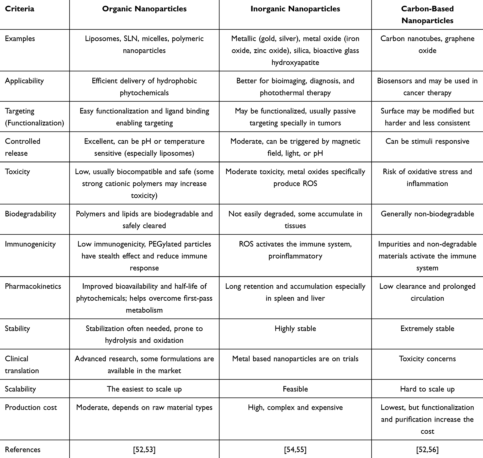 |
Table 1 A Detailed Comparison Between Organic, Inorganic, and Carbon-Based Nanoparticles |
Drugs and phytochemicals can be loaded into DDS through various techniques. Hydrophobic agents can be encapsulated into nanoparticles for stability, protection, and increase their size-to-surface area enhancing the solubility and bioavailability. DDS protects the loaded agents from enzymatic degradation and has a stealth effect protecting them from early phagocytosis clearance and excretion.52 Moreover, it decreases the toxicity and side effects associated with highly potent drugs thus increasing their biocompatibility.53 Nanoparticles may be functionalized at their surface by targeting ligands permitting site-specific delivery. DDS also provides sustained drug release decreasing the dosing intervals and drug concentration and stabilizing its concentration in the therapeutic range for a longer time.
Phytochemicals release from nanoparticles can be triggered by pH, temperature, and enzyme reactions. Co-delivery of multiple active agents including phytochemicals enhance the efficacy of the formulation. Additionally, DDS may be fabricated in hydrogels, nanofibers, and scaffolds that mimic the extracellular matrix or bone’s hierarchical structure in its porosity, mechanical strength, and bioactivity.57 This integration allows their localized drug therapy or implantation thus enhancing their therapeutic efficiency.
Phytochemicals Studied in Bone Research
Phytochemicals and natural products have been extensively studied for their potential to address bone-related issues and disorders. The following sections explore pure natural compounds and plant extracts that have been investigated in bone research. A summary of these phytochemicals is provided in Table 2.
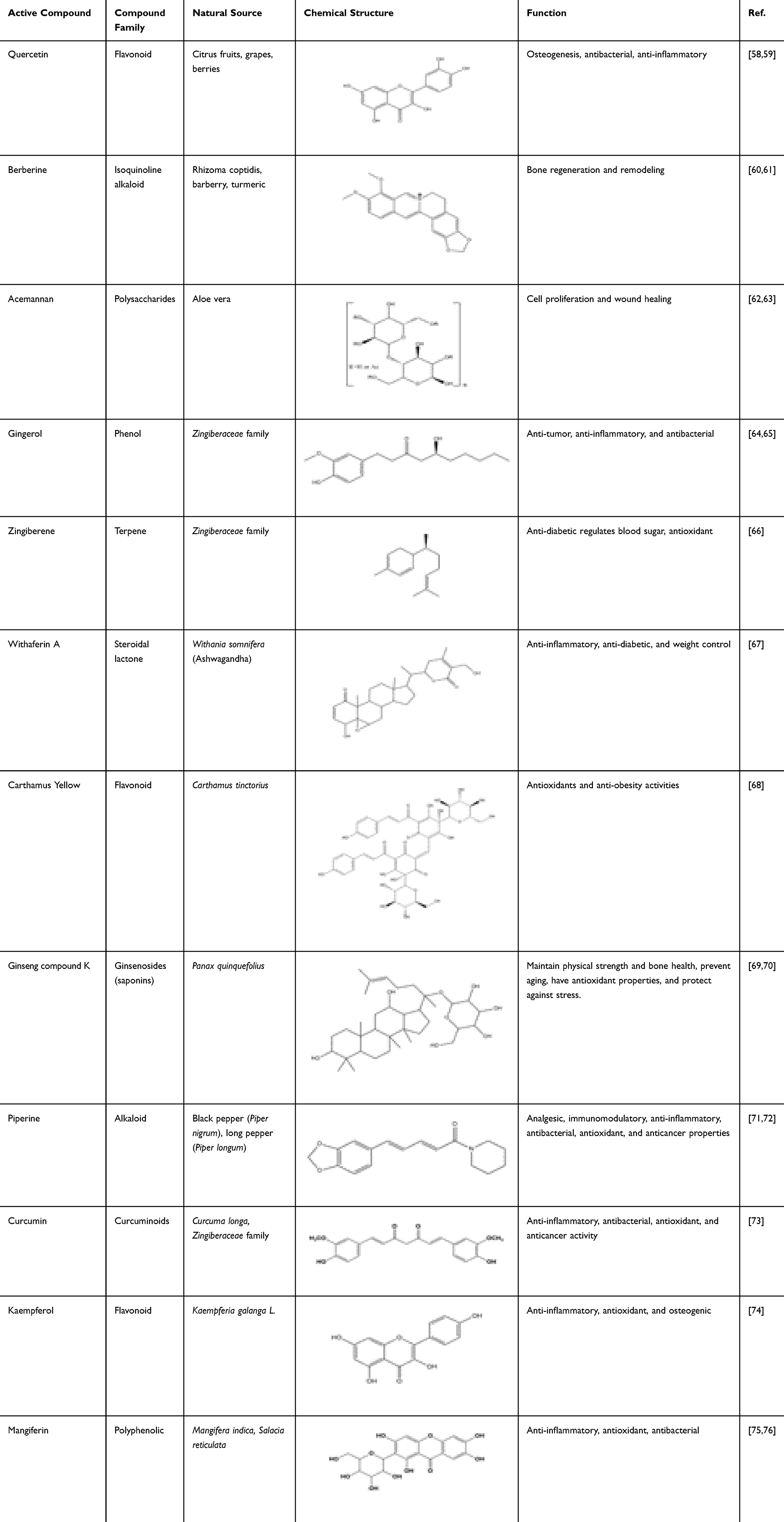 |
Table 2 List of Some Phytochemicals, Compound Class, Their Natural Source, Chemical Structure, and Biological Function |
Quercetin
Quercetin is a poorly soluble flavonoid that requires drug delivery systems (DDS) to enhance its therapeutic potential. Quercetin has been used as a cardioprotective, gastroprotective, neuroprotective, anti-diabetic, and osteogenic agent; due to its anti-inflammatory, antibacterial activity, and antioxidant properties.77–80 In two different studies, bone marrow stem cells (BMSC) were treated with quercetin in the concentration range 1–5 µM; results demonstrated an increase in cell proliferation, differentiation into osteoblasts, increase in the level of osteogenic-related genes such as ALP, RUNX2, and OPN, as well as enhanced antioxidant activity.78,81 Quercetin/SLN and lipid nanoparticle/quercetin complexed within calcium phosphate cement improved the bioavailability of orally administered quercetin as well as enhanced bone formation and remodeling in rats and restored bone loss and bone microstructure of osteoporotic rats, respectively.82,83 HA/quercetin nanoparticles increased the proliferation and differentiation of MG-63 cells (a human osteoblast-like cell line).84 In a synthetic polymeric electrospun nanofiber scaffold of polycaprolactone (PCL)/PVP, quercetin doped Mg/Ca/silicate significantly increased ALP and collagen and showed good antibacterial activity against Gram positive bacteria.85
Furthermore, a paste of bioactive glass (BG)/hyaluronic acid/alginate/quercetin had a dose-dependent osteogenic activity on human MSC.77 Calcium silicate calcium sulfate and quercetin fabrication within PCL increased calcium deposition in vitro.86 In addition, polydopamine (PDA) was used as a linker to bind quercetin to poly(L-lactide) (PLLA) 3D printed scaffold, which increased the expression of osteogenic genes in vitro.79 High-concentration quercetin inhibited cell proliferation in vitro, while low-concentration quercetin/silk fibroin (SF)/HA scaffold and quercetin/calcium sulfate hemihydrate/HA composite implanted in rat calvaria defect model and rat tibia defect, respectively,87,88 showed new bone formation covering around 80% of the defected area. On the other hand, a dual layer’s electrospinning scaffold of icariin/PDA/poly(lactic-co-glycolic) acid (PLGA)/chitosan and quercetin/PDA/PLLA/HA/chitosan tested for bone-cartilage defect repair had minor effects on the 4th week after implantation, while better results after 8 and 12 weeks. However, the chondrogenesis effect was not significant, and quercetin had little effect on COL1.89
As an anti-inflammatory agent, quercetin decreases M1 macrophage levels which are associated with pro-inflammatory cytokine production, by inhibiting pathways like NF-κB and decreasing the proinflammatory cytokines including tumor necrosis factor-α (TNFα), IL-1β, IL-6, and inducible nitric oxide synthase (iNOS). It also promotes M2 macrophage polarization, which is linked to anti-inflammatory and tissue repair functions, by enhancing pathways like STAT6 and increasing anti-inflammatory Arg1, IL-4, and IL-10.58,80,90 Crude quercetin could counteract the inflammatory effects of TNF-α on BMSC and cell apoptosis, as well as increase bone mineral density (BMD), enhance bone microstructure, and improve the elasticity and load-bearing of bones in postmenopausal osteoporotic rats.91 Yang and his colleagues formulated a phyto-nanocomposite of quercetin/mesoporous- bioactive glass (BG) that inhibited the effect of lipopolysaccharide on M1. Moreover, downregulated the proinflammatory miRNA miR-21a-5p thus inhibiting NF-κB, as well as performing indirect osteogenic and angiogenic activity.59 miR-21a has been reported to be involved in periodontitis, it upregulates NF-κB signaling thus its deficiency may result in a decrease in alveolar bone loss.59
A study by Xu et al has shown a 3D-printed scaffold of natural polymer alginate mixed with basic calcium phosphate nanosphere and quercetin increased ALP, RUNX2, and OCN levels. Moreover, it caused a shift from M1 to M2 creating a less inflammatory environment and supporting healing in vitro. In vivo, quercetin enhanced bone formation, improved bone density, and fostered a regenerative environment, supporting bone health.80 Similar results were found with BMSC and RAW264.7 cells treated with quercetin/HA/poly(glycolide-co-caprolactone) (PGCL) nanocomposite porous-microspheres.90 Likewise, a hydrogel of hyaluronic acid/polycaprolactone-co-lactide-PEG-polycaprolactone-co-lactide loaded with quercetin/SLN restored the osteogenic-osteolytic balance in vivo by inhibiting M1 macrophages and proinflammatory cytokines and increased polarization of M2 and IL-4.58
Quercetin-loaded-Zein microspheres were crosslinked using gallic acid to chitosan/Basil seed gum (BSG) 3D hydrogel, which had antioxidant and antibacterial effects.92 Another hydrogel combining SF and chitosan encapsulating vancomycin and quercetin/PLGA nanoparticles showed a decrease in osteomyelitis, better bone healing and repair in rat calvaria and tibial defects, and reduced bacterial activity.93
Berberine
Berberine (BER) is an isoquinoline alkaloid, proven to have many therapeutic activities including anti-inflammatory, antibiotic, antidiarrhea, anti-diabetic, lipid-lowering effect, and osteogenesis.60,94,95 However, BER has low solubility and bioavailability which constrain its therapeutic applications, thus it has been incorporated in many DDS. BER encapsulated into PEG/soybean/ethylene glycol nanoparticles and SF/Ag/calcium phosphate ceramics found to increase ALP levels in vitro.96,97 BER was incorporated with porous calcium phosphate ceramic and found to enhance bone regeneration and increase the levels of ALP, OCN, BMP-2, and RUNX2 in vitro. Moreover, when implemented into a calvaria defect of ovariectomized female rats, BER increased the BMD, bone volume/tissue volume (BV/TV), and the area and number of new bones in vivo.98 BER/chitosan-based nanoparticles and PCL/BER/SF nanofibrous scaffold both have anti-apoptotic activity, the first reversed the cartilage destruction in osteoarthritis model, while the later enhanced bone formation in type 2 diabetes rat model, could alleviate mitochondrial dysfunction and decreased ROS levels.99,100 A polymeric microsphere of PLGA/HA loaded with BER and IGF-1, a growth factor associated with bone regeneration, showed a synergistic osteogenic differentiation, mineralization, and bone formation in skull defect model with elevation in osteogenic markers such as OCN, OPN, COL1, and RUNX2.101
Xie et al compared two oral doses of BER 50 and 100 mg/kg/day in diabetic rat models induced by a combination of streptozotocin and a high-fat diet. They found that the lower dose had minimal effect on bones while the higher dose increased the plasma OCN levels significantly and reduced the TRAP levels, a marker for osteoclastogenesis.95 Pioglitazone (Pio) is a thiazolidinedione that increases insulin sensitivity but has a negative effect on bones. BER at 100 mg/kg/day dose was administered orally to male type 2 diabetic rats with/without Pio; BER counteracted the negative effect of Pio, increased the RANKL and OPG mRNA, and inhibited the RANKL osteoclast formation while increasing the expression of OCN and RUNX2.102 However, another group of scientists did not totally agree with these results. They argued that the previous results were recorded due to the high dose of BER and long treatment duration. They administered 50 mg/kg/day of BER to type 1 diabetic rats orally for 4 weeks, which they indicate is equivalent to 2.5 years in humans and it should be enough treatment period in rats. However, they found that the BER effect on bones was not significant and could not be used as a prevention of bone resorption in diabetes.103
BER extract was examined for its antibacterial effect against Porphyromonas gingivalis and it strongly suppressed its growth. In the same study, the BER extract on concentrations of 1–10 µM was used to treat BMSC and was able to increase the expression of some bone-related genes such as ALP, COL 1, OCN, OPN, and OSX and was found to act through the Wnt/β-catenin signaling pathway.104 This conclusion was also verified when the BER was incorporated with HA into cellulose acetate electrospun nanofibers.94
A dose-dependent effect of BER on osteogenesis and bone healing in vitro and in vivo with a 3D PCL/gelatin/BER nanofiber scaffold,105 and PCL/COL electrospun nanofiber when implanted in rats calvaria defects for 8 weeks. The PCL/COL/BER-based nanofiber promoted cell differentiation, increased BMD and enhanced new bone and collagen formation.61
Piperine
Piperine (Pip) is a bioactive water-insoluble alkaloid with therapeutic activity like analgesic, and immunomodulatory.71,72 Hence, Pip has been widely used in traditional medicine for inflammation, degenerative diseases, and gastrointestinal, and respiratory disorders.72,106 MC3T3-E1 cells treated with Pip showed an increase in ALP, Runx2, mineralization levels, distal-less homeobox 5 (Dlx5), and an inhibitor of DNA-binding 1 (Id1), which promotes osteoblast differentiation.71 Dlx5 is a bone-induced transcription factor regulated by BMP2 playing an essential role in osteoblast differentiation.107,108 Similarly, Id1 is a transcription factor responsible for cell differentiation, cell cycle progression, and apoptosis which is also upregulated by BMP and stimulates osteoblast differentiation.109,110
Similar results were obtained when Pip extract (5µg/mL) was applied on human Wharton’s Jelly mesenchymal stem cells (WJMSCs), where cell apoptosis was reduced and Ca deposition, mineralization, and osteogenic genes were upregulated.72 Pip not only induces osteoblast differentiation but also inhibits osteoclastogenesis. This was demonstrated by Li et al investigation in ovariectomized-osteoporotic mice models, recording that orally administered Pip promoted new bone formation, increased minerals deposition, biomechanical parameters, and BMD in a non-dose-dependent manner.111 Moreover, another study focused on the effect on osteoclasts showed that the water extract of Piper longum decreased TRAP levels in a dose-dependent manner while increasing the viability of bone marrow cells. Additionally, the extract exhibited direct inhibitory action on osteoclast precursors as it suppressed the expression of c-Fos and Nfatc1 which are important factors for osteoclast differentiation but increased the inhibitory Mafb and Irf8. Overall inhibiting the RANKL osteoclastogenesis pathway in vitro, as well as enhancing BMD and bone microstructure in vivo.106
Additionally, Pip/SLN gel formulation relieved inflammation measured by a reduction in TNF-α and reduced paw volume in tested rats.112 Pip/bovine serum albumin nanoparticles administered intraperitoneally to adjuvant-induced arthritis rats significantly alleviated joint and bone inflammation, and decreased IL-17, and bone erosion; interestingly, Pip was more potent in the suppression of fibrin deposition.113 Although a higher concentration or faster release should be tested, Pip-based nanofibrous PCL electrospun scaffolds were implanted in tibia defects and showed little improvement in bone regeneration.114 Due to its pharmacological activity, Pip was combined with the chemotherapeutic doxorubicin (DOX) to check its anticancer activity and its ability to increase the sensitivity of DOX on cancer cells. Pip/DOX combination showed synergistic effects on two osteosarcoma cell lines U2OS and 143B cells, where cell proliferation was dramatically inhibited, and tumor volume and weight were significantly reduced.115 The study also claims that Pip can protect and reduce the cardiotoxicity of DOX.
Curcumin
Curcumin (Cur) is a hydrophobic curcuminoid recognized for its anti-inflammatory, antibacterial, antioxidant, and anticancer activity,73 therefore, curcumin has been extensively investigated in such research. Cur has very low bioavailability and is usually fabricated in DDS. Cur was encapsulated into liposomes and loaded into calcium phosphate 3D printed scaffold, interestingly showing both osteogenesis effect on human fetal osteoblast cells (hFOB) and anticancer effect on osteosarcoma MG63 cells. This may be explained by the fact that Cur prevents the phosphorylation of inhibitory kappa beta (Iκβ) which will keep binding the NF-κB preventing its activation and thus promoting tumor cell apoptosis.116 In another study, the PCL/polyethylene glycol (PEG)/Cur/tricalcium phosphate (TCP) scaffold demonstrated superior results over PLGA/PEG/Cur/TCP for in vitro Cur release, biocompatibility, and cell adsorption capability. The 3D printed PCL/PEG/Cur/TCP scaffold promoted osteoid and blood vessel formation significantly on femur defect.117 Senthil and Çakır formulated a bone apatite composed of demineralized bone matrix, calcium sulfate hemihydrate, Cur nanoparticles, and Ag nanoparticles which has good antibacterial activity and good potential for dental tissues repair.118
In a different approach, Pip inhibits hepatic glucuronidation and drug metabolisms, thus Cur was combined with Pip to enhance its bioavailability. Orally administered Cur/Pip for periodontitis treatment showed a significant increase in TGF-β, COL1, IL-10, notable bone repair, and healing gingival tissues, while decreasing NF-κB.119 An herbal preparation combining Cur, Pip, papain, and bromelain was tested in vivo in adult and embryonic zebrafish models. Cur increased bone mineralization by 40% in the adult zebrafish model at a concentration of 250 nM, while acting as a protective agent against prednisolone-induced osteoporosis, moreover, counteracted its effect by increasing ALP levels and decreasing TRAP in the embryonic model.120 Cur/Pip polymeric micelles loaded into TCP 3D-printed scaffold showed good chemoprotective activity and enhanced osteoblast proliferation and differentiation in vitro.121
A degenerative osteoarthritis rat model was used to check curcumin’s effects on chondrocytes. Cur alone did not improve cell proliferation or migration of chondrocytes, however, interestingly when co-cultured with BMSC it did. Moreover, Cur/BMSC increased the COL2, SOX9, and Aggrecan on mRNA and protein levels and decreased the osteoarthritis score in vivo.22
Conventional anticancer drugs and treatments have many limitations and side effects, thus new methods, agents, and combinations are always examined and developed. Photothermal therapy (PTT) is the use of locally generated heat from a laser to kill cancer cells. Curcumin was incorporated in PLGA microspheres suspended in a thermosensitive hydrogel and applied in vitro on K7M2wt osteosarcoma cells, BMSC, and in vivo. The hydrogel showed anticancer activity in vitro with enhancing BMSC differentiation into osteoblast measured by the increase in ALP and mineralization, and sustained release of Cur in vivo along with thermal treatment that led to tumor cell apoptosis.122 A similar approach was used with superparamagnetic iron oxide nanoparticle (SPION) coated with Cur immersed in a polymeric layer and found a synergistic effect of Cur and hyperthermia, especially at 41°C.123
Kaempferol
Kaempferol is a yellow crystalline, slightly water-soluble, natural flavonoid. Green leafy vegetables, tea, apple, Kaempferia galanga, and Polygonum tinctorium are rich of kaempferol. As a flavonoid, kaempferol has antioxidant, anti-inflammatory, antimicrobial, and antitumor activity.74,124 Kaempferol counteracted cell apoptosis, cell cycle arrest, and proliferation inhibition effect of dexamethasone glucocorticoid. Furthermore, it increased osteogenic factors (ALP, OSX, RUNX2, cyclin D1) and activated the cellular proliferation and differentiation p38 MAPK pathway.125 In osteoporotic rats, kaempferol/metformin combined treatment resulted in increasing bone formation markers while suppressing bone resorption markers, along with enhancing angiogenesis.124 Micro-nano titanium implant doped with kaempferol showed anti-osteoporotic ability promoting osteogenesis and bone regeneration.126 In two different studies, kaempferol was encapsulated into albumin nanoparticles and formulated in PCL nanofibrous scaffold, once for bone regeneration and another for cartilage repair and showed promising results.127,128 Kaempferol was loaded into zein-coated bioactive glass scaffold and implanted in rat defect model causing new bone formation and regeneration with elevation in OCN, OPN, COL1 genes and Ca deposition.129 Moreover, kaempferol loaded gelatin nanoparticles coated with hyaluronic acid showed positive effects on osteoarthritis rat model by reducing the inflammation, inhibiting matrix degradation and subchondral sclerosis, and restored cartilage thickness.130
Kaempferol at a concentration of 1µM enhanced the proliferation and differentiation of periodontal ligament stem cells (PDLSC) through the Wnt/β-catenin signaling pathway.131 Moreover, at a concentration of 10µM kaempferol enhanced osteogenesis via increasing SOX2 levels which inhibited the transcription of miR-124-3p in rBMSC. miR-124-3p plays an essential role in inhibiting osteogenic differentiation through the inactivation of the PI3K/Akt/mTOR pathway.132 Another miRNA that was downregulated by kaempferol is miR-10a-3p in osteoporotic ovariectomized rats. miR-10a-3p binds the chemokine CXCL12, which regulates the balance between bone formation and resorption, preventing osteogenic differentiation.74 In the same study, kaempferol increased OSX, CXCL12, and RUNX2 levels in vitro and enhanced the BMD and bone microstructure in vivo.
Mangiferin
Mangiferin (MAN) is a xanthone glucoside, a polyphenolic compound, mainly presented and extracted from mango trees (Mangifera indica), and Salacia reticulata.133 MAN has a noticeable antioxidant, anti-inflammatory, and antiviral activity and has been reported to decrease bone destruction and osteoblastic ferroptosis.75,134–136 As many other phytochemicals, MAN is poorly soluble in water and thus has low bioavailability, therefore, MAN-based nanomedicine could permit its therapeutic utilization in various health conditions.133,135
Pure MAN was found to increase ALP and RUNX2 levels while decreasing TRAP in vitro.75 Oral administration of pure MAN to bilateral ovariectomized mice restored the osteogenic marker levels to the normal range and alleviated osteoporosis and He et al suggest that MAN acts through the AXL/ERK5 pathway.137 MAN-enriched bicalcium phosphate cement loaded with manganese and HA increased osteogenic gene markers.138 Furthermore, immunostimulatory CpG oligonucleotide/MAN incorporated into calcium alginate hydrogel proved to have anti-inflammatory and antibacterial activity against Porphyromonas gingivalis, as well as promoted new bone formation and inhibited osteoclastogenesis in vitro.134 PLGA/MAN scaffold promoted bone regeneration in alveolar bone defect in diabetic rats.139 Moreover, pretreatment of MAN significantly alleviated dexamethasone-induced injury and inflammation and restored the osteogenic gene expression (ALP, OPN, OCN, OPG).76 A MAN hybrid nanocomposite of chitosan-silica-zinc oxide nanoparticles increased mineralization and apatite formation in vitro.140 The anti-inflammatory properties of MAN were investigated through hyaluronic acid/methotrexate nano-drug targeting cancer cells,141 and MAN-loaded nanotransethosome gel for rheumatoid arthritis.142
Plant Extracts Use in Bone
Cissus Quadrangularis Extract
One of the most studied plants for bone therapy as an osteogenic and anti-osteoporotic agent is Cissus quadrangularis (CQ) from the Vitaceae family known as Hadjod. CQ was first used in traditional medicine, as people started using the stem for bone regeneration and fracture healing.143 This bioactivity may be attributed to the synergistic effect of its phytochemicals; since CQ is rich in vitamins, steroids, tannins, carotenes, polysaccharides, ascorbic acid, potassium, iron, zinc, and calcium.144,145 Therefore, CQ has been tested and proven to have antioxidant, anti-microbial, anti-inflammatory, and osteogenic activity.145,146
Several in vitro studies on stem cells showed that CQ enhanced their differentiation into osteoblasts, increased mineralization, and in some cases increased osteogenic genes (RUNX2 and OCN) expression. The CQ in these studies were incorporated into polymeric scaffolds such as alginate/O-carboxymethyl chitosan/CQ,147 CQ/gelatin/pectin/β-TCP,148 and electrospun fibrous PCL/CQ/graphene oxide (GO) scaffold.146 Other in vivo studies have also been conducted, Gupta et al prepared nano-HA, α-calcium sulfate hemihydrate nano-cement loaded with CQ and found that it enhanced proliferation and differentiation of C2C12 and MC3T3-E1 cells in vitro as well as enhanced bone formation and defect healing with better Ca and COL deposition in vivo after 8 weeks of treatment.149
Although the TCP/PDA-CQ 3D printed scaffold did not show good results in vitro, as the ALP levels increased only in dynamic cell culture while calcium deposition did not form in such media, the in vivo results showed new bone formation with increased osteoinductivity and osteogenesis after only 4 weeks of implantation.150 PCL/CQ nanoparticles exhibited an increase in ALP level, COL deposition, and osteoid accumulation in vivo in a 6-week study.151 In another study, a PCL/GO/CQ electrospun scaffold coated with human umbilical cord-derived mesenchymal stem cells (hUCMSC) was implanted into calvaria defect, after 12 weeks of the implantation results showed some improvement in osteoblast differentiation, defect closure, ALP and OCN levels compared to other groups including PCL/hUCMSC and PCL/GO/CQ.144
CQ crude extract was administered orally to female ovariectomized mice in 500 mg/kg/day dose to study its anti-osteoporotic effect. The 45 day-long-study revealed that CQ extract enhances bone mass by increasing BMD, reducing the differentiation and activity of osteoclasts, as well as increasing the anti-inflammatory cytokines producing cells Breg and Tregs while suppressing the osteoclastogenic Th17 cells.21
Aloe Vera
Aloe barbadensis Miller, commonly known as Aloe vera (AV), contains many different active compounds, especially in the gel part. This includes vitamins, lignin, saponin, proteins, minerals, and polysaccharides.62 AV has two main active polysaccharides, Acemannan and Glucomannan, as well as a growth hormone gibberellin which increases cell proliferation through the activation of fibroblast growth factor receptors.152 AV has anti-inflammatory, antibacterial, tissue regeneration ability, and cooling effect, so it is widely used for wound and burn healing.62,63 Therefore, researchers have tested it for bone regeneration ability.
A titanium implant was coated with chitosan/AV/HA incorporated with silicon (Si+4) and silver (Ag+) ions; AV exhibited some antibacterial function in vitro. As well as Ag/Si/HA/AV titanium implant also increased osteoid formation significantly and improved mineralization and bone volume in vivo after 5 weeks of implantation.153 In studying the effect of AV on MG-63 cells, AV was incorporated into electrospun PCL/PEG nanofibers and it caused an increase in Ca deposition.62 It was also formulated with starch/BG/Quail eggshell in a freeze-dried formulation and showed an increase in OCN and OPN levels.63 Interestingly, the AV/HA/Mg porous composite showed a good antibacterial effect against Gram-positive B. subtilis but not against Gram-negative E. coli.152 The incorporation of AV into a PLA 3D-printed scaffold significantly enhanced bone cell attachment (adsorption) and proliferation compared to PLA alone.154 However, loading AV into a collagen sponge containing mesenchymal stem cells derived from human dental pulp stem cells (hDPSCs) showed no significant improvement in osteogenic activity, as indicated by unchanged OPN levels, regardless of the presence of AV.155 While AV may exhibit promising wound-healing and anti-inflammatory properties, its impact on bone healing and regeneration was not notably significant.
Ginger Extract
Ginger (Gin) is a member of the Zingiberaceae family, which is widely found in Asian countries. Its roots are rich in phytochemicals, including phenolic compounds like gingerol and terpenes such as zingiberene and β-sesquiphellandrene.64 Gin has been traditionally used for its anti-inflammatory and antibacterial function in many medical conditions.64 And so, it has been extensively studied as an immune modulator, anti-tumor, anti-diabetic, anti-inflammatory, and antibacterial.65
In bone-related research, Gin was combined with curcumin extract in a COL/HA scaffold, which had anti-inflammatory activity as well as increased bioactivity and biocompatibility, and the expression of RUNX2 and COL1 was upregulated.65 COL/β-TCP scaffold loaded with Gin was implanted in a mandibular defect in rats with and without synovial membrane mesenchymal stem cells (SM-MSC), the scaffolds showed an anti-inflammatory effect, upon the addition of SM-MSC bone repair was accelerated.64 In another study, Gin and garlic extracts were loaded in a 3D printed PCL/β-TCP/PEG scaffold which was implanted into a rat distal femur model. This scaffold increased the osteoid formation after 4 weeks of implantation, mineralization, and COL1, and decreased bone resorption while showing new blood vessel formation after 12 weeks of implantation.156 In periodontitis model, Gin-exosome like nanoparticles reduced the inflammation, exhibited antioxidant activity and increased the proliferation of collagen fibers.157 Likewise, 6-shogaol, an active ingredient of ginger, prevented osteoclastogenesis and alveolar bone resorption, decreased IL-1β and TNF-α and inflammation in mice periodontitis.158
In addition, a completely natural ointment composed of Gin/turmeric/chili pepper/rose oil distributed in an oily base paste of sesame/black seed/olive oil was prepared for musculoskeletal pain relief and bone repair. The formula was tested in vitro on different cell types and in clinical studies; its results showed an increase in osteogenic-related genes like RUNX2, COL1, COL2, OCN, and OPN, and decreased inflammation markers and signs.159
Other Natural Agents Used in Bone-Related Research
The field horsetail (HT), scientifically known as Equisetum arvense, has been used in herbal medicine to treat different cardiovascular conditions and inflammation.160 The analysis of HT extract found that it contains many active components; such as β-sitosterol, campesterol, flavonols such as kaempferol and quercetin, lignin, and many minerals mainly silica, potassium, calcium, and phosphate.160 This combination of active materials encouraged researchers to test HT for bone regeneration and repair ability. HT was fabricated with PLA/HA using an electrospinning technique and showed an increase in ALP levels and calcium deposition in vitro.161 HT extract may be useful in bone tissue-engineered scaffolds due to its silica, calcium content, anti-inflammatory, and antibacterial activity.160 However, more in vitro and in vivo studies should be carried out to justify HT extract use and mechanism of action.
Ashwagandha (Withania somnifera) contains a steroidal lactone withaferin A (WA), which has anti-inflammatory, anti-diabetic, and weight control functions.67 WA incorporation in bone cement/chitosan microparticles was found to increase MC3T3-E1 cell proliferation and ALP levels.162 WA/alginate/BCP microspheres have also increased osteogenic markers in vitro.163 WA inhibited adipogenesis, favored osteogenesis, and improved bone microstructure in bone marrow cells of obese rats.67
Safflower (Carthamus tinctorius) main active compound is hydroxy-safflower yellow A (HSYA) or Carthamus yellow. As its name indicates, it has a yellow color and thus is used as a coloring agent in food and cosmetics. HSYA has some antioxidants and anti-obesity activities.68 In in vitro osteoarthritis model, HSYA alleviated the oxidative stress and reduced both IL-1β and TNF-α.164 HSYA was examined for its angiogenesis ability against HUVEC-12 cells and osteogenesis against BMSCs, which proved to increase blood vessel count and hypoxia-inducible factor-1α (HIF-1α), vascular endothelial growth factor (VEGF), and angiopoietin-2 as well as osteogenic-related factors ALP, Runx2, and OPN.165 When incorporated into a 3D printed scaffold of BG/chitosan/alginate, HSYA promoted angiogenesis and osteogenesis of BMSC and rat cranial defect.68 Further, when administered by gavage to osteoporotic female rats model, HSYA restored the balance between osteoclast and osteoblast, increased COL1 and 2, and reduced the expression of carbonic anhydrase 2.166 N-(p-Coumaroyl) serotonin and N-feruloyl serotonin were extracted from the safflower plant and tested on osteoarthritis, where they restored the cartilage dysfunction by inhibiting the Inhibitor of kappa B (IκB) degradation and thus blocking NF-κB pathway.167
As for other types of honey, manuka honey (MH) which is a special honey produced specifically from the manuka tree, Leptospermum scoparium, MH is full of phenolic compounds and has antioxidant, antibacterial, and anti-inflammatory properties.168 MH/BG/zein protein scaffold was prepared and its antibacterial activity against Staphylococcus aureus was confirmed, but due to its burst release the formulation should be optimized.169 MH effect on bone regeneration in vivo was examined by Robertson et al who found that MH did not have any positive effects on bone regeneration.170
Ginseng from the Araliaceae family, is rich in saponins called ginsenosides and other phenolic compounds, vitamins, and minerals. Ginseng is well known to maintain physical strength and bone health, prevent aging due to its antioxidant properties, and protect against stress.69,70 The main ginsenoside, ginseng compound K (CK) was found to promote cell proliferation and the expression of osteogenic genes OCN, OPN, and COL1 expression when incorporated with chitosan/biphasic calcium phosphate microspheres.171 CK-treated rats with bone femoral fracture presented faster healing and healthier bone formation as well as triggered angiogenesis.69 A titanium nanotube modified with ginseng extracts and implanted in an edentulous mandibular defect site showed increased bone formation with high BMD, BMP-2, BMP-7, and COL1 expression.70
On the other hand, other natural compounds were also fabricated on scaffolds for bone engineering purposes but did not show significant effects. For example, Linum usitatissimum known as flaxseed or linseed is rich in omega-3 polyunsaturated fatty acids, lignans, fibers, and magnesium and has been known to have bone formation ability.172 Flaxseed was incorporated in HA/alginate hydrogel and neither had a significant antioxidant effect nor a positive effect on cell proliferation.173 Cinnamon oil was loaded into the BG scaffold and showed good antibacterial and antioxidant activity. However, the scaffold was not biocompatible with MG-63 cells.174 Although Lemongrass oil (LGO) is assumed to have antibacterial and antioxidant activity, S. aureus viability did not decrease below 70% when cultured on chitosan/hydroxypropyl methylcellulose/HA/LGO scaffold even with high concentrations.175 Pomegranate peel extract was incorporated into PCL electrospun fibers and was biocompatible with bone marrow-derived stem cells, but further studies are required to check its favorable additive effects on bones.176 Brucine obtained from Strychnine semen was integrated into the 3D-printed scaffold of PLLA/Polyglycolide due to its known antibacterial, anti-inflammatory, and analgesic effects. Despite proving the antibacterial activity, brucine had toxic effects on MG-63 cells.177
Commercially Available Natural Supplements
Nowadays, since people’s demand has increased for natural products and naturally derived compounds instead of synthetic drugs, many dietary supplement companies have become World Health Organization (WHO), and Food and Drug Administration (FDA) approved to synthesize nutraceuticals. Some examples of the natural compounds and extracts mentioned in this review that are commercially available in the market are listed in Table 3.
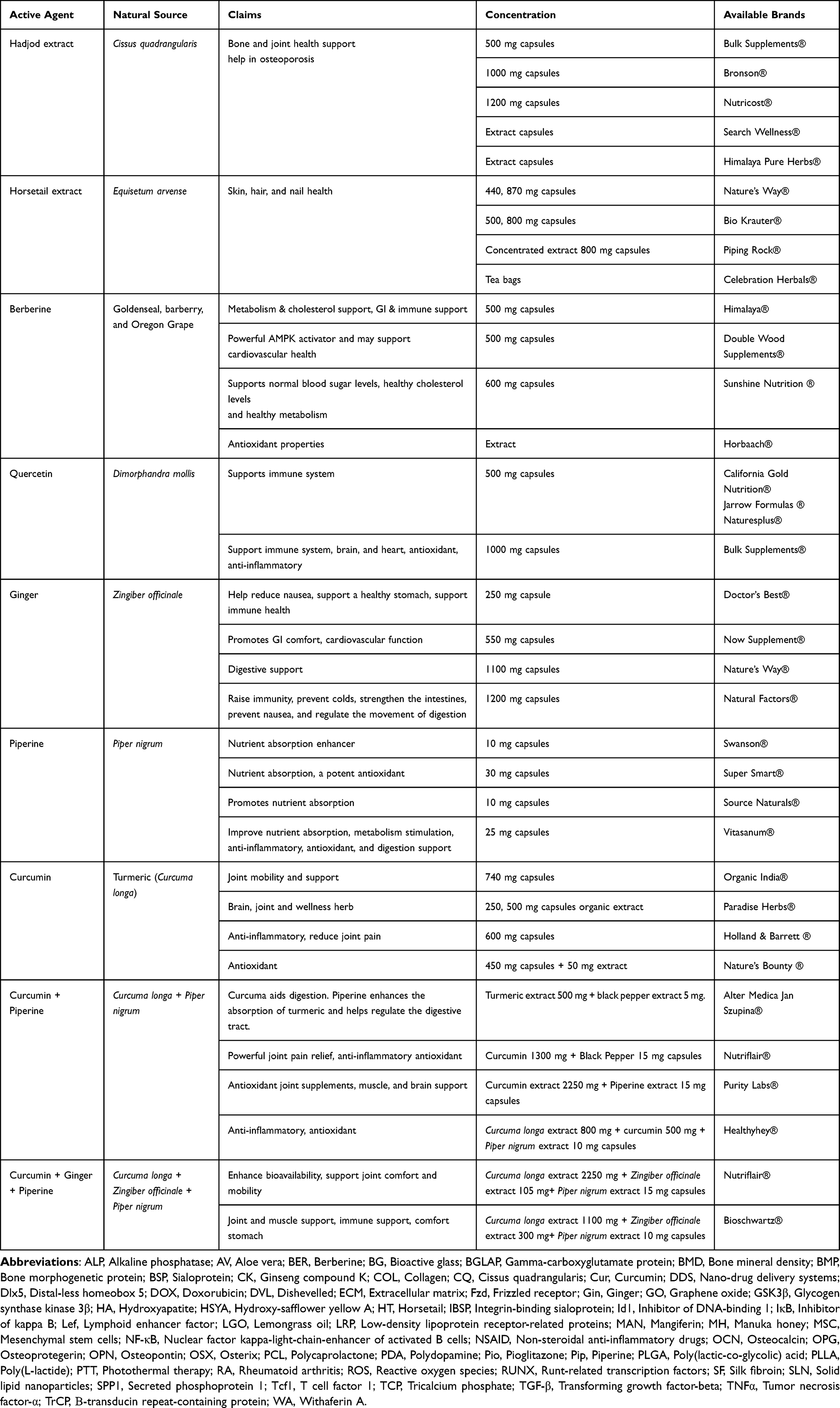 |
Table 3 List of Some Commercially Available Natural Extracts and Compounds |
Challenges of Using Natural Medicinal Plants
People have utilized natural medicinal plants in traditional medicine for centuries, especially before the development of synthetic drugs. These plants proved to have therapeutic activity and were beneficial up to certain limits. However, medicinal plants and natural agents use have several limitations and challenges. Firstly, most active compounds exhibit poor water solubility, low intestinal permeability, and rapid metabolism and elimination shortening their half-lives. These factors collectively result in poor pharmacokinetics of natural compounds. Moreover, the short half-lives require more frequent administration or the development of sustained release formulations such as nanoparticles.
In addition, standardization of dosage is a persistent challenge. Plant composition can vary between plant parts used like roots, stems, leaves, or fruits, or due to inter/intraspecies variation, time of harvesting, or even environmental and geographical factors such as weather, rain, soil, and solar radiation.5,49 This lack of consistency makes it difficult to determine optimal dosing regimens and can lead to variability in therapeutic outcomes.
Another major concern is the safety and toxicity limits. The whole plant crude extract or powder contains a mixture of different compounds that vary in composition and concentrations, making it challenging to determine their safety and potential interactions. Even though plants are considered relatively safer than synthetic drugs, there is no completely safe substance, and as Paracelsus stated all substances are poisonous.11 Many phytochemicals lack comprehensive toxicological profiles, and long-term studies assessing chronic use, potential accumulation in tissues, and organ-specific toxicities are limited. Additionally, natural compounds may interact with conventional drugs potentially leading to adverse drug interactions or altered pharmacodynamics.
Moreover, it is difficult to determine the efficacy and optimize the dose of a plant mixture. Therefore, recognition, spotting then separation of the active component could be a better choice. However, it is tedious, time and money-consuming, needs to be optimized, and is a multi-step process.49 Also, the stability of the plant extracts and active components is low, and they may not withstand process conditions such as high temperatures or organic compounds. And thus, carefully adjusted and well-monitored conditions and procedures should be used.
Additionally, different extraction techniques may result in different compositions and concentrations of the same extracted sample.120 That is why there is no guarantee of the reproducibility of the plants’ activity or composition analysis results and leading to batch-to-batch variations. All these factors raised the surge to make a standardization of plant taxonomy, used parts, extraction and separation methods, as well as quality control over medicinal plant studies.5,11
In addition, most of the natural compounds are marketed as dietary supplements rather than pharmaceuticals without clear standard testing and approval strategies from the regulatory agencies. This regulatory ambiguity makes it challenging to ensure the safety, efficacy, and quality of these products.
Still, the identification of natural active compounds and their use in pure form is advantageous and beneficial and overcomes most of these challenges. In addition, incorporating the medicinal compound or plant extract into ceramics or polymeric scaffolds, micro/nanoparticles including liposomes, micelles, nanotubes, mesoporous silica or bioactive glass can lead to its controlled release, targeted delivery, and reduced systemic side effects.
Conclusion
To summarize, nature is rich in phytochemicals with potent biological activities and potential to be developed into drugs. Traditionally, many plants and herbs were used as therapeutic agents especially when there were no synthetic drugs available for certain health conditions. Plants full of flavonoids, terpenoids, and alkaloids possess important antioxidants, anti-inflammatory, and anticancer activity. Looking closer into bone therapy, different phytochemicals proved to promote osteogenesis and inhibit osteoclastogenesis activity stimulating bone formation and maintaining bone remodeling balance.
Nano-formulations such as quercetin/SLNs, berberine-calcium phosphate ceramics, and curcumin/liposome embedded 3D printed scaffolds showed enhanced cellular uptake, targeted delivery, and sustained release, significantly improving bone tissue responses. Animal studies consistently showed that phytochemical loaded nano-preparations improve bone mineral density, promote new bone formation, restore microarchitecture, and reduce inflammatory markers in models of osteoporosis, osteoarthritis, and bone defects. Fabricating these phytochemicals or plants into nanoparticles or formulating them as nanomedicines boosts their function and benefits.
Although utilizing natural compounds as therapy is challenging, it is still a promising alternative for synthetic drugs with high adverse effects. However, translating these promising findings into clinical practice requires extensive research. Key priorities include the standardization of phytochemical content, precise dosage optimization, thorough assessment of long-term safety, and navigation of regulatory requirements. Comprehensive preclinical and clinical trials are essential to confirm their therapeutic effectiveness and ensure safe clinical application.
Acknowledgment
The authors gratefully acknowledge the support provided by the College of Graduate Studies at the United Arab Emirates University (UAEU) through research funding (Grant No. 131031). Figures include images and icons adapted from BioIcons (https://bioicons.com) under CC BY-SA and MIT licenses and Servier Medical Art (https://smart.servier.com/) under CC BY 4.0 (https://creativecommons.org/licenses/by/4.0/).
Disclosure
The author(s) report no conflicts of interest in this work.
References
1. Chaachouay N, Zidane L. Plant-derived natural products: a source for drug discovery and development. Drugs Drug Candidates. 2024;3(1):184–207. doi:10.3390/DDC3010011
2. Galanakis CM. Preface. Nutraceutical Funct Food Components Eff Innov Process Tech. 2017;xvii–xviii. doi:10.1016/B978-0-12-805257-0.00001-6
3. Huang WY, Cai YZ, Zhang Y. Natural phenolic compounds from medicinal herbs and dietary plants: potential use for cancer prevention. Nutr Cancer. 2009;62(1):1–20. doi:10.1080/01635580903191585
4. Tetali SD. Terpenes and isoprenoids: a wealth of compounds for global use. Planta. 2019;249(1):1–8. doi:10.1007/S00425-018-3056-X
5. Mukherjee PK. Quality evaluation of herbal medicines: challenges and opportunities. Qual Control Eval Herb Drugs. 2019;53–77. doi:10.1016/B978-0-12-813374-3.00003-X
6. Palazzolo S, Bayda S, Hadla M, et al. The clinical translation of organic nanomaterials for cancer therapy: a focus on polymeric nanoparticles, micelles, liposomes and exosomes. Curr Med Chem. 2017;25(34):4224–4268. doi:10.2174/0929867324666170830113755
7. Dadwal A, Baldi A, Kumar Narang R. Nanoparticles as carriers for drug delivery in cancer. Cells Nanomed Biotechnol. 2018;46(sup2):295–305. doi:10.1080/21691401.2018.1457039
8. Feng X. Chemical and biochemical basis of cell-bone matrix interaction in health and disease. Curr Chem Biol. 2009;3(2):189–196. doi:10.2174/187231309788166398
9. Bienko M, Radzki RP, Wawrzyniak A, Balawender K. Structural and metabolic changes in bone. Anim. 2022;12(15):1946. doi:10.3390/ANI12151946
10. Bhushan S, Singh S, Maiti TK, et al. Scaffold fabrication techniques of biomaterials for bone tissue engineering: a critical review. Bioengineering. 2022;9(12):728. doi:10.3390/bioengineering9120728
11. Bose S, Sarkar N, Banerjee D. Natural medicine delivery from biomedical devices to treat bone disorders: a review. Acta Biomater. 2021;126:63–91. doi:10.1016/J.ACTBIO.2021.02.034
12. Mohsin S, Kaimala S, Sunny JJ, Adeghate E, Brown EM. Type 2 diabetes mellitus increases the risk to hip fracture in postmenopausal osteoporosis by deteriorating the trabecular bone microarchitecture and bone mass. J Diabetes Res. 2019;2019(1):3876957. doi:10.1155/2019/3876957
13. Mohsin S, Kaimala S, AlTamimi EKY, Tariq S, Adeghate E. In vivo labeling of bone microdamage in an animal model of type 1 diabetes mellitus. Sci Rep. 2019;9(1):1–12. doi:10.1038/s41598-019-53487-6
14. Mohsin S, Brock F, Kaimala S, et al. A pilot study: effect of irisin on trabecular bone in a streptozotocin-induced animal model of type 1 diabetic osteopathy utilizing a micro-CT. PeerJ. 2023:11:e16278. doi:10.7717/PEERJ.16278
15. Baker R, Narla R, Baker JF, Wysham KD. Risk factors for osteoporosis and fractures in rheumatoid arthritis. Best Pract Res Clin Rheumatol. 2022;36(3):101773. doi:10.1016/J.BERH.2022.101773
16. Llorente I, García-Castañeda N, Valero C, González-álvaro I, Castañeda S. Osteoporosis in rheumatoid arthritis: dangerous liaisons. Front Med. 2020;7:601618. doi:10.3389/FMED.2020.601618
17. Gorka J, Taylor-Gjevre RM, Arnason T. Metabolic and clinical consequences of hyperthyroidism on bone density. Int J Endocrinol. 2013;2013(1):638727. doi:10.1155/2013/638727
18. Bassett JHD, O’Shea PJ, Sriskantharajah S, et al. Thyroid hormone excess rather than thyrotropin deficiency induces osteoporosis in hyperthyroidism. Mol Endocrinol. 2007;21(5):1095–1107. doi:10.1210/ME.2007-0033
19. Mohsin S, Baniyas MMYH, AlDarmaki RSMH, Tekes K, Kalász H, Adeghate EA. An update on therapies for the treatment of diabetes-induced osteoporosis. Expert Opin Biol Ther. 2019;19(9):937–948. doi:10.1080/14712598.2019.1618266
20. Drake MT, Clarke BL, Khosla S. Bisphosphonates: mechanism of action and role in clinical practice. Mayo Clin Proc. 2008;83(9):1032. doi:10.4065/83.9.1032
21. Azam Z, Sapra L, Baghel K, et al. Cissus quadrangularis (Hadjod) Inhibits RANKL-induced osteoclastogenesis and augments bone health in an estrogen-deficient preclinical model of osteoporosis via modulating the host osteoimmune system. Cells. 2023;12(2):216. doi:10.3390/CELLS12020216
22. Zhang R, Zhang Q, Zou Z, et al. Curcumin supplementation enhances bone marrow mesenchymal stem cells to promote the anabolism of articular chondrocytes and cartilage repair. Cell Transplant. 2021:30. doi:10.1177/0963689721993776
23. Derwich M, Górski B, Amm E, Pawłowska E. Oral glucosamine in the treatment of temporomandibular joint osteoarthritis: a systematic review. Int J Mol Sci. 2023;24(5):4925. doi:10.3390/IJMS24054925
24. Sawynok J. Topical analgesics in neuropathic pain. Curr Pharm Des. 2005;11(23):2995–3004. doi:10.2174/1381612054865019
25. Abdelnabi H, Alshaer W, Azzam H, Alqudah D, Al-Samydai A, Aburjai T. Loading of capsaicin-in-cyclodextrin inclusion complexes into PEGylated liposomes and the inhibitory effect on IL-8 production by MDA-MB-231 and A549 cancer cell lines. Zeitschrift fur Naturforsch - Sect C J Biosci. 2021;76(11):503–514. doi:10.1515/ZNC-2021-0018
26. Nagaoka I, Igarashi M, Sakamoto K. Biological activities of glucosamine and its related substances. Adv Food Nutr Res. 2012;65:337–352. doi:10.1016/B978-0-12-416003-3.00022-6
27. Varghese S, Theprungsirikul P, Sahani S, Hwang N, Yarema KJ, Elisseeff JH. Glucosamine modulates chondrocyte proliferation, matrix synthesis, and gene expression. Osteoarthr Cartil. 2007;15(1):59–68. doi:10.1016/J.JOCA.2006.06.008
28. Schemitsch EH. Size matters: defining critical in bone defect size! J Orthop Trauma. 2017;31:S20–S22. doi:10.1097/BOT.0000000000000978
29. Nauth A, McKee MD, Einhorn TA, Watson JT, Li R, Schemitsch EH. Managing bone defects. J Orthop Trauma. 2011;25(8):462–466. doi:10.1097/BOT.0B013E318224CAF0
30. Roberts TT, Rosenbaum AJ. Bone grafts, bone substitutes and orthobiologics: the bridge between basic science and clinical advancements in fracture healing. Organogenesis. 2012;8(4):114. doi:10.4161/ORG.23306
31. Campana V, Milano G, Pagano E, et al. Bone substitutes in orthopaedic surgery: from basic science to clinical practice. J Mater Sci Mater Med. 2014;25(10):2445. doi:10.1007/S10856-014-5240-2
32. Leteve M, Passuti N. Current concepts in bone graft substitutes. New J Glas Ceram. 2018;08(03):39–54. doi:10.4236/njgc.2018.83004
33. Wang W, Yeung KWK. Bone grafts and biomaterials substitutes for bone defect repair: a review. Bioact Mater. 2017;2(4):224–247. doi:10.1016/j.bioactmat.2017.05.007
34. Wang RN, Green J, Wang Z, et al. Bone Morphogenetic Protein (BMP) signaling in development and human diseases. Genes Dis. 2014;1(1):87–105. doi:10.1016/j.gendis.2014.07.005
35. Katagiri T, Watabe T. Bone Morphogenetic Proteins. Cold Spring Harb Perspect Biol. 2016;8(6):a021899. doi:10.1101/CSHPERSPECT.A021899
36. Arriaga MA, Ding MH, Gutierrez AS, Chew SA. The application of microRNAs in biomaterial scaffold-based therapies for bone tissue engineering. Biotechnol J. 2019;14(10):1900084. doi:10.1002/BIOT.201900084
37. Hu L, Chen W, Qian A, Li YP. Wnt/β-catenin signaling components and mechanisms in bone formation, homeostasis, and disease. Bone Res. 2024;12(1):1–33. doi:10.1038/s41413-024-00342-8
38. Cai YZ, Sun M, Xing J, Luo Q, Corke H. Structure–radical scavenging activity relationships of phenolic compounds from traditional Chinese medicinal plants. Life Sci. 2006;78(25):2872–2888. doi:10.1016/J.LFS.2005.11.004
39. Wu T, Zang X, He M, Pan S, Xu X. Structure-activity relationship of flavonoids on their anti- Escherichia coli activity and inhibition of DNA gyrase. J Agric Food Chem. 2013;61(34):8185–8190. doi:10.1021/JF402222V
40. Modak B, Leonor Contreras M, González-Nilo F, Torres R. Structure–antioxidant activity relationships of flavonoids isolated from the resinous exudate of Heliotropium sinuatum. Bioorg Med Chem Lett. 2005;15(2):309–312. doi:10.1016/J.BMCL.2004.10.081
41. Melidou M, Riganakos K, Galaris D. Protection against nuclear DNA damage offered by flavonoids in cells exposed to hydrogen peroxide: the role of iron chelation. Free Radic Biol Med. 2005;39(12):1591–1600. doi:10.1016/J.FREERADBIOMED.2005.08.009
42. de Melo LFM, de Aquino-Martins VGQ, da Silva AP, Oliveira Rocha HA, Scortecci KC. Biological and pharmacological aspects of tannins and potential biotechnological applications. Food Chem. 2023;414:135645. doi:10.1016/J.FOODCHEM.2023.135645
43. Amalraj A, Pius A, Gopi S, Gopi S. Biological activities of curcuminoids, other biomolecules from turmeric and their derivatives – a review. J Tradit Complement Med. 2016;7(2):205. doi:10.1016/J.JTCME.2016.05.005
44. Zálešák F, Bon DJYD, Pospíšil J. Lignans and Neolignans: plant secondary metabolites as a reservoir of biologically active substances. Pharmacol Res. 2019;146:104284. doi:10.1016/J.PHRS.2019.104284
45. Ain QU, Khan H, Mubarak MS, Pervaiz A. Plant alkaloids as antiplatelet agent: drugs of the future in the light of recent developments. Front Pharmacol. 2016;7(SEP). doi:10.3389/FPHAR.2016.00292
46. Bui VH, Rodríguez-López CE, Dang TTT. Integration of discovery and engineering in plant alkaloid research: recent developments in elucidation, reconstruction, and repurposing biosynthetic pathways. Curr Opin Plant Biol. 2023;74:102379. doi:10.1016/J.PBI.2023.102379
47. Laghezza Masci V, Bernardini S, Modesti L, Ovidi E, Tiezzi A. Medicinal plants as a source of alkaloids. Microorg Sustain. 2019;15:85–113. doi:10.1007/978-981-13-9566-6_5
48. Masyita A, Mustika Sari R, Dwi Astuti A, et al. Terpenes and terpenoids as main bioactive compounds of essential oils, their roles in human health and potential application as natural food preservatives. Food Chem X. 2022;13:100217. doi:10.1016/J.FOCHX.2022.100217
49. Atanasov AG, Zotchev SB, Dirsch VM, et al. Natural products in drug discovery: advances and opportunities. Nat Rev Drug Discov. 2021;20(3):200–216. doi:10.1038/s41573-020-00114-z
50. Yu LX, Amidon GL, Polli JE, et al. Biopharmaceutics classification system: the scientific basis for biowaiver extensions. Pharm Res. 2002;19(7):921–925. doi:10.1023/A:1016473601633
51. Khan KU, Minhas MU, Badshah SF, Suhail M, Ahmad A, Ijaz S. Overview of nanoparticulate strategies for solubility enhancement of poorly soluble drugs. Life Sci. 2022;291:120301. doi:10.1016/J.LFS.2022.120301
52. Campora S, Ghersi G. Recent developments and applications of smart nanoparticles in biomedicine. Nanotechnol Rev. 2022;11(1):2595–2631. doi:10.1515/NTREV-2022-0148
53. Dave V, Tak K, Sohgaura A, Gupta A, Sadhu V, Reddy KR. Lipid-polymer hybrid nanoparticles: synthesis strategies and biomedical applications. J Microbiol Methods. 2019;160:130–142. doi:10.1016/J.MIMET.2019.03.017
54. Fadeel B, Garcia-Bennett AE. Better safe than sorry: understanding the toxicological properties of inorganic nanoparticles manufactured for biomedical applications. Adv Drug Deliv Rev. 2010;62(3):362–374. doi:10.1016/J.ADDR.2009.11.008
55. Ehlerding EB, Chen F, Cai W. Biodegradable and renal clearable inorganic nanoparticles. Adv Sci. 2016;3(2):1500223. doi:10.1002/ADVS.201500223
56. Maiti D, Tong X, Mou X, Yang K. Carbon-based nanomaterials for biomedical applications: a recent study. Front Pharmacol. 2019;9:1401. doi:10.3389/FPHAR.2018.01401
57. Hassan M, Abdelnabi HA, Mohsin S. Harnessing the potential of PLGA nanoparticles for enhanced bone regeneration. Pharm. 2024;16(2):273. doi:10.3390/PHARMACEUTICS16020273
58. Zhou P, Yan B, Wei B, et al. Quercetin-solid lipid nanoparticle-embedded hyaluronic acid functionalized hydrogel for immunomodulation to promote bone reconstruction. Regen Biomater. 2023:10. doi:10.1093/RB/RBAD025
59. Yang SY, Hu Y, Zhao R, et al. Quercetin-loaded mesoporous nano-delivery system remodels osteoimmune microenvironment to regenerate alveolar bone in periodontitis via the miR-21a-5p/PDCD4/NF-κB pathway. J Nanobiotechnology. 2024;22(1):1–19. doi:10.1186/S12951-024-02352-4
60. Qin Z, Han Y, Du Y, et al. Bioactive materials from berberine-treated human bone marrow mesenchymal stem cells promote alveolar bone regeneration by regulating macrophage polarization. Sci China Life Sci. 2024;67(5):1010–1026. doi:10.1007/S11427-023-2454-9
61. Ma L, Yu Y, Liu H, et al. Berberine-releasing electrospun scaffold induces osteogenic differentiation of DPSCs and accelerates bone repair. Sci Rep. 2021;11(1):1–12. doi:10.1038/s41598-020-79734-9
62. Dehghan F, Gholipour-Kanani A, Kamali Dolatabadi M, Bahrami SH. Nanofibrous composite from polycaprolactone-polyethylene glycol-aloe vera as a promising scaffold for bone repairing. J Appl Polym Sci. 2022;139(26):e52463. doi:10.1002/APP.52463
63. Soltani M, Alizadeh P. Aloe vera incorporated starch-64S bioactive glass-quail egg shell scaffold for promotion of bone regeneration. Int J Biol Macromol. 2022;217:203–218. doi:10.1016/J.IJBIOMAC.2022.07.054
64. Tanideh N, Bordbar A, Bordbar H, et al. Evaluation of the bone formation potential of collagen/ß-TCP/ginger extract scaffold loaded with mesenchymal stem cells in rat animal model: a stereological study. J Maxillofac Oral Surg. 2022;23(5):1331–1342. doi:10.1007/S12663-022-01829-9
65. Khodabandeh Z, Tanideh N, Aslani FS, et al. A comparative in vitro and in vivo study on bone tissue engineering potential of the collagen/nano-hydroxyapatite scaffolds loaded with ginger extract and curcumin. Mater Today Commun. 2022;31:103339. doi:10.1016/J.MTCOMM.2022.103339
66. Raina J, Firdous A, Singh G, Kumar R, Kaur C. Role of polyphenols in the management of diabetic complications. Phytomedicine. 2024;122:155155. doi:10.1016/J.PHYMED.2023.155155
67. Tripathi AK, Sardar A, Rai N, et al. Withaferin A ameliorated the bone marrow fat content in obese male mice by favoring osteogenesis in bone marrow mesenchymal stem cells and preserving the bone mineral density. ACS Pharmacol Transl Sci. 2024;7(9):2621–2636. doi:10.1021/ACSPTSCI.3C00356
68. Deng Z, Chen J, Lin B, et al. A novel 3D printed bioactive scaffolds with enhanced osteogenic inspired by ancient Chinese medicine HYSA for bone repair. Exp Cell Res. 2020;394(2):112139. doi:10.1016/j.yexcr.2020.112139
69. Ding L, Gu S, Zhou B, et al. Ginsenoside compound K enhances fracture healing via promoting osteogenesis and angiogenesis. Front Pharmacol. 2022;13:855393. doi:10.3389/FPHAR.2022.855393
70. Kang MH, Lee SJ, Lee MH. Bone remodeling effects of Korean red ginseng extracts for dental implant applications. J Ginseng Res. 2020;44(6):823–832. doi:10.1016/J.JGR.2020.05.003
71. Kim DY, Kim EJ, Jang WG. Piperine induces osteoblast differentiation through AMPK-dependent Runx2 expression. Biochem Biophys Res Commun. 2018;495(1):1497–1502. doi:10.1016/J.BBRC.2017.11.200
72. Sanap A, Joshi K, Shah T, Tillu G, Bhonde R. Pre-conditioning of mesenchymal stem cells with piper longum L. augments osteogenic differentiation. J Ethnopharmacol. 2021;273:113999. doi:10.1016/J.JEP.2021.113999
73. Gupta SC, Patchva S, Aggarwal BB. Therapeutic roles of curcumin: lessons learned from clinical trials. AAPS J. 2012;15(1):195. doi:10.1208/S12248-012-9432-8
74. Liu H, Yi X, Tu ST, Cheng C, Luo J. Kaempferol promotes BMSC osteogenic differentiation and improves osteoporosis by downregulating miR-10a-3p and upregulating CXCL12. Mol Cell Endocrinol. 2021;520:111074. doi:10.1016/J.MCE.2020.111074
75. Sekiguchi Y, Mano H, Nakatani S, et al. Mangiferin positively regulates osteoblast differentiation and suppresses osteoclast differentiation. Mol Med Rep. 2017;16(2):1328–1332. doi:10.3892/mmr.2017.6752
76. Ding LZ, Teng X, Zhang ZB, Zheng CJ, Chen SH. Mangiferin inhibits apoptosis and oxidative stress via BMP2/Smad-1 signaling in dexamethasone-induced MC3T3-E1 cells. Int J Mol Med. 2018. doi:10.3892/ijmm.2018.3506
77. Sohrabi M, Hesaraki S, Shahrezaee M, Shams-Khorasani A. The release behavior and in vitro osteogenesis of quercetin-loaded bioactive glass/hyaluronic acid/sodium alginate nanocomposite paste. Int J Biol Macromol. 2024;280:136094. doi:10.1016/J.IJBIOMAC.2024.136094
78. Wang N, Wang L, Yang J, Wang Z, Cheng L. Quercetin promotes osteogenic differentiation and antioxidant responses of mouse bone mesenchymal stem cells through activation of the AMPK/SIRT1 signaling pathway. Phyther Res. 2021;35(5):2639–2650. doi:10.1002/PTR.7010
79. Chen S, Zhu L, Wen W, Lu L, Zhou C, Luo B. Fabrication and evaluation of 3D printed poly(l -lactide) scaffold functionalized with quercetin-polydopamine for bone tissue engineering. ACS Biomater Sci Eng. 2019;5(5):2506–2518. doi:10.1021/ACSBIOMATERIALS.9B00254/
80. Xu R, Yin J, Li L, et al. 3D-printed scaffolds of porous amorphous calcium phosphate nanospheres loaded with quercetin for promoting bone repair via synergistic osteogenesis and immunoregulation. ACS Appl Nano Mater. 2024;7(9):10573–10590. doi:10.1021/ACSANM.4C01020
81. Pang XG, Cong Y, Bao NR, Li YG, Zhao JN. Quercetin stimulates bone marrow mesenchymal stem cell differentiation through an estrogen receptor-mediated pathway. Biomed Res Int. 2018;2018:1–11. doi:10.1155/2018/4178021
82. Kamal NH, Heikal LA, Ali MM, Aly RG, Abdallah OY. Development and evaluation of local regenerative biomimetic bone-extracellular matrix scaffold loaded with nano-formulated quercetin for orthopedic fractures. Biomater Adv. 2023;145:213249. doi:10.1016/J.BIOADV.2022.213249
83. Ahmad N, Banala VT, Kushwaha P, et al. Quercetin-loaded solid lipid nanoparticles improve osteoprotective activity in an ovariectomized rat model: a preventive strategy for post-menopausal osteoporosis. RSC Adv. 2016;6(100):97613–97628. doi:10.1039/C6RA17141A
84. Forte L, Torricelli P, Boanini E, et al. Antioxidant and bone repair properties of quercetin-functionalized hydroxyapatite: an in vitro osteoblast–osteoclast–endothelial cell co-culture study. Acta Biomater. 2016;32:298–308. doi:10.1016/J.ACTBIO.2015.12.013
85. Preethi AM, Bellare JR. Concomitant effect of quercetin- and magnesium-doped calcium silicate on the osteogenic and antibacterial activity of scaffolds for bone regeneration. Antibiot. 2021;10(10):1170. doi:10.3390/ANTIBIOTICS10101170
86. Huang KH, Chen CY, Chang CY, Chen YW, Lin CP. The synergistic effects of quercetin-containing 3D-printed mesoporous calcium silicate/calcium sulfate/poly-ε-caprolactone scaffolds for the promotion of osteogenesis in mesenchymal stem cells. J Formos Med Assoc. 2021;120(8):1627–1634. doi:10.1016/J.JFMA.2021.01.024
87. Song JE, Tripathy N, Lee DH, Park JH, Khang G. Quercetin inlaid silk fibroin/hydroxyapatite scaffold promotes enhanced osteogenesis. ACS Appl Mater Interfaces. 2018;10(39):32955–32964. doi:10.1021/ACSAMI.8B08119
88. Ren M, Wang X, Hu M, et al. Enhanced bone formation in rat critical-size tibia defect by a novel quercetin-containing alpha-calcium sulphate hemihydrate/nano-hydroxyapatite composite. Biomed Pharmacother. 2022;146:112570. doi:10.1016/J.BIOPHA.2021.112570
89. Dai G, Xu C, Han B, et al. Treatment of bone-cartilage defects with dual-layer tissue-engineered scaffolds loaded with icariin and quercetin. J Biomed Mater Res Part A. 2024;112(12):2170–2186. doi:10.1002/JBM.A.37753
90. Han C, Guo M, Bai J, et al. Quercetin-loaded nanocomposite microspheres for chronologically promoting bone repair via synergistic immunoregulation and osteogenesis. Mater Des. 2022;222:111045. doi:10.1016/J.MATDES.2022.111045
91. Yuan Z, Min J, Zhao Y, et al. Quercetin rescued TNF-alpha-induced impairments in bone marrow-derived mesenchymal stem cell osteogenesis and improved osteoporosis in rats. Am J Transl Res. 2018;10(12):4313–4321.
92. Al-Musawi MH, Rashidi M, Mohammadzadeh V, Albukhaty S, Mahmoudi E, Ghorbani M. Development of a novel scaffold based on basil seed gum/chitosan hydrogel containing quercetin-loaded zein microshphere for bone tissue engineering. J Polym Environ. 2023;31(11):4738–4751. doi:10.1007/S10924-023-02913-Y
93. Jafarbeglou M, Meimandi-Parizi A, Derakhshandeh A, et al. Silk fibroin/chitosan thiourea hydrogel scaffold with vancomycin and quercetin-loaded PLGA nanoparticles for treating chronic MRSA osteomyelitis in rats. Int J Pharm. 2024;666:124826. doi:10.1016/J.IJPHARM.2024.124826
94. Shaban NZ, Kenawy MY, Taha NA, Abd El-Latif MM, Ghareeb DA. Cellulose acetate nanofibers: incorporating hydroxyapatite (ha), ha/berberine or ha/moghat composites, as scaffolds to enhance in vitro osteoporotic bone regeneration. Polymers. 2021;13(23):4140. doi:10.3390/POLYM13234140/S1
95. Xie H, Wang Q, Zhang X, et al. Possible therapeutic potential of berberine in the treatment of STZ plus HFD-induced diabetic osteoporosis. Biomed Pharmacother. 2018;108:280–287. doi:10.1016/J.BIOPHA.2018.08.131
96. Minh NTH, Anh TT, Duong LTT, Linh NV, Phuong NTM. Berberine encapsulated nanoparticles stimulate osteoblast differentiation in vitro. Vietnam J Biotechnol. 2020;18(4):633–641. doi:10.15625/1811-4989/18/4/15311
97. Hu C, Wu L, Zhou C, et al. Berberine/Ag nanoparticle embedded biomimetic calcium phosphate scaffolds for enhancing antibacterial function. Nanotechnol Rev. 2020;9(1):568–579. doi:10.1515/NTREV-2020-0046
98. Wang D, Zhang P, Mei X, Chen Z. Repair calvarial defect of osteoporotic rats by berberine functionalized porous calcium phosphate scaffold. Regen Biomater. 2021;8(3). doi:10.1093/RB/RBAB022
99. Zhou Y, Liu SQ, Peng H, Yu L, He B, Zhao Q. In vivo anti-apoptosis activity of novel berberine-loaded chitosan nanoparticles effectively ameliorates osteoarthritis. Int Immunopharmacol. 2015;28(1):34–43. doi:10.1016/J.INTIMP.2015.05.014
100. Ming Y, He X, Zhao Z, et al. Nanocarrier-assisted delivery of berberine promotes diabetic alveolar bone regeneration by scavenging ROS and improving mitochondrial dysfunction. Int J Nanomed. 2024;19:10263–10282. doi:10.2147/IJN.S475320
101. Chen L, Tian M, Yang J, Wu Z. Berberine-encapsulated poly(lactic-co-glycolic acid)–hydroxyapatite (PLGA/HA) microspheres synergistically promote bone regeneration with DOPA-IGF-1 via the IGF-1R/PI3K/AKT/mTOR pathway. Int J Mol Sci. 2023;24(20):15403. doi:10.3390/IJMS242015403
102. Adil M, Mansoori MN, Singh D, Kandhare AD, Sharma M. Pioglitazone-induced bone loss in diabetic rats and its amelioration by berberine: a portrait of molecular crosstalk. Biomed Pharmacother. 2017;94:1010–1019. doi:10.1016/J.BIOPHA.2017.08.001
103. Londzin P, Kocik S, Kisiel-Nawrot E, et al. Lack of berberine effect on bone mechanical properties in rats with experimentally induced diabetes. Biomed Pharmacother. 2022;146:112562. doi:10.1016/J.BIOPHA.2021.112562
104. Zhang R, Yang J, Wu J, et al. Berberine promotes osteogenic differentiation of mesenchymal stem cells with therapeutic potential in periodontal regeneration. Eur J Pharmacol. 2019;851:144–150. doi:10.1016/J.EJPHAR.2019.02.026
105. Ehterami A, Abbaszadeh-Goudarzi G, Haghi-Daredeh S, et al. Bone tissue engineering using 3-D polycaprolactone/gelatin nanofibrous scaffold containing berberine: in vivo and in vitro study. Polym Adv Technol. 2022;33(2):672–681. doi:10.1002/PAT.5549
106. Gu DR, Yang H, Kim SC, Hwang YH, Ha H. Water extract of piper longum linn ameliorates ovariectomy-induced bone loss by inhibiting osteoclast differentiation. Nutr. 2022;14(17):3667. doi:10.3390/NU14173667
107. Lee MH, Kim YJ, Kim HJ, et al. BMP-2-induced Runx2 expression is mediated by Dlx5, and TGF-β1 opposes the BMP-2-induced osteoblast differentiation by suppression of Dlx5 expression. J Biol Chem. 2003;278(36):34387–34394. doi:10.1074/jbc.M211386200
108. Jang WG, Kim EJ, Lee KN, Son HJ, Koh JT. AMP-activated protein kinase (AMPK) positively regulates osteoblast differentiation via induction of Dlx5-dependent Runx2 expression in MC3T3E1 cells. Biochem Biophys Res Commun. 2011;404(4):1004–1009. doi:10.1016/J.BBRC.2010.12.099
109. Peng Y, Kang Q, Luo Q, et al. Inhibitor of DNA binding/differentiation helix-loop-helix proteins mediate bone morphogenetic protein-induced osteoblast differentiation of mesenchymal stem cells. J Biol Chem. 2004;279(31):32941–32949. doi:10.1074/jbc.M403344200
110. Kim HJ, Chung H, Yoo YG, et al. Inhibitor of DNA binding 1 activates vascular endothelial growth factor through enhancing the stability and activity of hypoxia-inducible factor-1α. Mol Cancer Res. 2007;5(4):321–329. doi:10.1158/1541-7786.MCR-06-0218
111. Li C, Li Y, Zhang L, Zhang S, Yao W, Zuo Z. The protective effect of piperine on ovariectomy induced bone loss in female mice and its enhancement effect of osteogenic differentiation via Wnt/β-catenin signaling pathway. J Funct Foods. 2019;58:138–150. doi:10.1016/J.JFF.2019.04.048
112. Bhalekar MR, Madgulkar AR, Desale PS, Marium G. Formulation of piperine solid lipid nanoparticles (SLN) for treatment of rheumatoid arthritis. Drug Dev Ind Pharm. 2017;43(6):1003–1010. doi:10.1080/03639045.2017.1291666
113. Gholijani N, Azarpira N, Abolmaali SS, et al. Piperine and piperine-loaded albumin nanoparticles ameliorate adjuvant-induced arthritis and reduce IL-17 in rats. Exp Mol Pathol. 2024;140:104937. doi:10.1016/J.YEXMP.2024.104937
114. Oliveira SRP, Lima GG, de Azevedo MMF, et al. Controlled drug release from policaprolactone-piperine electrospun scaffold for bone tissue engineering. J Drug Deliv Sci Technol. 2024;91:105188. doi:10.1016/J.JDDST.2023.105188
115. Qi Y, Yao L, Liu J, Wang W. Piperine improves the sensitivity of osteosarcoma cells to doxorubicin by inducing apoptosis and inhibiting the PI3K/AKT/GSK-3β pathway. J Orthop Surg Res. 2023;18(1):1–12. doi:10.1186/S13018-023-03642-7
116. Sarkar N, Bose S. Liposome-encapsulated curcumin-loaded 3D printed scaffold for bone tissue engineering. ACS Appl Mater Interfaces. 2019;11(19):17184–17192. doi:10.1021/ACSAMI.9B01218/
117. Bose S, Sarkar N, Banerjee D. Effects of PCL, PEG and PLGA polymers on curcumin release from calcium phosphate matrix for in vitro and in vivo bone regeneration. Mater Today Chem. 2018;8:110–120. doi:10.1016/J.MTCHEM.2018.03.005
118. Senthil R, Çakır S. Nano apatite growth on demineralized bone matrix capped with curcumin and silver nanoparticles: dental implant mechanical stability and optimal cell growth analysis. J Oral Biosci. 2024;66(1):232–240. doi:10.1016/J.JOB.2023.12.004
119. Guimaraes-Stabili MR, de Aquino SG, De almeida Curylofo F, et al. Systemic administration of curcumin or piperine enhances the periodontal repair: a preliminary study in rats. Clin Oral Investig. 2019;23(8):3297–3306. doi:10.1007/S00784-018-2755-9
120. Carnovali M, Ramoni G, Banfi G, Mariotti M. Herbal preparation (Bromelain, Papain, Curcuma, Black Pepper) enhances mineralization and reduces glucocorticoid-induced osteoporosis in Zebrafish. Antioxidants. 2021;10(12):1987. doi:10.3390/ANTIOX10121987
121. Bose S, Sarkar N, Majumdar U. Micelle encapsulated curcumin and piperine-laden 3D printed calcium phosphate scaffolds enhance in vitro biological properties. Colloids Surf B Biointerfaces. 2023;231:113563. doi:10.1016/J.COLSURFB.2023.113563
122. Tan B, Wu Y, Wu Y, et al. Curcumin-microsphere/IR820 hybrid bifunctional hydrogels for in situ osteosarcoma chemo- co-thermal therapy and bone reconstruction. ACS Appl Mater Interfaces. 2021;13(27):31542–31553. doi:10.1021/ACSAMI.1C08775
123. Khodaei A, Jahanmard F, Madaah Hosseini HR, et al. Controlled temperature-mediated curcumin release from magneto-thermal nanocarriers to kill bone tumors. Bioact Mater. 2022;11:107–117. doi:10.1016/J.BIOACTMAT.2021.09.028
124. Zhang Z, Xu W, Zhang Z, et al. The bone-protective benefits of kaempferol combined with metformin by regulation of osteogenesis-angiogenesis coupling in OVX rats. Biomed Pharmacother. 2024;173:116364. doi:10.1016/J.BIOPHA.2024.116364
125. Xie B, Zeng Z, Liao S, Zhou C, Wu L, Xu D. Kaempferol ameliorates the inhibitory activity of dexamethasone in the osteogenesis of MC3T3-E1 cells by JNK and p38-MAPK pathways. Front Pharmacol. 2021;12:739326. doi:10.3389/FPHAR.2021.739326
126. Wang A, Yuan W, Song Y, Zang Y, Yu Y. Osseointegration effect of micro-nano implants loaded with kaempferol in osteoporotic rats. Front Bioeng Biotechnol. 2022;10:842014. doi:10.3389/FBIOE.2022.842014
127. Moorthy T, Hathim BM, NagaMahesh CHM, et al. Controlled release of kaempferol from porous scaffolds augments in-vitro osteogenesis in human osteoblasts. J Drug Deliv Sci Technol. 2023;83:104396. doi:10.1016/J.JDDST.2023.104396
128. Gupta N, Kamath SM, Rao SK, et al. Kaempferol loaded albumin nanoparticles and dexamethasone encapsulation into electrospun polycaprolactone fibrous mat – concurrent release for cartilage regeneration. J Drug Deliv Sci Technol. 2021;64:102666. doi:10.1016/J.JDDST.2021.102666
129. Ranjbar FE, Farzad-Mohajeri S, Samani S, et al. Kaempferol-loaded bioactive glass-based scaffold for bone tissue engineering: in vitro and in vivo evaluation. Sci Rep. 2023;13(1):1–14. doi:10.1038/s41598-023-39505-8
130. Lee CY, Chang YC, Yang KC, Lin Y, Wu ATH, Tseng CL. Development and functional evaluation of a hyaluronic acid coated nano-formulation with kaempferol as a novel intra-articular agent for knee osteoarthritis treatment. Biomed Pharmacother. 2024;175:116717. doi:10.1016/J.BIOPHA.2024.116717
131. Nie F, Zhang W, Cui Q, Fu Y, Li H, Zhang J. Kaempferol promotes proliferation and osteogenic differentiation of periodontal ligament stem cells via Wnt/β-catenin signaling pathway. Life Sci. 2020;258:118143. doi:10.1016/J.LFS.2020.118143
132. Gan L, Leng Y, Min J, Luo XM, Wang F, Zhao J. Kaempferol promotes the osteogenesis in rBMSCs via mediation of SOX2/miR-124-3p/PI3K/Akt/mTOR axis. Eur J Pharmacol. 2022;927:174954. doi:10.1016/J.EJPHAR.2022.174954
133. Pleguezuelos-Villa M, Nácher A, Hernández MJ, Ofelia Vila Buso MA, Ruiz Sauri A, Díez-Sales O. Mangiferin nanoemulsions in treatment of inflammatory disorders and skin regeneration. Int J Pharm. 2019;564:299–307. doi:10.1016/J.IJPHARM.2019.04.056
134. Gu Y, Hu Y, Huang S, et al. CpG ODN/mangiferin dual delivery through calcium alginate hydrogels inhibits immune-mediated osteoclastogenesis and promotes alveolar bone regeneration in mice. Biol. 2023;12(7):976. doi:10.3390/BIOLOGY12070976
135. Wang Q, Mei S, Manivel P, Ma H, Chen X. Zinc oxide nanoparticles synthesized using coffee leaf extract assisted with ultrasound as nanocarriers for mangiferin. Curr Res Food Sci. 2022;5:868–877. doi:10.1016/J.CRFS.2022.05.002
136. Deng X, Lin B, Wang F, Xu P, Wang N. Mangiferin attenuates osteoporosis by inhibiting osteoblastic ferroptosis through Keap1/Nrf2/SLC7A11/GPX4 pathway. Phytomedicine. 2024;124:155282. doi:10.1016/J.PHYMED.2023.155282
137. He J, Wang X, Zhao D, Geng B, Xia Y. Mangiferin promotes osteogenic differentiation and alleviates osteoporosis in the ovariectomized mouse via the AXL/ERK5 pathway. Front Pharmacol. 2022;13:1028932. doi:10.3389/FPHAR.2022.1028932
138. Swain S, Koduru JR, Rautray TR. Mangiferin-enriched mn–hydroxyapatite coupled with β-TCP scaffolds simultaneously exhibit osteogenicity and anti-bacterial efficacy. Mater. 2023;16(6):2206. doi:10.3390/MA16062206
139. Li H, Liao H, Bao C, Xiao Y, Wang Q. Preparation and evaluations of mangiferin-loaded PLGA scaffolds for alveolar bone repair treatment under the diabetic condition. AAPS Pharm Sci Tech. 2017;18(2):529–538. doi:10.1208/S12249-016-0536-9
140. Demeyer S, Athipornchai A, Pabunrueang P, Trakulsujaritchok T. Development of mangiferin loaded chitosan-silica hybrid scaffolds: physicochemical and bioactivity characterization. Carbohydr Polym. 2021;261:117905. doi:10.1016/J.CARBPOL.2021.117905
141. Wang H, Shao W, Lu X, et al. Synthesis, characterization, and in vitro anti-tumor activity studies of the hyaluronic acid-mangiferin-methotrexate nanodrug targeted delivery system. Int J Biol Macromol. 2023;239:124208. doi:10.1016/J.IJBIOMAC.2023.124208
142. Adin SN, Gupta I, Rashid MA, Alhamhoom Y, Aqil M, Mujeeb M. Nanotransethosomes for enhanced transdermal delivery of mangiferin against rheumatoid arthritis: formulation, characterization, invivo pharmacokinetic and pharmacodynamic evaluation. Drug Deliv. 2023;30(1). doi:10.1080/10717544.2023.2173338
143. Nair PR, Sreeja S, Sailaja GS. In vitro biomineralization and osteogenesis of Cissus quadrangularis stem extracts: an osteogenic regulator for bone tissue engineering. J Biosci. 2021;46(4):1–14. doi:10.1007/S12038-021-00206-X
144. Kashte S, Dhumal R, Chaudhary P, Sharma RK, Dighe V, Kadam S. Bone regeneration in critical-size calvarial defect using functional biocompatible osteoinductive herbal scaffolds and human umbilical cord Wharton’s Jelly-derived mesenchymal stem cells. Mater Today Commun. 2021;26:102049. doi:10.1016/J.MTCOMM.2021.102049
145. Tamburaci S, Kimna C, Tihminlioglu F. Novel phytochemical Cissus quadrangularis extract–loaded chitosan/Na-carboxymethyl cellulose–based scaffolds for bone regeneration. J Bioact Compat Polym. 2018;33(6):629–646. doi:10.1177/0883911518793913
146. Kashte S, Sharma RK, Kadam S. Layer-by-layer decorated herbal cell compatible scaffolds for bone tissue engineering: a synergistic effect of graphene oxide and Cissus quadrangularis. J Bioact Compat Polym. 2020;35(1):57–73. doi:10.1177/0883911519894667
147. Soumya S, Sajesh KM, Jayakumar R, Nair SV, Chennazhi KP. Development of a phytochemical scaffold for bone tissue engineering using Cissus quadrangularis extract. Carbohydr Polym. 2012;87(2):1787–1795. doi:10.1016/J.CARBPOL.2011.09.094
148. Liao L, Zhu W, Tao C, Li D, Mao M. Cissus quadrangularis L extract-loaded tricalcium phosphate reinforced natural polymer composite for guided bone regeneration. J Mater Sci Mater Med. 2023;34(7):1–13. doi:10.1007/S10856-023-06739-X/FIGURES/9
149. Gupta A, Kumar Mehta S, Qayoom I, Gupta S, Singh S, Kumar A. Biofunctionalization with cissus quadrangularis phytobioactives accentuates nano-hydroxyapatite based ceramic nano-cement for neo-bone formation in critical sized bone defect. Int J Pharm. 2023;642:123110. doi:10.1016/J.IJPHARM.2023.123110
150. Robertson SF, Bose S. Enhanced osteogenesis of 3D printed β-TCP scaffolds with Cissus Quadrangularis extract-loaded polydopamine coatings. J Mech Behav Biomed Mater. 2020;111:103945. doi:10.1016/J.JMBBM.2020.103945
151. Prabhu SS, Aparna IN, Mutalik S, et al. Osteoinductive effect of the nanoparticulate form of Cissus quadrangularis ethanolic extract on implant surface in experimental animals. Beni-Suef Univ J Basic Appl Sci. 2024;13(1):1–17. doi:10.1186/S43088-024-00463-3
152. Aggarwal D, Sharma S, Gupta M. Porous Mg–hydroxyapatite composite incorporated with aloe barbadensis miller for scaphoid fracture fixation: a natural drug loaded orthopedic implant. Appl Sci. 2024;14(4):1512. doi:10.3390/APP14041512
153. Banerjee D, Bose S. Effects of aloe vera gel extract in doped hydroxyapatite-coated titanium implants on in vivo and in vitro biological properties. ACS Appl Bio Mater. 2019;2(8):3194–3202. doi:10.1021/ACSABM.9B00077
154. Donate R, Alemán-Domínguez ME, Monzón M, Yu J, Rodríguez-Esparragón F, Liu C. Evaluation of aloe vera coated polylactic acid scaffolds for bone tissue engineering. Appl Sci. 2020;10(7):2576. doi:10.3390/APP10072576
155. Soares IMV, de Fernandes GVO, Cavalcante LC, et al. The influence of aloe vera with mesenchymal stem cells from dental pulp on bone regeneration: characterization and treatment of non-critical defects of the tibia in rats. J Appl Oral Sci. 2019:27:e20180103. doi:10.1590/1678-7757-2018-0103
156. Bose S, Banerjee D, Vu AA. Ginger and garlic extracts enhance osteogenesis in 3D printed calcium phosphate bone scaffolds with bimodal pore distribution. ACS Appl Mater Interfaces. 2022;14(11):12964–12975. doi:10.1021/ACSAMI.1C19617
157. Xie Q, Gu J, Sun Y, et al. Therapeutic potential of ginger exosome-like nanoparticles for alleviating periodontitis-induced tissue damage. Int J Nanomed. 2024;19:11941–11956. doi:10.2147/IJN.S483091
158. Kim YG, Kim MO, Kim SH, et al. 6-Shogaol, an active ingredient of ginger, inhibits osteoclastogenesis and alveolar bone resorption in ligature-induced periodontitis in mice. J Periodontol. 2020;91(6):809–818. doi:10.1002/JPER.19-0228
159. Izadyari Aghmiuni A, Heidari Keshel S, Aghababai A, Zahraei M, Rezaei-tavirani M. The design of natural hybrid biomaterial to promote osteogenic differentiation, collagen i and II expression and relief of musculoskeletal pains: bone tissue-engineering applications (in-vitro and clinical studies). Arab J Chem. 2024;17(6):105766. doi:10.1016/J.ARABJC.2024.105766
160. Aguayo-Morales H, Sierra-Rivera CA, Claudio-Rizo JA, Cobos-Puc LE. Horsetail (Equisetum hyemale) extract accelerates wound healing in diabetic rats by modulating IL-10 and MCP-1 release and collagen synthesis. Pharm. 2023;16(4):514. doi:10.3390/PH16040514
161. Khakestani M, Jafari SH, Zahedi P, Bagheri R, Hajiaghaee R. Physical, morphological, and biological studies on PLA/nHA composite nanofibrous webs containing Equisetum arvense herbal extract for bone tissue engineering. J Appl Polym Sci. 2017;134(39):45343. doi:10.1002/APP.45343
162. Qayoom I, Teotia AK, Meena M, et al. Enhanced bone mineralization using hydroxyapatite-based ceramic bone substitute incorporating Withania somnifera extracts. Biomed Mater. 2020;15(5):055015. doi:10.1088/1748-605X/AB8835
163. Priyadarshini I, Swain S, Koduru JR, Rautray TR. Electrically polarized Withaferin A and alginate-incorporated biphasic calcium phosphate microspheres exhibit osteogenicity and antibacterial activity in vitro. Mol. 2022;28(1):86. doi:10.3390/MOLECULES28010086
164. Ju S, Liu P, Tan L, et al. Hydroxysafflor yellow A regulates inflammation and oxidative stress by suppressing the HIF-1α/JAK/STAT3 signaling pathway to attenuate osteoarthritis. Rev Bras Farmacogn. 2023;33(5):1022–1030. doi:10.1007/S43450-023-00429-Z
165. Tang Z, Xie H, Jiang S, et al. Safflower yellow promotes angiogenesis through p-VHL/ HIF-1α/VEGF signaling pathway in the process of osteogenic differentiation. Biomed Pharmacother. 2018;107:1736–1743. doi:10.1016/j.biopha.2018.06.119
166. Wang Y, Li X, Deng F, Yin R. Hydroxy-safflower yellow a alleviates osteoporosis in ovariectomized rat model by inhibiting carbonic anhydrase 2 activity. Front Pharmacol. 2021;12. doi:10.3389/fphar.2021.734539
167. Han SJ, Lim MJ, Lee KM, et al. Safflower seed extract attenuates the development of osteoarthritis by blocking NF-κB signaling. Pharm. 2021;14(3):258. doi:10.3390/PH14030258
168. Johnston M, McBride M, Dahiya D, Owusu-Apenten R, Nigam PS. Antibacterial activity of Manuka honey and its components: an overview. AIMS Microbiol. 2018;4(4):655. doi:10.3934/MICROBIOL.2018.4.655
169. Arango-Ospina M, Lasch K, Weidinger J, Boccaccini AR. Manuka honey and zein coatings impart bioactive glass bone tissue scaffolds antibacterial properties and superior mechanical properties. Front Mater. 2021;7. doi:10.3389/fmats.2020.610889
170. Robertson EM, Hixon KR, McBride‐Gagyi SH, Sell SA. Bioactive impact of manuka honey and bone char incorporated into gelatin and chitosan cryogels in a rat calvarial fracture model. J Biomed Mater Res Part B Appl Biomater. 2023;111(10):1763–1774. doi:10.1002/jbm.b.35283
171. Thangavelu M, Adithan A, John Peter JS, et al. Ginseng compound K incorporated porous Chitosan/biphasic calcium phosphate composite microsphere for bone regeneration. Int J Biol Macromol. 2020;146:1024–1029. doi:10.1016/j.ijbiomac.2019.09.228
172. Batool I, Altemimi AB, Munir S, et al. Exploring flaxseed’s potential in enhancing bone health: unveiling osteo-protective properties. J Agric Food Res. 2024;15:101018. doi:10.1016/J.JAFR.2024.101018
173. Mohammadpour M, Samadian H, Moradi N, et al. Fabrication and characterization of nanocomposite hydrogel based on alginate/nano-hydroxyapatite loaded with linum usitatissimum extract as a bone tissue engineering scaffold. Mar Drugs. 2021;20(1):20. doi:10.3390/MD20010020
174. Unalan I, Fuggerer T, Slavik B, Buettner A, Boccaccini AR. Antibacterial and antioxidant activity of cinnamon essential oil-laden 45S5 bioactive glass/soy protein composite scaffolds for the treatment of bone infections and oxidative stress. Mater Sci Eng C. 2021;128:112320. doi:10.1016/J.MSEC.2021.112320
175. Ali HU, Iqbal DN, Iqbal M, et al. HPMC crosslinked chitosan/hydroxyapatite scaffolds containing Lemongrass oil for potential bone tissue engineering applications. Arab J Chem. 2022;15(7):103850. doi:10.1016/J.ARABJC.2022.103850
176. Sadek KM, Mamdouh W, Habib SI, El Deftar M, Habib ANA. In vitro biological evaluation of a fabricated polycaprolactone/pomegranate electrospun scaffold for bone regeneration. ACS Omega. 2021;6(50):34447–34459. doi:10.1021/ACSOMEGA.1C04608/
177. Li J, Hu S, Feng P, et al. Brucine sulfate, a novel bacteriostatic agent in 3d printed bone scaffold systems. Polym. 2024;16(10):1428. doi:10.3390/POLYM16101428
 © 2025 The Author(s). This work is published and licensed by Dove Medical Press Limited. The
full terms of this license are available at https://www.dovepress.com/terms
and incorporate the Creative Commons Attribution
- Non Commercial (unported, 4.0) License.
By accessing the work you hereby accept the Terms. Non-commercial uses of the work are permitted
without any further permission from Dove Medical Press Limited, provided the work is properly
attributed. For permission for commercial use of this work, please see paragraphs 4.2 and 5 of our Terms.
© 2025 The Author(s). This work is published and licensed by Dove Medical Press Limited. The
full terms of this license are available at https://www.dovepress.com/terms
and incorporate the Creative Commons Attribution
- Non Commercial (unported, 4.0) License.
By accessing the work you hereby accept the Terms. Non-commercial uses of the work are permitted
without any further permission from Dove Medical Press Limited, provided the work is properly
attributed. For permission for commercial use of this work, please see paragraphs 4.2 and 5 of our Terms.