Back to Journals » Journal of Experimental Pharmacology » Volume 15
Phytochemical Screening and Antimicrobial Activity Evaluation of Selected Medicinal Plants in Ethiopia
Authors Dubale S ![]() , Kebebe D
, Kebebe D ![]() , Zeynudin A
, Zeynudin A ![]() , Abdissa N
, Abdissa N ![]() , Suleman S
, Suleman S
Received 11 August 2022
Accepted for publication 24 January 2023
Published 8 February 2023 Volume 2023:15 Pages 51—62
DOI https://doi.org/10.2147/JEP.S379805
Checked for plagiarism Yes
Review by Single anonymous peer review
Peer reviewer comments 3
Editor who approved publication: Prof. Dr. Héctor Mora-Montes
Sileshi Dubale,1,2 Dereje Kebebe,1 Ahmed Zeynudin,3 Negera Abdissa,4 Sultan Suleman1
1School of Pharmacy and Laboratory of Drug Quality (JuLaDQ), Jimma University, Jimma, Oromia, Ethiopia; 2Department of Pharmacy, Mattu University, Mattu, Oromia, Ethiopia; 3Medical Laboratory School, Jimma University, Jimma, Oromia, Ethiopia; 4Department of Chemistry, Wallaga University, Nekemte, Oromia, Ethiopia
Correspondence: Sileshi Dubale; Sultan Suleman, Email [email protected]; [email protected]
Background: The emergence and spread of resistant microbes continue to be a major public health concern. Effective treatment alternatives, particularly from traditionally used medicinal plants, are needed.
Objective: The main objective of this study was to conduct phytochemical screening and antimicrobial activity evaluation of selected traditionally used medicinal plants in Ethiopia.
Methods: The ethnomedicinal use value frequency index (FI) was used to select twelve medicinal plants. Phytochemical classes of compounds were screened using different standard methods. Anti-microbial activities of plant extracts were evaluated against Klebsiella pneumoniae, Pseudomonas aeruginosa, Staphylococcus aureus, Escherichia coli, and Candida albicans. Minimum inhibitory concentrations were measured using the broth micro-dilution method. The data were analyzed using Statistical Package for the Social Sciences (SPSS) version 21.0 and the findings were presented descriptively and using non parametric one-way ANOVA analysis (Kruskal–Wallis/Ddunn’s test).
Results: The phytochemical constituents identified were flavonoids, alkaloids, glycosides, phenols, saponins, steroids, and terpenoids, with flavonoids, alkaloids, and phenols being the most abundant. The crude extracts and chloroform fractions of the extracts showed an activity against the tested strains. The crude extract of Thalictrum rhynchocarpum Quart.-Dill. and A.Rich root demonstrated superior activity against all the tested strains with the lowest minimum inhibitory concentrations of 0.48 μg/mL against Staphylococcus aureus and Escherichia coli; 0.98 μg/mL against Klebsiella pneumoniae, Pseudomonas aeruginosa; and 3.90 μg/mL against Candida albicans, which are even better than the reference drug, gentamicin and clotrimazole.
Conclusion: The majority of evaluated medicinal plants demonstrated remarkable activity against tested microbial strains, which can be attributed to the presence of secondary metabolites of different classes of compounds. The finding provided scientific evidence for the use of these traditionally used medicinal plants.
Keywords: traditional medicine, medicinal plants, phytochemical screening, antimicrobial activity, minimum inhibitory concentration
Introduction
The emergence and spread of drug-resistant microbes has threatened the activity of available drugs and remains the major cause of treatment failure.1–3 The burden of morbidity and mortality has been inclined towards developing countries due to the increased prevalence of risk factors associated with economic transition.4,5 Antibiotics that were once thought to be miracle cures are now unable to treat resistant bacteria. Numerous multidrug-resistant microbes have been identified as virulently dangerous bacteria.6,7 Over recent years, the number of new approved antimicrobial medicines has dropped greatly, and the supply of effective antimicrobials is anticipated to run out shortly.3,8,9 Traditionally used medicinal plants represent the ancient and remain an indispensable source of novel and effective pharmaceutical products. The capacity to use active substances derived from plants or their synthetic equivalents in medicine has improved with the development of phytochemistry and pharmaceutical chemistry. This is due to the fact that medicinal plants have a greater variety and novelty of chemicals than any other sources.
Africa has an immensely rich biodiversity and knowledge base in the use of plants to treat various ailments, including infectious diseases. In fact, the World Health Organization (WHO) estimates that due to their easy availability, low cost, and socio-cultural background, over 80% of the population in sub-Saharan Africa relies solely on traditional medicine derived from plants for their primary health-care needs.9–12 However, these resources have hardly been investigated scientifically. In Ethiopia, some of the studies presented on medicinal plants were limited to an ethnomedical survey, and the results were listed with incomplete descriptions.13–15
In various regions of Ethiopia different plant species are traditionally utilized for the treatment and prevention of both human and animal illnesses. Justicia schimperiana (Hochst. ex Nees), Croton macrostachyus (Hochst. ex Delile), Albizia gumifera (J.F.Gmel.) C. A. Sm, Clematis hirsuta Guill. and Perr, Solanum nigrum L, Dodonaea angustifolia L.f., Crinum abyssinicum Hochst. ex A. Rich, Dracaena steudneri Engl., Pycnostachys abyssinica Fresen, Trichilia dregaeha Sand, are the most commonly used medicinal plants by TMPs.17–21 Therefore, it is of paramount importance to focus on antimicrobial drug discovery from medicinal plants, particularly from those which are widely used by traditional healers for the mitigation of infectious diseases.
Materials and Methods
Study Design
Qualitative phytochemical screening and in-vitro antimicrobial investigation were conducted from 1st June to 31st September 2021.
Plant Material
A comprehensive ethnomedicinal survey was conducted in southwest Ethiopia. Based on the information from the traditional healers and evidence of traditional use value frequency index, twelve medicinal plants were selected and their plant materials were collected from Ilu Aba Bor Zone forest, Oromia, southwest Ethiopia (34° 52’ 30” E to 36° 5’ 30” E longitudes and 7° 27’ 30” N to 8° 49’ 30” N latitude). The selected plant species include Justicia schimperiana (Hochst. ex Nees) root, Croton macrostachyus (Hochst. ex Delile) stem bark, Albizia gumifera (J.F.Gmel.) C. A. Sm stem bark, Clematis hirsuta Guill. and Perr. whole part, Solanum nigrum L. fruit, Dodonaea angustifolia L.f. leaf, Crinum abyssinicum Hochst. ex A. Rich root bulb, Dracaena steudneri Engl. root, Pycnostachys abyssinica Fresen root, Trichilia dregaeha Sand stem bark, Momordica foetida Schumach. et Thonn leaf, and Thalictrum rhynchocarpum Quart.-Dill. and A.Rich. root. The collected plant samples were allowed to dry at room temperature under the shade; their identification was carried out by a botanist; and the voucher specimens (P1/2021-P12/2021) have been deposited at Mattu University Herbarium.
Materials, Chemicals and Reagents
Materials
Beakers, conical flask, measuring cylinders (different size), glass funnels, glass stirrer, cotton wool, spatula, bunsen burner, top mettler weighing balance, test tubes, stainless steel tray, thermostat water bath, oven, aluminum foil paper, hand gloves, mortar and pestle, analytical weighing balance, test-tube holder, refrigerator, meter rule, bottles, cabinet tripod stand, wire gauze, capillary tubes, filter paper, autoclave, UV box with UV lamp, and TLC paper.
Chemicals and Reagents
Analytical standards of chloroform, methanol, n-hexane ethyl acetate (Lneos Solvents Belgium), ferric chloride, HCl, Mayer-Wagner reagent (2.5 gm of iodine is dissolved in 12.5 gm of KI2 with 250 mL of distilled water), magnesium ribbon, NaOH, sulfuric acid, potassium ferricyanide (K2Fe (CN6)), dimethyl sulfoxide (DMSO) (Mettler-Toledo India Pvt. Ltd), Mueller Hinton Broth (Thermo ScientificTM), gentamycin (Bactigen FDC Limited) and clotrimazole (Glenmark Pharmaceuticals Ltd). Jimma University Laboratory of Drug Quality (JuLaDQ), organic chemistry, and microbiology labs of Jimma University provided all the chemicals and reagents.
Test Organism
Four bacterial strains; Klebsiella pneumoniae (ATCC 700603), Pseudomonas aeruginosa (ATCC 27853), Staphylococcus aureus (ATCC43300), Escherichia coli (ATCC 25922), and one fungal strain, Candida albicans (ATCC 90028) were obtained from Ethiopian Public Health Institute (EPHI) and used to examine the antimicrobial activity of the plant extracts.
Extraction and Fractionation
The air-dried and pulverized plant materials were extracted with chloroform/methanol 1:1 (v/v) three times for 24 hours each. The extracts were concentrated using a rotary evaporator at a temperature of 40°C to obtain crude extracts, which were subjected to phytochemical screening and antimicrobial evaluation. The crude extracts were suspended in water and further partitioned successively with n-hexane, chloroform, and methanol. Each fraction of the plant extracts were then concentrated using rotary evaporator; scanted and dried by putting it in warm mental mantle using desiccator to remove the solvent residue based on previous studies.22–25
Phytochemical Screening
The confirmatory qualitative phytochemical screening of plant extracts was performed to identify the main classes of compounds (tannins, saponins, flavonoids, alkaloids, phenols, glycosides, steroids, and terpenoids) present in the extracts following standard protocols.26–28
Test for Tannins
About 200 mg of the plant extract was boiled with 10 mL of distilled water; and 0.1% Ferric chloride was added to the mixture; which was then observed for blue-black coloration indicating the presence of tannins.
Test for Alkaloids
The plant extract was dissolved in 100 mL of water, filtered, and cooked in steam with 2 mL of the filtrate and three drops of 1% HCl. Then, 1 mL of the heated mixture was combined with 6 mL of the Mayer-Wagner reagent. The appearance of a cream or brown-red colored precipitate indicated the presence of alkaloids.
Test for Saponins
About 0.5 milliliters of the extract and 5 mL of distilled water were combined and agitated. Then, the formation of foam confirmed the presence of saponins.
Test for Flavonoids and Glycosides
200 mg of the plant extract was mixed with 10 mL of ethanol and filtrated. Two mL of the filtrate, concentrated HCl, and magnesium ribbon were mixed. The formation of a pink or red color indicates the presence of flavonoids. Adding 1 mL of distilled water and NaOH to 0.5 mL of crude extract, the formation of a yellowish color indicated the presence of glycosides.
Test for Steroids
About 1 mL of the crude extract was combined with 10 mL of chloroform and 10 mL of sulfuric acid, and the formation of the bilayer (red top layer and greenish bottom layer) reveals the presence of steroids.
Test for Terpenoids
The presence of terpenoids was determined by the formation of a reddish-brown color in the test for terpenoids, which included mixing of 0.5 mL of crude extract with 2 mL of chloroform and 3 mL of sulfuric acid.
Test for Phenols
About 1 mL of the extract was combined with three drops of FeCl3, and 1 mL of K2Fe (CN6). The formation of greenish-blue forms confirmed the presence of phenols.
Thin Layer Chromatography (TLC) Test
Thin layer chromatography was performed on TLC plate (aluminum silica gel pre-coated with layer thickness of 0.2 mm) using hexane/ethyl acetate mixtures (8:2) as an eluent. Spots were applied using capillary tube 1.5 cm from the bottom marked by a line ruled using a pin. The sample spotted on the plate was allowed to dry before the plate was placed into the chromatographic tank which was covered immediately. When the solvent reaches the top of the plate, the plate was removed, marked and dried. The number of the spots was detected under UV at 254 and 366 nm wavelengths and spraying with spotting reagent, using iodine vapor.26,27,29
Antibacterial Activity Evaluation
Minimum inhibitory concentration (MIC) values of the extracts and fractions were determined using broth micro dilution method.30–33 Eppendorf tube was filled with 1gm of samples including the crude extracts and fractions of each plant. About 1 mL of dimethyl sulfoxide (DMSO) was added to each tube containing plant extracts. The samples were vortexed in a geometric progression from 1000 µg/mL up to final dilution of 0.24 µg/mL. In all tubes, 100 μL of sterile Mueller Hinton Broth (MHB) culture was introduced. The microbial strain (2*108 CFU/mL) was inoculated into MHB liquid culture medium. Gentamycin and clotrimazole reference drugs were used as a positive control for bacteria strains and C. albicans, respectively. Negative controls consist of the tubes containing only the culture medium on the one hand, and the tubes containing a mixture of broth culture and bacteria or the fungus on the other hand. After 24 hours of incubation at 37°C, the turbidity was observed as an indication of growth. All tests were performed in triplicate to confirm the activity. The minimum inhibitory concentration (MIC), which is defined to be the lowest concentration of the sample that prevents the growth of bacteria was calculated.
Statistical Analysis
Non parametric one way ANOVA analysis, Kruskal–Wallis/Ddunn’s test were used for comparison of overall association of the plant species as well as fractions of the extracts on the values of MIC.34,35 Statistical significance was defined at a level of 0.05 and the data was described with a confidence interval of 95%.
Result
The Ethnomedicinal Information of Selected Plants
Among the twelve plant species selected, five (42%) were trees and three (25%) were herbs. The roots of the plants were the most commonly used, followed by stem bark. The selected plant species were usually utilized to treat different perceived infections. Justicia schimperiana, A. gumifera, S. nigrum, C. abyssinicum traditionally used for the treatment of neglected tropical diseases such as leishmaniasis, trypanosomiasis, onchocerciasis and scabies. C. hirsuta, C. macrostachyus, and T. rhynchocarpum were claimed to be used for the treatment of gastrointestinal infections. And C. abyssinicum, D. steudneri and M. foetida were used the treatments of wound infections. The majorities of the TMPs provide the plant materials as fresh, crushed or powdered and applied on the affected part or administered orally by mixing them with milk, honey, butter, coffee, or water (Table 1).
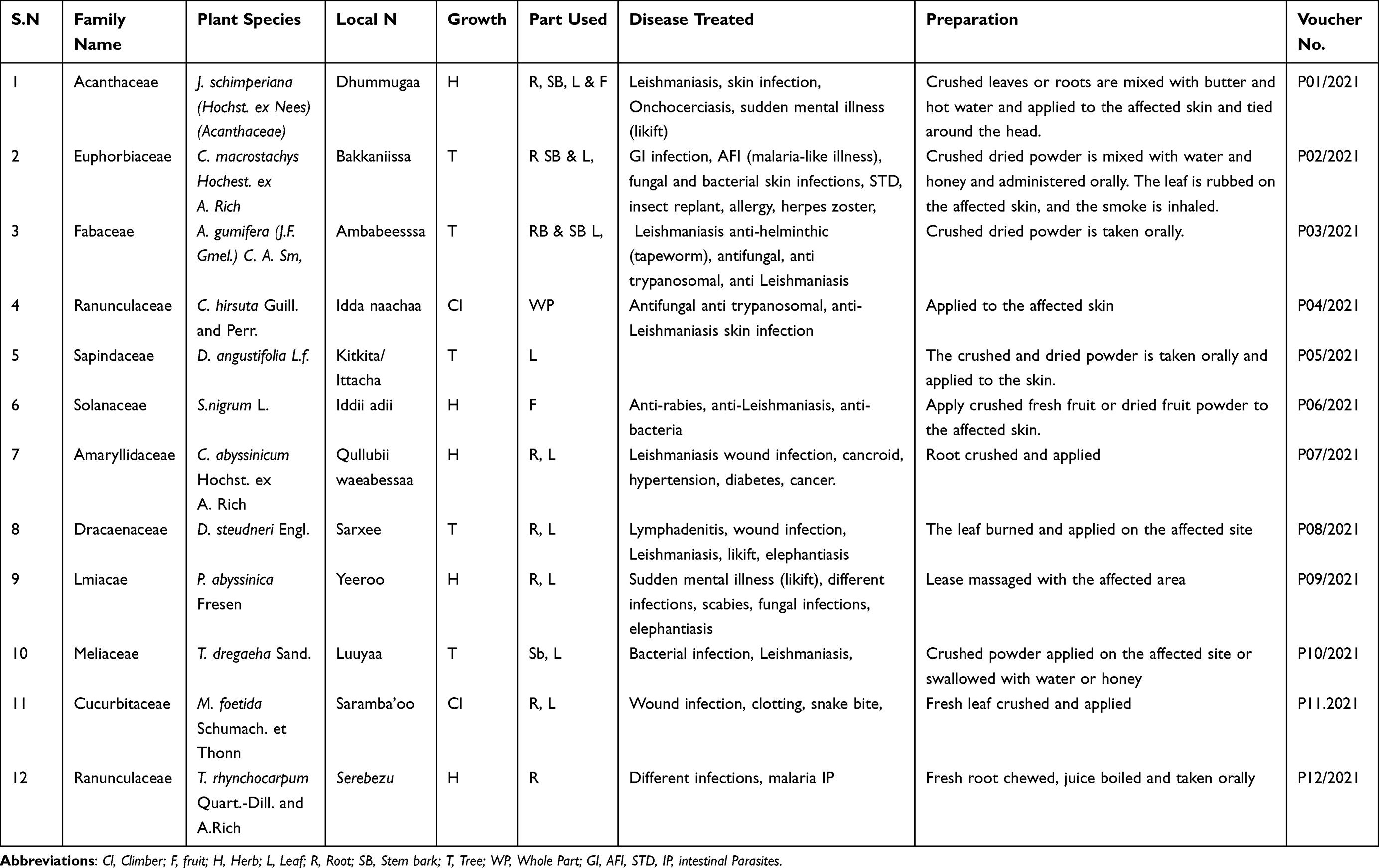 |
Table 1 Ethnomedicinal Information of the Selected Medicinal Plants |
Phytochemical Screening
All selected plant extracts were presented with notable positive phytochemical results (Table 2), which were evidenced with remarkable color changes. Flavonoids, alkaloids and phenols were the most abundant classes of compounds in majorities of the screened plants. Flavonoids were exhibited highly positive with significantly visible color change in J. schimperiana root, C. macrostachyus stem bark, A. gumifera stem bark, C. hirsuta whole part, T. dregaeha stem bark, C. abyssinicum root bulb and T. rhynchocarpum root. Alkaloid was the next most class of compound which presented in C. macrostachyus, C. hirsuta C. abyssinicum and T. rhynchocarpum root. Phenols were the third phytochemicals presented in J. schimperiana, C. macrostachyus, A. gumifera, C. hirsute, C. abyssinicum and T. rhynchocarpum root. Thin layer chromatography also confirmed the presence of different phytochemical components.
 |
Table 2 Phytochemical Screening Results of Crude Extract of Selected Plant Species |
Antimicrobial Activities
Among the selected plant species, all fractions of the extracts from T. rhynchocarpum root presented with the greatest efficacy against all tested strains. Particularly, the crude extract of T. rhynchocarpum exhibited with MIC 0.98 μg/mL against K. pneumoniae and P. aeruginosa and 0.48 μg/mL against S. aureus and E. coli which is even greater than that of the control drugs, gentamicin and clotrimazole (Table 3). Extracts from J. schimperiana and C. macrostachyus also demonstrated remarkable activity against tested microbial strains. The chloroform fraction of J. schimperiana root presented the highest activity with MIC of 3.8 μg/mL against S. aureus and E. coli. The crude extract of C. macrostachys exhibited 3.9 μg/mL against K. pneumoniae, 7.8 μg/mL against S. aureus, and E. coli. The chloroform fraction of C. macrostachys also demonstrated the lowest MIC with 7.8 μg/mL against K. pneumoniae, P. aeruginosa and S.aureus. A Kruskal–Wallis/Ddunn’s statistical test showed a significant difference between the tested samples and fractions of the plant extracts on MIC with tested plant species (H(df:11) = X2:180.45, p = 0.000) (Table 4).
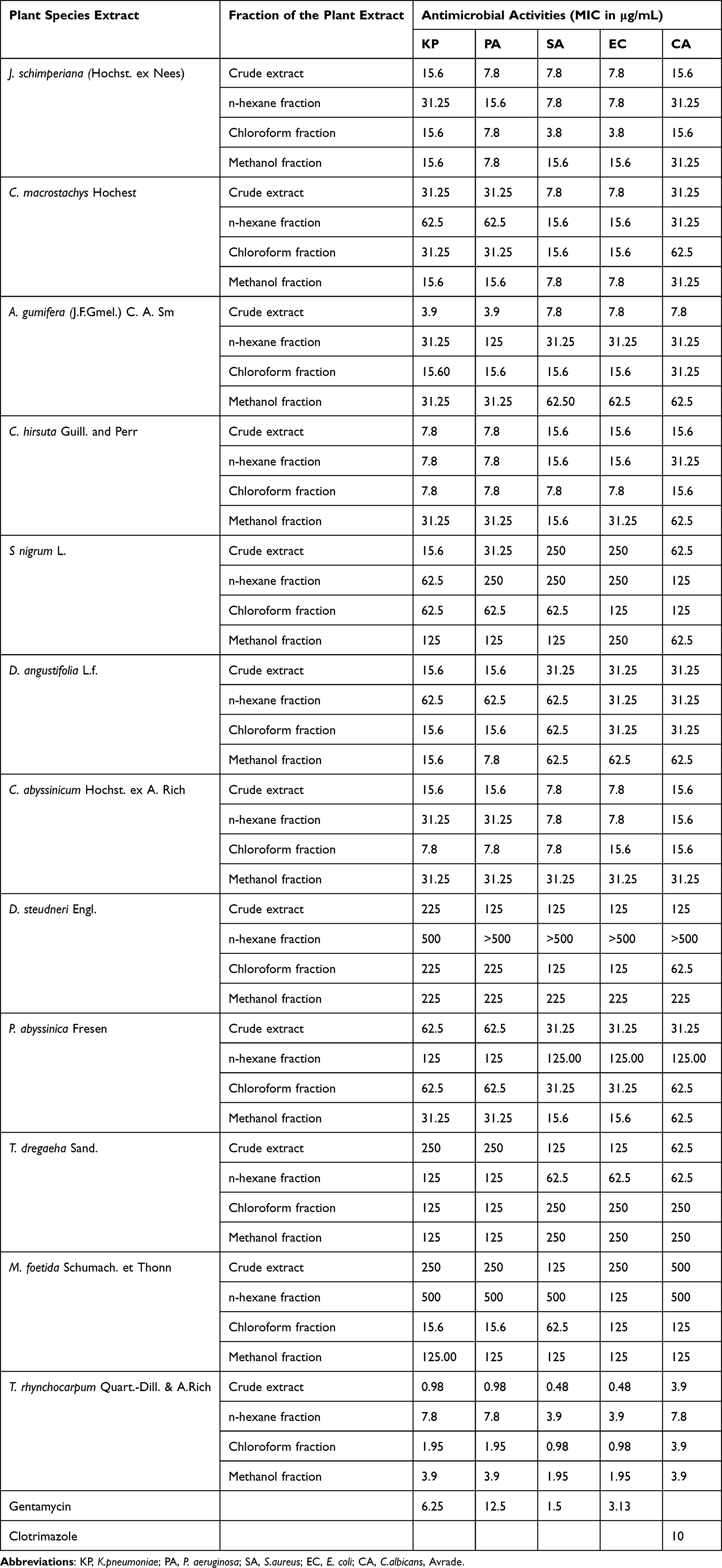 |
Table 3 Percentage Yields and Antimicrobial Activities Test of Selected Plant Crude Extract and Different Solvent Fraction |
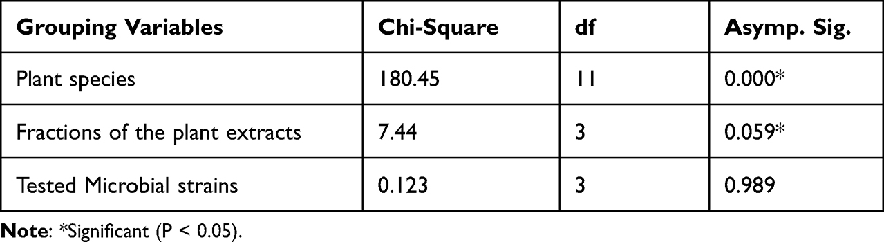 |
Table 4 SPSS Output of Kruskal–Wallis/Ddunn’s Report of MIC of Each Extract Against Selected Strain Among Grouping Variable Test Statistics |
Discussion
Plant extracts have demonstrated high-level activity against pathogens due to the enormous variety of phytochemicals. There are limited detailed examinations of these plants for their potential role as phytochemical entities and antimicrobial therapy.16,20,21 Antibiotic resistance, harmful side effects, and the high costs of synthetic drug development are shifting the focus to plant-derived medicines.4,7,30 This study identified potential plant species traditionally utilized to treat a variety of infections, including tropical infectious diseases, gastrointestinal, skin, and wound infections. The majority of the investigated plants were found to contain different phytochemical classes of compounds including flavonoids, alkaloids, phenols, glycosides, and steroids; which was confirmed by TLC results presented with multiple spots at different RF values. Among screened classes of compounds flavonoids, alkaloid and phenols were the phytochemicals with significant visible color changes. Justicia schimperiana, C. macrostachyus, A. gumifera, C. hirsuta, T. dregaeha, C. abyssinicum and T. rhynchocarpum were the plant species containing flavonoids, alkaloids and phenols. This finding is similar to the findings of other studies elsewhere.7,19,30,36
As illustrated in Table 3, most of the evaluated plant extracts demonstrated remarkable activity against selected microbial strains, with the lowest in-vitro inhibitory concentration (<10 μg/mL). The crude extracts of T. rhynchocarpum root demonstrated the greatest activity, with the lowest MIC of 0.48 μg/mL against S. aureus and E. coli and 0.98 μg/mL K. pneumonia and P. aeruginosa. The finding is consistent with reports indicating the antimicrobial efficacy of this medicinal plant for treatment of microbial infections.30,37 The chloroform fraction of J. schimperiana also demonstrated antibacterial activity with the lowest MIC value of 3.8 μg/mL against S. aureus and E. coli. Except K.pneumoniae and C.albicans its MIC is less than 10 μg/mL, which in line with other similar studies.30,38–40 Extracts from C. macrostachyus also exhibited activity against S. aureus and E. coli. These findings are consistent with previous report that the plants have antimicrobial activity.21,41
Extracts from A. gumifera, D. angustifolia, C. abyssinicum, P. abyssinica, and C. hirsuta showed moderate activity against tested microbial strains, with MIC values ranging from 10 μg/mL to 100 μg/mL, which is comparable to previous studies.42,43 In contrast with some previous studies, extracts from S. nigrum, D. steudneri, T. dregaeha, and M. foetida showed insignificant activity against tested strains.16,44,45 The difference could probably be due to differences in preparation methods, the season of plant collection, and/or environmental variations.
The tested plant extracts showed difference in activity between each fraction. Most studied plants’ crude extracts and chloroform fractions were found to be more effective against the tested strains of microbes. A crude extract of T. rhynchocarpum presented with the greater activity with MIC of 0.48 μg/mL against S. aureus and E. coli and 0.98 μg/mL against K. pneumoniae and P. aeruginosa. Justicia schimperiana crude extract was more active against P. aeruginosa, S. aureus, and E. coli with MIC of 7.8 μg/mL. Thalictrum rhynchocarpum chloroform fractions exhibited the lowest MIC: 0.98 μg/mL against S. aureus, and E. coli; 1.95 μg/mL against K. pneumoniae and P. aeruginosa and 3.9 μg/mL against C. albicans. Similarly, other studies have shown the presence of differences in activities of the different solvent fractions. Some of the phytochemical components such as terpenoids, alkaloids, flavonoids, and phenols were more extracted in the chloroform fraction, which exhibited the highest activity and broadest spectrum of antimicrobial activities against S. aureus, P. aeruginosa, and E. coli.30,44,46,47 Literature reveals that the phenolic components of medicinal plant extracts are crucial secondary metabolites responsible for efficient anti-microbial capabilities. The structure-activity relationship of phenol has been proven for p-hydroxy benzoic acid and different functional groups with ester side chains demonstrate excellent antibacterial activity.10,43,48 Flavonoids are also more effective against different microbial strains than conventional medications. Naturally occurring polyphenolic chemicals distinguished by their flavan nucleus, which makes them an important component in a variety of pharmacological applications.46,48,49 It is believed that the structure-activity relationship in the antimicrobial effect of alkaloids should be further examined because it is a very large group of compounds, and many issues have not yet been clarified. Some studies, however, have discovered that hydroxyl groups at specific positions on its aromatic rings improve antibacterial activity.42,47 All of the crude plant extracts included in this study contained one or more secondary metabolites. Therefore, the observed biological activity profile could be due to either the individual class of compounds present in each plant or the synergistic effect of each class of compounds.38,43,49 Finally, a Kruskal–Wallis H statistical test showed that there was significant difference between the tested plant species with H (df: 11) = (X2:180.45, p = 0.000) and fractions of the plant extracts H (df: 3), X2 = 7.44, p = 0.059 on MIC. But the difference in microbial strains has no significant association with the difference in MIC of the extracts.
Conclusion and Recommendations
The current ethnomedicinal survey revealed that the majority of the selected plant species were trees and herbs in growth habit. These plant species were claimed by THs as being utilized to treat different infections, including leishmaniasis, onchocerciasis, GI, wound, and skin infections. The major phytochemical classes of compounds with visible color changes and TLC spots were phenols and alkaloids. Flavonoids were remarkably exhibited with significant visible color change in J. schimperiana root, C. macrostachyus stem bark, A. gumifera stem bark T. rhynchocarpum root. Alkaloid is the next most abundant class of compound present in C. macrostachyus, C. hirsuta and C. abyssinicum. And phenols were the third phytochemicals which were present in J. schimperiana, C. macrostachyus, A. gumifera, T. rhynchocarpum root. All fractions of the extracts of T. rhynchocarpum root presented with the greatest activity against all selected strains with the lowest MICs; J. schimperiana root had the second highest activity against P. aeruginosa, S. aureus, and E. coli. All fractions of C. macrostachyus stem bark also demonstrated more activity against S. aureus and E. coli with the mean lowest MIC. The crude extract and chloroform fraction of the examined plant species had the maximum efficiency. Solanum nigrum, D. steudneri, T. dregaeha, and M. foetida were shown to be ineffective against tested strains with MICs greater than 100 μg/mL. The biological activity profile seen in each plant can be attributed to either the various classes of chemicals present or the synergistic impact that each class of compounds. The findings support scientific evidence for the usage of these plants as groundwork in traditional knowledge and point to a bright future for antibacterial drug research. Further pharmacological studies are required to be conducted using other microbial strains for effective plant species. Toxicological tests, in vivo bioactivity studies, and molecular characterization should be conducted on plant species that exhibit significant activity.
Abbreviations
ANOVA, Analysis of Variance; EPHI, Ethiopia Public Health Institution; DCM, Dichloro Methane; DMSO, Dimethyl Sulfoxide; IRB, Institutional Review Board; NTD, Neglected Tropical Infectious Diseases; MIC, Minimum Inhibitory Concentration; RF, Retention Factor; TLC, Thin Layer Chromatography; TMP, Traditional Medicine Practitioner; TM, Traditional Medicine; V/V, Volume by volume; WHO, World Health Organization.
Ethics Approval
The ethical clearance was obtained from the Jimma University Health Science Institutional Review Board (IRB) with approval letter reference number JHRPG/720/2020.
Acknowledgment
The authors would like to thank Jimma University for funding the study and providing reagents. Our gratitude also goes to the Ethiopian Public Health Institute (EPHI) for kindly providing us with the standard microbial strains.
Disclosure
All authors reported that there was no conflict of interest.
References
1. Mahady G. Medicinal plants for the prevention and treatment of bacterial infections. Curr Pharm Des. 2005;11(19):2405–2427. doi:10.2174/1381612054367481
2. Cheuka PM, Mayoka G, Mutai P, Chibale K. The role of natural products in drug discovery and development against neglected tropical diseases. Molecules. 2017;22(1). doi:10.3390/molecules22010058
3. Lacmata ST, Kuete V, Dzoyem JP, et al. Antibacterial activities of selected Cameroonian plants and their synergistic effects with antibiotics against bacteria expressing MDR phenotypes. Evid Based Complement Altern Med. 2012;2012(2010):1–11. doi:10.1155/2012/623723
4. Aslam B, Wang W, Arshad MI, et al. Antibiotic resistance: a rundown of a global crisis. Infect Drug Resist. 2018;11:1645–1658. doi:10.2147/IDR.S173867
5. Fenollar F, Mediannikov O. Emerging infectious diseases in Africa in the 21st century. N Microbes N Infect. 2018;26:S10–S18. doi:10.1016/j.nmni.2018.09.004
6. Schultz F, Anywar G, Tang H, et al. Targeting ESKAPE pathogens with anti-infective medicinal plants from the Greater Mpigi region in Uganda. Sci Rep. 2020;10(1):1–19. doi:10.1038/s41598-020-67572-8
7. Fonkeng LS, Mouokeu RS, Tume C, et al. Anti ‑ Staphylococcus aureus activity of methanol extracts of 12 plants used in Cameroonian folk medicine. BMC Res Notes. 2015;8:4–9. doi:10.1186/s13104-015-1663-1
8. Mahmoud AS, Thao N, Mario A. Potential of herbal drug and antibiotic combination therapy: a new approach to treat multidrug resistant bacteria. Pharmaceutica Analytica Acta. 2016;7(11). doi:10.4172/2153-2435.1000523
9. World Health Organization. Traditional Medicine Strategy 2014–2023. World Health Organization; 2013:1–76.
10. Aylate A, Agize M, Ekero D, Kiros A, Ayledo G, Gendiche K. In-vitro and in-vivo antibacterial activities of Croton macrostachyus methanol extract against E. coli and S. aureus. Adv Anim Vet Sci. 2017;5(3):107–114. doi:10.14737/journal.aavs/2017/5.3.107.114
11. Cheesman MJ, Ilanko A, Blonk B, Cock IE. Developing new antimicrobial therapies: are synergistic combinations of plant extracts / compounds with conventional antibiotics the solution? Pharmacogn Rev. 2019;11(22):57–72. doi:10.4103/phrev.phrev
12. Lee JA, Uhlik MT, Moxham CM, Tomandl D, Sall DJ. Modern phenotypic drug discovery is a viable, neoclassic pharma strategy. J Med Chem. 2012;55(10):4527–4538. doi:10.1021/jm201649s
13. Alves RRN, Rosa IML. Biodiversity, traditional medicine and public health: where do they meet? J Ethnobiol Ethnomed. 2007;3(1). doi:10.1186/1746-4269-3-14
14. Habtamu A, Mekonnen Y. Antibacterial potential of the 80 % methanol and chloroform extracts of Clematis hirsuta. Afri J Pharm Pharmacol. 2017;11(16):204–208. doi:10.5897/AJPP2016.4540
15. Abdullahi AA. Trends and challenges of traditional medicine in Africa. Afri J Trad Complement Altern Med. 2011;8(5SUPPL.):115–123. doi:10.4314/ajtcam.v8i5S.5
16. Belay G, Tariku Y, Kebede T, Hymete A. Ethnopharmacological investigations of essential oils isolated from five Ethiopian medicinal plants against eleven pathogenic bacterial strains. Phytopharmacology. 2011;1(5):133–143.
17. Abera B. Medicinal plants used in traditional medicine by Oromo people, Ghimbi District, Southwest Ethiopia. J Ethnobiol Ethnomed. 2014;10(1):1–15. doi:10.1186/1746-4269-10-40
18. Megersa M, Tamrat N. Medicinal plants used to treat human and livestock ailments in Basona Werana District, North Shewa Zone, Amhara Region, Ethiopia. Evid Based Complement Altern Med. 2022;2022. doi:10.1155/2022/5242033
19. Abebe W, Zhang W, Zhang S, Xie G. Chemical composition and antimicrobial activity of essential oil from Justicia schimperiana. J Pharmacogn Natural Prod. 2018;04(02):2–4. doi:10.4172/2472-0992.1000154
20. Mesfin F, Seta T, Assefa A. An ethnobotanical study of medicinal plants in Amaro Woreda, Ethiopia. Ethnobotany Res Appl. 2014;12:341–354. doi:10.17348/era.12.0.341-354
21. Meresa A, Ashebir R, Gemechu W, Teka F. Ethno medicinal uses, phytochemistry and anti-malarial effect of Croton Ethno medicinal uses, phytochemistry and anti- malarial effect of Croton macrostachyus (Bisana): a review. Polymers. 2019;11. doi:10.3390/polym11101682
22. Sayed E, Atwaa H, Shahein MR, et al. Antimicrobial activity of some plant extracts and their applications in homemade tomato paste and pasteurized cow milk as natural preservatives. J Biomol Struct Dynamics. 2022;1–16. doi:10.1080/07391102.2022.2130987
23. Al-rimawi F, Imtara H, Khalid M, Salah Z, Parvez MK. Assessment of antimicrobial, anticancer, and antioxidant activity of Verthimia iphionoides plant extract. Processes. 2022;10:2375. doi:10.3390/pr10112375
24. Ali H, Nguta J, Musila F, Ole-mapenay I, Matara D, Mailu J. Evaluation of antimicrobial activity, cytotoxicity, and phytochemical composition of Ocimum americanum L. (Lamiaceae). Evid Based Complement Altern Med. 2022;2022:154.
25. Luitel S, Dahal RK, Dahal RK. In vitro antimicrobial activity of some medicinal plants against human pathogenic bacteria. J Trop Med. 2019;2019. doi:10.1155/2019/1895340
26. Lawal AM, Lawan MM, Apampa SA. Phytochemical analysis and thin layer chromatography profiling of crude extracts from GuieraSenegalensis (Leaves). J Biotechnol Biomed Sci. 2019;3(3):7–12.
27. Solanki SL, Modi CM, Patel HB, Patel UD. Phytochemical screening and thin-layer chromatography of six medicinal plants from the surroundings of Junagadh, Gujarat, India. J Pharmacogn Phytochem. 2019;8(4):3122–3126.
28. Wilde TJJD, Ben IO, Woode E, Abotsi WKM. Preliminary phytochemical screening and in vitro antioxidant properties of Trichilia monadelpha (Thonn.) JJ De Wilde (Meliaceae). J Med Biomed Sci. 2013;2:6–15.
29. Hassan IA, Nasiru IA, Malut AM, Abdulkadir S, Ali AS. Phytochemical studies and thin layer chromatography of leaves and flower extracts of S enna siamea lam for possible biomedical applications. J Pharmacogn Phytother. 2015;7:18–26. doi:10.5897/JPP2014.0337
30. Mayeku PW, Hassanali A, Kiremire BT, Odalo JO, Hertweck C. Anti-bacterial activities and phytochemical screening of extracts of different parts of Thalictrum rhynchocarpum. Afri J Trad Complement Altern Med. 2013;10(5):341–344. doi:10.4314/ajtcam.v10i5.20
31. Belay G, Tariku Y, Kebede T, Hymete A, Mekonnen Y. Antibacterial activity of five oil bearing Ethiopian medicinal plants against eleven pathogenic bacterial strains. Med Plants. 2011;3(4):293–299. doi:10.5958/j.0975-4261.3.4.049
32. Razmavar S, Abdulla MA, Ismail SB, Hassandarvish P. Antibacterial activity of leaf extracts of Baeckea frutescens against methicillin-resistant Staphylococcus aureus. Biomed Res Int. 2014;2014:1–5. doi:10.1155/2014/521287
33. Fentahun M, Ayele Y, Amsalu N, Alemayehu A, Amsalu G. Antibacterial evaluation and phytochemical analysis of selected medicinal plants against some pathogenic enteric bacteria in Gozamin District, Ethiopia. J Pharmacovigilance. 2017;5(5). doi:10.4172/2329-6887.1000244
34. Cheruiyot KR, Olila D, Kateregga J. In-vitro antibacterial activity of selected medicinal plants from Longisa Region of Bomet District, Kenya. Afri Health Sci. 2009;9:115.
35. Ribeiro SM, Fratucelli ÉDO, Bueno PCP, et al. Antimicrobial and antibiofilm activities of Casearia sylvestris extracts from distinct Brazilian biomes against Streptococcus mutans and Candida albicans. BMC Complement Altern Med. 2019;2019:1–16.
36. Ayele TT, Regasa MB, Delesa DA. Antibacterial and antagonistic activity of selected traditional medicinal plants and herbs from East Wollega Zone against clinical isolated human pathogens. Sci Technol Arts Res J. 2016;4(3):175. doi:10.4314/star.v4i3.26
37. Maroyi A. Pharmacological properties of Croton macrostachyus hochst. ex delile: a comprehensive review. Altern Med. 2017;2017:147.
38. Gurunathan A, Subramanium P, Maran S. In vitro antimicrobial activity of leaf, stem and root extracts of the medicinal plant species, Thalictrum javanicum blume against certain human pathogens. Int J Pharma Pharma Sci. 2013;5(SUPPL.4):71–75.
39. Asfere Y, Kebede A, Zinabu D. In-vitro antimicrobial activities and phytochemical screening of selected plant extracts against some medically and agriculturally important pathogens. Eur J Med Plants. 2020;31(10):167–189. doi:10.9734/ejmp/2020/v31i1030293
40. Nascimento GGF, Locatelli J, Freitas PC, Silva GL. Antibacterial activity of plant extracts and phytochemicals on antibiotic-resistant bacteria. Braz J Microbiol. 2000;31(4):247–256. doi:10.1590/S1517-83822000000400003
41. Oluwakemi Sola A. Antibacterial screening of crude fractions of calotropis procera (Linn) and Gc-Ms profile of fractions obtained from the extracts of calotropis procera. Biomed J Sci Tech Res. 2019;18(2). doi:10.26717/bjstr.2019.18.003124
42. Othman L, Sleiman A, Abdel-Massih RM. Antimicrobial activity of polyphenols and alkaloids in Middle Eastern plants. Front Microbiol. 2019;10. doi:10.3389/fmicb.2019.00911
43. Ads EN, Hassan SI, Rajendrasozhan S, Hetta MH, Aly SH, Ali MA. Isolation, structure elucidation and antimicrobial evaluation of natural pentacyclic triterpenoids and phytochemical investigation of different fractions of Ziziphus spina-christi (L.) Stem Bark Using LCHRMS analysis. Molecules. 2022;27(6):1–14. doi:10.3390/molecules27061805
44. Odeleye OM, Oyedeji AO. Antibacterial activity of crude and fractions of Momordica foetida leaf extracts. Int J Biomed Pharma Sci. 2008;2(2):75–78.
45. Lulekal E, Rondevaldova J, Bernaskova E, et al. Antimicrobial activity of traditional medicinal plants from Ankober District, North Shewa Zone, Amhara Region, Ethiopia. Pharm Biol. 2014;52(5):614–620. doi:10.3109/13880209.2013.858362
46. Farhadi F, Khameneh B, Iranshahi M, Iranshahy M. Antibacterial activity of flavonoids and their structure–activity relationship: an update review. Phytother Res. 2019;33(1):13–40. doi:10.1002/ptr.6208
47. Mabhiza D, Chitemerere T, Mukanganyama S. Antibacterial properties of alkaloid extracts from Callistemon citrinus and Vernonia adoensis against Staphylococcus aureus and Pseudomonas aeruginosa. Int J Med Chem. 2016;2016:1–7. doi:10.1155/2016/6304163
48. Tungmunnithum D, Thongboonyou A, Pholboon A. Flavonoids and other phenolic compounds from medicinal plants for pharmaceutical and medical aspects: an overview. Medicines. 2018. doi:10.3390/medicines5030093
49. Tagousop CN, Tamokou JDD, Ekom SE, Ngnokam D, Voutquenne-Nazabadioko L. Antimicrobial activities of flavonoid glycosides from Graptophyllum grandulosum and their mechanism of antibacterial action. BMC Complement Altern Med. 2018;18(1):1–10. doi:10.1186/s12906-018-2321-7
 © 2023 The Author(s). This work is published and licensed by Dove Medical Press Limited. The
full terms of this license are available at https://www.dovepress.com/terms
and incorporate the Creative Commons Attribution
- Non Commercial (unported, 3.0) License.
By accessing the work you hereby accept the Terms. Non-commercial uses of the work are permitted
without any further permission from Dove Medical Press Limited, provided the work is properly
attributed. For permission for commercial use of this work, please see paragraphs 4.2 and 5 of our Terms.
© 2023 The Author(s). This work is published and licensed by Dove Medical Press Limited. The
full terms of this license are available at https://www.dovepress.com/terms
and incorporate the Creative Commons Attribution
- Non Commercial (unported, 3.0) License.
By accessing the work you hereby accept the Terms. Non-commercial uses of the work are permitted
without any further permission from Dove Medical Press Limited, provided the work is properly
attributed. For permission for commercial use of this work, please see paragraphs 4.2 and 5 of our Terms.