Back to Journals » Pharmacogenomics and Personalized Medicine » Volume 13
Pharmacogenomics and Pharmacogenetics: In Silico Prediction of Drug Effects in Treatments for Novel Coronavirus SARS-CoV2 Disease
Authors Cafiero C, Re A ![]() , Micera A
, Micera A ![]() , Palmirotta R
, Palmirotta R ![]() , Monaco D, Romano F, Fabrizio C, Di Francia R, Cacciamani A
, Monaco D, Romano F, Fabrizio C, Di Francia R, Cacciamani A ![]() , Surico PL
, Surico PL ![]() , D'Amato G, Pisconti S
, D'Amato G, Pisconti S
Received 12 July 2020
Accepted for publication 9 September 2020
Published 13 October 2020 Volume 2020:13 Pages 463—484
DOI https://doi.org/10.2147/PGPM.S270069
Checked for plagiarism Yes
Review by Single anonymous peer review
Peer reviewer comments 2
Editor who approved publication: Dr Martin H Bluth
Concetta Cafiero,1,* Agnese Re,2,* Alessandra Micera,3,* Raffaele Palmirotta,4 Delio Monaco,5 Francesca Romano,6 Claudia Fabrizio,7 Raffaele Di Francia,8 Andrea Cacciamani,3 Pier Luigi Surico,9 Gerardo D’Amato,10 Salvatore Pisconti1
1Medical Oncology, SG Moscati Hospital, Taranto, Italy; 2CNR-IASI, Catholic University of Sacred Heart, Rome, Italy; 3Research Laboratories in Ophthalmology, IRCCS — Fondazione Bietti, Rome, Italy; 4Department of Biomedical Sciences and Clinical Oncology, Oncogenomic Research Center, Aldo Moro University of Bari, Bari, Italy; 5Radiology, SG Moscati Hospital, Taranto, Italy; 6Department of Precision Medicine, Luigi Vanvitelli University of Campania, Napoli, Italy; 7Infectious Disease Unit, SG Moscati Hospital, Taranto, Italy; 8Italian Association of Pharmacogenomics and Molecular Diagnostics, Ancona, Italy; 9Oncology and Hematology Department, F Miulli Hospital, Acquaviva delle Fonti, Italy; 10Endocrine and Metabolic Surgery, A Gemelli Polyclinic Foundation, Rome, Italy
*These authors contributed equally to this work
Correspondence: Alessandra Micera
Research Laboratories in Ophthalmology, IRCCS — Fondazione Bietti, 6 via di Santo Stefano Rotondo, Rome 00184, Italy
Tel +39-06-4554-1191
Email [email protected]
Raffaele Palmirotta
Department of Biomedical Sciences and Clinical Oncology, Oncogenomic Research Center, Aldo Moro University of Bari, Italy
Email [email protected]
Abstract: The latest developments in precision medicine allow the modulation of therapeutic approaches in different pathologies on the basis of the specific molecular characterization of the patient. This review of the literature coupled with in silico analysis was to provide a selected screening of interactions between single-nucleotide polymorphisms (SNPs) and drugs (repurposed, investigational, and biological agents) showing efficacy and toxicityin counteracting Covid-19 infection. In silico analysis of genetic variants related to each drug was performed on such databases as PharmGKB, Ensembl Genome Browser, www.drugs.com, and SNPedia, with an extensive literature review of papers (to May 10, 2020) on Covid-19 treatments using Medline, Embase, International Pharmaceutical Abstracts, PharmGKB, and Google Scholar. The clinical relevance of SNPs, known as both drug targets and markers, considering genetic variations with known drug responses, and the therapeutic consequences are discussed. In the context of clinical treatment of Covid-19, including infection prevention, control measures, and supportive care, this review highlights the importance of a personalized approach in the final selection of therapy, which is probably essential in the management of the Covid-19 pandemic.
Keywords: Covid-19, SARS-CoV2, drug effects, pharmacogenomics, pharmacogenetics, in silico, ADR
Introduction
Covid-19, an emergency all over the world, is caused by severe acute respiratory syndrome coronavirus 2 (SARS-CoV2), a positive-sense RNA virus with higher mutation rates than DNA viruses.1,2 Laboratory tests on respiratory tract specimens (swab/saliva/tears PCR and serological tests) and computed tomography (CT; for chest or other tissue) should stage as soon as possible the presence of disease and its evolution/progression.3,4 SARS-CoV2 binds principally to the cell-membrane ACE2 receptor through the viral structural spike (S) protein, although other “virus doors” have been suggested (integrins and Toll-like receptors)5 (Figure 1). At the intracellular level, the virus activates cellular processes to produce viral proteins that replicate the virus’s genetic material, providing potential targets for drug therapy.1,5,6
 |
Figure 1 Overview of Covid-19 infection: SARS-CoV2–host interaction and tissue manifestation. Schematic representation of SARS-CoV2 virus interacting with mucosa epithelia (framed) and gradual sprouting to gut, kidney, brain, and lung tissue. “Virus doors” reportedwere ACE2, integrin, TLR3, or TMPRSS2. Overall symptoms are listed. Briefly, most common Covid-19 symptoms occur at the upper respiratory tract, together with asthenia and general malaise. Early, less common clinical features include gastrointestinal symptoms (nausea, anorexia, diarrhea), anosmia and dysgeusia. Severe cases encompass pneumonia or bronchopneumonia, severe acute respiratory syndrome, renal failure, up to death. The lower respiratory tract participation and complications are more frequent in people aged over 50 with pre-existing chronic diseases of cardiovascular and/or respiratory system, as well as some autoimmune diseases (diabetes). Cardiac involvement represents another potentially life-threatening complication, caused by direct viral damage, hypoxic injury, microvascular dysfunction and disclosing with diverse clinical pictures (arrhythmias, myocarditis, and heart failure). Finally, virus can reach central nervous system causing central (acute cerebrovascular diseases, impairment of consciousness) and peripheral complications. The evolution of infection from early detection severe complications and death (shaded arrow). As reported, molecular and serological, as well as imaging tests, are useful depending on disease evolution: real-time PCR applied to nasopharyngeal swabs, venous blood as well as conjunctival swabs can track disease ongoing, while early-released IgM and late-released IgG might assess for immune response, in line with cytokine storm panel (IL6, TNFα, IFNγ, and IL10) typifying tissue/organ complications. |
SARS-CoV2 Infection and Main Clinical Aspects
Covid-19 features, diagnostic route, and early, mild, and severe Covid-19 symptoms are summarized in Figure 1. Virus entry and multiple signaling pathways (Ca2+ release, cSrc, FAK, MAPK, and PI3K) occur upon Spike2–RGD–integrin interaction.7 A “cytokine storm” is locally released (IL1β, IL6, IL8, TNFα, MIP1α, and VEGF), as first observed in patients showing fatal complications.5 This specific panel was used to screen patients requiring prompt intervention to avoid acute respiratory distress syndrome (ARDS).6 In a recent study, candidate drug targets for nonstructural proteins (3-chymotrypsin–like protease, papain-like protease, RNA-dependent RNA polymerase), viral access, and related immuno regulatory pathways have been prospected for evaluation.6,8,9 A wide range of pulmonary manifestations can be observed, varying from mild respiratory symptoms (cough or sore throat) to severe pneumonia, up to a sudden development of respiratory failure.10 Silent hypoxemia is a peculiar aspect frequent in frail and elderly patients that usually precedes the onset of an overt acute and severe respiratory syndrome.11,12 Chest CT imaging is strongly recommended for an early diagnosis of novel coronavirus pneumonia.13,14 CT findings, including patterns associated with less frequent signs, eg, lobular, bronchial, pleural, and subpleural involvement, as well as pleural lymphadenopathy and pericardial effusion, have been described.15 SARS-CoV2 initially causes airspace exudates, interstitial edema, hyaline membranes, and inflammatory infiltrates in the alveoli.16 Other than the deleterious lung effects, systemic SARS-CoV2 has a significant impact on the hematopoietic system and tissue homeostasis.17 Initially, lymphopenia showed interesting prognostic value, for both neutrophil:lymphocyte and platelet:lymphocyte ratios in severe SARS-CoV2 cases. Lymphocyte parameters (count dynamics), some inflammatory indices (LDH, CRP, and IL6), and some recent circulating biomarkers (high serum procalcitonin and ferritin) have been suggested for identifying cases with poor prognosis (prompt intervention and improved outcomes). As observed, blood hypercoagulability, high D-dimer levels (particularly associated with disease worsening), protraction of both prothrombin (PT) time, activated partial thromboplastin time (aPTT), and disseminated intravascular coagulation require constant surveillance to allow prompt intervention. In addition to upper-airway contact, conjunctivitis may be an ocular manifestation of SARS-CoV2 infection, if quickly detected.18 In December 2019, a Chinese ophthalmologist reported an unusual viral conjunctivitis. To date, several Internet articles and scientific reports have documented the potential use of eye swabs as a tool to screen virus infection, although contrasting data are available, most probably related to progression of infection and virus detection.19 In January 2020, a worldwide expert in infectious diseases referred to conjunctivitis during an inspection of Wuhan and tested positive for SARS-CoV2, but quickly recovered from infection. This led to the idea of “eye infection” as a possible alternative route of SARS-CoV2 transmission, alternative to the respiratory one.20 Ocular secretion might represent a reservoir of virus at the early stages of contagious.18 When pinkeye occurs as a sign of conjunctivitis, a differential diagnosis can be of great utility to screen symptomatic and even presymptomatic individuals.21
Genetic Background, Individual Responses, and SNP Association
Recently, pharmacogenomics (the effects of a single genetic marker) and pharmacogenetics (the collective influence of variability across the genome to modulate an individual’s drug response) have received great attention for their abilities to provide a new way to select drugs for personalized therapy (optimal dosing for maximizing drug efficacy or minimizing the risk of toxicity).22,23 Through drug therapy, pharmacogenetics might influence both pharmacokinetics and pharmacodynamics with respect to dosing, formulation sensitivity, and adverse drug reactions (ADRs), as well as drug-hypersensitivity reactions (allergic, pseudoallergic, and exaggerated pharmacological reactions to medications), determining an enhanced immunologic reaction or inflammatory response (side effects).24 Genetic assessment can predict the occurrence of drug-related toxic effects. Single-nucleotide polymorphisms (SNPs) are the most common type of polymorphism found in the human genome, and represent the main reason for 90% of all types of genetic variations among individuals. An example of an ADR is the hypersensitivity reaction occurring in SNP carriers of the HLA-B*5701 allele who receive the antiviral abacavir for treatment of HIV.25,26 Covid-19 therapy is wide-ranging and multiple, and risk factors for ADRs can occur. Therefore, optimal doses, duration of treatment, side effects. and long-term outcomes are critical aspects of Covid-19 therapy.27 CoV-mediated inflammation may be counteracted by anti-inflammatory cytokines, including IL1 family members, IL6, and TNFα.28 Since no specific drug/therapy for Covid-19 treatment has been US Food and Drug Administration (FDA)-approved, an array of drugs approved for other diseases and under investigation (including off-label and biological drugs) have been included in clinical trials.29,30 As observed, the metabolic pathways of these drugs included various polymorphic cytochrome P450 enzymes, strongly suggesting genotyping for the CYP2D6*2, CYP2D6*3, CYP2D6*4, and CYP2D6*10 alleles.31,32 As an example, no predictability in the metabolic function of the CYP3A5*3 allele can result in alternative mRNA splicing with a trunked protein, due to the formation of an untimely stop codon. Moreover, haplotype CYP3A5*3 has been related to a reduced clearance of both ritonavir and lopinavir (substrates) (Table 1). Of interest is the association between polymorphisms inABCC and therapy efficacy, as the drug transporters are one of the primary mechanisms related to subtherapeutic antiretroviral-drug concentrations.33 Prominent research has highlighted the relationship between the ABCB1 polymorphism (3435 C>T) and hepatotoxicity risk after antiviral treatments, and few additional studies were found regarding other nucleoside analogues.33 More recently, the presence of grade 3–4 hyperbilirubinemia is directly proportional to the homozygosity, heterozygosity, and wild-type genotyping for the UGT1A1*28 allele in patients who receive ritonavir and lopinavir.34 Therefore, the attempt of this literature review coupled with in silico analysis was to provide a selected screening of drugs showing efficacy and toxicity effects useful for counteracting Covid-19 infection.
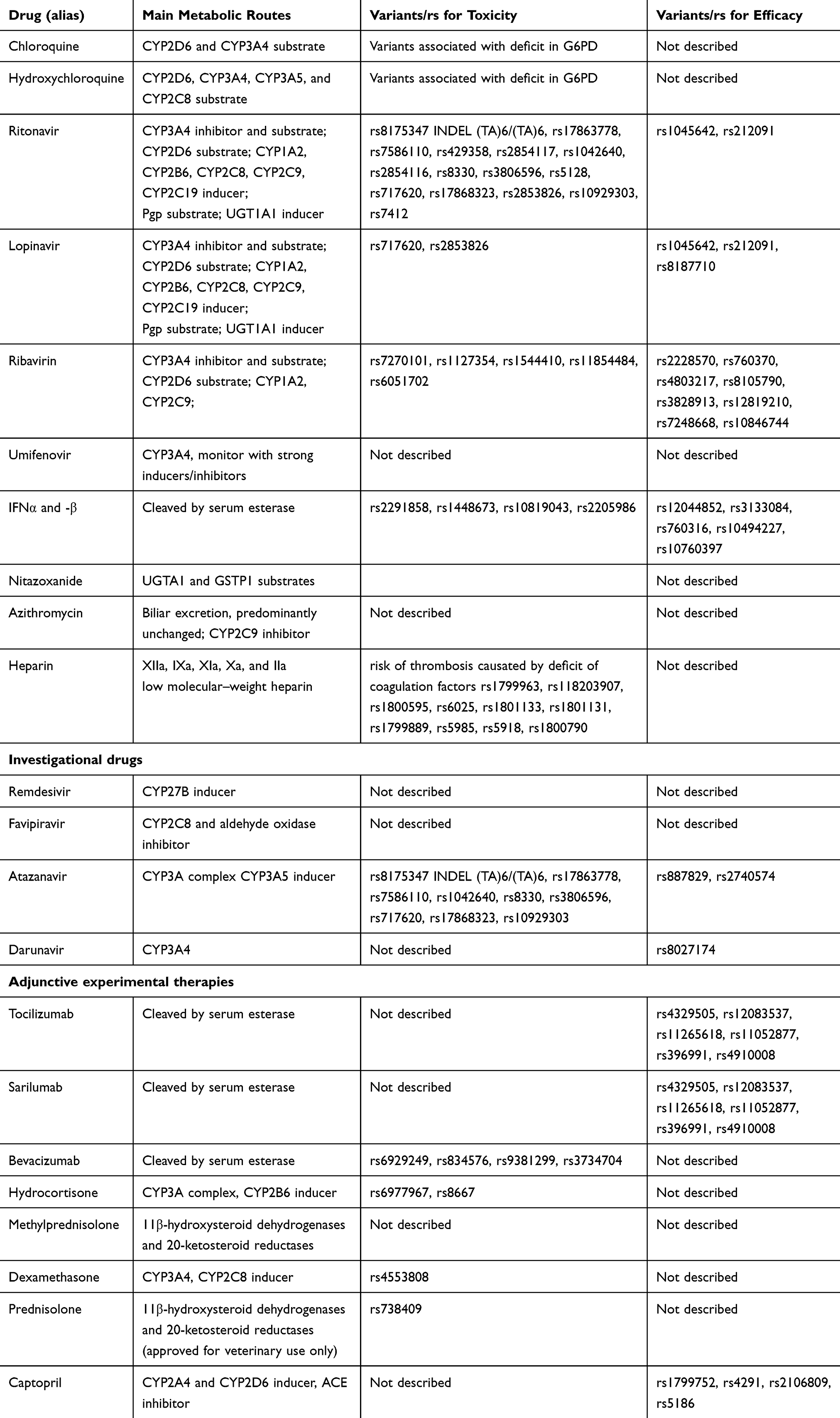 |
Table 1 The Most Promising and Repurposed Drugs |
Methods
An extensive literature review of papers published until May 10, 2020 was performed based on a standard procedure (Medline, Embase, International Pharmaceutical Abstracts, PharmGKB. and Google Scholar).35 Search terms were “Covid-19”, “novel coronavirus”, “SARS-CoV2”, “pharmacogenetics”, “treatment/s”, “adverse side effects”, “therapy”, “lung”, “ocular”, “pulmonary infection”, “drugs”, “drug response”, “virus”, “candidate drugs”, “potential inhibitors”, “protease inhibitors”, “personalized medicine”, “individual therapy”, “pneumonia”, “ACE”, “heparin”, “vasculitis”, “conjunctivitis”, “rhinitis”, “hematological complication” and “main metabolic routes”, either alone or in combination. From this manually performed analysis, drugs reported in at least two studies or in a clinical trial were included. Ongoing clinical trials and the index of studies of Covid-19 were identified using the search term “coronavirus infection” on ClinicalTrials.gov and the Chinese Clinical Trial Registry (http://www/chictr.org/enindex.aspx). In silico analysis of genetic variants related to each drug was performed on dedicated databases, such as PharmGKB (Table 2), Ensembl Genome Browser, www.drugs.com, and SNPedia. The allelic frequency of each variant average in all populations was based on data from the 1000 Genomes Project phase 3. We considered SNPs related to both efficacy and toxicity response with allele frequencies ≥5%, as already described in previous studies.36,37 The quality of each study was assured, and resulting information included study design, baseline characteristics of disease, treatment regimens, and allelic frequencies of the genetic variant.
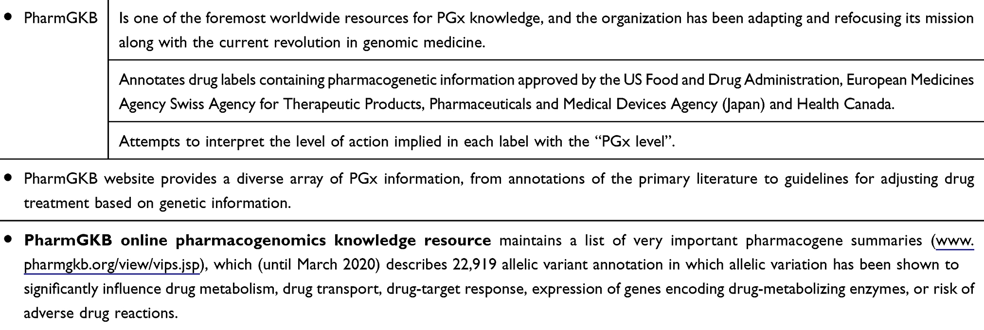 |
Table 2 Overview of PharmGKB |
Results
Drugs in use as routine therapy or in clinical trials for Covid-19 include steroids and antiviral and biological humanized neutralizing antibodies against some proinflammatory cytokines, such as IL1, IL6, IFN, and TNFα, in addition to supportive measures and symptomatic treatment, according to the severity of the disease.38 Data analysis on registered clinical trials of Covid-19 in the US is available at https://clinicaltrials.gov. Nowadays, a great number of agents have been evaluated for potential use in Covid-19 management, and only a fewhave been included within international and/or local protocols.38 According to the US National Institutes of Health, no pharmacological agent has earned approval for safe and effective use yet, mainly because of the lack of evidence in favor or against these agents (https://covid19treatmentguidelines.nih.gov). One important aspect of a therapeutic approach consists in the choice of the right drugs throughout the natural history of disease. Indeed, a three-phase pathogenetic model has been proposed, with different clinical and laboratory features, each requiring a specific treatment based on the changing role over time,of direct viral damage and host inflammatory response in the disease course. This model describes an early stage (stage I), coinciding with incubation and/or mild flulike symptoms, during which antiviral drugs might reach high effectiveness, a moderate stage (stage II) characterized by pulmonary involvement without IIa or with IIb hypoxia, which may benefit from the use of antivirals and anti-inflammatory therapy (including steroids), and lastly a severe stage (stage III), in which a dysregulated, systemic hyperinflammatory response takes place, thus requiring the administration of immunomodulating agents, several of which are currently under investigation, and in some cases already in use within management protocols.37
Gene variants associated with pharmacological responses to drugs are reported in a dedicated database — PharmGKB (https://www.pharmgkb.org) — allowing the identification of relationships between genetic variations (eg, SNPs, indels, repeats, haplotypes) and individual drug responsiveness.40,41 Herein, in silico pharmacogenetic analysis shows the potential clinical efficacy and/or toxicity of the major drugs selected for Covid-19 treatment. The main drugs proposed for Covid-19 treatments and reference sequence (rs) related to ADRs and efficacy are shown in Table 1. Genes and related SNPs (rs) associated with drug effects and their study annotation are summarized in Table 3.42–64 Major clinical information regarding “variant drug” responsiveness (clinical annotations) is highlighted. Bloodstream fluidity requires a tidy balance among factors favoring flow of blood and thrombosis. Any modification in this physiological balance triggers pathological conditions: deficit of coagulation factors favor bleeding, while genetic mutations of coagulation factors can determine a thrombotic risk framework characterized by differences in genetic variants of coagulation (Table 4). All drugs used as therapeutic options and their study annotation based on pharmacogenetic data are discussed.
 | 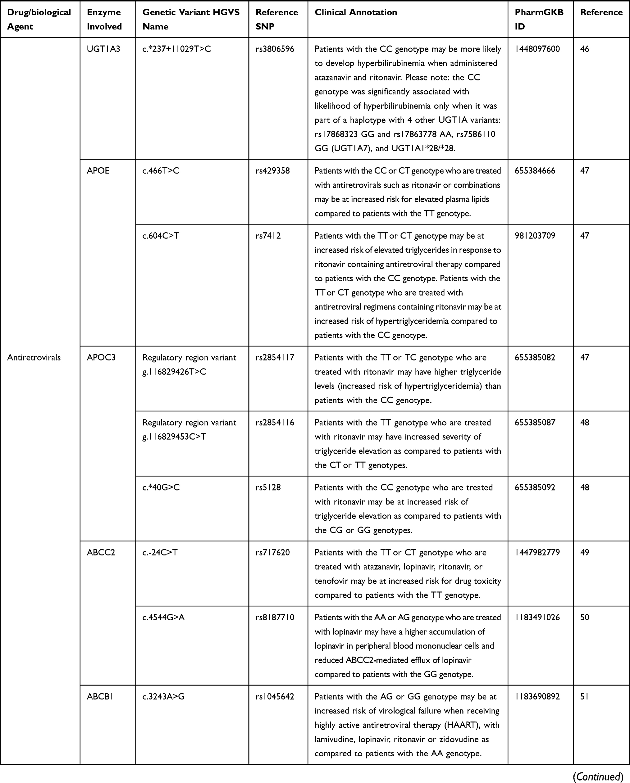 |  |  |  |  |  |
Table 3 List of Genes and Reference SNPs (Rs: Reference Sequences) Associated with Drug Effects. Study Annotation Based on Data from Pharmgkb.org HGVS: Human Genome Variation Society (http://varnomen.hgvs.org) |
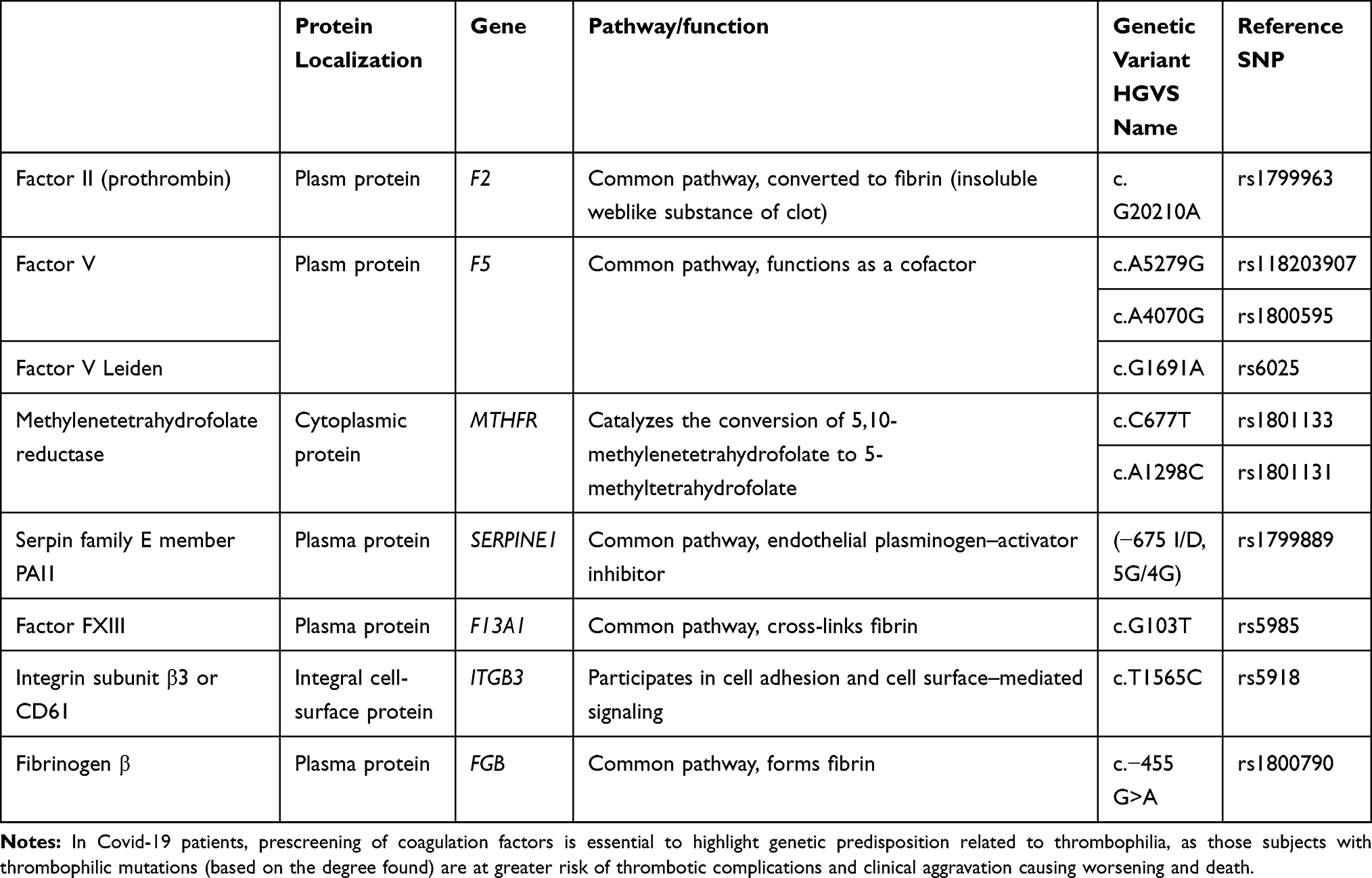 |
Table 4 List of Coagulation Factors with Relative Protein Localization, Pathway, Function, Nucleotide Variants, and Reference SNPs (Reference Sequences) Involved in Thrombophilic Disorders |
The Most Promising and Repurposed Drugs
Chloroquine (Cq) and Hydroxychloroquine (Hcq)
Cq is used for chemoprophylaxis of malaria and amebiasis, while Hcq is used to treat autoimmune disease.8 The mechanism of action includes the blocking of viral entry through the inhibition of glycosylation of host receptors, proteolytic processing, and endosomal acidification.65 Effects of immunomodulation on cytokine production and inhibition of autophagy/lysosomal activity have been also reported.65–67 Several randomized controlled trials (RCTs) are currently investigating Cq and Hcq in Covid-19 treatment (https://clinicaltrials.gov). Based on current data, both drugs are recommended for treatment of unhospitalized Covid-19 patients, and the effects of cardiotoxicity in immunosuppressed subjects or patients with kidney or liver problems are known. Recommendations for oral administration in Covid-19 treatment are 500 mg twice a day and 400 mg twice a day, followed by 200 mg twice a day for Cq and Hcq, respectively.68 Blood monitoring is required, as hemolytic anemia might occur, particularly when the drug is delivered in association with other drugs that cause hemolysis.69 It should not to be underestimated the extent to which both drugs may cause hemolysis in glucose-6-phosphate dehydrogenase (G6PD)-deficient individuals. Of note, about 39% of Covid-19 patients suffering from lupus have failed to respond or were even intolerant to Hcq.70 Moreover, long-term Cq–Hcq treatment induces retinal toxicity, and particularly when Hcq-related retinopathy is diagnosed, the retinal damage endures, even after cessation of therapy.71 High doses or prolonged administration of Hcq, even at recommended doses, may increase the risk of ocular toxicity (paracentral scotomas, color-vision changes, corneal/ciliary body/conjunctival as well as retinal abnormalities), and visual disturbances (retinal and macular toxicity).72 This would imply that accurate eye screening for confirming SARS-CoV2 presence in early conjunctivitis or monitoring ocular structure during Covid-19 therapy is recommended to counteract early or even prevent eventual signs of ocular drug toxicity, including the retinopathy.73
Lopinavir/Ritonavir
The oral combination of these two agents (FDA-approved and currently authorised as anti-HIV medicine) demonstrate activity against other novel coronaviruses.74,75 Several RCTs examining lopinavir/ritonavir in Covid-19 are in progress.76 Recommendations for administration in Covid-19 treatment is 400 mg/100 mg twice daily for up to 14 days.77 Adverse effects of lopinavir/ritonavir include several gastrointestinal complains, such as nausea, vomiting, and diarrhea, and hepatotoxicity, pancreatitis, and cardiac conduction abnormalities.78 These adverse effects are increased in 20%–30% of Covid-19 patients with elevated transaminases.79 Previous studies have shown that these ADRs with lopinavir–ritonavir combination are related to several polymorphisms present in the genes UGT1A1, UGT1A7, APOE, and APOC3 (Table 1).
Ribavirin
This antiviral drug (guanine analogue) inhibits viral RNA–dependent RNA polymerase. Ribavirin activity against Covid-19 disease is limited and requires high doses or combination therapy to be effective in humans. No evidence exists for inhaled ribavirin for Covid-19 treatment — no benefit over enteral or intravenous administration79 — though several studies have demonstrated possible harm due to adverse effects (hematologic and liver toxicity).80 Ribavirin causes severe dose-dependent hematologic toxicity.81 The inconclusive efficacy and toxicity results suggest that ribavirin has limited value in treatment of Covid-19. Case–control pharmacogenetic association studies indicate different polymorphisms in the ITPA, VDR, and SLC28A2 genes that are related to toxicity and adverse effects, while an increased pharmacological response is associated when some variants of the VDR, SLC29A1, IFNL3, and MICB–OASL genes are present. It must be noted that numerous studies have undertaken pharmacogenetic evaluations on populations undergoing ribavirin therapy combined with PEG-IFNα.82
Umifenovir
This is an antiviral agent that exerts a mechanism of action targeting the S protein–ACE2 interaction, inhibiting membrane fusion of the viral envelope.83 The recommendation for umifenovir oral dose (200 mg every 8 hours, 7–14 days) for influenza treatment was studied for Covid-19 therapy (NCT04260594). In Covid-19 patients treated with umifenovir, adverse effects include allergic-reaction gastrointestinal upset and elevated transaminases, although there are no studies that describe an association of these episodes with genetic variants.27,83
Miscellaneous Agents
IFNα/β
IFNα and -β have been studied for novel CoVs.84 Several studies reported clinical outcomes in combination with ribavirin and/or lopinavir/ritonavir.85 Adverse effects have been described in almost every organ, clearly dose-dependent.84,85 Several toxic mechanisms for IFNα/β have been investigated in recent years. Much remains still to be elucidated, although most side effects disappear on dose reduction or interruption of treatment. Previous genome-wide association studies on the response to IFNβ in neurological degenerative diseases suggest an increase in therapeutic response with the presence of several SNPs in the genes CD58, ZNF697, and FHIT. In parallel, ADRs have been observed with the presence of variants of the GAPVD1 and IRF6 genes. As of now, the use of interferons to treat Covid-19 disease is controversial.86
Nitazoxanide/Camostat Mesylate
Although an antihelminthic agent, nitazoxanide has shown antiviral activity with a favorable safety profile.87,88 Camostat mesylate prevents novel CoV–cell entry through inhibition of the host surface transmembrane protease/serine (TMPRSS2), as observed by in vitro studies.89 Neither drug has pharmacogenetic studies regarding efficacy or toxicity effects associated with genetic variants.
Azithromycin
This macrolide antibiotic is used extensively in patients with viral infections to prevent severe respiratory tract infections. Azithromycin–Hcq combination shows a synergistic effect on Covid-19 disease in vitro at concentrations comparable with that observed in human lung,90 and has been found useful in combination with remdesivir, lopinavir/ritonavir and IFNβ. To date, the interaction between this macrolide and genetic factors is unknown. However, due to the potential risk of prolonged QT interval, remarkably increased by the mutual interaction between these two agents, their concomitant use is not recommended by several guidelines, unless within the setting of controlled trials (https://covid19treatmentguidelines.nih.gov).
Low Molecular–Weight Heparin
This anticoagulant is used to reduce thromboembolic events in patients with Covid-19 infection. As known, the SARS-CoV2 pandemic is characterized by the development of ARDS that results from acute inflammation within the alveolar space and prevention of normal gas exchange. Indeed, huge deposits of fibrin in the lung parenchyma and air spaces have been reported. The raising of proinflammatory cytokines within the lung leads to recruitment of leukocytes, increasing the local inflammatory response, and since the coagulation route contributes to reducing pathogen invasion by improving compartmentalization, this anticoagulant treatment has potential risk in those Covid-19 patients with no significant coagulopathy. The drug enoxaparin can be started as soon as the day of Covid-19 diagnosis and continued over 14 days, after baseline assessment and monitoring of PT, aPTT, complete blood-cell count, and creatinine levels. Clinical aggravation causing worsening and death in Covid-19 inpatients appears to be thrombotic. Therefore, special attention should be devoted to both risks and benefits of using heparin in Covid-19 patients. Prescreening is essential to highlight genetic predisposition related to thrombophilia, as these subjects with thrombophilic mutations (based on the degree found) are at greater risk of thrombotic complications.
Investigational Drugs
Remdesivir
This is an RNA-polymerase inhibitor (GS-5734), a retroviral drug belonging to the class of nucleotide analogues, and a monophosphate prodrug that was discovered during a screening study of antimicrobials with antiviral activity, showing promise against the Ebola virus.91,92 Recently approved by the FDA for the treatment of patients with Covid-19, remdesivir is recommended as a single 200 mg dose, followed by 100 mg daily infusion.93 Besides the drug’s clinical use in the treatment of Ebola, several clinical trials are ongoing to evaluate the antiviral activity of remdesivir in patients with Covid-19 (NCT04292899, NCT04292730, NCT04257656, NCT04252664, and NCT04280705).91 No pharmacogenetic indications are present, allowing modulation of dosage for therapy. Due to its toxicity, remdesivir is not currently FDA-approved, and must be obtained via compassionate use, expanded access, or enrollment in a clinical trial.
Favipiravir
Favipiravir ribofuranosyl-5′-triphosphate is a retroviral drug acting as an inhibitor of viral replication (RNA-polymerase inhibitor T705). This agent demonstrates broad activity against Ebola infection and other RNA viruses.94 Favipiravir is recommended at a dose of 2,400–3,000 mg every 12 hours (two doses) followed by maintenance doses of 1,200–1,800 mg every 12 hours. Favipiravir is overall well tolerated, although a mild adverse-event profile for higher-dose regimens has been observed.95–97 The high doses used result in hyperuricemia, diarrhea, elevated transaminases, and reduction in neutrophil count, but nothing is known about any pharmacogenetic correlations. Favipiravir presents efficacy in the treatment of Covid-19 disease, but to date limited clinical experience has reported support for its use. No pharmacogenetic indications are present, allowing modulation of dosage for therapy using this drug.
Atazanavir and Darunavir
These drugs are well-known protease inhibitors used for the treatment of AIDS, with good efficacy and safety profiles. While atazanavir is combined with other antiretrovirals, darunavir is used in a fixed-dose combination with cobicistat, a new pharmacokinetic enhancer. With respect to the other protease inhibitors, atazanavir is less likely to cause lipodystrophy and is preferentially used in combination with other antiviral agents (ritonavir), providing antiviral potency equivalent to lopinavir, although concomitant use of ritonavir decreases the efficacy of atazanavir. Valid pharmacogenetic correlations of the efficacy of these antivirals can be deduced from studies carried out on HIV patients in which the efficacy of polymorphisms in the UGT1A1, CYP3A4, CYP3A5, and SLCO3A1 genes has been demonstrated. In contrast, cases of toxicity and ADRs are related to sequence variants in the UGT1A1, UGT1A7, UGT1A3, APOE, and APOC3 genes.44–48,51,55,56,98 In some settings, darunavir–cobicistat may be used in the presence of tolerability or availability of the lopinavir–ritonavir combination (https://covid19treatmentguidelines.nih.gov). Concerning the metabolism of Azatavir, the variants CYP3A5*1, CYP3A5*3, CYP3A5*6, CYP3A5*7 in CYP3A5 (rs776746) have been reported. Briefly, individuals carrying one or two copies of the *1 allele may metabolize atazanavir more rapidly than individuals with one or more copies of the *3, *6, or *7 alleles.98
Adjunctive Experimental Therapies
Anticytokine or Immunomodulatory Agents (Tocilizumab, Sarilumab, and Bevacizumab)
Monoclonal antibodies directed against key inflammatory cytokines represent another potential class of adjunctive anti–Covid-19 therapies.99,100 IL6 is a key driver of this dysregulated inflammation.100 The use of monoclonal antibodies against IL6 shows a dampening of this process and improves clinical outcomes. Tocilizumab, a monoclonal antibody IL6-receptor antagonist, is used in severe Covid-19 cases with success. Adult doses are 400 mg or 8 mg/kgin one or two doses, with second dose 8–12 hours after the first if response is inadequate.101 Sarilumab is another IL6-receptor antagonist approved for rheumatoid arthritis. It is being studied in multiple centers for hospitalized patients with severe Covid-19 (NCT04315298; www.news.sanofi.us/2020-03-16-Sanofi-and-Regeneron-begin-global-Kevzara-R-sarilumabclinical-trial-program-in-patients-with-severe-COVID-19). Other monoclonal antibodies or immunomodulatory agents in several clinical trials are available for expanded access, such as bevacizumab (anti-VEGF medication; NCT04275414), fingolimod (immunomodulator approved for multiple sclerosis; NCT04280588) and eculizumab (antibody inhibiting terminal complement; NCT04288713). ADRs have been reported in subjects carrying variants in the IL6R, CD69, FCGR3, and GALNT18 genes.
Corticosteroids (Hydrocortisone, Prednisolone/Methylprednisolone, and Dexamethasone)
Corticosteroids are used to reduce lung inflammatory responses that in many cases evolve into acute lung injury and ARDS. At present, the effects of corticosteroids in patients affected by Covid-19 have been poorly described, so our considerations are based on observations in other viral pneumonia types, such as SARS and MERS.102 These previous studies do not indicate any association between corticosteroid use and increased survival, but slower viral clearance in the respiratory and blood tracts, hyperglycemia (high blood sugar), vascular necrosis, and psychotic episodes.103 Data are still unclear in patients with SARS-CoV2 infection. Although it remains controversial, the possibility of using corticosteroid treatment in patients with moderate or severe ARDS can be useful to reduce mortality. Low (ie, 25 mg/day) and moderate (ie, 140 mg/day) doses of corticosteroids reduce mortality in patients with acquired pneumonia,104 but at present the risk of secondary infections, excessive persistence of viral load, and long-term complications using high-dose corticosteroids cannot be excluded. Dexamethasone is a potent CYP3A4 inducer. To date, the main studies on the pharmacogenetic aspects of steroid drugs have indicated a correlation between adverse events and variants of the ATF5, MIR3683, CTNNB1, and PNPLA3 genes.61–63
ACE Inhibitors
As stated, SARS-CoV2 has the ability to introduce and infect the host cell using the ACE2 receptor.5 This has alarmed several researchers, who hypothesized how ACE inhibitors and angiotensin-receptor blockers could interfere negatively or positively with the viral infection process.89,105 Since ACE inhibitors upregulate ACE2 receptors, possible worsening of disease under ACE inhibitor therapy has been hypothesized. By contrary, angiotensin-receptor blockers could theoretically prevent the entry of the virus at the cellular level.106 Some RCTs specific for captopril used alone or in combination in patients with Covid-19 with severe pneumonia are currently under investigation in order to understand ARDS (NCT04355429, https://clinicaltrials.gov). Since ACE inhibitors are among the most frequently used drugs in medical practice, consequent knowledge on related pharmacogenetic aspects is now widely known. The polymorphisms that modulate the activity of these drugs in ACE, ACE2, and AGTR1 genes have been widely studied and described in several recent studies.
Discussion
The Covid-19 pandemic is due to the novel pathogenic coronavirus SARS-CoV2, which emerged in China and spread quickly worldwide.1,2 Therapeutic options for Covid-19 are wide-ranging, and some drugs have gained emergency-use authorization from the FDA and/or European Medicines Agency. At present, patients are treated with symptomatic therapy and vital support in severe cases. Several international efforts are aimed at the investigation and development of antiviral agents, other immunotherapies, and vaccine strategies. Pharmacogenomics might be a promising tool in the development of more appropriate therapies (including drug management) and the prevention of fatal-complication onset due to ADRs.27 Pharmacogenomics can predict from the beginning the effect of a specific drug formulation in terms of efficacy/toxicity with respect to individual genetic background and minimize exposure to drugs potentially less/ineffective other than toxic (precision medicine).22,23 As known, drug formulations can elicit different cell/tissue responses depending on individual genetic background. This is possible because each subject can have variations in nucleotide sequences belonging to genes encoding for enzymes involved in the activity of the drug that has been considered for therapy.22–24 This aspect appears of great importance in patients having several comorbidities or simply “fragile” old patients.
As there is no specific cure for Covid-19, we reviewed all the therapies (drugs) actually in use to counteract SARS-CoV2 effects and the related adverse effects to certain drugs (antiviral, antimalarial, and several biological humanized agents able to reduce the levels of some cytokines belonging to the cytokine storm), as reported by RCTs.29–34,38–41 Response to these drugs was extremely complex, and numerous cases of toxicities and antiretroviral drug resistance (viral mutations) have been reported.38 A possible explanation of this variability might be the presence of factors that modify pharmacokinetics/pharmacodynamics and the activity of the virus itself (viral pharmacodynamics).40 In addition, viral pharmacodynamics and mutagenesis are still unclear with regard to Covid-19 drug resistance.38
Genetic factors might account at least in part for the unpredictability of therapy among Covid-19 patients.38 Merely, a significant number of SNPs in genes encoding proteins implicated in the transport and metabolization of drugs may be responsible for the wide variability in drug pharmacokinetics and toxicity.32 Consistently, this review, coupled with a wide in silico analysis on the relevance of identification of SNPs involved in drug metabolism, provides a list of specific drug-associated SNPs associated with efficacy (green) or toxicity (red), useful in predicting individual response to therapy in Covid-19 patients. This would imply the possibility of checking variants in biological samples collected and evaluated before the beginning of therapy with the aim of predicting the outcome of a single or combined Covid-19 therapy. To our knowledge, although a considerable number of ADR episodes in Covid-19 patients have to date been described in the literature, there has been no pharmacogenetic study attempting to correlate the clinical outcomes of drug treatment with gene variants.27 The identification of gene variants is only the first step in a complex process prior to applicability into clinical practice. In fact, the clinical application of pharmacogenetic analysis requires previous studies confirming its validity and usefulness. To support this, essential measures applied to genetic tests are required: analytical and clinical validity and clinical utility. While analytical validity defines test accuracy, sensitivity, and specificity, guaranteeing that a “positive” or “negative” result corresponds with the real presence or absence of the sequence variant investigated, clinical validity represents the ability to identify the clinical phenotype of interest, evaluating clinical sensitivity and specificity, and positive or negative predictive values, or in other cases, the association measured as a risk or odds ratio.107,108 Of note, clinical utility is related to the evidence that genetic testing can provide useful information for the diagnostic process, better measures for clinical outcomes, and odds ratios for patient-management decision-making (precision medicine).107,108 As such, a pharmacogenetic test — potentially useful for patient treatment — must improve clinical outcomes. Evidence on the clinical utility of a pharmacogenetic assay is obtained by experimental studies, preferably RCTs. A valid method to evaluate clinical utility is the use of prospective trials on randomized subjects undergoing genetic testing or not to compare the same treatment between the two groups. Similarly, prospective trials on genetically stratified groups are also mandatory to comparing treatment outcomes between different groups.108,109 In other cases, clinical utility is determined by a “chain of indirect evidence” linking the results of a genetic test to intermediate data that are associated with improved clinical outcomes.108,110 However, pharmacogenetic research remains an expanding field, and to date there is no unanimous consensus on the best appropriate study designs to uniquely evaluate drug–response variability related to genetic variations.109 Finally, several national drug agencies are carefully evaluating risk:benefit ratios in individual cases, carefully considering the concomitant pathologies (long QT syndrome, major arrhythmias, liver or kidney failure, electrolyte disorders), pharmacological associations (in particular for drugs that increase the QT), and above all the clinical anamnesis and identification with genetic diagnosis of favism (G6PD deficiency).
In our opinion, this is the first study to suggest the application of personalized medicine tools during the treatment of SARS-CoV2 infection. All identified SNPs, including allelic efficacy/toxicity, selected from an accurate in silico analysis have been identified for the most promising and repurposed drugs, and the investigational and adjunctive experimental drugs have been reported in both tables and figures.42–64 Particularly, in Figure 2 we graphically represent the frequency of SNP alleles related to an efficacy response (green bars) or toxicity (red bars), useful in predicting individual response to therapy with the main therapies used for Covid-19 patients. In this study, tocilizumab showed varying efficacy, while inside the antiviral group a divergent response was observed. Therefore, to understand from the beginning specific susceptibility (efficacy/toxicity) to a drug, as displayed by the presence of specific functional clusters in the genetic background of the patient under treatment, might assist the specialist toward a more “specific” selection of therapeutic agent. This would result in a more appropriate therapeutic response, with fewer ADRs. As reported in other therapies, this individualized approach appears of great utility, and particularly for the Covid-19 pandemic could improve the choice of more efficient therapy. The screening is less invasive for the patient, as it is possible by venous blood or buccal-cell swab using real-time PCR analysis.
 |
Figure 2 Graphical representation of the frequency of SNPs’ alleles related to toxicity (red bars) and efficacy (green bars) response, useful in predicting individual response to therapy with the main drugs used for the therapy of Covid-19 patients. Allelic frequencies have been recovered by 1000 Genomes Project (https://www.internationalgenome.org/home), Ensemble (http://www.ensembl.org/index.html), dbSNPs (https://www.ncbi.nlm.nih.gov/snp/), gnomAD v2.1.1 (https://gnomad.broadinstitute.org/). The gray bar refers to SNP rs1799752 for which there is no correlation information. The information for SNP rs8027174 refers to both Atazanavir and Darunavir. |
Conclusion
Any attempt to find more suitable tests, identify asymptomatic/presymptomatic and/or confirm symptomatic subjects, and therapeutic agents/strategies to sustain therapeutic decisions in Covid-19–affected patients appears mandatory. Herein, we performed a wide in silico study of genetic variants associated with the main drugs in use for Covid-19 therapy, providing a list of genetic variants of efficacy/toxicity. This study highlights the clinical utility of a pharmacogenetic tool in planning personalized treatments that are likely to become essential in the pharmacological management of Covid-19 patients.
Acknowledgments
AM and AC thank the Italian Ministry of Health and Fondazione Roma (Italy).
Author Contributions
CC, AR, AM, RP, and SP conceived the study and participated in its design and coordination. CC, AR, AM, and RP contributed to data collection and analysis. All authors made a significant contribution to the work reported, whether in its conception, study design, execution, acquisition of data, analysis and interpretation, or all these areas, took part in drafting, revising, or critically reviewing the article, gave final approval to the version to be published, have agreed on the journal to which the article has been submitted, and agree to be accountable for all aspects of the work.
Disclosure
The authors declare that the research was conducted in the absence of any commercial or financial relationships that could be construed as a potential conflict of interest and report no conflicts of interest for this work.
References
1. Chen Y, Liu Q, Guo D. Emerging coronaviruses: genome structure, replication, and pathogenesis. J Med Virol. 2020;92:418–423. doi:10.1002/jmv.25681
2. Rothan HA, Byrareddy SN. The epidemiology and pathogenesis of coronavirus disease (COVID-19) outbreak. J Autoimmun. 2020;109:102433. doi:10.1016/j.jaut.2020.102433
3. Padoan A, Sciacovelli L, Basso D, et al. IgA-Ab response to spike glycoprotein of SARS-CoV-2 in patients with COVID-19: a longitudinal study. ClinicaChimica Acta. 2020;507:164–166. doi:10.1016/j.cca.2020.04.026
4. Sethuraman N, Jeremiah SS, Ryo A. Interpreting diagnostic tests for SARS-CoV-2. JAMA. 2020. doi:10.1001/jama.2020.8259
5. Tay MZ, Poh CM, Rénia L, MacAry PA, Ng LFP. The trinity of COVID-19: immunity, inflammation and intervention. Nat Rev Immunol. 2020;20(6):363–374. doi:10.1038/s41577-020-0311-8
6. Ye Q, Wang B, Mao J. The pathogenesis and treatment of the `Cytokine Storm’ in COVID-19. J Infect. 2020;80(6):607–613. doi:10.1016/j.jinf.2020.03.037
7. Coroneoa MT. The eye as the discrete but defensible portal of coronavirus infection. Ocul Surf. 2020;S1542–0124(20):30089. doi:10.1016/j.jtos.2020.05.011
8. Savarino A, Boelaert JR, Cassone A, Majori G, Cauda R. Effects of chloroquine on viral infections: an old drug against today’s diseases? Lancet Infect Dis. 2003;3:722–727. doi:10.1016/s1473-3099(03)00806-5
9. Al-Bari MAA. Targeting endosomal acidification by chloroquine analogs as a promising strategy for the treatment of emerging viral diseases. Pharmacol Res Perspect. 2017;5:e00293. doi:10.1002/prp2.293
10. Müller NL, Ooi GC, Khong PL, Nicolaou S. Severe acute respiratory syndrome: radiographic and CT findings. AJR Am J Roentgenol. 2003;181:3–8. doi:10.2214/ajr.182.1.1820039
11. Ajlan AM, Ahyad RA, Jamjoom LG, Alharthy A, Madani TA. Middle East respiratory syndrome coronavirus (MERS-CoV) infection: chest CT findings. AJR Am J Roentgenol. 2014;203(4):782–787. doi:10.2214/AJR.14.13021
12. Salehi S, Abedi A, Balakrishnan S, Gholamrezanezhad A. Coronavirus Disease 2019 (COVID-19): A Systematic Review of Imaging Findings in 919 Patients. AJR Am J Roentgenol. 2020;14:1–7. doi:10.2214/AJR.20.23034
13. Han R, Huang L, Jiang H, Dong J, Peng H, Zhang D. Early clinical and CT manifestations of Coronavirus disease 2019 (COVID-19) pneumonia. AJR Am J Roentgenol. 2020;1–6. doi:10.2214/AJR.20.22961
14. Ai T, Yang Z, Hou H, et al. Correlation of chest CT and RT-PCR testing in Coronavirus Disease 2019 (COVID-19) in China: a report of 1014 cases. Radiology. 2020:200642. doi:10.1148/radiol.2020200642.
15. Wu J, Wu X, Zeng W, et al. Chest CT findings in patients with Coronavirus Disease 2019 and its relationship with clinical features. Invest Radiol. 2020;55(5):257–261. doi:10.1097/RLI.0000000000000670
16. Xu Z, Shi L, Wang Y, et al. Pathological findings of COVID-19 associated with acute respiratory distress syndrome. The Lancet Respir Med. 2020;8:420–422. doi:10.1016/S2213-2600(20)30076-X
17. Terpos E, Ntanasis-Stathopoulos I, Elalamy I, et al. Hematological findings and complications of COVID-19. Am J Hematol. 2020;95(7):834–847. doi:10.1002/ajh.25829
18. Hu K, Patel J, Patel BC. Ophthalmic manifestations of Coronavirus (COVID-19). In: StatPearls. Treasure Island (FL): StatPearls Publishing; 2020.
19. Ulhaq ZS, Soraya GV. The prevalence of ophthalmic manifestations in COVID-19 and the diagnostic value of ocular tissue/fluid. Graefes Arch Clin Exp Ophthalmol. 2020;258(6):1351–1352. doi:10.1007/s00417-020-04695-8
20. Wu P, Duan F, Luo C, et al. Characteristics of ocular findings of patients with Coronavirus Disease 2019 (COVID-19) in Hubei Province, China. JAMA Ophthalmol. 2020;138(5):575–578. doi:10.1001/jamaophthalmol.2020.1291
21. Romano MR, Montericcio A, Montalbano C, et al. Facing COVID-19 in Ophthalmology Department. Curr Eye Res. 2020;45(6):653–658. doi:10.1080/02713683.2020.1752737
22. Roederer MW. NAVAGATE: a rubric to move from pharmacogenomics science to pharmacogenomics practice. Pharmacogenomics. 2012;13(11):1307–1313. doi:10.2217/pgs.12.110
23. Zhang W, Zhou HH. Translational approach for pharmacogenomics and personalized medicine. Yao XueXue Bao. 2011;46:1–5.
24. Khan DA. Pharmacogenomics and adverse drug reactions: primetime and not ready for primetime tests. J Allergy Clin Immunol. 2016;138(4):943–955. doi:10.1016/j.jaci.2016.08.002
25. Mallal S, Nolan D, Witt C, et al. Association between presence of HLA-B*5701, HLA-DR7, and HLA-DQ3 and hypersensitivity to HIV-1 reverse-transcriptase inhibitor abacavir. Lancet. 2002;359:727–732. doi:10.1016/s0140-6736(02)07873-x
26. Hetherington S, Hughes AR, Mosteller M, et al. Genetic variations in HLA-B region and hypersensitivity reactions to abacavir. Lancet. 2002;359:1121–1122. doi:10.1016/S0140-6736(02)08158-8
27. Sun J, Deng X, Chen X, et al. Incidence of adverse drug reactions in COVID-19 patients in China: an active monitoring study by Hospital Pharmacovigilance system. Clin Pharmacol Ther. 2020. doi:10.1002/cpt.1866
28. Conti P, Ronconi G, Caraffa A, et al. Induction of pro-inflammatory cytokines (IL-1 and IL-6) and lung inflammation by Coronavirus-19 (COVI-19 or SARS-CoV-2): anti-inflammatory strategies. J Biol RegulHomeost Agents. 2020;34(2):1. doi:10.23812/CONTI-E
29. TMA E-A, Stockand JD. Recent progress and challenges in drug development against COVID-19 coronavirus (SARS-CoV-2) - an update on the status. Infect Genet Evol. 2020;83:104327. doi:10.1016/j.meegid.2020.104327
30. McCreary EK, Pogue JM. Coronavirus disease 2019 treatment: a review of early and emerging options. Open Forum Infect Dis. 2020;7(4):ofaa105. doi:10.1093/ofid/ofaa105
31. Haas DW, Smeaton LM, Shafer RW, et al. Pharmacogenetics of long-term responses to antiretroviral regimens containing Efavirenz and/or Nelfinavir: an Adult Aids Clinical Trials Group Study. J Infect Dis. 2005;192:1931–1942. doi:10.1086/497610
32. Di Francia R, et al. Selected pharmacogenetic panel test for toxicity prevention of drug-drug interactions between Highly Active Antiretroviral Therapy (HAART) and antiblastic chemotherapy. WCRJ. 2015;2:e492.
33. Ritchie MD, Haas DW, Motsinger AA, et al. Drug transporter and metabolizing enzyme gene variants and nonnucleoside reverse-transcriptase inhibitor hepatotoxicity. Clin Infect Dis. 2006;43(6):779–782. doi:10.1086/507101
34. Rodríguez-Nóvoa S, Martín-Carbonero L, Barreiro P, et al. Genetic factors influencing atazanavir plasma concentrations and the risk of severe hyperbilirubinemia. AIDS. 2007;21:41–46. doi:10.1097/QAD.0b013e328011d7c1
35. Falagas ME, Pitsouni EI, Malietzis GA, Pappaet G. Comparison of pubmed, scopus, web of science, and google scholar: strengths and weaknesses. FASEB J. 2008;22:338–342. doi:10.1096/fj.07-9492LSF
36. Crews KR, Hicks JK, Pui CH, Relling MV, Evans WE. Pharmacogenomics and individualized medicine: translating science into practice. Clin Pharmacol Ther. 2012;92:
37. Ahsan T, Urmi NJ, Sajib AA. Heterogeneity in the distribution of 159 drug-response related SNPs in world populations and their genetic relatedness. PLoSOne. 2020;15(1):e0228000. doi:10.1371/journal.pone.0228000
38. Sanders JM, Monogue ML, Jodlowski TZ, Cutrell JB. Pharmacologic treatments for Coronavirus Disease 2019 (COVID-19): a review. JAMA. 2020;323:1824–1836. doi:10.1001/jama.2020.6019
39. Siddiqi HK, Mehra MR. COVID-19 illness in native and immunosuppressed states: a clinical-therapeutic staging proposal. J Heart Lung Transplant. 2020;39(5):405–407. doi:10.1016/j.healun.2020.03.012
40. Thorn CF, Klein TE, Altman RB. PharmGKB: the pharmacogenomics knowledge base. Methods Mol Biol. 2013;1015:311–320. doi:10.1007/978-1-62703-435-7_20
41. Mitra-Ghosh T, Callisto SP, Lamba JK, et al. PharmGKB summary: lamotrigine pathway, pharmacokinetics and pharmacodynamics. Pharmacogenet Genomics. 2020;30(4):81–90. doi:10.1097/FPC.0000000000000397
42. den Dunnen JT, Dalgleish R, Maglott DR, et al. HGVS recommendations for the description of sequence variants: 2016 update. Hum Mutat. 2016;37(6):564–569. doi:10.1002/humu.22981
43. Kitts A, Sherry S. The Single Nucleotide Polymorphism Database (dbSNP) of Nucleotide Sequence Variation. McEntyre J, Ostell J editors, The NCBI Handbook; 2002. Bethesda (MD): National Center for Biotechnology Information (US). Available from: https://www.ncbi.nlm.nih.gov/books/NBK21088/.
44. Culley CL, Kiang TKL, Gilchrist SE, Ensom MHH. Effect of the UGT1A1*28 allele on unconjugated hyperbilirubinemia in HIV-positive patients receiving Atazanavir: a systematic review. Ann Pharmacother. 2013;47(4):561–572. doi:10.1345/aph.1R550
45. Nishijima T, Tsuchiya K, Tanaka N, et al. Single-nucleotide polymorphisms in the UDP-glucuronosyltransferase 1A-3ʹ untranslated region are associated with atazanavir-induced nephrolithiasis in patients with HIV-1 infection: a pharmacogenetic study. J Antimicrob Chemother. 2014;69:3320–3328. doi:10.1093/jac/dku304
46. Johnson DH, Venuto C, Ritchie MD, et al. Genomewide association study of atazanavir pharmacokinetics and hyperbilirubinemia in AIDS Clinical Trials Group protocol A5202. Pharmacogenet Genomics. 2014;24:195–203. doi:10.1097/FPC.0000000000000034
47. Bush WS, Crosslin DR, Owusu-Obeng A, et al. Genetic variation among 82 pharmacogenes: the PGRNseq data from the eMERGE network. Clin Pharmacol Ther. 2016;100:160–169. doi:10.1002/cpt.350
48. Lankisch TO, Moebius U, Wehmeier M, et al. Gilbert’s disease and atazanavir: from phenotype to UDP-glucuronosyltransferase haplotype. Hepatology. 2006;44:1324–1332. doi:10.1002/hep.21361
49. Tarr PE, Taffé P, Bleiber G, et al. Swiss HIV Cohort Study. Modeling the influence of APOC3, APOE, and TNF polymorphisms on the risk of antiretroviral therapy-associated lipid disorders. J Infect Dis. 2005;191:1419–1426. doi:10.1086/429295
50. Foulkes AS, Wohl DA, Frank I, et al. Associations among race/ethnicity, ApoC-III genotypes, and lipids in HIV-1-infected individuals on antiretroviral therapy. PLoS Med. 2006;3:e52. doi:10.1371/journal.pmed.0030052
51. da Rocha IM, Gasparotto AS, Lazzaretti RK, Notti RK, Sprinz E, Mattevi VS. Polymorphisms associated with renal adverse effects of antiretroviral therapy in a Southern Brazilian HIV cohort. Pharmacogenet Genomics. 2015;25(11):541–547. doi:10.1097/FPC.0000000000000169
52. Elens L, Tyteca D, Panin N, et al. Functional defect caused by the 4544G>A SNP in ABCC2: potential impact for drug cellular disposition. Pharmacogenet Genomics. 2011;21(12):884–893. doi:10.1097/FPC.0b013e32834d672b
53. Coelho AV, Silva SP, de Alencar LC, et al. ABCB1 and ABCC1 variants associated with virological failure of first-line protease inhibitors antiretroviral regimens in Northeast Brazil patients. J Clin Pharmacol. 2013;53:1286–1293. doi:10.1002/jcph.165
54. Tanaka Y, Nishida N, Sugiyama M, et al. Genome-wide association of IL28B with response to pegylated interferon-alpha and ribavirin therapy for chronic hepatitis C. Nat Genet. 2009;41:1105–1109. doi:10.1038/ng.449
55. Mpeta B, Kampira E, Castel S, et al. Differences in genetic variants in lopinavir disposition among HIV-infected Bantu Africans. Pharmacogenomics. 2016;17(7):679–690. doi:10.2217/pgs.16.14
56. Moltó J, Xinarianos G, Miranda C, et al. Simultaneous pharmacogenetics-based population pharmacokinetic analysis of darunavir and ritonavir in HIV-infected patients. Clin Pharmacokinet. 2013;52:543–553. doi:10.1007/s40262-013-0057-6
57. Mahurkar S, Moldovan M, Suppiah V, et al. Response to interferon-beta treatment in multiple sclerosis patients: a genome-wide association study. Pharmacogenomics J. 2017;17:312–318. doi:10.1038/tpj.2016.20
58. Kowalec K, Wright GEB, Drögemöller BI, et al. Common variation near IRF6 is associated with IFN-β-induced liver injury in multiple sclerosis. Nat Genet. 2018;50:1081–1085. doi:10.1038/s41588-018-0168-y
59. Torbati S, Karami F, Ghaffarpour M, Zamani M. Association of CD58 polymorphism with multiple sclerosis and response to interferon ß therapy in a subset of Iranian population. Cell J. 2015;16:506–513. doi:10.22074/cellj.2015.505
60. Li M, Mulkey F, Jiang C, et al. Identification of a genomic region between SLC29A1 and HSP90AB1 associated with risk of bevacizumab-induced hypertension: CALGB 80405 (Alliance). Clin Cancer Res. 2018;24:4734–4744. doi:10.1158/1078-0432.CCR-17-1523
61. Gutierrez-Camino Á, Umerez M, Lopez-Lopez E, et al. Involvement of miRNA polymorphism in mucositis development in childhood acute lymphoblastic leukemia treatment. Pharmacogenomics. 2018;19:1403–1412. doi:10.2217/pgs-2018-0113
62. Favis R, Sun Y, van de Velde H, et al. Genetic variation associated with bortezomib-induced peripheral neuropathy. Pharmacogenet Genomics. 2011;21:121–129. doi:10.1097/FPC.0b013e3283436b45
63. Gutierrez-Camino A, Martin-Guerrero I, Garcia-Orad A. PNPLA3 rs738409 and hepatotoxicity in children with b-cell acute lymphoblastic leukemia: a validation study in a Spanish cohort. Clin Pharmacol Ther. 2017;102:906. doi:10.1002/cpt.756
64. Thorn CF, Klein TE, Altman RB. PharmGKB summary: very important pharmacogene information for angiotensin-converting enzyme. Pharmacogenet Genomics. 2010;20:143–146. doi:10.1097/FPC.0b013e3283339bf3
65. Devaux CA, Rolain JM, Colson P, Raoult D. New insights on the antiviral effects of chloroquine against coronavirus: what to expect for COVID-19? Int J Antimicrob Agents. 2020;55(5):105938. doi:10.1016/j.ijantimicag.2020.105938
66. Zhou D, Dai SM, Tong Q. COVID-19: a recommendation to examine the effect of hydroxychloroquine in preventing infection and progression. J Antimicrob Chemother. 2020;75(7):1667–1670. doi:10.1093/jac/dkaa114
67. Gao J, Tian Z, Yang X. Breakthrough: chloroquine phosphate has shown apparent efficacy in treatment of COVID-19 associated pneumonia in clinical studies. Biosci Trends. 2020;14:72–73. doi:10.5582/bst.2020.01047
68. Colson P, Rolain JM, Lagier JC, Brouqui P, Raoult D. Chloroquine and hydroxychloroquine as available weapons to fight COVID-19. Int J Antimicrob Agents. 2020;55(4):105932. doi:10.1016/j.ijantimicag.2020.105932
69. Mohammad S, Clowse MEB, Eudy AM, Criscione-Schreiber LG. Examination of hydroxychloroquine use and hemolytic anemia in G6PDH-deficient patients. Arthritis Care Res (Hoboken). 2018;70:481–485. doi:10.1002/acr.23296
70. Wahie S, Daly AK, Cordell HJ, et al. Clinical and pharmacogenetic influences on response to hydroxychloroquine in discoid lupus erythematosus: a retrospective cohort study. J Invest Dermatol. 2011;131:1981–1986. doi:10.1038/jid.2011.167
71. Browning DJ. Hydroxychloroquine and chloroquine retinopathy: screening for drug toxicity. Am J Ophthalmol. 2002;133:649–656. doi:10.1016/s0002-9394(02)01392-2
72. Leung LS, Neal JW, Wakelee HA, Sequist LV, Marmor MF. Rapid onset of retinal toxicity from high-dose hydroxychloroquine given for cancer therapy. Am J Ophthalmol. 2015;160:799–805. doi:10.1016/j.ajo.2015.07.012
73. Romano MR, Raimondi R, Montericcio A, Allegrini D. Hydroxychloroquine and ritonavir for COVID-19 infection: a possible synergic toxicity for retinal pigmented epithelium. Graefes Arch Clin Exp Ophthalmol. 2020;1. doi:10.1007/s00417-020-04727-3
74. Chu CM, Cheng VC, Hung IF, et al. HKU/UCH SARS Study Group. Role of lopinavir/ritonavir in the treatment of SARS: initial virological and clinical findings. Thorax. 2004;59:252–256. doi:10.1136/thorax.2003.012658
75. de Wilde AH, Jochmans D, Posthuma CC, et al. Screening of an FDA-approved compound library identifies four small-molecule inhibitors of Middle East respiratory syndrome coronavirus replication in cell culture. Antimicrob Agents Chemother. 2014;58:4875–4884. doi:10.1128/AAC.03011-14
76. Chan KS, Lai ST, Chu CM, et al. Treatment of severe acute respiratory syndrome with lopinavir/ritonavir: a multicentre retrospective matched cohort study. Hong Kong Med J. 2003;9:399–406.
77. Cao B, Wang Y, Wen D, et al. A trial of lopinavir-ritonavir in adults hospitalized with severe Covid-19. N Engl J Med. 2020;382(19):1787–1799. doi:10.1056/NEJMoa2001282
78.. Wu C, Chen X, Cai Y, et al. Risk factors associated with acute respiratory distress syndrome and death in patients with Coronavirus Disease 2019 Pneumonia in Wuhan, China. JAMA Intern Med. 2020:e200994. doi:10.1001/jamainternmed.2020.0994.
79. Foolad F, Aitken SL, Shigle TL, et al. Oral versus aerosolized ribavirin for the treatment of respiratory syncytial virus infections in hematopoietic cell transplant recipients. Clin Infect Dis. 2019;68:1641–1649. doi:10.1093/cid/ciy760
80. Stockman LJ, Bellamy R, Garner P. SARS: systematic review of treatment effects. PLoS Med. 2006;3:e343. doi:10.1371/journal.pmed.0030343
81. Muller MP, Dresser L, Raboud J, et al. Adverse events associated with high-dose ribavirin: evidence from the toronto outbreak of severe acute respiratory syndrome. Pharmacotherapy. 2007;27:494–503. doi:10.1592/phco.27.4.494
82. Hadziyannis SJ, Sette H
83. Lian N, Xie H, Lin S, Huang J, Zhao J, Lin Q. Umifenovir treatment is not associated with improved outcomes in patients with coronavirus disease 2019: a retrospective study. Clin Microbiol Infect. 2020;26(7):917–921. doi:10.1016/j.cmi.2020.04.026
84. Sallard E, Lescure FX, Yazdanpanah Y, Mentre F, Peiffer-Smadja N. Type 1 interferons as a potential treatment against COVID-19. Antiviral Res. 2020;178:104791. doi:10.1016/j.antiviral.2020.104791
85. Hung IF, Lung KC, Tso EY, et al. Triple combination of interferon beta-1b, lopinavir–ritonavir, and ribavirin in the treatment of patients admitted to hospital with COVID-19: an open-label, randomised, Phase 2 trial. The Lancet. 2020;395(10238):1695–1704. doi:10.1016/S0140-6736(20)31042-4
86. Totura AL, Bavari S. Broad-spectrum coronavirus antiviral drug discovery. Expert Opin Drug Discov. 2019;14(4):397–412. doi:10.1080/17460441.2019.1581171
87. Wang M, Cao R, Zhang L, et al. Remdesivir and chloroquine effectively inhibit the recently emerged novel coronavirus (2019-nCoV) in vitro. Cell Res. 2020;30(3):269–271. doi:10.1038/s41422-020-0282-0
88. Rossignol JF. Nitazoxanide, a new drug candidate for the treatment of Middle East respiratory syndrome coronavirus. J Infect Public Health. 2016;9(3):227–230. doi:10.1016/j.jiph.2016.04.001
89. Hoffmann M, Kleine-Weber H, Schroeder S, et al. SARS-CoV-2 cell entry depends on ACE2 and TMPRSS2 and is blocked by a clinically proven protease inhibitor. Cell. 2020;181(2):271–280.e8. doi:10.1016/j.cell.2020.02.052
90. Andreani J, Le Bideau M, Duflot I, et al. In vitro testing of combined hydroxychloroquine and azithromycin on SARS-CoV-2 shows synergistic effect. Microb Pathog. 2020;145:104228. doi:10.1016/j.micpath.2020.104228
91. Al-Tawfiq JA, Al-Homoud AH, Memish ZA. Remdesivir as a possible therapeutic option for the COVID-19. Travel Med Infect Dis. 2020;34:101615. doi:10.1016/j.tmaid.2020.101615
92. Siegel D, Hui HC, Doerffler E, et al. Discovery and synthesis of a phosphoramidate prodrug of a pyrrolo[2,1-f][triazin-4-amino] adenine C-nucleoside (GS-5734) for the treatment of Ebola and emerging viruses. J Med Chem. 2017;60(5):1648–1661. doi:10.1021/acs.jmedchem.6b01594
93. Cao YC, Deng QX, Dai SX. Remdesivir for severe acute respiratory syndrome coronavirus 2 causing COVID-19: an evaluation of the evidence. Travel Med Infect Dis. 2020;35:101647. doi:10.1016/j.tmaid.2020.101647
94. Furuta Y, Komeno T, Nakamura T. Favipiravir (T-705), a broad spectrum inhibitor of viral RNA polymerase. Proc Jpn Acad Ser B Phys Biol Sci. 2017;93(7):449–463. doi:10.2183/pjab.93.027
95. Dong L, Hu S, Gao J. Discovering drugs to treat coronavirus disease 2019 (COVID-19). Drug Discov Ther. 2020;14(1):58–60. doi:10.5582/ddt.2020.01012
96. Chinello P, Petrosillo N, Pittalis S, et al. INMI Ebola Team. QTc interval prolongation during favipiravir therapy in an Ebolavirus-infected patient. PLoS Negl Trop Dis. 2017;11(12):e0006034. doi:10.1371/journal.pntd.0006034
97. Kumagai Y, Murakawa Y, Hasunuma T, et al. Lack of effect of favipiravir, a novel antiviral agent, on QT interval in healthy Japanese adults. Int J Clin Pharmacol Ther. 2015;53(10):866–874. doi:10.5414/CP202388
98. Savic RM, Barrail-Tran A, Duval X, et al. ANRS 134-COPHAR 3 Study Group. Effect of adherence as measured by MEMS, ritonavir boosting, and CYP3A5 genotype on atazanavir pharmacokinetics in treatment-naive HIV-infected patients. Clin Pharmacol Ther. 2012;92(5):575–583. doi:10.1038/clpt.2012.137
99. Zhou F, Yu T, Du R, et al. Clinical course and risk factors for mortality of adult inpatients with COVID-19 in Wuhan, China: a retrospective cohort study. The Lancet. 2020;395:1054–1062.
100. Mehta P, McAuley DF, Brown M, et al. HLH Across Speciality Collaboration, UK. COVID-19: consider cytokine storm syndromes and immunosuppression. The Lancet. 2020;395(10229):1054–1062. doi:10.1016/S0140-6736(20)30566-3
101. Xu X, Han M, Li T, et al. Effective treatment of severe COVID-19 patients with tocilizumab. Proc Natl Acad Sci USA. 2020;117(20):10970–10975. doi:10.1073/pnas.2005615117
102. Russell CD, Millar JE, Baillie JK. Clinical evidence does not support corticosteroid treatment for 2019-nCoV lung injury. The Lancet. 2020;395(10223):473–475. doi:10.1016/S0140-6736(20)30317-2
103. Arabi YM, Mandourah Y, Al-Hameed F, et al. Saudi Critical Care Trial Group. Corticosteroid therapy for critically ill patients with Middle East respiratory syndrome. Am J Respir Crit Care Med. 2018;197(6):757–767. doi:10.1164/rccm.201706-1172OC
104. Yan J, Liu A, Huang J, Wu J, Fan H. Research progress of drug treatment in novel Coronavirus pneumonia. AAPS Pharm Sci Tech. 2020;21(4):130. doi:10.1208/s12249-020-01679-z
105. Gurwitz D. Angiotensin receptor blockers as tentative SARS-CoV-2 therapeutics. Drug Dev Res. 2020;81(5):537–540. doi:10.1002/ddr.21656
106. Saavedra JM. Angiotensin receptor blockers and COVID-19. Pharmacol Res. 2020;156:104832. doi:10.1016/j.phrs.2020.104832
107. Teutsch SM, Bradley LA, Palomaki GE, et al., EGAPP Working Group. The Evaluation of Genomic Applications in Practice and Prevention (EGAPP) initiative: methods of the EGAPP Working Group. Genetics Medi. 2009;11(1):3–14. doi:10.1097/GIM.0b013e318184137c
108. National Academies of Sciences, Engineering, and Medicine; Health and Medicine Division; Board on Health Care Services; Board on the Health of Select Populations; Committee on the Evidence Base for Genetic Testing. An Evidence Framework for Genetic Testing. Washington (DC): National Academies Press (US); 2017Mar 27: 3. https://www.ncbi.nlm.nih.gov/books/NBK425803/.
109. Smit RAJ, Noordam R, le Cessie S, Trompet S, Jukema JW. A critical appraisal of pharmacogenetic inference. Clin Genet. 2018;93(3):498–507. doi:10.1111/cge.13178
110. Dotson WD, Bowen MS, Kolor K, Khoury MJ. Clinical utility of genetic and genomic services: context matters. Genet Med. 2016;18(7):672–674. doi:10.1038/gim.2015.153
 © 2020 The Author(s). This work is published and licensed by Dove Medical Press Limited. The
full terms of this license are available at https://www.dovepress.com/terms
and incorporate the Creative Commons Attribution
- Non Commercial (unported, 3.0) License.
By accessing the work you hereby accept the Terms. Non-commercial uses of the work are permitted
without any further permission from Dove Medical Press Limited, provided the work is properly
attributed. For permission for commercial use of this work, please see paragraphs 4.2 and 5 of our Terms.
© 2020 The Author(s). This work is published and licensed by Dove Medical Press Limited. The
full terms of this license are available at https://www.dovepress.com/terms
and incorporate the Creative Commons Attribution
- Non Commercial (unported, 3.0) License.
By accessing the work you hereby accept the Terms. Non-commercial uses of the work are permitted
without any further permission from Dove Medical Press Limited, provided the work is properly
attributed. For permission for commercial use of this work, please see paragraphs 4.2 and 5 of our Terms.