Back to Journals » Clinical, Cosmetic and Investigational Dermatology » Volume 15
Persistent Facial Flushing in a Patient with Telangiectasia Macularis Eruptiva Perstans: An Unusual but Should Emphasized Clinical Finding
Authors He X, Wang B, Jia X, Li Y, Yan H, Mu Q , Chen S
Received 22 April 2022
Accepted for publication 4 July 2022
Published 9 July 2022 Volume 2022:15 Pages 1309—1312
DOI https://doi.org/10.2147/CCID.S371921
Checked for plagiarism Yes
Review by Single anonymous peer review
Peer reviewer comments 2
Editor who approved publication: Dr Jeffrey Weinberg
Xigetu He,1,* Bailing Wang,1,* Xiujuan Jia,1 Yanfei Li,2 Hongxia Yan,1 Qiri Mu,1 Shana Chen3
1Department of Dermatology, International Mongolian Hospital of Inner Mongolia, Hohhot, 010020, People’s Republic of China; 2Department of Dermatology, The First Hospital of Hohhot, Hohhot, 010010, People’s Republic of China; 3Department of Hematology, International Mongolian Hospital of Inner Mongolia, Hohhot, 010020, People’s Republic of China
*These authors contributed equally to this work
Correspondence: Qiri Mu, Department of Dermatology, International Mongolian Hospital of Inner Mongolia, China, 83 University East Street, Hohhot, Inner Mongolia, People’s Republic of China, Email [email protected] Shana Chen, Department of Hematology, International Mongolian Hospital of Inner Mongolia, 83 University East Street, Hohhot, Inner Mongolia, People’s Republic of China, Email [email protected]
Abstract: Facial flushing is one of the common conditions in dermatology, which affects the aesthetic of patients to a great extent, and even leads to psychological and economic burdens. The most common causes of facial flushing are often inflammatory skin diseases such as rosacea, contact dermatitis, and others, but the facial flushing as a sign can also be the cutaneous manifestation of systemic disease. Telangiectasia macularis eruptiva perstans (TMEP) is a rare disease associated with mast cells. Here, we describe an unusual clinical finding with persistent facial flushing in a patient with TMEP.
Keywords: facial flushing, telangiectasia macularis eruptiva perstans, cutaneous mastocytosis, dermoscopy, mast cells
Introduction
Telangiectasia macularis eruptiva perstans (TMEP) is a rare form of cutaneous mastocytosis with potential systemic involvement that is often neglected in clinical practice, the classic clinical presentations of cutaneous lesions are usually as red-brown telangiectatic macules were shown.1,2 The pathogenesis and pathophysiology of TMEP are complex and remained still a mystery and not completely clear to date. In many cases, the identification of TMEP is faced many challenges and is often ignored by dermatologists. Since its atypical clinical presentation may also be misidentified as an allergic reaction, a skin biopsy is usually required for a definite diagnosis, and a dermoscopic examination may also be helpful to provide potential clues.3,4 Here, we describe an unusual clinical finding that presented persistent facial flushing in a patient with TMEP.
Case Report
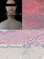
A 44-year-old male presented with a 10-year history of persistent facial flushing with transient aggravation after stimulation by external factors such as exercise and drinking. Photosensitive dermatitis and rosacea are suspected to have been diagnosed but poorly with the response with minocycline, antihistamine, topical or oral corticosteroids, and topical tacrolimus. Any systemic symptoms and similar family history were denied, including fever, joint pain, and only slight itching in the lesion area. Upon physical examination, facial redness and flushing were shown, in addition to the facial area, the erythematous-brownish macules and patches were widely distributed in the trunk and limbs with a few telangiectasias found unexpectedly although the patient did not describe it (Figure 1A and C). Laboratory tests, including whole blood cell count, C-reactive protein and erythrocyte sedimentation rate, serum immunoglobulin E level, peripheral blood tumor markers, antinuclear antibody spectrum, complement C3, and C4, were all within the normal range, and no positive findings were found. Dermoscopy imaging demonstrated the branching linear vessels of the facial lesion area and the branching linear vessels in a reticular pattern with a brown pigmentation network of the back area (Figure 1B and D). The patient refused to perform a skin biopsy of the face for aesthetic purposes; thus, a skin biopsy was performed on the back area, the specimen obtained from the biopsy was sent to further histological examination and showed telangiectasia in the upper dermis with perivascular mononuclear cell infiltration, Toluidine staining display the presence of mast cells (Figure 1E–H; Hematoxylin and Eosin-staining, Toluidine-staining, original magnification ×200). The clinical, dermoscopic, and histological findings were consistent with a diagnosis of telangiectasia macularis eruptiva perstans (TMEP). Persistent facial flushing was suggested to associate with TMEP, which is an unusual clinical observation. The patient refused to receive further treatment and is still in long-term follow-up.
Discussion
TMEP is a rare, benign, and indolent subtype and form of cutaneous mastocytosis and usually involved the skin that is distributed over the trunk and extremities and potential systemic involvement, but the removal of TMEP from the current classification of diagnostic criteria for cutaneous mastocytosis was suggested, the clinical classic and typical cutaneous lesions are usually presented as red-brown telangiectatic macules, although the pathogenesis and pathophysiology have remained unclear, the abnormal hyperplasia of mast cells was still considered.2,5,6 Our case demonstrates a patient with TMEP which was confirmed by skin biopsy; interestingly, the patient seeking medical help for the symptom of facial redness and flushing, which is an unusual clinical presentation and observation in patients with TMEP. Although the most common cause of facial flushing is climacteric, cutaneous diseases such as rosacea, and other inflammatory skin diseases, some malignant tumors or rare diseases can also lead to facial flushings, such as carcinoid syndrome, pheochromocytoma and medullary carcinoma of the thyroid.7 Interestingly, in the case we reported, facial flushing was considered associated with TMEP and although the mechanism remains completely unclear, it is inferred that facial flushing may be caused by the activation and degranulation of mast cells. Moreover, we obtain the dermoscopic images of the cutaneous lesions and demonstrated the branching linear vessels of the facial lesion area and the branching linear vessels in a reticular pattern with a brown pigmentation network of the back area, these findings corresponded to the dilated superficial capillaries surrounded by mast cells in the dermis at the level of histopathology.4
In conclusion, TMEP is a rare disease, atypical clinical manifestations of TMEP may also present and its clinical diagnosis faces challenges, skin biopsy, and histopathological examination may be necessary, but noninvasive imaging technology also shows advantages in the auxiliary diagnosis and noninvasive imaging tools may help diagnose TMEP. Clinicians, especially dermatologists, should pay attention to the atypical clinical manifestations of TMEP, especially for seeking medical help with facial flushing, and pay more attention to some details of physical examination to consider the possibility of TMEP diagnosis. Facial flushing is a very common clinical challenge that dermatologists, especially cosmetic dermatologists need to face, in most cases, the frequent causes are contact dermatitis, rosacea, and other inflammatory skin diseases, and facial flushing caused by systemic diseases should be considered. Although TMEP is a rare disease, facial flushing secondary TMEP should be emphasized in clinical practice. It will help dermatologists recognize the common clinical manifestations of rare diseases and avoid the neglect and wrong diagnosis of this disease. In addition, most importantly, dermatologists often use laser treatment in patients with facial flushing, but such patients with TMEP should be treated with caution, because this may lead to mast cell activation and induce a series of unnecessary clinical troubles.
Ethics Statement
The authors certify that they have obtained all appropriate patient consent forms. The patient gave written informed consent for publication of clinical information and photographs. No ethical committee approval was required because the data were analyzed in a retrospective manner.
Funding
This work was supported by The Natural Science Foundation of Inner Mongolia Autonomous Region of China (2021ZD15, 2021ZD16).
Disclosure
The authors declare no conflict of interest.
References
1. Michelerio A, Grassi S, Elena C, et al. Telangiectasia macularis eruptiva perstans: a neglected type of mastocytosis with exclusively cutaneous involvement? A case series. Eur J Dermatol. 2019;29(2):174–178. doi:10.1684/ejd.2019.3532
2. Severino M, Chandesris M-O, Barete S, et al. Telangiectasia macularis eruptiva perstans (TMEP): a form of cutaneous mastocytosis with potential systemic involvement. J Am Acad Dermatol. 2016;74(5):885–91.e1. doi:10.1016/j.jaad.2015.10.050
3. Vidal C, Del Rio E, Suárez-Peñaranda J, et al. Telangiectasia macularis eruptiva perstans presented as a pseudoallergic food reaction. J Investig Allergol Clin Immunol. 2000;10(4):248–250.
4. Kumar S, Jakhar D, Misri R. Dermoscopy of Telangiectasia macularis eruptiva perstans. Indian Dermatol Online J. 2020;11(1):131–132. doi:10.4103/idoj.IDOJ_312_18
5. Lawrence M, Sampson J, Oliver F, et al. Telangiectasia macularis eruptiva perstans of the scalp mimicking angiosarcoma. Am J Dermatopathol. 2021;43(12):e277–e279. doi:10.1097/DAD.0000000000002070
6. Hartmann K, Escribano L, Grattan C, et al. Cutaneous manifestations in patients with mastocytosis: consensus report of the European competence network on mastocytosis; the American Academy of Allergy, Asthma & Immunology; and the European Academy of Allergology and Clinical Immunology. J Allergy Clin Immunol. 2016;137(1):35–45. doi:10.1016/j.jaci.2015.08.034
7. Izikson L, English JC
 © 2022 The Author(s). This work is published and licensed by Dove Medical Press Limited. The full terms of this license are available at https://www.dovepress.com/terms.php and incorporate the Creative Commons Attribution - Non Commercial (unported, v3.0) License.
By accessing the work you hereby accept the Terms. Non-commercial uses of the work are permitted without any further permission from Dove Medical Press Limited, provided the work is properly attributed. For permission for commercial use of this work, please see paragraphs 4.2 and 5 of our Terms.

