Back to Journals » Clinical Ophthalmology » Volume 8
Periocular necrotizing fasciitis following retrobulbar injection
Authors Gelaw Y, Abateneh A
Received 25 November 2013
Accepted for publication 18 December 2013
Published 28 January 2014 Volume 2014:8 Pages 289—292
DOI https://doi.org/10.2147/OPTH.S58259
Checked for plagiarism Yes
Review by Single anonymous peer review
Peer reviewer comments 2
Yeshigeta Gelaw, Aemero Abateneh
Department of Ophthalmology, College of Public Health and Medical Sciences, Jimma University, Jimma, Ethiopia
Abstract: Necrotizing fasciitis is a rare, rapidly progressive severe bacterial soft tissue infection with a high mortality rate. While necrotizing fasciitis classically involves the trunk, groin/perineum, lower limbs, and postoperative wound sites, primary involvement of the eyelids is a rare but well known entity. We present a 33-year-old female patient who developed periocular necrotizing fasciitis after local retrobulbar anesthesia injection and facial block for cataract surgery in the left eye and canthotomy/cantholysis for treatment of moderate retrobulbar hemorrhage in the same eye. Surgical debridement was done and necrotic foul-smelling eyelid and deep orbital tissues were removed, and culture grew Staphylococcus aureus. Despite initial surgical debridement and intravenous antibiotic therapy, the disease progressed rapidly; orbital exenteration was considered, but the patient declined the surgery and self-discharged. Periocular necrotizing fasciitis remains predominantly a clinical diagnosis, and is often missed early in its presentation because of the difficulty in distinguishing it from other common soft tissue infections, especially in the presence of surgical wounds and retrobulbar hemorrhage. A high index of suspicion, early recognition, and prompt therapeutic interventions are indispensable for optimal visual outcome and patient survival.
Keywords: periocular necrotizing fasciitis, eyelid necrosis, deep fascia, surgical debridement, fasciitis
Introduction
Necrotizing fasciitis (NF) is a serious life-threatening, progressive, rapidly spreading inflammatory infection located in the deep fascia that rapidly destroys the skin and soft tissue beneath it,1,2 and is most commonly caused by a rapidly spreading infection of Streptococcus pyogenes in the subcutaneous plane.3 The overall mortality rate is 25%–30%, more than 70% of cases are associated with toxic shock syndrome, and nearly 100% of cases are fatal without surgical intervention.1,2,4
NF usually affects the extremities, abdomen, or perineum, and facial involvement is rare owing to the excellent blood supply of this region.4,5 The organisms most closely linked to NF are group A beta-hemolytic streptococci (type II NF), with or without a coexisting staphylococcal infection, although these bacteria are isolated in only a minority of the cases.2–4 Type II NF typically occurs in healthy young immunocompetent individuals.2,4 The rarer form (type I NF) predominantly affects immunocompromised or debilitated patients and is caused by polymicrobial infections and the usual pathogens are obligate and facultative anaerobes.2–4 The periorbital infection is mainly caused by beta-hemolytic streptococci alone (50%), occasionally in combination with Staphylococcus aureus (18%).4
Periocular NF is characterized by acute onset of a painful edematous, erythematous, nonspecific rash. As the infection spreads subcutaneously along the facial planes, the pain becomes severe, whereas the skin is often anesthetic.4,5 The patient feels sick and develops a low to medium grade fever associated with hypotension and tachycardia out of proportion to the temperature elevation.4,5 This is true in most cases, but patients can appear systemically quite well, at least initially.6 Within the next 48 hours the lid skin assumes a violaceous hue which may progress to fluid-filled bullae and later to black necrotic patches. Within 4–5 days, frank cutaneous gangrene develops, and by 8–10 days, the skin sloughs and becomes gangrenous.4
To our knowledge, there are no case reports of periocular NF after retrobulbar injection and/or canthotomy. Here we report the first case of periocular NF preceded by local retrobulbar anesthesia injection for cataract extraction, to highlight the importance of a high index of suspicion for early recognition and prompt intervention in this potentially vision-threatening and life-threatening disease.
Case report
A 33-year-old female patient presented to the eye clinic at Jimma University Hospital with bilateral painless progressive loss of vision, and on examination was found to have bilateral presenile cataract, so was admitted to the eye ward to have left eye cataract extraction. On the following day, she underwent standard preoperative preparation of the left eye and was taken to the operating theater. The left eyelid and periorbital area were cleaned with 70% alcohol. During retrobulbar injection of 4 mL of lidocaine hydrochloride 2% and epinephrine 1:100,000, she developed moderate retrobulbar hemorrhage which warranted cancellation of the planned cataract surgery. Tubicort® (Tubilux Pharma S.p.A, Rome, Italy) (hydrocortisone 0.5% + oxytetracycline 0.9%) eye suspension was instilled and the eye was patched. She was given a 500 mg oral loading dose of acetazolamide and a maintenance dose of 250 mg qid, and sent to the eye ward for bed rest and follow-up.
On the next day, she had diffuse, warm, tense, and tender reddish blistering eye lid swelling associated with headache and periorbital pain, but her body temperature was normal. With the impression of severe retrobulbar hemorrhage, lateral canthotomy and cantholysis were done to relieve the pressure, and she was given paracetamol 1 g tablets as required and continued acetazolamide. However, the globe was still tense. Hence, surgical evacuation of orbital hematoma through anterior orbitotomy was attempted but no significant hematoma was found. Post-orbitotomy, intravenous cloxacillin 500 mg qid and ceftriaxone 1 g twice daily was started. Chloramphenicol eye ointment was applied twice daily. Vital signs were stable (pulse rate 90 per minute, respiratory rate 16 per minute, temperature 36.5°C, blood pressure 90/70 mmHg).
On subsequent days (3 and 4), the upper and lower eyelids of the left eye and the surrounding face became blue-gray and necrotic, with loss of touch and pain sensation over the swelling and around it (Figure 1). The patient was unable to open the lid, so visual acuity was not measured. At this stage, necrotizing fasciitis was considered and intravenous metronidazole was added.
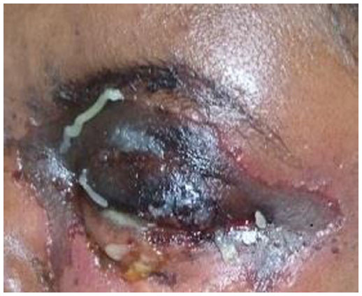 | Figure 1 Blue-gray colored eye lid with underlying tissue necrosis. |
On day 5, surgical debridement was done and necrotic foul-smelling tissues over the upper lid, lower lid, and deep orbital tissue under the lateral canthus were removed (Figure 2). A specimen was sent for culture which grew S. aureus. Other hematological and serological tests (for fasting blood sugar, white blood cell count with differential, erythrocyte sedimentation rate, and venereal disease research laboratory and human immunodeficiency virus) were unrevealing.
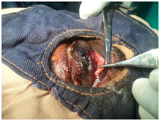 | Figure 2 Intraoperative appearance of advancing wound. |
Intravenous cloxacillin, ceftriaxone, and metronidazole, as well as daily wound care and dressing with Furacin® (Universal Twin Labs, Mumbai, India) ointment was continued but there was no improvement. On day 13, redebridement was done; at this time, the bulbar conjunctiva, sclera, and orbital tissue were necrotic and vision was no light perception. The clinical decision for orbital exenteration was taken and discussed with the patient and her family, but she declined and self-discharged against medical advice and repeated counseling, and did not return for further follow-up.
The patient had no previous or current nasal discharge, history of trauma, previous facial/nasal/ocular wound, known drug allergy, history of drug usage, or personal or family history of known systemic diseases (diabetes mellitus, hypertension, or cardiac, renal, or liver disease).
Discussion
Periocular NF or primary involvement of the eyelids is a rare but well known entity1,7 and is seen mainly in adults with a female predominance (54%); about half (47%) of the patients are previously healthy.4 This is in agreement with our case involving a healthy adult female patient. The infection may follow local blunt trauma (17%), as well as penetrating injuries (22%) and face surgery (11%), whereas no cause is identified in about one third of cases (28%). It may also be secondary to immunosuppression, diabetes, alcoholism, and other factors.2,3,7,8 In our case, retrobulbar anesthesia injection was identified as a risk factor. The evolution of the lesion seen in our patient is in line with the clinical description of NF in the literature4–10 and hence supports the diagnosis of periocular NF.
The clinical diagnosis of necrotizing fasciitis is further substantiated by the operative findings, which included the presence of grayish necrotic fascia, lack of resistance of normally adherent superficial fascia to blunt dissection, lack of bleeding of the fascia during dissection, and the presence of foul-smelling “dishwater” pus.5 This was seen in our case, further supporting the diagnosis.
However, other possible and serious causes of periocular necrotizing fasciitis, including preseptal cellulitis, orbital cellulitis, cavernous sinus thrombosis, and rhino-orbital mucormycosis should be considered and ruled out. Although it is quite difficult to distinguish periocular NF from preseptal cellulitis and orbital cellulitis, rapid progression and eventual cyanosis of the involved tissue and formation of serous fluid-filled bullae are clues to common non-necrotizing preseptal cellulitis, while the presence of dusky erythema in an area of cellulitis helps to differentiate orbital cellulitis from periorbital NF.11 In underdeveloped countries like Africa, Latin America, Asia, and Eastern Europe,12 periocular cutaneous anthrax should also be considered in the differential diagnosis, and physicians must be vigilant about inquiring for a history of contact with anthrax-infected animals or animal products. Moreover, the presence of severe pain and loss of sensation over the affected area, which are features suggestive of necrotizing fasciitis,1,13 will enable it to be distinguished, and cutaneous anthrax has characteristic black eschar formation, but at a later stage.8,13
Management of periorbital NF is centered on early recognition of clinical signs and symptoms and this mandates aggressive multidisciplinary treatment to avoid severe and permanent disfigurement, and loss of vision and mortality is linked to misdiagnosis or delay in diagnosis.4 If prompt diagnosis is established, treatment by extensive surgical debridement and systemic antibiotics (with or without the use of hyperbaric oxygen therapy) can prevent a fulminant course with a fatal outcome.2,4,14–16 An optimal antibiotic regimen for methicillin-susceptible S. aureus skin and soft tissue infection is a higher dose of oxacillin (1–2 g intravenously, every 4 hours) with clindamycin (600 mg/kg intravenously, every 8 hours or 300–450 mg orally three times daily)17 because of oxacillin’s excellent diffusion in soft tissue and its antitoxin properties. Our patient was given cloxacillin, ceftriaxone, and metronidazole because oxacillin and clindamycin were not available. Use of hyperbaric oxygen therapy can reduce the extent of hypoxic leukocyte dysfunction occurring within an area of hypoxia and infection, and provide oxygenation to otherwise ischemic areas, thus blocking the progression of NF18 and perhaps saving more of the eyelid and orbital tissue.
Some survivors might need extensive surgical intervention like orbital exenteration as reported by Lazzeri et al4 in their review of the published literature. Our patient also developed deep orbital tissue necrosis with eye involvement; exenteration was planned but the patient declined and self-discharged against medical advice and did not come back for follow-up.
In conclusion, periocular necrotizing fasciitis remains principally a clinical diagnosis, and is often missed early in its presentation because of the paucity of specific cutaneous signs early in its evolution, and the difficulty in differentiating it from other common soft tissue infections, especially in the presence of a surgical wound and retrobulbar hemorrhage. The rarity of the disease may also result in misdiagnosis due to lack of experience on the part of the clinician involved. A high index of suspicion, early recognition, and prompt therapeutic intervention are essential for an optimal visual outcome, cosmetic result, and patient survival. It is essential that physicians are aware of the clinical presentation of this disease and its possible risk factors, including minor surgical trauma with a needle.
Acknowledgment
We would like to thank the ophthalmologists in the department of ophthalmology at Jimma University for their contribution to the management of this patient.
Disclosure
The authors report no conflicts of interest in this work.
References
Kanski JJ. Clinical Ophthalmology: A Systematic Approach. 5th ed. Edinburgh, UK: Butterworth Heinemann; 2003. | |
Hayek S, Ibrahim A, Atiyeh B. The diagnosis and management of necrotizing fasciitis. Wounds Int. 2011;2(4). Available from: http://www.woundsinternational.com/practice-development/the-diagnosis-and-management-of-necrotising-fasciitis/page-1. Accessed December 19, 2013. | |
Urschel JD. Necrotizing soft tissue infections. Postgrad Med J. 1999;75:645–649. | |
Lazzeri D, Lazzeri S, Figus M, et al. Periorbital necrotizing fasciitis. Br J Ophthalmol. 2010;94:1577–1585. | |
Wong CH, Wang YS. The diagnosis of necrotizing fasciitis. Curr Opin Infect Dis. 2005;18:101–106. | |
Wong CH, Chang HC, Pasupathy S, et al. Necrotizing fasciitis: clinical presentation, microbiology and determinants of mortality. J Bone Joint Surg Am. 2003;85A:1454–1460. | |
American Academy of Ophthalmology. Basic and Clinical Science Course. Orbit, Eyelids, and Lacrimal System. Section 7, 2004–2005. San Francisco, CA, USA: American Academy of Ophthalmology; 2004. | |
Brittain CJ, Penwarden A, Mearza A, Verity D. Moraxella as a cause of necrotizing fasciitis of the eyelid. Eye. 2006;20:1312–1314. | |
Luksich JA, Holds JB, Hartstein ME. Conservative management of necrotizing fasciitis of the eyelids. Ophthalmology. 2002;109:2118–2122. | |
Miller LG, Perdreau-Remington F, Rieg G, et al. Necrotizing fasciitis caused by community-associated methicillin-resistant Staphylococcus aureus in Los Angeles. N Engl J Med. 2005;352:1445–1453. | |
Holliman RE, Catford GV. Periorbital necrotizing fasciitis with loss of vision. J Infect. 1986;13:35–36. | |
Lew DP. Bacillus anthracis (anthrax). In: Mandell GL, Bennett JE, Dolin R, editors. Principles and Practice of Infectious Diseases. 5th ed. New York, NY, USA: Churchill Livingstone; 2000. | |
Gelaw Y, Asaminew T. Periocular cutaneous anthrax in Jimma Zone, Southwest Ethiopia: a case series. BMC Res Notes. 2013;6:313. | |
Wang G, Egbert J, McCulley TJ. Periorbital necrotizing fasciitis. Invest Ophthalmol Vis Sci. 2005;46:E-Abstract 5059. | |
Raja V, Job R, Hubbard A, Moriarty B. Periorbital necrotizing fasciitis: delay in diagnosis results in loss of lower eyelid. Int Ophthalmol. 2008;28:67–69. | |
Marshall DH, Jordan DR, Gilberg SM, Harvey J, Arthurs BP, Nerad JA. Periocular necrotizing fasciitis: a review of five cases. Ophthalmology. 1997;104:1857–1862. | |
Stevens DL, Bisno AL, Chambers HF, et al. Practice guidelines for the diagnosis and management of skin and soft-tissue infections. Clin Infect Dis. 2005;41:1373–1406. | |
Undersea and Hyperbaric Medical Society. Necrotizing soft tissue infections. Durham, NC, USA: Undersea and Hyperbaric Medical Society. Available from: http://membership.uhms.org/?page=NSTI. Accessed November, 2013. |
 © 2014 The Author(s). This work is published and licensed by Dove Medical Press Limited. The
full terms of this license are available at https://www.dovepress.com/terms
and incorporate the Creative Commons Attribution
- Non Commercial (unported, 3.0) License.
By accessing the work you hereby accept the Terms. Non-commercial uses of the work are permitted
without any further permission from Dove Medical Press Limited, provided the work is properly
attributed. For permission for commercial use of this work, please see paragraphs 4.2 and 5 of our Terms.
© 2014 The Author(s). This work is published and licensed by Dove Medical Press Limited. The
full terms of this license are available at https://www.dovepress.com/terms
and incorporate the Creative Commons Attribution
- Non Commercial (unported, 3.0) License.
By accessing the work you hereby accept the Terms. Non-commercial uses of the work are permitted
without any further permission from Dove Medical Press Limited, provided the work is properly
attributed. For permission for commercial use of this work, please see paragraphs 4.2 and 5 of our Terms.