
Back to Journals » Clinical Ophthalmology » Volume 20
Overview of Intracameral Drug Delivery Systems in Glaucoma
Authors Shi KS  , Abubaker Y , Lang MV , Dönmez Gün R, Banoub RG, Ahuja AS , Wagner IV , Dorairaj SK
Received 17 September 2025
Accepted for publication 9 February 2026
Published 20 February 2026 Volume 2026:20 568183
DOI https://doi.org/10.2147/OPTH.S568183
Checked for plagiarism Yes
Review by Single anonymous peer review
Peer reviewer comments 6
Editor who approved publication: Dr Scott Fraser
Kelvin SY Shi,1 Yazan Abubaker,1 Mary V Lang,1,2 Raziye Dönmez Gün,1,3 Raphael G Banoub,1 Abhimanyu S Ahuja,4 Isabella V Wagner,5 Syril K Dorairaj1
1Department of Ophthalmology, Mayo Clinic, Jacksonville, FL, USA; 2Department of Ophthalmology, Mayo Clinic Alix School of Medicine, Jacksonville, FL, USA; 3Department of Ophthalmology, Kartal Doctor Lütfi Kirdar City Hospital, Istanbul, Turkey; 4Department of Ophthalmology, Casey Eye Institute, Portland, OR, USA; 5Department of Medicine, University of Florida College of Medicine, Gainesville, FL, USA
Correspondence: Syril K Dorairaj, Department of Ophthalmology, Mayo Clinic, 4500 San Pablo Road S, Jacksonville, FL, 32224, USA, Tel +1-904-953-7110, Email [email protected]
Abstract: Glaucoma is the leading cause of irreversible blindness worldwide, and intraocular pressure (IOP) remains the only proven modifiable risk factor for slowing disease progression. Although topical medications are the standard first-line therapy, their effectiveness is limited by poor adherence and low ocular bioavailability. These limitations have prompted the development of non-topical drug delivery systems (DDSs) that aim to provide consistent long-term IOP control without reliance on patient-administered drops. Among these approaches, intracameral DDSs have received increasing attention for their ability to deliver medication directly to the anterior chamber. This review aims to provide clinicians with an overview of the design, clinical performance, and key considerations of the two FDA-approved intracameral implants, Durysta® (BimSR) and iDose® TR (iDose), as well as investigational devices including ENV515, OTX-TIC, and PA5108. Currently, clinical trials indicate that these devices can achieve sustained reductions in IOP with efficacy comparable to topical prostaglandins (PGA), and real-world studies similarly report reductions in both IOP and medication burden. However, several limitations remain, including concerns about corneal endothelial cell loss (CECL), variability in treatment response among patients with more advanced disease, and the limited availability of long-term safety and durability data. In addition, randomized comparative studies evaluating intracameral DDSs against other glaucoma interventions, such as minimally invasive glaucoma surgery (MIGS), are scarce. Intracameral DDSs represent a developing area within glaucoma management, and further evidence is needed to clarify their long-term safety and their appropriate role within personalized treatment strategies.
Keywords: glaucoma, drug delivery, intracameral, medical devices
Introduction
Glaucoma, characterized by the progressive degeneration of retinal ganglion cells leading to irreversible optic neuropathy and vision loss, is projected to become increasingly prevalent, with estimates suggesting it will affect more than 110 million people worldwide by 2040.1–3 Currently, IOP is the most identifiable and the only modifiable and proven strategy to slow glaucoma’s progression.2,3 For several decades, topical IOP-lowering medications have been the first-line therapy for most patients with glaucoma.4 However, real-world effectiveness is limited. Despite multiple classes of topical agents, between 24% and 59% of patients fail to achieve full therapeutic benefit due to incorrect instillation, preservative-related ocular discomfort, and reluctance to report nonadherence.5 In addition, the ocular bioavailability of most commercial eye drops is typically <5%. The lipid-rich corneal epithelium and endothelium restrict hydrophilic drug penetration, whereas the water-dense stroma impedes hydrophobic drugs diffusion, collectively limiting delivery to intraocular targets.6–8
Given the challenges with topical eye medications, non-topical DDSs have emerged as a promising alternative by potentially improving adherence and clinical outcomes while avoiding the risks of surgical interventions, prompting considerable investigation into their use for glaucoma management.3,9 Non-topical DDSs can be broadly categorized into intraocular and extraocular platforms.3 Intraocular approaches include intraocular lens implants and intracameral PGA implants, while extraocular methods encompass drug-eluting punctal plugs, conjunctival fornix inserts, contact lenses, and nanoparticles.3
Among these, intracameral PGA implants have gained particular attention because they represent the only non-topical DDSs with products currently approved by the US Food and Drug Administration (FDA).3,10 Therefore, a focused review of this class of non-topical drug delivery systems offers a clinically relevant and timely perspective. This article aims to summarize the design, clinical trial outcomes, and key considerations of both FDA-approved (BimSR and iDose) and investigational intracameral DDSs (ENV515, OTX-TIC, PA5108). (Table 1).
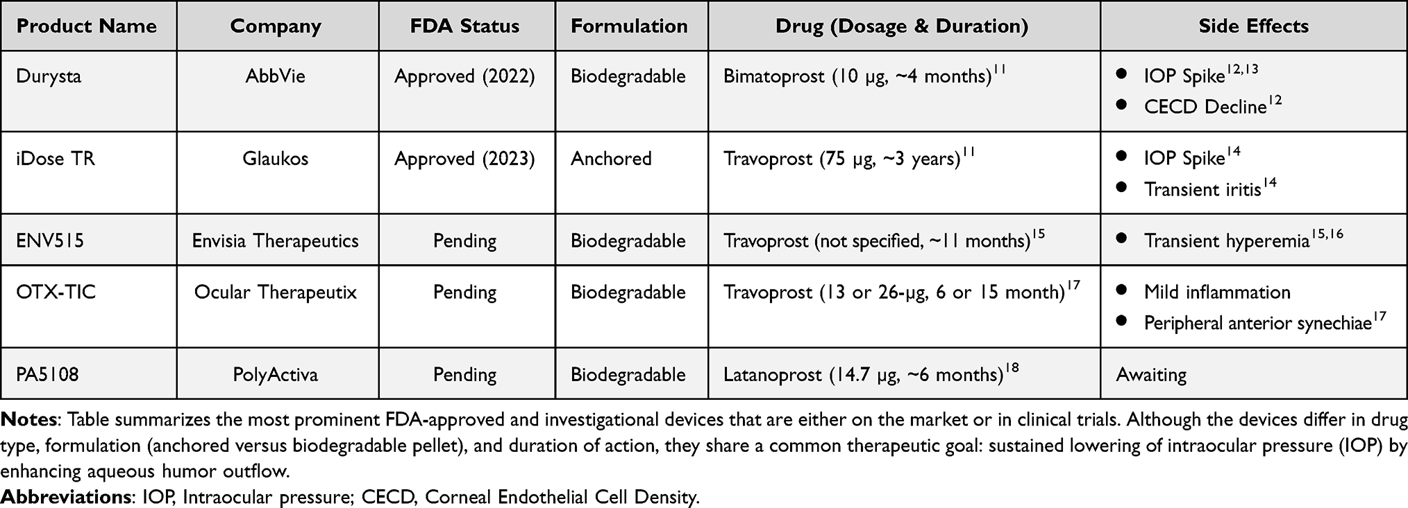 |
Table 1 FDA-Approved and Investigational Intracameral Drug Delivery Systems (DDSs) for Glaucoma Therapy |
Method
This narrative review was conducted to synthesize current evidence on intracameral PGA DDSs for the management of primary open-angle glaucoma (POAG) and ocular hypertension (OHT). A comprehensive literature search was performed using PubMed, Embase, and ClinicalTrials.gov to identify relevant publications from January 2000 to January 2025. The search strategy incorporated the following terms and Boolean operators: “glaucoma,” “ocular hypertension,” “intracameral implant,” “sustained-release,” “drug delivery system,” “bimatoprost implant,” “travoprost implant,” “ENV515,” “OTX-TIC,” “PA5108,” and “PolyActiva.” Both peer-reviewed manuscripts and conference abstracts were included to capture emerging investigational devices.
Eligible studies included randomized controlled trials, prospective and retrospective clinical studies, real-world analyses, preclinical evaluations, and review articles that described the design, pharmacology, mechanism of action, clinical performance, or safety of intracameral DDSs. Studies focused exclusively on topical, periocular, or posterior segment drug delivery routes were excluded unless they provided contextual insights relevant to anterior chamber implants.
Data on device design, drug release characteristics, pharmacokinetics, IOP outcomes, duration of effect, safety events, and procedural considerations were extracted and qualitatively synthesized. Given the heterogeneity across study designs, populations, and outcome measures, no formal meta-analysis was performed. Instead, findings were narratively summarized to highlight shared principles, differences between platforms, and implications for clinical practice. The review emphasizes FDA-approved devices and investigational systems currently in early- or late-phase development.
Bimatoprost Sustained-Release Pellet (BimSR; Biodegradable)
Design
The BimSR (Durysta®, AbbVie, North Chicago, IL, USA) is an intracameral implant containing 10 μg of bimatoprost, a type of PGA commonly used topically as a first-line treatment for POAG and OHT.11,19,20 The total dose of bimatoprost delivered is comparable to that of a single drop of 0.03% bimatoprost ophthalmic solution.21 Approved by the FDA in 2022, the implant is positioned in the iridocorneal angle and gradually releases the medication over a period of three to four months.11 The device is a small cylinder measuring about 200 µm in diameter and 1.1 mm in length.11,20 It is composed of a biodegradable matrix of poly-lactic acid and polylactic-co-glycolic acid that enables controlled degradation and sustained release.20 The implant is delivered into the anterior chamber using a single-use 28-gauge applicator that can be deployed at the slit lamp or in the operating room.11,20 By delivering the drug close to the iris–ciliary body, the intended effect was to reduce exposure to conjunctival tissues, thereby potentially lowering the risk of adverse effects.22
Developmental Studies and Clinical Trials
Early evaluation of the BimSR was conducted in a 24-month, Phase I/II, open-label, multicenter study that used a paired-eye comparison design to assess four dose strengths of the implant in successive cohorts of patients with POAG.19 In this trial, each patient received a 6-, 10-, 15-, or 20-µg implant in one eye while the fellow eye continued treatment with topical bimatoprost 0.03%.19 During the first 12 weeks, mean IOP reductions ranged from 7.4 to 9.8 mmHg across the implant doses and 8.4 mmHg in the fellow eyes treated with topical therapy, from baseline values of 23.6 to 24.4 mmHg (all p < 0.001), confirming ≥20% IOP reduction and demonstrating clinical significance.19 Most adverse events occurred within the first two days; conjunctival hyperemia was most common in treated eyes (34.7%), likely related to povidone–iodine preparation.19,23 After this period, the overall adverse event incidence was similar between study and fellow eyes, with fewer PGA-related effects in implant-treated eyes than in topically treated eyes: conjunctival hyperemia (17.3% vs 28.0%), eyelash growth (0% vs 6.7%), and iris hyperpigmentation (0% vs 4.0%).19 Implants typically swelled to 76–125% of baseline size by month 6 before regressing; by 12 months 26 eyes’ implants were ≤25% of original size or no longer visible in 6 of the patients, and at month 24, gonioscopy in 34 patients showed complete biodegradation in 26.5% of study eyes, with 75% of residual implants ≤25% of original size.19
Following these early findings, two larger multicenter randomized Phase III trials were launched to evaluate long-term efficacy and safety. ARTEMIS-1 was a 20-month subject- and evaluator-masked parallel-group trial in which 10-µg or 15-µg implants were administered at day 1, week 16, and week 32, and compared with twice-daily timolol in 198 eyes per arm.20 Baseline mean IOPs of 24.0, 24.2, and 23.9 mmHg decreased to 17.4, 17.1, and 17.5 mmHg at week 12, and remained comparable at week 52 with mean diurnal IOPs of 18.1 ± 4.0, 17.9 ± 3.9, and 17.2 ± 3.4 mmHg.20 ARTEMIS-2 employed a similar 20-month design and enrolled 528 patients with 176 eyes per treatment arm. Similar IOP reduction efficacy was established, with baseline diurnal IOPs of 23.7, 23.9, and 23.9 mmHg reduced to 18.2 ± 4.0, 17.6 ± 3.5, and 17.2 ± 3.4 mmHg at month 20 for the 10-µg, 15-µg, and timolol groups, respectively.24 Across both ARTEMIS trials, a clear dose- and exposure-dependent pattern of progressive corneal endothelial cell density (CECD) decline emerged with repeated implant administration. In ARTEMIS-1, ≥20% endothelial loss by month 20 occurred in 10.2% of eyes treated with 10-µg and 21.8% treated with 15-µg, compared with 0.5% in the timolol arm, with rates increasing after each successive administration.20 In ARTEMIS-2, endothelial loss followed a similar cumulative pattern, reaching 3.8 and 5.8% in the 10-µg cohort and 11.6 and 14.6% in the 15-µg cohort after the third administration and at 20 month, respectively.24 Serious ocular adverse events also occurred more frequently in implant-treated eyes than in timolol-treated eyes, with rates of 3.4% and 7.4% in the 10-µg and 15-µg groups compared with none in the control group (p = 0.030 and p < 0.001).24
Complementing these parallel-group studies, a multicenter randomized paired-eye 24-month phase III trial compared the 10-µg BimSR with 360° selective laser trabeculoplasty (SLT) in 183 patients with OAG or OHT inadequately controlled on topical therapy.12 Each patient received an implant in one eye and SLT in the fellow eye. Baseline IOPs were similar (25.2 ± 2.99 mmHg in implant eyes and 25.1 ± 3.00 mmHg in SLT eyes). Least-squares mean IOP reductions at weeks 4, 12, and 24 were 6.8 ± 0.28, 6.9 ± 0.30, and 6.9 ± 0.27 mmHg with the implant, compared with 6.2 ± 0.28, 6.4 ± 0.30, and 6.5 ± 0.28 mmHg with SLT, fulfilling criteria for both statistical and clinical noninferiority based on 95% confidence intervals for between-treatment differences. The proportion of eyes avoiding rescue therapy remained similar at both one and two years (implant: 67.5% and 50.2%; SLT: 68.7% and 60.6%).12 In contrast, corneal safety outcomes consistently favored SLT. At month 24, mean percentage change in central CECD was −6.2 ± 1.13% in implant-treated eyes (−7.9 ± 2.04% with fixed retreatment vs −5.2 ± 1.35% with flexible retreatment) compared with −3.1 ± 0.43% in SLT-treated eyes, and ≥20% endothelial cell loss (ECL) occurred in 8.0% versus 3.4% of eyes, respectively.12 These results align with the ARTEMIS trials, indicating that while BimSR achieved IOP reduction comparable to established treatments, repeated administration introduces corneal endothelial risk that is not observed with SLT or topical timolol therapy.
Real World Data
In a multicenter Phase IV prospective observational study of the BimSR, 217 patients with POAG or OHT were followed for 18 months, with prespecified follow-up assessments at 6, 12, and 18 months after implantation.13 At baseline, the mean IOP was 17.2 mmHg, and 89.1% of patients were using at least one topical IOP–lowering medication, with a mean of 1.8 medications.13 After receiving the implant, 89% of primary eyes required no additional IOP-lowering treatment by month 6.13 By month 12, 84% remained treatment-free, and by month 18, 78% still required no additional therapy.13 Mean IOP decreased by approximately 1–2 mmHg across all follow-up time points, with mean IOP values at 6, 12, and 18 months clustering around the target IOP of 15.2 mmHg.13 Correspondingly, the average number of topical IOP-lowering medications declined from 1.8 at baseline to 0.9 by month 12 and remained low through month 18.13 The most common adverse effects were increased IOP, dry eye, and a 3.5% decline in CECD during the study period.13 In a qualitative sub-study, 25 patients completed interviews at 4 to 6 months after implantation. Of these, 84% reported satisfaction with treatment outcomes, one patient (4%) reported dissatisfaction due to increased IOP after surgery, and 76% expressed a preference for treatment with the BimSR implant alone when offered a choice between implant therapy, topical drops, or combination treatment.13
Additional outcomes were evaluated in a retrospective observational study conducted at Duke Eye Center following a single administration of the BimSR implant.25 A total of 92 eyes from 63 patients were included. Glaucoma severity was mild in 11% of eyes, moderate in 30%, and severe in 54%. Mean baseline IOP was 18.34 ± 5.71 mmHg. Mean IOP was numerically lower at all follow-up visits through 12 months but did not reach statistical significance, with changes of −1.07 ± 4.48 mmHg at month 1 (p = 0.053), −0.70 ± 4.72 mmHg at month 3 (p = 0.188), −0.30 ± 4.26 mmHg at month 6 (p = 0.456), −0.38 ± 4.25 mmHg at month 9 (p = 0.407), and −1.34 ± 4.11 mmHg at month 12 (p = 0.111). In contrast, the mean number of topical IOP–lowering medications decreased significantly from a baseline of 2.05 ± 1.21, with reductions of −0.81 ± 0.95, −0.75 ± 0.74, −0.63 ± 0.97, −0.70 ± 0.99, and −0.67 ± 1.04 at months 1, 3, 6, 9, and 12, respectively (all p < 0.001). Interestingly, medication reduction was statistically significant only at month 1 in eyes with severe glaucoma, whereas reductions remained significant across all time points in eyes with mild to moderate disease. Adverse effects were uncommon and occurred in 4.3% of eyes, including three cases of mild corneal edema and one case of recurrent anterior chamber inflammation. During follow-up, 19 eyes (20.7%) required additional laser or surgical intervention at a mean of 6.0 ± 4.5 months, and most had severe glaucoma with prior incisional surgery.25
Clinical Considerations
Evidence from both Phase III ARTEMIS trials and the paired-eye SLT comparison study consistently indicates that CECD decline is the principal long term safety consideration associated with the BimSR, particularly with repeated exposure12,20,24 Rather than being isolated observations, these safety signals have been consistent across studies and become more pronounced with repeated implantation, ultimately leading the FDA to restrict approval to a single administration.11 In the ARTEMIS-2 trial, after the third injection, approximately 6% of patients experienced worsening ECL, and about 3% required implant removal during the 20-month follow-up.11,24 Similarly, in the SLT comparison trial, significant CECD loss (defined as ≥20% decrease from baseline) was more frequent in implant-treated eyes than in SLT-treated eyes (8% vs 3%, respectively).12 Together, these findings indicate a dose-, exposure-, and retreatment-dependent endothelial risk, reinforcing the need for careful patient selection. Individuals with low baseline endothelial counts or other endothelial stressors may be particularly vulnerable. For this reason, clinicians should obtain a pre-treatment CECD measurement and monitor periodically thereafter, especially if retreatment outside the approved single-use label is contemplated.
Another clinically relevant consideration is the risk of increased IOP following implantation. In the phase III SLT comparison trial, increased IOP was the most frequently reported ocular adverse event, occurring in 24.6% of implant-treated eyes.12 Notably, these events often clustered around 90 days post-implantation, coinciding with waning drug release and suggesting a rebound phenomenon as the pharmacologic effect diminished.12 Similarly, in the phase IV prospective observational study, increased IOP was the most frequently reported adverse event, occurring in 23 eyes, although classification relied on investigator judgment without a predefined IOP threshold.13 Clinicians should therefore counsel patients regarding the possibility of IOP elevation and plan follow-up accordingly, with a low threshold for reinstating topical therapy or escalating treatment if pressure rises.
Disease severity further influences the clinical utility of the BimSR. As noted by the Duke observational study, medication burden for those with mild to moderate disease, in severe cases the benefit often lasts only about one month.25 Patients with mild to moderate disease experienced a longer period of significant medication reduction compared with those with severe disease.25 In up to one-third of patients with severe glaucoma, the implant produced no appreciable effect and surgical intervention was required within three months.25 In the remaining two-thirds, the implant may have delayed surgery, but the observed IOP reduction was not statistically significant. As a result, a clear causal relationship between the implant and surgical delay cannot be established.25 Therefore, for patients with advanced glaucoma, the BSRP is unlikely to serve as a primary management option by the clinician.
Taken together, available evidence indicates that while the BimSR can meaningfully reduce topical medication burden and provide clinically relevant IOP lowering in appropriately selected patients, its use requires thoughtful integration into individualized treatment plans. Optimal candidates are those with early to moderate disease, preserved endothelial health, and challenges with adherence or topical intolerance. Clinicians should maintain vigilant follow-up focused on endothelial health and IOP trends and be prepared to modify therapy as drug effect diminishes. In contemporary practice, the BimSR is best positioned as an adjunctive, adherence-supporting intervention rather than a long-term standalone solution for patients requiring sustained, aggressive pressure control.
Travoprost Intracameral Implant (iDose; Anchored)
Design
iDose (iDose® TR, Glaukos, Aliso Viejo, CA, USA) is a trabecular meshwork implant designed for the intracameral treatment of POAG and OHT. It was first approved by the FDA in December 2023.3 The implant contains 75 µg of preservative-free travoprost, which is another class of PGA and is approximately 25,000 times more concentrated than the travoprost found in the 0.004% ophthalmic eyedrop formulation.3 The drug is released in a slow, sustained manner, regulated by the thickness of a nanoporous ethylene vinyl acetate (EVA) membrane.11
Structurally, iDose consists of a biocompatible titanium reservoir measuring approximately 0.5 mm in diameter and 1.8 mm in length, with 1.2 mm residing in the anterior chamber.26 The device has four main parts: a cap, an elution membrane, a drug reservoir, and a scleral anchor.26 It is preloaded in a sterile, single-use inserter for direct implantation.26
Two designs exist for iDose: a slow-eluting (SE) and a fast-eluting (FE) model. These differ only in the thickness of the EVA membrane, which modulates the drug release rate.10 The SE model is the commercially available version, while the FE model is currently limited to research use.11
Developmental Studies and Clinical Trials
In a randomized, double-masked, multicenter phase IIB clinical trial, 154 patients with open-angle glaucoma or ocular hypertension were assigned in a 1:1:1 ratio to receive either a single FE iDose implant in the study eye, a single SE iDose implant in the study eye, or twice-daily topical timolol in the study eye, resulting in approximately one-third of patients in each group with 51, 54, and 49 patients, respectively.27 Baseline mean diurnal IOPs were 25.35 ± 3.55 mmHg in the FE group, 24.82 ± 3.94 mmHg in the SE group, and 24.77 ± 3.01 mmHg in the timolol group.27 From week 10 through month 36, mean 8:00 AM IOP reductions ranged from 7.6 to 8.8 mmHg in the FE group, 7.3 to 8.0 mmHg in the SE group, and 7.3 to 7.9 mmHg in the timolol group, with reductions from baseline statistically significant at all visits in each arm (p < 0.0001).27 At months 12, 24, and 36, more patients in both the FE and SE groups were able to remain on the same or a lower number of pressure-lowering topical medications compared with the timolol group.27 The safety profile was favorable. There were no implant dislodgements or removals, no cases of conjunctival hyperemia or fat atrophy, no treatment-related discontinuations, and no serious adverse events in the study eye. Corneal endothelial cell counts (CECC) also remained stable across groups. At 36 months, the difference in CECC between the FE and timolol groups was −37.9 cells/mm2 (p = 0.1730), and between the SE and timolol groups was −18.3 cells/mm2 (p = 0.4581).27
Following this, a randomized multicenter phase III double masked parallel-group trial compared a single iDose implant with twice daily topical timolol in adults with OAG or OHT.14 A total of 590 patients were randomized once again to three cohorts: FE implant group (n = 200), SE implant group (n = 197), or timolol group (n = 193), with one study eye per patient.14 Baseline unmedicated mean diurnal IOP was similar across groups at 24.18 ± 2.78, 24.02 ± 2.81, and 24.12 ± 2.68 mmHg in the FE, SE, and timolol arms, respectively.14 IOP was assessed at six distinct time points, namely day ten, week six, and month three at 8 o’clock and 10 o’clock in the morning.14 Across these six measurements, average IOP reductions ranged from 6.6 to 8.4 mmHg in the FE group, 6.6 to 8.5 mmHg in the SE group, and 6.5 to 7.7 mmHg in the timolol group.14 At the final measurement, taken at month 3 at 10 AM, 56.5% of patients in the FE group, 54.8% in the SE group, and 57.0% in the timolol group achieved at least a 25% IOP reduction from baseline, confirming clinical significance.14 Adverse ocular events occurred in 43 eyes (21.5%) in the FE group, 53 eyes (27.2%) in the SE group, and 21 eyes (10.8%) in the timolol group, and the majority were mild or moderate in severity. Severe IOP elevations were observed in two patients in each implant group and were not reported in the timolol group. CECC counts remained relatively stable through month three, with mean percent changes of −2.69 ± 5.78% in the FE group, −1.93 ± 4.37% in the SE group, and −0.96 ± 5.04% in the timolol group, and no eye in any group met or exceeded the predefined safety threshold of a 30% reduction from baseline.14
Real World Data
A non-randomized, retrospective, unmasked consecutive case series included all standalone iDose implantation performed by a single glaucoma surgeon in the United States, with a maximum follow-up of three months for 54 eyes. All patients had undergone prior treatment with SLT, BimSR, MIGS, or cyclophotocoagulation.26 After implantation, mean IOP decreased from 19.6 ± 3.8 mmHg preoperatively to 13.2 ± 2.9 mmHg at one month (32.7% reduction, p = 0.001) and to 13.1 ± 2.5 mmHg at three months (33.2% reduction, p = 0.001).26 The mean number of topical medications declined from 0.28 ± 0.71 at baseline to 0.0 ± 0.0 at three months (100% reduction, p = 0.006).
The mean IOP reduction in this real-world cohort was 6.5 mmHg, slightly lower than the reductions observed in Phase IIB trials (7.3–8.0 mmHg) and Phase III trials (6.6–8.5 mmHg).14,26,27 This difference may be due to the medication washout period used in pivotal trials, as well as the higher mean baseline IOP of approximately 24 mmHg in those studies. It is well established that higher preoperative IOP is associated with greater postoperative reduction.28 Despite these baseline differences, the postoperative outcomes were comparable. At three months, 100% of patients in this real-world series remained free of topical medication, closely matching the 95.8% observed in the Phase III trial.14,28
Adverse events were uncommon. One eye developed mild iritis at one to two weeks postoperatively and was successfully treated with a tapering course of prednisolone acetate 1% (PredForte). Importantly, there were no cases of ocular hyperemia, which is typically reported in 27% to 44% of patients using topical PGA.26 No cases of iris or periorbital pigmentation, peripheral anterior synechiae, or periorbital fat atrophy were observed. These findings are consistent with those reported in Phase IIB and Phase III pivotal trials of iDose.14,26,27 While these observations align with the safety findings reported in Phase IIB and Phase III trials of the travoprost implant, the sample size and limited follow up warrant cautious interpretation when extrapolating safety expectations more broadly.
Clinical Considerations
Currently, the most common adverse effects associated with iDose and occurring in more than 2% of patients are increased IOP, iritis, and conjunctival or ocular hyperemia.29 While the 36-month Phase IIB trial showed stable CECC, endothelial cell health remains a clinically relevant parameter, and continued monitoring is warranted as longer-term experience and broader post-market data become available.26
Economic considerations represent another key practical consideration when positioning iDose in clinical practice. Starting in the first quarter of 2024, Glaukos announced a wholesale acquisition price of approximately $13,950 per iDose, compared with $1,950 for the BSRP.3,30 By contrast, topical travoprost medication costs about $64 for a 2.5-mL bottle, which lasts approximately one month when used in one eye, amounting to roughly $2,300 over a 36-month period.30 In fact, studies by Gazzard et al and Guedes et al have suggested that laser or surgical treatments may be more cost-effective, particularly in younger patients or those with early-stage glaucoma.11,31,32 Despite these considerations, the prolonged drug delivery and adherence advantages offered by iDose may justify its higher upfront cost in carefully selected patients who prioritize long-term drop independence.30
Another important limitation is that current evidence does not clearly define whether iDose performs differently across varying stages of glaucoma. Existing trials have not stratified outcomes by disease severity, leaving uncertainty regarding differential efficacy in mild, moderate, or advanced disease. In addition, direct comparative studies with other glaucoma treatments are lacking, and broader long-term post-market data will be necessary to fully characterize durability, safety, and comparative effectiveness. Although the available evidence remains encouraging, these gaps underscore the need for further investigation before the optimal clinical role of iDose can be fully defined.
Travoprost Extended-Release (ENV515; Biodegradable)
Design
ENV515 (Envisia Therapeutics, Morrisville, NC, USA) is a sustained-release biodegradable implant designed for the long-term management of IOP in patients with POAG or OHT and is currently awaiting FDA approval.18,33 The implant is administered via intracameral injection into the anterior chamber and provides continuous travoprost release with a targeted duration of six to twelve months.18,34,35 It is composed of poly(ester amide) (PEA), a biodegradable polymer that supports controlled, zero-order drug release kinetics. Using Particle Replication In Non-wetting Templates (PRINT®) technology, the implant is fabricated as a rod optimized for intracameral delivery.18 ENV515’s intraocular applicator was developed to deliver these rod-shaped implants via intracameral injection. To achieve this, it was constructed from medical-grade materials with a stainless-steel shaft and a single-lumen hypodermic needle actuated by a scroll wheel, and it was terminally sterilized and verified to be sterile, biocompatible, and consistently functional with low insertion force.36
Developmental Studies and Clinical Trials
Preclinical evaluation of ENV515 in six hypertensive Beagle dogs demonstrated sustained and clinically meaningful IOP reduction following intracameral implantation. The implant administrations were conducted in Beagle dogs by intracameral injections via a custom injector and resulted in 100% success rate for implant delivery. Gonioscopy and anterior chamber OCT imaging showed stability without movement in the iridocorneal angle in hypertensive Beagle dogs. Gonioscopy and anterior chamber optical coherence tomography confirmed stable positioning of the implant within the iridocorneal angle without migration in hypertensive animals. Baseline IOP in the 6 hypertensive Beagle dogs was 23.4 ± 1.0 mmHg. Over a 24-week treatment period, mean IOP decreased by 7.2 ± 0.5 mmHg, corresponding to a 30 ± 2% reduction from baseline (p < 0.001).34
Building on these results, a Phase IIA clinical trial in 21 glaucoma patients found that ENV515 achieved IOP-lowering effects comparable to once-daily topical PGA therapy by day 25. In this trial, one eye received ENV515 and the fellow eye received topical medication. By day 25, ENV515 achieved IOP lowering comparable to topical therapy, meeting the primary efficacy endpoint. The mean change from baseline in diurnal IOP was −6.7 mmHg in ENV515-treated eyes and −6.6 mmHg in topically treated eyes, with both reductions statistically significant compared with baseline (p < 0.001).15
A subsequent 12-month dose-ranging study further evaluated ENV515 in 15 patients, including 5 patients in a low-dose cohort and 10 patients in a high-dose cohort, all of whom had prior exposure to topical PGA medication. In each case, one eye received ENV515 while the fellow eye received topical timolol 0.5% once daily. In the low-dose cohort, baseline IOP was 26.1 ± 2.2 mmHg, and by month 11 mean IOP had decreased by 6.7 ± 3.7 mmHg (p < 0.005), equivalent to a 25% reduction.16 The high-dose ENV515 group demonstrated an additional 1.1 mmHg greater IOP reduction at 28 days compared with the low-dose group.
Across both clinical studies, no serious adverse events were reported, and the most common adverse event was early-onset transient conjunctival hyperemia.15,16
Travoprost Intracameral Hydrogel (OTX-TIC; Biodegradable)
Design
OTX-TIC (Ocular Therapeutix, Bedford, MA, USA) is a biodegradable intracameral device composed of a hydrogel matrix embedded with travoprost microparticles.33 The implant uses hydrogel depot technology to achieve sustained, zero-order release of travoprost for approximately four to six months.18,37 Implanted in the anterior chamber, as illustrated in Figure 1, OTX-TIC is currently being evaluated by FDA for its efficacy, safety, and tolerability in patients with POAG and OHT.35
 |
Figure 1 Intraocular Placement of the OTX-TIC. Image from a Phase I trial showing a partially degraded OTX-TIC implant. The red oval highlights the location of the OTX-TIC, where the implant remains intact with visible microparticles, indicating ongoing presence despite partial degradation. |
Developmental Studies and Clinical Trials
In preclinical studies with normotensive beagle dogs, four groups were formed: placebo, one 26-µg implant, two 26-µg implants, or one 13-µg implant, all placed in the right eye (Groups 1 through 4, respectively). OTX-TIC was generally well tolerated across all tested doses, with no clinically significant changes in corneal thickness or CECD following repeated intracameral administration. At nine months, mean corneal thickness for the right and left eyes was 601 µm and 592 µm in Group 1, 589 µm and 591 µm in Group 2, 606 µm and 604 µm in Group 3, and 584 µm and 588 µm in Group 4. Mean CECD at nine months for the right and left eyes was 2455 and 2419 cells/mm2 in Group 1, 2449 and 2404 cells/mm2 in Group 2, 2462 and 2423 cells/mm2 in Group 3, and 2547 and 2534 cells/mm2 in Group 4, respectively.38 Across dosing groups, OTX-TIC produced sustained IOP reductions lasting up to six months, with decreases from baseline of 13.1% in Group 2, 8.4% in Group 3, and 26.9% in Group 4; however, the study did not report absolute baseline IOP values, limiting interpretation of the magnitude of pressure reduction.
Following this, a multicenter prospective Phase I trial then assessed the safety and efficacy of OTX-TIC in patients with POAG and OHT.17 The contralateral eye continued topical travoprost therapy (Travatan), while study eyes were divided into three cohorts: low-dose (3 eyes), high-dose (4 eyes), and a still-enrolling fast-degrading low-dose cohort. While no baseline IOP was given, the implant lowered IOP by 7–10 mmHg, with the effect lasting up to 15 months in the low-dose cohort and 6 months in the high-dose cohort. No significant differences in IOP control were observed compared with the non-study eyes, and no significant differences in mean IOP change from baseline were noted between the low-dose and high-dose cohorts at 6 months. The safety profile was favorable, with the most common adverse events being mild inflammation and peripheral anterior synechiae, although the abstract did not specify the number of affected eyes. CECC and implant stability remained unchanged during follow-up. The implant typically degraded within five to seven months.17
Latanoprost Intracameral Implant (PA5108; Biodegradable)
Design
PA5108 (PolyActiva, Melbourne, VIC, Australia) is a rod-shaped biodegradable implant designed for insertion into the anterior chamber using a 27-gauge needle, providing sustained-release of latanoprost over approximately six months and is currently awaiting FDA approval (Figure 2A and B).18 Upon insertion, drug release begins immediately, delivering a consistent daily dose throughout the treatment period. This is achieved through a proprietary polymeric drug delivery platform that enables zero-order kinetics without an initial burst, ensuring steady dosing and targeted delivery to the site of action.18
 |
Figure 2 PA5108 Implant and Intraocular Positioning. (A) The red arrow indicates the rod-shaped biodegradable implant, which is designed for intracameral insertion using a 27-gauge needle. The 14.7 µg PA5108 implant was manufactured for the Phase IIA clinical trial and provides sustained release of latanoprost through a proprietary polymeric drug delivery platform. (B) The red circle highlights the intraocular positioning of the 14.7 µg PA5108 implant within the patient’s anterior chamber at 12 weeks following initial administration during the Phase IIA trial. |
Developmental Studies and Clinical Trials
In a preclinical study involving glaucomatous dogs, three different formulations of latanoprost implants, including the current PA5108, demonstrated sustained and clinically meaningful IOP reduction with a favorable safety profile. Treated animals had a mean baseline IOP of 24.3 ± 3.6 mmHg, and all active implants achieved IOP lowering comparable to once-daily topical latanoprost while also reducing diurnal IOP fluctuations. Central corneal thickness remained stable throughout the 24-week follow-up, with PA5108-specific measurements of 639 µm at baseline and 648 µm at week 24, and no clinical signs of intraocular inflammation were observed.39 Retinal thickness, optic nerve head morphology, electroretinographic responses, and visual performance assessments showed no treatment-related adverse effects, supporting the ocular safety and sustained efficacy of intracameral latanoprost delivery in this glaucomatous canine model.39
Building on these results, PolyActiva announced certain findings from a Phase IIA clinical trial in which PA5108 produced consistent and statistically significant IOP reductions. The implant was noted to achieve IOP reductions of ≥20% over six months.40 The implant was fully biodegradable and no treatment-related toxicity was reported. Implant persistence was observed for at least 21 weeks, with complete biodegradation occurring by week 40.40
Conclusion
Sustained-release intracameral DDSs represent an important advancement in glaucoma management, offering a potential alternative to traditional topical medications by providing consistent IOP control and reducing daily treatment burden. FDA-approved devices such as BimSR and iDose are already in clinical use, and additional intracameral PGA DDSs are progressing through clinical trials, underscoring the growing relevance of this therapeutic class. These innovations have the potential to reshape current treatment paradigms by improving adherence and delivering more predictable pharmacologic effects.
Challenges remain, particularly with respect to corneal endothelial safety and the need for robust long-term real-world safety data for the approved products. Furthermore, although current studies of anchored intracameral implant devices have not demonstrated severe or rapid ECL, these findings should not be interpreted as definitive reassurance. Clinicians should remain vigilant by routinely monitoring CECD, especially in eyes with preexisting endothelial compromise, prior intraocular surgery, or other risk factors. Continued attention to CECD is critical to ensure early identification of potential adverse effects as broader and longer-term clinical experience accumulates.
In addition to safety considerations, evidence directly comparing intracameral implants with other established glaucoma interventions remains limited. Although BimSR has been evaluated against SLT, further randomized controlled trials are needed to compare intracameral implants with additional glaucoma treatments such as MIGS, MicroPulse, and trabeculectomy. Such comparative studies will be essential for defining the optimal clinical placement of intracameral DDSs and guiding more personalized treatment decisions.
Looking ahead, the integration of intracameral DDSs with existing interventional devices, including the potential development of drug-eluting trabecular bypass implants such as the iStent® infinite, may further broaden treatment options and support more individualized care. However, these approaches will require rigorous clinical evaluation to determine their safety, durability, and true additive benefit over current therapies. Collectively, ongoing advances in intracameral DDSs suggest the possibility of meaningful improvements in adherence and long-term outcomes. Yet their ultimate impact on glaucoma management will depend on continued accumulation of high-quality evidence, careful patient selection, and sustained post-marketing surveillance.
Consent for Publication
No personal identifying information is contained within this review article. All images have been deidentified.
Acknowledgment
No funding or grant support was obtained for this manuscript.
Author Contributions
All authors made a significant contribution to the work reported, whether that is in the conception, study design, execution, acquisition of data, analysis and interpretation, or in all these areas; took part in drafting, revising or critically reviewing the article; gave final approval of the version to be published; have agreed on the journal to which the article has been submitted; and agree to be accountable for all aspects of the work.
Disclosure
The authors declare no conflicts of interest in this work.
References
1. Tham YC, Li X, Wong TY, Quigley HA, Aung T, Cheng CY. Global prevalence of glaucoma and projections of glaucoma burden through 2040: a systematic review and meta-analysis. Ophthalmology. 2014;121(11):2081–11. doi:10.1016/j.ophtha.2014.05.013
2. Asrani SG, McGlumphy EJ, Al-Aswad LA, et al. The relationship between intraocular pressure and glaucoma: an evolving concept. Prog Retin Eye Res. 2024;103:101303. doi:10.1016/j.preteyeres.2024.101303
3. Tan SS, Wong TT. Drug delivery systems in glaucoma – current innovations and future perspectives. Asia-Pac J Ophthalmol. 2025;14(2):100163. doi:10.1016/j.apjo.2025.100163
4. Wagner IV, Stewart MW, Dorairaj SK. Updates on the Diagnosis and Management of Glaucoma. Mayo Clin Proc Innov Qual Outcomes. 2022;6(6):618–635. doi:10.1016/j.mayocpiqo.2022.09.007
5. Zaharia AC, Dumitrescu OM, Radu M, Rogoz RE. Adherence to Therapy in Glaucoma Treatment-A Review. J Pers Med. 2022;12(4):514. doi:10.3390/jpm12040514
6. Yang Y, Lockwood A. Topical ocular drug delivery systems: innovations for an unmet need. Exp Eye Res. 2022;218:109006. doi:10.1016/j.exer.2022.109006
7. Grassiri B, Zambito Y, Bernkop-Schnürch A. Strategies to prolong the residence time of drug delivery systems on ocular surface. Adv Colloid Interface Sci. 2021;288:102342. doi:10.1016/j.cis.2020.102342
8. Imperiale JC, Acosta GB, Sosnik A. Polymer-based carriers for ophthalmic drug delivery. J Control Release. 2018;285:106–141. doi:10.1016/j.jconrel.2018.06.031
9. Chong RS, Su DH, Tsai A, et al. Patient acceptance and attitude toward an alternative method of subconjunctival injection for the medical treatment of glaucoma. J Glaucoma. 2013;22(3):190–194. doi:10.1097/IJG.0b013e318237c6c4
10. Teymoorian S, Craven ER, Nguyen L, Werts E. Real-World Study of the Effectiveness and Safety of Intracameral Bimatoprost Implant in a Clinical Setting in the United States. Clin Ophthalmol. 2024;18:187–199. doi:10.2147/opth.S445005
11. Elhusseiny AM, Aref AA. Sustained Release Therapies with the Prostaglandin Analogues Intracameral Implants. Ophthalmol Ther. 2024;13(7):1833–1839. doi:10.1007/s40123-024-00965-4
12. Kolko M, Tatham AJ, Lim KS, et al. Phase 3, Randomized, Comparison Study of Intracameral Bimatoprost Implant 10 µg and Selective Laser Trabeculoplasty. Am J Ophthalmol. 2025;272:19–37. doi:10.1016/j.ajo.2024.12.026
13. Mann E, Kammer JA, Sawhney G, et al. Prospective 18-Month Study of Bimatoprost Intracameral Implant in Patients with Open-Angle Glaucoma or Ocular Hypertension in US Clinical Practice. Drugs. 2025;85(3):397–414. doi:10.1007/s40265-025-02157-1
14. Sarkisian Jr SR, Ang RE, Lee AM, et al. Phase 3 Randomized Clinical Trial of the Safety and Efficacy of Travoprost Intraocular Implant in Patients with Open-Angle Glaucoma or Ocular Hypertension. Ophthalmology. 2024;131(9):1021–1032. doi:10.1016/j.ophtha.2024.02.022
15. Kesav NP, Young CEC, Ertel MK, Seibold LK, Kahook MY. Sustained-release drug delivery systems for the treatment of glaucoma. Int J Ophthalmol. 2021;14(1):148–159. doi:10.18240/ijo.2021.01.21
16. Mansberger SL, Conley J, Verhoeven RS, et al. Interim Analysis of Low Dose ENV515 Travoprost XR with 11 Month Duration Followed by Dose Escalation and 28 Day Efficacy Evaluation of High Dose ENV515. Invest Ophthalmol Visual Sci. 2017;58(8):2110.
17. Goldstein MH, Goldberg D, Walters TR, Vantipalli S, Braun E, Metzinger JL. Evaluating Safety, Tolerability and Efficacy of an Intracameral Hydrogel-Based Travoprost Implant in Subjects with Glaucoma - Phase 1 Trial. Invest Ophthalmol Visual Sci. 2020;61(7):4266.
18. Al-Qaysi ZK, Beadham IG, Schwikkard SL, Bear JC, Al-Kinani AA, Alany RG. Sustained release ocular drug delivery systems for glaucoma therapy. Expert Opin Drug Deliv. 2023;20(7):905–919. doi:10.1080/17425247.2023.2219053
19. Craven ER, Walters T, Christie WC, et al. 24-Month Phase I/II Clinical Trial of Bimatoprost Sustained-Release Implant (Bimatoprost SR) in Glaucoma Patients. Drugs. 2020;80(2):167–179. doi:10.1007/s40265-019-01248-0
20. Medeiros FA, Walters TR, Kolko M, et al. Phase 3, Randomized, 20-Month Study of Bimatoprost Implant in Open-Angle Glaucoma and Ocular Hypertension (ARTEMIS 1). Ophthalmology. 2020;127(12):1627–1641. doi:10.1016/j.ophtha.2020.06.018
21. Medeiros FA, Sheybani A, Shah MM, et al. Single Administration of Intracameral Bimatoprost Implant 10 µg in Patients with Open-Angle Glaucoma or Ocular Hypertension. Ophthalmol Ther. 2022;11(4):1517–1537. doi:10.1007/s40123-022-00527-6
22. Seal JR, Robinson MR, Burke J, Bejanian M, Coote M, Attar M. Intracameral Sustained-Release Bimatoprost Implant Delivers Bimatoprost to Target Tissues with Reduced Drug Exposure to Off-Target Tissues. J Ocul Pharmacol Ther. 2019;35(1):50–57. doi:10.1089/jop.2018.0067
23. Kass MA, Heuer DK, Higginbotham EJ, et al. The Ocular Hypertension Treatment Study: a randomized trial determines that topical ocular hypotensive medication delays or prevents the onset of primary open-angle glaucoma. Arch Ophthalmol. 2002;120(6):701–713. doi:10.1001/archopht.120.6.701
24. Bacharach J, Tatham A, Ferguson G, et al. Phase 3, Randomized, 20-Month Study of the Efficacy and Safety of Bimatoprost Implant in Patients with Open-Angle Glaucoma and Ocular Hypertension (ARTEMIS 2). Drugs. 2021;81(17):2017–2033. doi:10.1007/s40265-021-01624-9
25. Choi EY, Johnson NA, Stinnett S, Rosdahl J, Moya F, Herndon LW. The Effect of Bimatoprost Implant on Glaucoma Patients: an Observational Study. J Glaucoma. 2024;33(6):431–436. doi:10.1097/ijg.0000000000002368
26. Teymoorian S, Kaur J. Travoprost Intracameral Implant in Eyes with Glaucoma or Ocular Hypertension: early Short-Term Real-World Outcomes. Clin Ophthalmol. 2025;19:157–166. doi:10.2147/opth.S498431
27. Berdahl JP, Sarkisian Jr SR, Ang RE, et al. Efficacy and Safety of the Travoprost Intraocular Implant in Reducing Topical IOP-Lowering Medication Burden in Patients with Open-Angle Glaucoma or Ocular Hypertension. Drugs. 2024;84(1):83–97. doi:10.1007/s40265-023-01973-7
28. Singh IP, Berdahl JP, Sarkisian Jr SR, et al. Long-Term Safety and Efficacy Evaluation of Travoprost Intracameral Implant Based on Pooled Analyses from Two Phase III Trials. Drugs. 2024;84(10):1299–1311. doi:10.1007/s40265-024-02074-9
29. Cheng CM, Rehmani C, Chin J. Prostaglandin Intracameral Implants for Ocular Hypertension and Open-Angle Glaucoma. J Pharm Technol. 2025;87551225251313707. doi:10.1177/87551225251313707
30. Huston J, Paauw M, Orey D, et al. Travoprost Intracameral Implant: a Review on the Novel Treatment Modality for Open-Angle Glaucoma and Ocular Hypertension. Ann Pharmacother. 2025;59(8):767–772. doi:10.1177/10600280241291911
31. Gazzard G, Konstantakopoulou E, Garway-Heath D, et al. Selective laser trabeculoplasty versus eye drops for first-line treatment of ocular hypertension and glaucoma (LiGHT): a multicentre randomised controlled trial. Lancet. 2019;393(10180):1505–1516. doi:10.1016/s0140-6736(18)32213-x
32. Guedes RAP, Guedes VMP, Gomes CEM, Chaoubah A. Maximizing cost-effectiveness by adjusting treatment strategy according to glaucoma severity. Medicine. 2016;95(52):e5745. doi:10.1097/md.0000000000005745
33. Shen Y, Sun J, Sun X. Intraocular nano-microscale drug delivery systems for glaucoma treatment: design strategies and recent progress. J Nanobiotechnology. 2023;21(1):84. doi:10.1186/s12951-023-01838-x
34. Navratil T, Garcia A, Tully J, et al. Preclinical Evaluation of ENV515 (travoprost) Intracameral Implant - Clinical Candidate for Treatment of Glaucoma Targeting Six-Month Duration of Action. Invest Ophthalmol Visual Sci. 2014;55(13):3548.
35. Belamkar A, Harris A, Zukerman R, et al. Sustained release glaucoma therapies: novel modalities for overcoming key treatment barriers associated with topical medications. Ann Med. 2022;54(1):343–358. doi:10.1080/07853890.2021.1955146
36. Navratil T, Delgado J, Garcia A, et al. Design and Development of ENV515 Intraocular Applicator for Intracameral Injections of ENV515 Glaucoma Extended Release (XR) Therapy. Invest Ophthalmol Visual Sci. 2016;57(12):3021.
37. Ö E K, Mekala P, Dave B, Kooner KS. Managing Ocular Surface Disease in Glaucoma Treatment: a Systematic Review. Bioengineering. 2024;11(10):1010. doi:10.3390/bioengineering11101010
38. Patel C, Blizzard CD, Hartman J, et al. Preclinical Safety and Tolerability of Repeated Intracameral Travoprost Implant (OTX-TIC) Administrations. Invest Ophthalmol Visual Sci. 2023;64(8):4299.
39. Komaromy AM, Koehl K, Harman CD, et al. Long-term intraocular Pressure (IOP) control by means of a novel biodegradable intracameral (IC) latanoprost free acid (LFA) implant. Invest Ophthalmol Visual Sci. 2017;58(8):4591.
40. Appell MB, Pejavar J, Pasupathy A, et al. Next generation therapeutics for retinal neurodegenerative diseases. J Control Release. 2024;367:708–736. doi:10.1016/j.jconrel.2024.01.063
 © 2026 The Author(s). This work is published and licensed by Dove Medical Press Limited. The
full terms of this license are available at https://www.dovepress.com/terms
and incorporate the Creative Commons Attribution
- Non Commercial (unported, 4.0) License.
By accessing the work you hereby accept the Terms. Non-commercial uses of the work are permitted
without any further permission from Dove Medical Press Limited, provided the work is properly
attributed. For permission for commercial use of this work, please see paragraphs 4.2 and 5 of our Terms.
© 2026 The Author(s). This work is published and licensed by Dove Medical Press Limited. The
full terms of this license are available at https://www.dovepress.com/terms
and incorporate the Creative Commons Attribution
- Non Commercial (unported, 4.0) License.
By accessing the work you hereby accept the Terms. Non-commercial uses of the work are permitted
without any further permission from Dove Medical Press Limited, provided the work is properly
attributed. For permission for commercial use of this work, please see paragraphs 4.2 and 5 of our Terms.