Back to Journals » OncoTargets and Therapy » Volume 10
Outcomes of adding induction chemotherapy to concurrent chemoradiotherapy for stage T3N0-1 nasopharyngeal carcinoma: a propensity-matched study
Authors Lan XW, Xiao Y, Zou XB, Zhang XM, Ouyang PY, Xie FY
Received 4 February 2017
Accepted for publication 3 May 2017
Published 1 August 2017 Volume 2017:10 Pages 3853—3860
DOI https://doi.org/10.2147/OTT.S133917
Checked for plagiarism Yes
Review by Single anonymous peer review
Peer reviewer comments 3
Editor who approved publication: Dr Ingrid Espinoza
Xiao-Wen Lan,1,2,* Yao Xiao,1,* Xue-Bin Zou,3,* Xiao-Min Zhang,1 Pu-Yun OuYang,1 Fang-Yun Xie1
1Department of Radiation Oncology, Sun Yat-Sen University Cancer Center, State Key Laboratory of Oncology in South China, Collaborative Innovation Center for Cancer Medicine, 2Department of Radiation Oncology, Sun Yat-Sen Memorial Hospital, Sun Yat-Sen University, 3Department of Ultrasound, Sun Yat-Sen University Cancer Center, State Key Laboratory of Oncology in South China, Collaborative Innovation Center for Cancer Medicine, Guangzhou, People’s Republic of China
*These authors contributed equally to this work
Objective: Our objective was to examine whether adding induction chemotherapy to concurrent chemoradiotherapy improved survival in stage III nasopharyngeal carcinoma (NPC) patients, especially in low-risk patients at stage T3N0-1.
Materials and methods: We retrospectively analyzed 687 patients with stage T3N0-1 NPC treated with intensity-modulated radiation therapy (IMRT) plus concurrent chemotherapy (CC) with or without induction chemotherapy (IC). Propensity score matching (PSM) method was used to select 237 pairs of patients from two cohorts. Overall survival (OS), locoregional relapse-free survival (LRFS), distant metastasis-free survival (DMFS), and progression-free survival (PFS) were assessed by using the Kaplan–Meier method, log-rank test, and Cox regression analysis.
Results: No significant survival differences were observed between IC plus CC and CC cohorts with similar 4-year OS (91.7% vs 92.6%, P=0.794), LRFS, (92.7% vs 96.8%, P=0.138), DMFS (93.5% vs 94.3%, P=0.582), and PFS (87.5% vs 91.1%, P=0.223). In a univariate analysis, lower Epstein–Barr virus deoxyribonucleic acid (EBV DNA; <4,000 copies/mL) significantly improved 4-year DMFS (95.5% vs 91.6%, P=0.044) compared with higher EBV DNA (≥4,000 copies/mL). No factors were associated with 4-year OS, LRFS, DMFS, and PFS in a multivariate analysis. IC plus CC group experienced higher rates of grade 3–4 leucopenia (P<0.001) and neutropenia (P<0.001).
Conclusion: The addition of IC to CC in stage T3N0-1 NPC patients treated with IMRT did not significantly improve their survival. The IC group experienced higher rates of grade 3–4 hematological toxicities. Therefore, further investigation is required.
Keywords: nasopharyngeal carcinoma, induction chemotherapy, intensity-modulated radiation therapy, propensity score matching, stage T3N0-1
Introduction
Nasopharyngeal carcinoma (NPC) occurs at a high incidence rate in Southern China, especially in Hong Kong and Guangdong.1 NPC exhibits high radiosensitivity, and radiotherapy (RT) is the primary and most effective treatment for pathologically confirmed NPC. Intensity-modulated radiation therapy (IMRT) has significantly improved local control and lowered radiation-induced toxicities compared with two-dimensional RT.2,3 A series of phase III clinical trials have established chemoradiotherapy as the standard treatment for locoregionally advanced NPC (LA-NPC) because it improves disease control and survival.4–10
However, whether the addition of induction chemotherapy (IC) would improve survival in LA-NPC patients remains controversial. Differing results have been found in several trials.11–14 Hui et al14 showed that overall survival (OS) improved by adding IC (94.1% vs 67.7%, P=0.012), whereas 3-year progression-free survival (PFS) did not significantly improve (P=0.12). Fountzilas et al12 and Tan et al11 reported no significant survival benefit in LA-NPC patients treated with IC. Sun et al13 recently published a phase III study showing that treatment with IC in LA-NPC patients (except T3-4N0) significantly improved 3-year failure-free survival, OS, and distant failure-free survival. Our recent retrospective study15 showed that the addition of IC significantly improved 5-year OS (P=0.022) and 5-year distant metastasis-free survival (DMFS; P=0.018) in stage IVa-b NPC patients treated with IMRT.
IC may be associated with high incidences of hematological acute toxicity and could affect chemoradiotherapy. A previous study showed that IC exhibited grade 3 and 4 toxicities, including leukopenia (P=0.046), neutropenia (P=0.029), and thrombocytopenia (P<0.001).11 Therefore, IC may not improve survival benefits but increase hematological acute toxicity in low-risk stage III NPC patients. In order to assess the benefit of IC, we retrospectively analyzed stage T3N0-1 NPC patients treated with IMRT plus concurrent chemotherapy (CC) with or without IC. The propensity score matching (PSM) method was used to mimic randomized trials to reduce potential bias.16,17
Materials and methods
Patients
A total of 687 patients with stage T3N0-1 NPC treated with IMRT plus CC with or without IC were retrospectively examined in our institution between January 19, 2005, and December 27, 2012. Before treatment, a complete history of each patient was noted. Clinical examinations of the head and neck, hematological studies and biochemical profiles, pretreatment plasma Epstein–Barr virus deoxyribonucleic acid (pre-EBV DNA) level, fiberoptic nasopharyngoscopy with biopsy, magnetic resonance imaging (MRI) of the nasopharynx and neck, chest radiography, abdominal sonography, and a whole body bone scan using single photon emission computed tomography (CT) or positron emission CT were performed for each patient. Plasma EBV DNA level was measured by using real-time quantitative polymerase chain reaction as previously described.18,19 All the patients were restaged according to the seventh edition of the International Union against Cancer/American Joint Committee on Cancer staging system for NPC.20 This study was approved by the Institutional Review Board of Sun Yat-Sen University Cancer Center. The requirement for written consent was waived as this is a retrospective study; however, oral consent was obtained via telephone.
Treatment
Radiation therapy
All the patients were treated using one daily fraction of IMRT for 5 days per week at our institution. Gross tumor volume (GTV) included the nasopharynx GTV (GTVnx) and the cervical lymph nodes GTV (GTVnd). High-risk clinical target volume (CTV-1) was defined as the GTVnx plus a 5–10 mm margin (2–3 mm posteriorly) to encompass the high-risk area and the whole nasopharynx, and low-risk clinical target volume (CTV-2) was defined as the CTV-1 plus a 5–10 mm margin (2–3 mm posteriorly) to encompass the low-risk area including parapharyngeal space, posterior parts of the nasal cavity, retropharyngeal nodal regions, clivus, pterygoid fossae, sphenoid sinus, pterygopalatine fossae, and the selective neck area. The prescribed doses were as follows: 66–72 Gy to the GTVnx, 60–68 Gy to the GTVnd, ≥60 Gy to the (CTV-1), and 54–56 Gy to the CTV-2 for >30–33 fractions.
Chemotherapy
Platinum-based chemotherapy was given to all the patients. IC consisted of docetaxel plus cisplatin (TP), paclitaxel plus carboplatin (TC), docetaxel plus cisplatin plus fluorouracil (TPF), or cisplatin/nedaplatin plus fluorouracil (PF) regimen for up to three cycles. Patients received a CC regimen of cisplatin/nedaplatin of 30–40 mg/m2 weekly or every 3 weeks during RT. The three weekly regimens consisted of 80–100 mg/m2 of cisplatin/nedaplatin, PF, or TP for up to seven cycles.
Follow-up
Patient follow-up was calculated from the first day of therapy to either the day of death or the last day of examination. The patients underwent physical examination, plasma EBV DNA level, endoscopy, MRI scans, chest radiography, abdominal sonography, and whole body bone scan every 3–6 months during the first 3 years and every 6–12 months thereafter until death. OS, locoregional relapse-free survival (LRFS), DMFS, and PFS were analyzed as end points, and these end points were measured from the date of the first therapy to the date of death, first locoregional relapse, distant metastasis, disease progression (locoregional relapse or distant metastasis), or the date of the last follow-up visit. Patients not having recent examination records were followed-up via telephone calls.
Statistical analysis
SPSS version 22.0 (IBM Corporation, Armonk, NY, USA) was used for statistical analyses. We used the PSM method to match the patients between the two groups (IC plus CC and CC) at the ratio of 1:1 based on propensity scores. The scores were computed by using logistic regression based on the following covariates: gender, age, World Health Organization (WHO) pathological types (types I + II and III), smoking history, NPC family history, pre-EBV DNA, chemotherapy strategy (IC plus CC and CC), and N category (ie, N0 and N1). The balance of the covariates between the two groups was examined by using independent-samples t-test (continuous variable), χ2 test, or Fisher’s exact test (categorical variable). A cutoff of 4,000 copies/mL of pre-EBV DNA was used to define low versus high levels because this threshold has previously been shown to be a good prognostic factor.21 The χ2 test or Fisher’s exact test was used to compare the patient characteristics. Survival rates were estimated by using the Kaplan–Meier method, and differences were compared by using the log-rank test. Multivariate analyses were performed by using the Cox proportional hazards model to calculate hazard ratios (HRs) and 95% CIs and to identify significant independent prognostic factors. In the multivariate analyses, the following parameters were included in the model as covariates for each analysis: gender, age (≤50 vs >50 years), WHO pathological types (types I + II and III), smoking history, NPC family history, N category (N0 and N1), pre-EBV DNA (≤4,000 vs >4,000 copies/mL), and chemotherapy strategy (IC plus CC and CC). Two-tailed P-values <0.05 were considered statistically significant.
Results
Patient characteristics
Before PSM, we compared the characteristics of all the 687 patients between two groups. The analysis showed that age, pre-EBV DNA, and N category were not balanced between the two groups (P=0.031, P<0.001, and P<0.001, respectively). In order to balance the characteristics and reduce potential bias, we selected 237 patient pairs from 687 T3N0-1 NPC patients by using PSM method. In the PSM cohort, the median age of patients was 44 years (range =21–70 years), including 330 men and 144 women with a ratio of 2.29:1. No significant differences were found in gender, age, pathology type, smoking, NPC family history, pre-EBV DNA, and N category between the IC plus CC group and the CC group (Table 1).
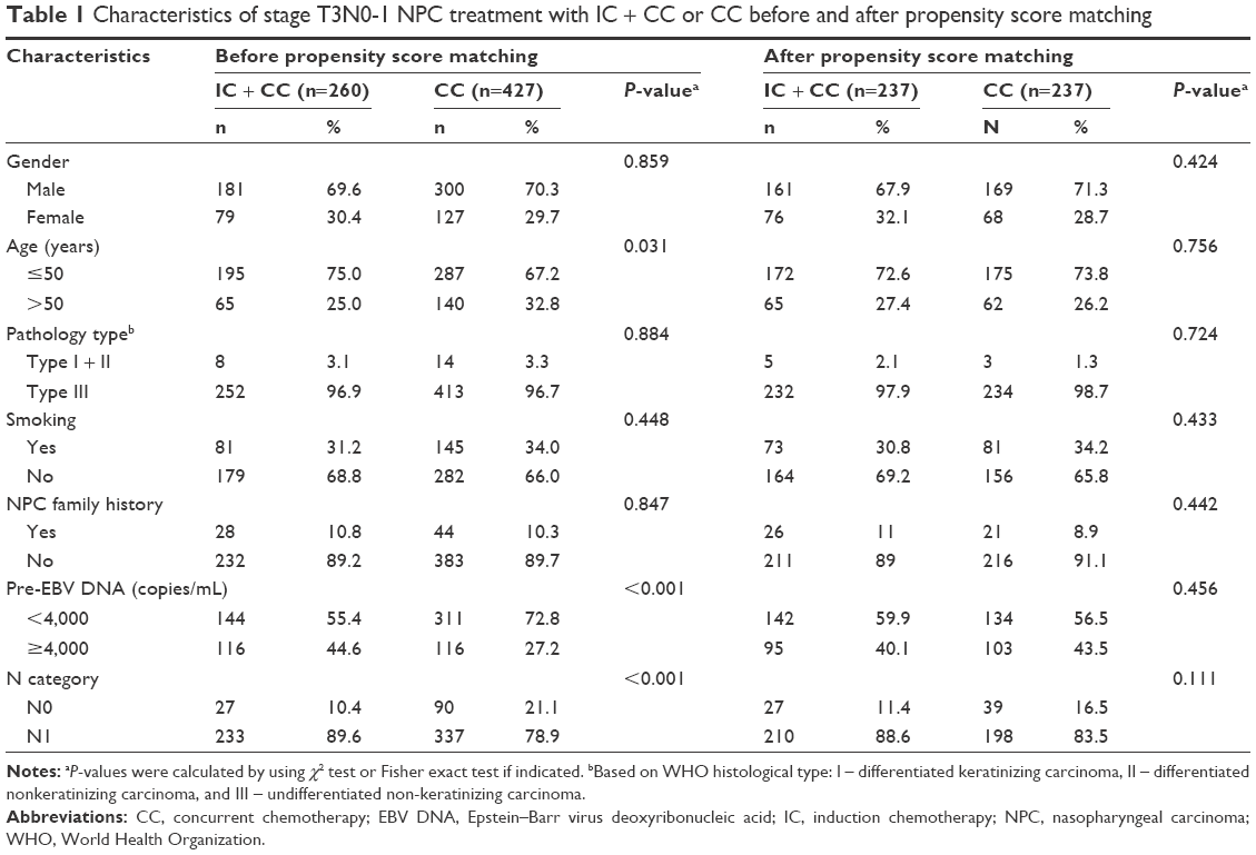 | Table 1 Characteristics of stage T3N0-1 NPC treatment with IC + CC or CC before and after propensity score matching |
Patterns of treatment failure
The median follow-up time was 49.4 months (range =1.4–117.7 months). Up to the last day of follow-up, 54 patients experienced disease progression. Furthermore, 33 of 54 (61.1%) patients showed disease progression in the IC plus CC group and 21 of 54 (38.9%) patients in the CC group. In total, 22 of 54 (40.7%) patients developed local/regional recurrence alone. A total of 5 of 22 (22.7%) patients in the IC plus CC group and 1 of 22 (4.5%) patients in the CC group developed both local and regional recurrence. Furthermore, 4 of 22 (18.2%) patients in the IC plus CC group and 5 of 22 (22.7%) patients in the CC group developed local recurrence, and 6 of 22 (27.3%) patients in the IC plus CC group and 1 of 22 (4.5%) patients in the CC group developed regional recurrence. In addition, 29 of 54 (53.7%) patients experienced distant metastases alone, which included 16 of 29 (55.2%) patients treated with IC plus CC and 13 of 29 (44.8%) patients treated with CC (P=0.565). Moreover, only two patients treated with IC plus CC and one patient treated with CC developed both distant metastases and recurrence. Table 2 shows the summary of treatment failure.
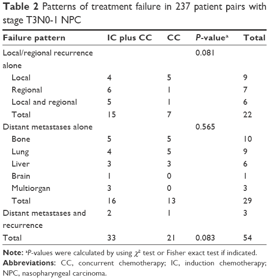 | Table 2 Patterns of treatment failure in 237 patient pairs with stage T3N0-1 NPC |
Prognostic value of IC
The 4-year OS, LRFS, DMFS, and PFS for the patient population were 92.1%, 94.7%, 93.9%, and 89.2%, respectively. Table 3 shows the summary of univariate analysis of prognostic factors, including gender, age, pathological type, smoking, NPC family history, pre-EBV DNA, chemotherapy strategy, and N category. Among these factors, gender, age, pathological type, smoking, NPC family history, and N category were not significantly associated with 4-year OS, LRFS, DMFS, and PFS. Patients with lower pre-EBV DNA (<4,000 copies/mL) demonstrated a significant improvement in 4-year DMFS (95.5% vs 91.6%, P=0.044) compared with those with higher EBV DNA (≥4,000 copies/mL). However, we did not detect a significant difference in pre-EBV DNA in 4-year OS, LRFS, and PFS rates. Patients treated with IC plus CC and CC alone resulted in similar 4-year OS (91.7% vs 92.6%, P=0.794; Figure 1A), 4-year LRFS (92.7% vs 96.8%, P=0.138; Figure 1B), 4-year DMFS (93.5% vs 94.3%, P=0.582; Figure 1C), and 4-year PFS (87.5% vs 91.1%, P=0.223; Figure 1D). Although there were no significant differences in survival improvement between the two groups, CC group was numerically superior to the IC plus CC group.
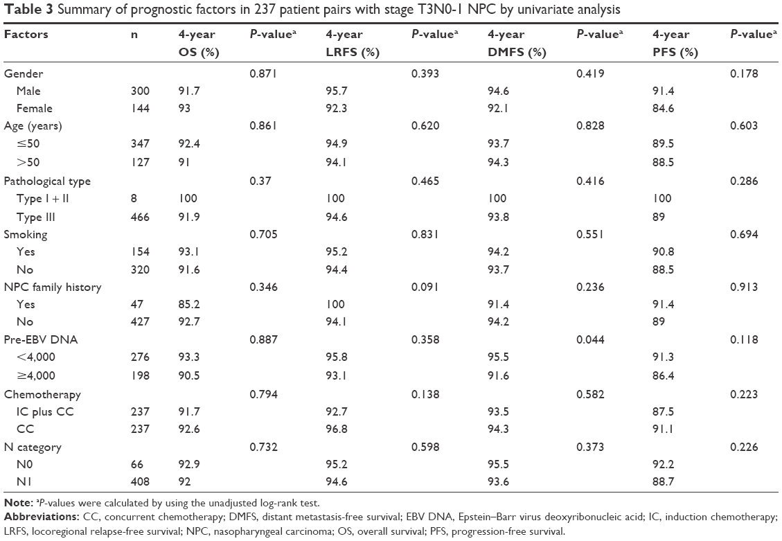 | Table 3 Summary of prognostic factors in 237 patient pairs with stage T3N0-1 NPC by univariate analysis |
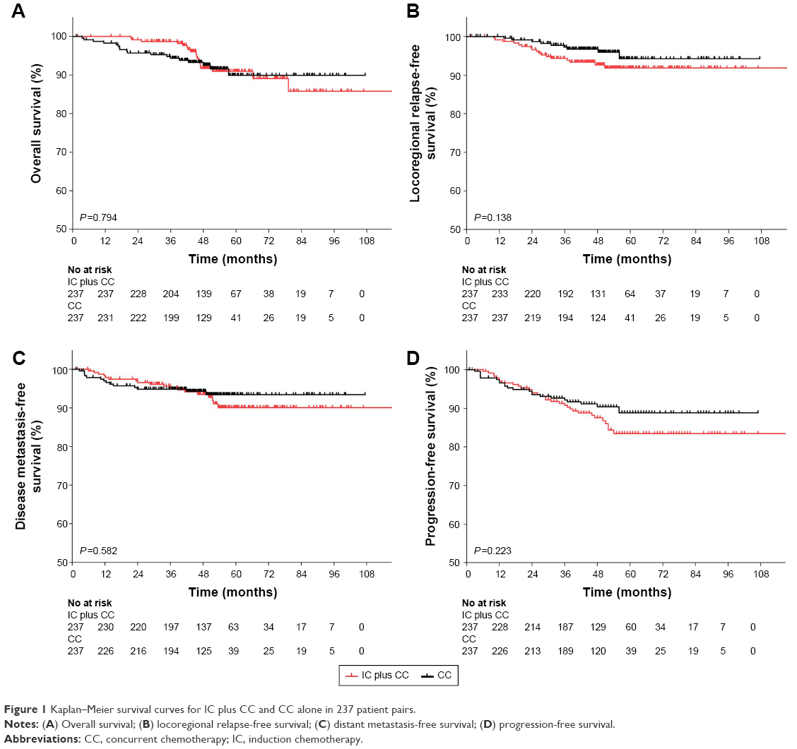 | Figure 1 Kaplan–Meier survival curves for IC plus CC and CC alone in 237 patient pairs. |
Multivariate analysis was performed to adjust for various prognostic factors. The factors were included as covariates in accordance with univariate analysis (Table 4). Consistent with the univariate analysis, gender, age, pathological type, smoking, NPC family history, chemotherapy strategy, and N category were not associated with 4-year OS, LRFS, DMFS, and PFS. Only pre-EBV DNA showed an approximately significant difference in 4-year DMFS (HR =1.05; 95% CI =0.38–0.83; P=0.05).
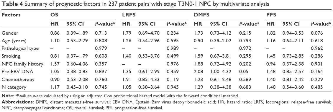 | Table 4 Summary of prognostic factors in 237 patient pairs with stage T3N0-1 NPC by multivariate analysis |
Grade 3–4 hematological toxicities
In the IC plus CC treatment group, 82 patients experienced grade 3–4 leucopenia, and 97 patients experienced grade 3–4 neutropenia. IC plus CC treatment significantly increased the incidence of grade 3–4 leucopenia (P<0.001) and neutropenia (P<0.001) compared with CC treatment. The rates of grade 3–4 anemia and thrombocytopenia exhibited no significant difference in either treatment group (Table 5).
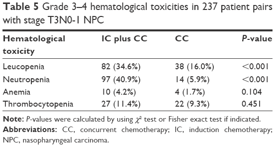 | Table 5 Grade 3–4 hematological toxicities in 237 patient pairs with stage T3N0-1 NPC |
Discussion
Previous phase II/III clinical trials showed conflicting outcomes when IC was added to concurrent chemoradiotherapy (CCRT) in LA-NPC patients.11–14 Tan et al’s11 and Hui et al’s14 studies included stage III–IVB NPC patients and Fountzilas et al’s study12 included stage IIB–IVB patients, but only Hui et al’s study significantly increased 3-year OS (94.1% vs 67.7%, P=0.012).14 When stage T3-4N0 NPC patients were excluded to enhance the power for survival, the trial showed a significant improvement in 3-year PFS (P=0.034), OS (P=0.029), and DMFS (P=0.031) in LA-NPC patients.13 Therefore, no published study exists which assesses the effect of IC in stage T3N0-1 patients, which could be defined as low-risk LA-NPC because of low T and N categories. Therefore, our retrospective study is the first to analyze IC plus CC treatment and CC treatment alone in stage T3N0-1 NPC patients treated with IMRT.
As it is a propensity-matched study, the groups were shown to be well matched for prognostic factors. Other than exploring conventional prognostic factors (ie, gender, age, WHO pathological type, chemotherapy strategy, and N category), we also considered some carcinogenic factors, including smoking history and family history of NPC. Our previous study22 revealed that pretreatment of cigarette smoking was a negative prognostic factor of death, locoregional recurrence, distant metastasis, and disease progression. Guo et al23 reported that the ever-smokers suffered a higher risk of locoregional disease recurrence compared with never-smokers in LA-NPC patients. Ouyang et al24 published that patients who had a first-degree family history of NPC had higher rates of OS and DMFS than those without a family history. However, smoking history and family history of NPC were not associated with survival in both univariate analysis and adjusted multivariate analysis. Plasma EBV DNA has been proven a prognostic biomarker in several studies.25–27 Leung et al28 demonstrated that high levels of EBV DNA pretreatment were associated with the incidence of distant metastasis. Moreover, pre-EBV DNA has been reported a prognostic factor using a cutoff of 4,000 copies/mL to define low versus high levels.21 Our results showed an increased risk of distant metastasis in patients with high EBV DNA (≥4,000 copies/mL) compared with those with low EBV DNA (<4,000 copies/mL), whereas there was no significant difference in 4-year OS, LRFS, and PFS.
Chemoradiotherapy is the standard treatment for LA-NPC. Chemoradiotherapy has been shown to significantly improve patient survival in several phase III randomized clinical trials.4–10 For patients with bulky and/or extensive nodal disease (N2-3), there is higher potential for metastasis; CC is not adequate.29,30 Moreover, T4-classified tumors are considered to result in a poor prognosis.31 Consequently, N2-3 and/or T4 patients can be defined as a high-risk group of LA-NPC; therefore, IC is needed to reduce metastasis and improve survival. Xu et al32 demonstrated that CCRT achieved higher 3-year DMFS rates (94.9% vs 80.1%, P=0.03) in N0-1 LA-NPC patients, but not in N2-3 tumors. In the present study, we chose stage T3N0-1 NPC patients treated with IMRT as a low-risk group to assess the value of IC. Results showed that IC plus CC did not improve survival compared with CC. However, the results showed that CC group was numerically superior to the IC plus CC group in 4-year OS (92.6% vs 91.7%), 4-year LRFS (96.8% vs 92.7%), DMFS (94.3% vs 93.5%), and PFS (91.1% vs 87.5%). We considered that IC increased hematological acute toxicities and reduced the tolerance of CC, which resulted in poor treatment intensity.
A previous trial showed that IC caused higher rates of grade 3 and 4 hematological toxicities, including leukopenia (52% vs 37%, P=0.046), neutropenia (24% vs 12%, P=0.029), and thrombocytopenia (14% vs 0%, P<0.01) compared with the chemoradiotherapy group in LA-NPC patients.11 Our study showed that IC did not improve survival benefits and increased hematological acute toxicities. The IC group experienced a significantly higher incidence of grade 3–4 leucopenia (34.6% vs 16%, P<0.001) and neutropenia (40.9% vs 5.9%, P<0.001). These hematological toxicities may compromise the delivery of subsequent CC with dose reduction.
The present study had several limitations. First, our study is a single institutional retrospective study in an endemic area; therefore, a selection bias existed. Second, although all the patients in the cohort received platinum-based chemotherapy, the regimen and cycles of IC and CC were in disunity. IC included TP, TC, TPF, or PF regimens, whereas CC regimens consisted of cisplatin/nedaplatin, PF, or TP. Furthermore, there was no IMRT dose-fractionation consensus in NPC treatment. Third, only acute hematological toxicities were evaluated. Nonhematological mucositis, vomiting, and late toxicities were not acquired because of the long time interval of the cohort, and some information was missing. Fourth, besides plasma pre-EBV DNA,33 pre-treatment serum lactate dehydrogenase was reported to be associated with distant metastasis,34,35 but we did not include this factor into our analysis. Finally, we assessed only 4-year survival; however, the investigation should be 5 years or longer to evaluate the survival.
Conclusion
IC did not significantly improve survival in stage T3N0-1 NPC patients treated with CC. Furthermore, IC increased the incidence of grade 3–4 hematological toxicities, and it is not recommended for low-risk LA-NPC patients. Further confirmation is warranted in prospective studies.
Acknowledgments
We thank the Sun Yat-Sen University Cancer Center for their assistance with trial monitoring, data management, and statistical analysis. We appreciate the anonymous editors and reviewers who reviewed this manuscript. This research did not receive any specific grant from funding agencies in the public, commercial, or not-for-profit sectors.
Disclosure
The authors report no conflicts of interest in this work.
References
Wee JT, Ha TC, Loong SL, Qian CN. Is nasopharyngeal cancer really a “Cantonese cancer”? Chin J Cancer. 2010;29(5):517–526. | ||
Peng G, Wang T, Yang KY, et al. A prospective, randomized study comparing outcomes and toxicities of intensity-modulated radiotherapy vs. conventional two-dimensional radiotherapy for the treatment of nasopharyngeal carcinoma. Radiother Oncol. 2012;104(3):286–293. | ||
Zhang MX, Li J, Shen GP, et al. Intensity-modulated radiotherapy prolongs the survival of patients with nasopharyngeal carcinoma compared with conventional two-dimensional radiotherapy: a 10-year experience with a large cohort and long follow-up. Eur J Cancer. 2015;51(17):2587–2595. | ||
Kwong DL, Sham JS, Au GK, et al. Concurrent and adjuvant chemotherapy for nasopharyngeal carcinoma: a factorial study. J Clin Oncol. 2004;22(13):2643–2653. | ||
Wu X, Huang PY, Peng PJ, et al. Long-term follow-up of a phase III study comparing radiotherapy with or without weekly oxaliplatin for locoregionally advanced nasopharyngeal carcinoma. Ann Oncol. 2013;24(8):2131–2136. | ||
Chan AT, Leung SF, Ngan RK, et al. Overall survival after concurrent cisplatin-radiotherapy compared with radiotherapy alone in locoregionally advanced nasopharyngeal carcinoma. J Natl Cancer Inst. 2005;97(7):536–539. | ||
Lin JC, Jan JS, Hsu CY, Liang WM, Jiang RS, Wang WY. Phase III study of concurrent chemoradiotherapy versus radiotherapy alone for advanced nasopharyngeal carcinoma: positive effect on overall and progression-free survival. J Clin Oncol. 2003;21(4):631–637. | ||
Chen Y, Sun Y, Liang SB, et al. Progress report of a randomized trial comparing long-term survival and late toxicity of concurrent chemoradiotherapy with adjuvant chemotherapy versus radiotherapy alone in patients with stage III to IVB nasopharyngeal carcinoma from endemic regions of China. Cancer. 2013;119(12):2230–2238. | ||
Lee AW, Tung SY, Chua DT, et al. Randomized trial of radiotherapy plus concurrent-adjuvant chemotherapy vs radiotherapy alone for regionally advanced nasopharyngeal carcinoma. J Natl Cancer Inst. 2010;102(15):1188–1198. | ||
Wee J, Tan EH, Tai BC, et al. Randomized trial of radiotherapy versus concurrent chemoradiotherapy followed by adjuvant chemotherapy in patients with American Joint Committee on Cancer/International Union against cancer stage III and IV nasopharyngeal cancer of the endemic variety. J Clin Oncol. 2005;23(27):6730–6738. | ||
Tan T, Lim WT, Fong KW, et al. Concurrent chemo-radiation with or without induction gemcitabine, Carboplatin, and Paclitaxel: a randomized, phase 2/3 trial in locally advanced nasopharyngeal carcinoma. Int J Radiat Oncol Biol Phys. 2015;91(5):952–960. | ||
Fountzilas G, Ciuleanu E, Bobos M, et al. Induction chemotherapy followed by concomitant radiotherapy and weekly cisplatin versus the same concomitant chemoradiotherapy in patients with nasopharyngeal carcinoma: a randomized phase II study conducted by the Hellenic Cooperative Oncology Group (HeCOG) with biomarker evaluation. Ann Oncol. 2012;23(2):427–435. | ||
Sun Y, Li WF, Chen NY, et al. Induction chemotherapy plus concurrent chemoradiotherapy versus concurrent chemoradiotherapy alone in locoregionally advanced nasopharyngeal carcinoma: a phase 3, multicentre, randomised controlled trial. Lancet Oncol. 2016;17(11):1509–1520. | ||
Hui EP, Ma BB, Leung SF, et al. Randomized phase II trial of concurrent cisplatin-radiotherapy with or without neoadjuvant docetaxel and cisplatin in advanced nasopharyngeal carcinoma. J Clin Oncol. 2009;27(2):242–249. | ||
Lan XW, Zou XB, Xiao Y, et al. Retrospective analysis of the survival benefit of induction chemotherapy in stage IVa-b nasopharyngeal carcinoma. PLoS One. 2016;11(8):e160758. | ||
D’Agostino RJ. Propensity score methods for bias reduction in the comparison of a treatment to a non-randomized control group. Stat Med. 1998;17(19):2265–2281. | ||
Austin PC. The use of propensity score methods with survival or time-to-event outcomes: reporting measures of effect similar to those used in randomized experiments. Stat Med. 2014;33(7):1242–1258. | ||
Shao JY, Zhang Y, Li YH, et al. Comparison of Epstein-Barr virus DNA level in plasma, peripheral blood cell and tumor tissue in nasopharyngeal carcinoma. Anticancer Res. 2004;24(6):4059–4066. | ||
Lo YM, Chan LY, Lo KW, et al. Quantitative analysis of cell-free Epstein-Barr virus DNA in plasma of patients with nasopharyngeal carcinoma. Cancer Res. 1999;59(6):1188–1191. | ||
Edge SB, Compton CC. The American Joint Committee on Cancer: the 7th edition of the AJCC cancer staging manual and the future of TNM. Ann Surg Oncol. 2010;17(6):1471–1474. | ||
Leung SF, Zee B, Ma BB, et al. Plasma Epstein-Barr viral deoxyribonucleic acid quantitation complements tumor-node-metastasis staging prognostication in nasopharyngeal carcinoma. J Clin Oncol. 2006;24(34):5414–5418. | ||
Ouyang PY, Su Z, Mao YP, et al. Prognostic impact of cigarette smoking on the survival of patients with established nasopharyngeal carcinoma. Cancer Epidemiol Biomarkers Prev. 2013;22(12):2285–2294. | ||
Guo SS, Huang PY, Chen QY, et al. The impact of smoking on the clinical outcome of locoregionally advanced nasopharyngeal carcinoma after chemoradiotherapy. Radiat Oncol. 2014;9:246. | ||
Ouyang PY, Su Z, Mao YP, Liang XX, Liu Q, Xie FY. Prognostic impact of family history in southern Chinese patients with undifferentiated nasopharyngeal carcinoma. Br J Cancer. 2013;109(3):788–794. | ||
An X, Wang FH, Ding PR, et al. Plasma Epstein-Barr virus DNA level strongly predicts survival in metastatic/recurrent nasopharyngeal carcinoma treated with palliative chemotherapy. Cancer. 2011;117(16):3750–3757. | ||
Peng H, Guo R, Chen L, et al. Prognostic impact of plasma Epstein-Barr virus DNA in patients with nasopharyngeal carcinoma treated using intensity-modulated radiation therapy. Sci Rep. 2016;6:22000. | ||
Lin JC, Wang WY, Chen KY, et al. Quantification of plasma Epstein-Barr virus DNA in patients with advanced nasopharyngeal carcinoma. N Engl J Med. 2004;350(24):2461–2470. | ||
Leung SF, Chan AT, Zee B, et al. Pretherapy quantitative measurement of circulating Epstein-Barr virus DNA is predictive of posttherapy distant failure in patients with early-stage nasopharyngeal carcinoma of undifferentiated type. Cancer. 2003;98(2):288–291. | ||
Lin JC, Liang WM, Jan JS, Jiang RS, Lin AC. Another way to estimate outcome of advanced nasopharyngeal carcinoma–is concurrent chemoradiotherapy adequate? Int J Radiat Oncol Biol Phys. 2004;60(1):156–164. | ||
Lee AW, Lau WH, Tung SY, et al. Preliminary results of a randomized study on therapeutic gain by concurrent chemotherapy for regionally-advanced nasopharyngeal carcinoma: NPC-9901 Trial by the Hong Kong Nasopharyngeal Cancer Study Group. J Clin Oncol. 2005;23(28):6966–6975. | ||
Chen L, Liu LZ, Chen M, et al. Prognostic value of subclassification using MRI in the t4 classification nasopharyngeal carcinoma intensity-modulated radiotherapy treatment. Int J Radiat Oncol Biol Phys. 2012;84(1):196–202. | ||
Xu T, Zhu G, He X, Ying H, Hu C. A phase III randomized study comparing neoadjuvant chemotherapy with concurrent chemotherapy combined with radiotherapy for locoregionally advanced nasopharyngeal carcinoma: updated long-term survival outcomes. Oral Oncol. 2014;50(2):71–76. | ||
Peng H, Chen L, Zhang Y, et al. Survival analysis of patients with advanced-stage nasopharyngeal carcinoma according to the Epstein-Barr virus status. Oncotarget. 2016;7(17):24208–24216. | ||
Wan XB, Wei L, Li H, et al. High pretreatment serum lactate dehydrogenase level correlates with disease relapse and predicts an inferior outcome in locally advanced nasopharyngeal carcinoma. Eur J Cancer. 2013;49(10):2356–2364. | ||
Zhou GQ, Ren XY, Mao YP, et al. Prognostic implications of dynamic serum lactate dehydrogenase assessments in nasopharyngeal carcinoma patients treated with intensity-modulated radiotherapy. Sci Rep. 2016;6:22326. |
 © 2017 The Author(s). This work is published and licensed by Dove Medical Press Limited. The
full terms of this license are available at https://www.dovepress.com/terms
and incorporate the Creative Commons Attribution
- Non Commercial (unported, 3.0) License.
By accessing the work you hereby accept the Terms. Non-commercial uses of the work are permitted
without any further permission from Dove Medical Press Limited, provided the work is properly
attributed. For permission for commercial use of this work, please see paragraphs 4.2 and 5 of our Terms.
© 2017 The Author(s). This work is published and licensed by Dove Medical Press Limited. The
full terms of this license are available at https://www.dovepress.com/terms
and incorporate the Creative Commons Attribution
- Non Commercial (unported, 3.0) License.
By accessing the work you hereby accept the Terms. Non-commercial uses of the work are permitted
without any further permission from Dove Medical Press Limited, provided the work is properly
attributed. For permission for commercial use of this work, please see paragraphs 4.2 and 5 of our Terms.