Back to Journals » International Journal of Nanomedicine » Volume 20
Os Draconis-Derived Nanoparticles Improve Insomnia Symptoms by Activating Calcium-Dependent 5-HT Release and the Vagal-NTS Pathway
Authors Liu Z, Wang Q, Fan X, Ye X, Wang Q, Huang Y, Wu C
Received 30 July 2025
Accepted for publication 22 November 2025
Published 2 December 2025 Volume 2025:20 Pages 14329—14341
DOI https://doi.org/10.2147/IJN.S553405
Checked for plagiarism Yes
Review by Single anonymous peer review
Peer reviewer comments 2
Editor who approved publication: Professor Lijie Grace Zhang
Zibo Liu,1 Qian Wang,1 Xinyun Fan,1 Xun Ye,1 Qinyu Wang,1 Yongliang Huang,2 Chunjie Wu3,4
1State Key Laboratory of Southwestern Chinese Medicine Resources, School of Pharmacy, Chengdu University of Traditional Chinese Medicine, Chengdu, 611137, People’s Republic of China; 2Pharmacy Department, the Affiliated Hospital of Chengdu University of Traditional Chinese Medicine, Chengdu, 610075, People’s Republic of China; 3Innovative Institute of Chinese Medicine and Pharmacy/Academy for Interdiscipline, Chengdu University of Traditional Chinese Medicine, Chengdu, 611137, People’s Republic of China; 4Sichuan Engineering Research Center for Endangered Medicinal Animals, Chengdu, 611137, People’s Republic of China
Correspondence: Chunjie Wu, Email [email protected] Yongliang Huang, Email [email protected]
Background: Os draconis (OD), a traditional Chinese medicine from fossil mammalian bones, effectively treats insomnia and anxiety. Its usage conflicts with the laws, causing a shortage. This research clarifies OD’s pharmacodynamic mechanisms, aiming to establish a scientific basis for developing novel artificial substitutes.
Methods: This research used a mouse chronic insomnia paradigm to assess an os draconis decoction (DOD). DOD was digested in vitro, and the resultant nanoparticles (OD-NPs) were analyzed by scanning electron microscopy, dynamic light scattering, X-ray diffraction, and Fourier transform infrared spectroscopy. The in vivo effects were evaluated using behavioral tests, Nissl staining, neuronal activity in the nucleus tractus solitarii (NTS), and plasma 5-HT levels. In vitro mechanistic investigations used FITC-labeled OD-NPs to detect cellular uptake. The research used calcium channel blockers to examine changes in intracellular Ca2⁺ concentration and critical protein expression in 5-HT-related pathways.
Results: After DOD treatment, significantly improved movement in the Open field, brain malondialdehyde (MDA) and plasma tumor necrosis factor-α (TNF-α) were reduced, increased hippocampal Nissl bodies, and alleviated neuronal damage. Digested DOD formed numerous sub-1000 nm spindle particles. Its composition remained carbonate hydroxyapatite (F-rich), but crystallinity decreased. DOD elevated plasma 5-HT and c-fos expression in the NTS. In vitro, OD-NPs were uptaken by cells, increasing supernatant 5-HT and cytosolic calcium. This upregulated TPH1 and DdC expression, a trend unaffected by Ca2⁺ channel blockade, unlike the filtrate group.
Conclusion: This is the first research to suggest that oral DOD is digested into OD-NPs, which are then internalized by enterochromaffin cells. This absorption initiates calcium signaling, boosts 5-HT release, and then activates the vagus nerve-NTS pathway, thereby regulating central nervous system activity. This research provides scientific proof for the clinical application of OD, lays the groundwork for the development of artificial alternatives, and generates ideas for future traditional Chinese medicine research.
Keywords: os draconis, apatite, nanoparticles, intestinal chromaffin cells, vagus, insomnia
Introduction
According to TCM records, Os draconis (OD) is derived from fossilized bones of ancient mammals including Hipparion, rhinoceroses, deer, cattle, and elephants. When milled into powder and decocted in water for oral administration, it has anxiolytic qualities and is clinically used for palpitations, hyperarousal, insomnia, and dream-disturbed sleep.1,2 As paleontological fossils, OD specimens are crucial materials for researching Earth’s evolution and biological history, necessitating strong regulatory safeguards that limit therapeutic use. After years of investigation, the researchers’ understanding of the composition and chemical constituents of OD has improved. Minerals like apatite are the most common phase components. There is a minor amount of calcite and quartz.3,4 The chemical elements contained are numerous, mostly containing Ca, P, Mg, Fe, Al, and other trace rare earth elements.5,6 However, there has been little research into its pharmacological effects. There is no clear scientific explanation for how it may alleviate insomnia symptoms after oral use. As a result, two significant difficulties remain: 1) Limited understanding of its mode of action and bioactive components; 2) A scarcity of clinically recognized substitutes.
Current research defines OD as largely composed of apatite-phase minerals, while controversy persists regarding whether its primary form is hydroxyapatite or carbonate-substituted hydroxyapatite, both belonging to the wider calcium phosphate family.7–9 Notably, calcium phosphates demonstrate significant bioactivity in biomedical applications, including drug delivery systems and biological materials, etc.10–13The intracellular transport of hydroxyapatite nanoparticles typically involves four steps: The process involves overcoming biological barriers, fusing nanoparticles with lysosomes, acidifying lysosomes to breakdown some nanoparticles, rupturing lysosomes to release calcium ions, hydrogen phosphate ions, and cargo into the cytoplasm, and removing excess calcium. Calcium ions serve as intracellular secondary messengers.14 In resting cells, the intracellular free Ca2+ content is always lower than the extracellular fluid. When the intracellular calcium ion concentration rises,15 it necessarily results in biological repercussions. Can OD form nano-scale OD calcium phosphate after grinding and decoction and passing through the gastrointestinal tract digestion, resulting in similar biological effects? This is something that should be investigated thoroughly.
Recent advances have identified the gut-brain axis as a critical research frontier. Accumulating evidence demonstrates that modifications in the gut milieu directly or indirectly impact brain circuits.16,17 At present, the gut influences the central nervous system mainly through three pathways: the vagus nerve, the endocrine system, and the immune system.18–20 Enterochromaffin cells (ECs), a subset of enteroendocrine cells, serve as the major epithelial chemosensors. Although quantitatively sparse, ECs produce more than 90% of peripheral 5-hydroxytryptamine (5-HT) in humans.21 The vast majority of gut-derived 5-HT is absorbed by circulating platelets. Following appropriate inputs, these platelets release 5-HT. A modest percentage of gut-derived 5-HT remains unbound in serum, operating as a hormone modulating multiple physiological processes, including intestinal inflammation, cardiovascular control, and nervous system activation.22–24 5-HT, a crucial neurotransmitter in the brain, is intimately linked to insomnia and the maintenance of slow-wave sleep in the body. It is challenging for 5-HT in peripheral blood to pass through the blood-brain barrier, though. According to recent research, EC cells have the ability to control the expression of 5-HT3 receptors in afferent sensory nerve fibers. The vagus nerve carries this signal, which has physiologic effects on the brain.16
Based on comprehensive literature review, our team previously proposed a scientific hypothesis: Following oral administration, the decoction of os draconis (DOD) undergoes gastrointestinal digestion, resulting in numerous nanoparticles. ECs in the stomach ingest and digest these particles, increasing intracellular Ca2⁺ concentrations and stimulating peripheral 5-HT production. This cascade activates intestine sensory nerve fibers, which then carry signals via the vagus nerve to the central nervous system. This research seeks to explain the pharmacodynamic material basis and mode of action of OD, laying the groundwork for the development of specific artificial alternatives.
Materials and Methods
Reagents
Os Draconis was purchased from Sichuan Xinhehua Traditional Chinese Medicine Decoction Pieces Co., LTD (Lot: D2408057); RIN-14B cells were purchased from Qingqi Biotechnology Development Co., LTD (Cat: BFN60805924, China); Artificial gastric juice (Cat: SL6600, Coolaber, China); Artificial small bowel fluid (Cat: SL6610A, Coolaber, China); Compound diazepam tablet (National Drug Approval Number: H10970219, Jining Ankang Pharmaceutical Co., Ltd, China); Dulbecco’s Modified Eagle Medium (Lot: 6124059, Gibco, USA); 0.25% trypsin (Lot: 25200056, Gibco, USA); Fetal bovine serum (FBS) (Cat: FSP500, Excell, China); phosphate buffered saline (PBS) (Cat: G4250-500ML, Servicebio, China); DMSO (Cat: D8371, Solarbio, China); HC-030031 (Cat: HY-15064, MCE, USA);Gabapentin (Cat: T0702, Topscience, China); Fluorescein 5-isothiocyanate (FITC) (Cat: HY-66019, MCE, USA); Total RNA isolation kit for cells (Cat: RE-03111, Foregene, China); RT EasyTM II (Cat: RT-01022, Foregene, China); Enhanced RIPA lysate (Cat: AR 0102, Boster, China); A mixture of broad-spectrum protease inhibitors (Cat: AR 1182, Boster, China); A mixture of broad spectrum phosphatase inhibitors (Cat: AR 1183, Boster, China); Tris Buffered Saline Tween (TBST) (Cat: BL315B); PAGE gel was used for rapid preparation of kits (Cat: PG111, Shanghai Yaenzyme Biomedical Technology Co., Ltd).; CCK-8 cell viability detection kit (Cat: BS350B, Biosharp, China); 5-HT ELISA kit (Cat: E-EL-0033, Elabscience, China); TNF-α ELISA kit (Cat: E-EL-M3063, Elabscience, China); MDA detection kit (Cat: S0131S, Beyotime, China); Fluo-4 calcium ion detection kit (Cat: S1061S, Beyotime, China); Polyvinylidene fluoride membrane (PVDF) (Cat: IPVH00010, Millipore, USA); Anti-Tryptophan Hydroxylase 1 Antibody (Cat: ET1610-37, Huabio, China); Serotonin transporter Rabbit Polyclonal Antibody (Cat: ER1916-44, Huabio, China); DOPA Decarboxylase Recombinant Rabbit Monoclonal Antibody (Cat: ET1704-94, Huabio, China); GAPDH Recombinant Rabbit Monoclonal Antibody (Cat: ET1601-4, Huabio, China); HRP-conjugated goat anti-Rabbit IgG (H+L) (Cat: AS014, Abclonal, China); BCA Protein concentration Assay kit (Cat: AR0197, Boster, China). Anti-c-Fos Mouse mAb (Cat: GB12069, Servicebio, China); Goat Anti-Mouse IgG H&L (Cat: bs-0296G, Bioss, China). Glutamic Acid Content Assay Kit (Cat: BC1585, Solarbio, China).
Preparation of DOD: OD was pulverized into coarse powder. A 100 g OD powder was decocted with 1 L of deionized water for 1 h under reflux. Collecting the supernatant liquid. The residue was re-decocted with 1 L of fresh deionized water for 0.5 h. Both liquids were combined, concentrated under reduced pressure to 200 mL, and standardized to a final concentration of 0.5 g/mL. Compound Diazepam Tablet Solution: Tablets were crushed and sieved through a 100-mesh screen. The powder was suspended in purified water to achieve a concentration of 1.71 mg/mL.
Mice and Treatment
Using the multi-platform water environment method, the mice were randomly placed on platforms with a diameter of approximately 3 cm in the experimental box. The distance between each platform was 3 cm. Tap water was injected into the experimental box, with the water surface height approximately 1 cm above the platform surface. The mice were deprived of sleep for 12 hours every day (from 9:00 to 21:00 the next day) for a total of 28 days. During the remaining time, they were returned to the incubator to rest.25–27 They were randomly divided into a control group, a model group, a positive group, and a DOD group. Each group consists of 10 animals. The positive group: The dosage for mice was 15.39 mg/kg, once a day, for 20 days. The DOD group: The dosage for mice was 4.505 g/kg. It was administered by gavage once a day for 20 days. The control group and the model group: Equal doses of distilled water were administered by gavage once a day for 20 days. (Figure 1A) The Animal Welfare Ethics approval number for this research is: 2025118.
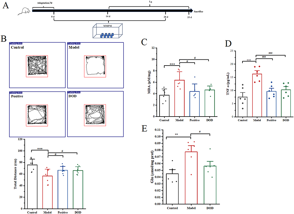 |
Figure 1 OD can improve the physiology of insomnia mice. (A) Schematic diagram of the model of chronic insomnia; (B) Open field test (n=8); (C) MDA content in brain tissue (n=6); (D) TNF-α content in plasma (n=6); (E) Glutamic acid content in brain tissue (n=6). Data are shown as mean ± SD, **P < 0.01, ***P < 0.001 vs control; #P < 0.05 vs Model, ###P < 0.001, vs Model. |
Open Field Test
Mice were introduced from a fixed position into a 50 cm × 50 cm open field and allowed to move freely. The movement of mice in each group was recorded for 5 min, and after each test, stool and urine stains were cleaned from the field and wiped with alcohol before the introduction of another mouse.
Nissl Staining and Immunohistochemical Analysis
The tissues were fixed in 4% paraformaldehyde for 24 h, dehydrated in a gradient of ethanol, embedded in paraffin, and sectioned at a thickness of 5 μm. The sections were stained with methyl violet and scanned to observe the morphological and structural changes of the hippocampus. The paraffin sections of NTS were successively subjected to deparaffinization, antigen retrieval, inactivation, blocking, primary antibody incubation, secondary antibody incubation, color development, counterstaining, and sealing. The sections were scanned with PANNORAMIC 150 (3DHISTECH, Hungary) and observed by CaseViewer 2.4 (3DHISTECH, Hungary) software.
Simulated Digestion in vitro
About 6 mL of 0.5g/mL DOD was added to 2 mL of artificial stomach liquid preheated at 37°C for 1.5 h, and then 3 mL of artificial small intestine liquid preheated at 37°C for 4 h. The digestion process was carried out in a 37°C water bath at 200 rpm.28 After centrifugation at 12000 × g for 20 min, the supernatant was discarded. The pellet was resuspended in an equal volume of PBS. The resulting suspension was then sterilized under UV light for 30 min. A portion of this suspension was centrifuged through a 3 kDa molecular weight cut-off (MWCO) ultrafiltration membrane at 5,000 × g for 20 min. The filtrate (< 3 kDa) was collected and subsequently used for administration to cells.
Characterization of OD-NPs
The DOD with simulated digestion (SD-DOD) was centrifuged at 12000 × g for 20 min, and the supernatant was removed and dried. The morphology was detected by scanning electron microscope (SEM) (ZEISS-Sigma 360, Germany): The accelerating voltage (5.00 kV) and resolution (100 nm). Particle size was determined by a dynamic light scattering instrument (Anton Paar-Litesizer DIF 500, Austria): The detection angle of dynamic light scattering is SideScatter. The phase was detected by powder X-ray diffraction (XRD) (Rigaku-Ultima IV, Japan). The 2θ angle was from 10° to 80°, the scanning rate was 10°/min, the step size 0.02°, and the samples were passed through a 200-mesh screen before starting the machine. Infrared spectra were detected by Fourier transform infrared spectroscopy (FT-IR) (SHIMADZU-IRTracer-100, Japan): the wavelength range was 4000 ~ 400cm−1, and the number of scans was 32.
Cell Culture and Treatment
RIN-14B cells were used to simulate intestinal chromaffin cells, medium: DMEM high glucose medium, 10% FBS, 1% bispecific antibody, cultured in a CO2 constant temperature incubator at 37°C, and the medium was changed every two days, and passaged when the cell density reached 70%–80%. Cell viability assay: In a 96-well plate, prepare a 100 μL cell suspension (approximately 1×10⁴ cells). Place the culture plate in incubator for pre-culture for 24 h (37°C, 5% CO2). The dosing concentration was configured according to the ratio of suspension: medium = 1:200, 1:100, 1:50, 1:25, 3:50, 1:10. For each well, replace the culture medium with 110 μL of medium containing different concentrations of the drug. Place the culture plate in the incubator for 24 h, then replace the medium and add 110 μL of the medium (containing 10 μL of CCK-8 solution) to each well. Incubate in the incubator for approximately 20 minutes. Measure the absorbance at 450 nm using an enzyme-linked spectrophotometer., For subsequent in vitro experiments were divided into the following four groups: control group; SD-DOD group (treated with 4% nanoparticle-containing supernatant); SD-DOD+CA group (treated with 4% nanoparticle-containing supernatant 150 nM Gabapentin 10um HC-030031); Ultra group (treated with 4% ultrafiltrate).
Measurement of Biochemical Indicators
The blood was collected in a blood collection container containing EDTA K2, centrifuged at 4°C at 1000×g for 15 min within 30 minutes after sample collection, the supernatant was subsequently removed, and the determination was carried out according to the instructions of the 5-HT and TNF-α assay kits. Brain tissue was accurately weighed, normal saline was added, and the ratio of weight: volume = 1:9 was used. Grinding beads were added for mechanical homogenization, centrifugation at 12000 g for 10 min, the supernatant was collected, and then diluted to an appropriate concentration with normal saline. The concentration of tissue protein was determined by BCA, and the instructions of the MDA and Glutamic acid content kit were followed. All measurements were performed using a multimode microplate reader (Thermo-Varioska, USA).
In vitro Cellular Uptake Assay of OD-NPs
About 20 mL of SD-DOD solution was centrifuged at 12000 rpm for 20 min, the supernatant was removed, 1 mg FITC was added, and the mixture was stirred overnight.29 Then, 3 washes were made using PBS, and finally PBS was suspended, and the dosing concentration was configured according to the ratio of suspension: medium 1:25, 37°C, incubated in an incubator for 6 h in the dark, and imaged on a confocal microscope (Leica-TCS SP8X, Germany).
Detection of Intracellular Ca2+ Signals
About 1×10⁵ cells were seeded into a confocal dish, and each group was given different drugs and incubated at 37°C for 12 h. After the specified time, aspirate the culture medium and wash the cells once with PBS. Add 200 μL of Fluo-4 staining solution and incubate at 37°C for 45 min in the dark. After incubation, acquire images using a confocal microscope (Ex/Em = 490/525nm).
Real-Time Fluorescence Quantitative PCR Analysis
Total RNA was extracted from the cells in the six-well plate using the column method, and cDNA was synthesized by a reverse transcription kit. Primers were synthesized based on the gene sequences in the following Table 1. Finally, polymerase chain reaction was performed on a fluorescence quantitative PCR instrument (BIO-RAD-CFX Conect, USA) to detect the fluorescence expression intensity.
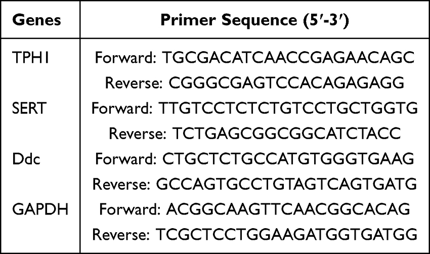 |
Table 1 PCR Primer Sequences |
Western Blot Analysis
The cells were washed with cold PBS, the RIPA lysate was added, and thoroughly homogenized on ice. The protein concentration was measured by the BCA method, and the membrane was transferred using a 0.45μm PVDF membrane by SDS-PAGE electrophoresis. The transformed membrane was closed at room temperature with 5% skim milk (TBST) for 1h. Add diluted primary antibody and incubate at room temperature for 3 hours; Wash with TBST at room temperature 3 times, 5 min each time. The second antibody was diluted with TBST, incubated at room temperature for 1h, and washed with TBST 3 times at room temperature for 5 min each time. ECL reagents A and B were mixed at a 1:1 ratio in a tube, and the PVDF membrane was fully contacted with the mixture. The image was acquired under ChemiScope 6100 (Shanghai Qinxiang Scientific Instruments Co., LTD.), and band intensity was analyzed using the ChemiScope software (Shanghai Qinxiang Scientific Instruments Co., LTD).
Statistical Analysis
The data were analyzed using IBM SPSS Statistics 26 and are displayed in Origin 2021 as the mean ± SD. One-way analyses of variance were used in our investigation to assess statistical significance. Statistical significance was considered as a P-value less than 0.05.
Results
OD Can Improve Sleep Disorders in Mice with Insomnia
After continuous modeling, except for the normal group, all mice in all groups showed photophobia, reduced activity, and drowsiness. Compared with the normal group, the mice in the model group had a significant decrease in the distance traveled in the open field (Figure 1B), significantly reduce the content of MDA in brain tissue and TNF-α in plasma (Figure 1C and D), and the neurons in the hippocampal CA3 region showed nuclear condensation, cell body shrinkage and deformation, and Nissl body disappearance (Figure 2). After 20 days of continuous administration, compared with the model group, both the positive drug and the DOD group could significantly increase the distance traveled in the mine (Figure 1B), significantly reduce the content of MDA and Glutamic acid in brain tissue and TNF-α in plasma (Figure 1C–E), increase the number of Nissl body in the hippocampal CA3 region, and exhibited fewer pathological changes in neuronal structure (Figure 2).
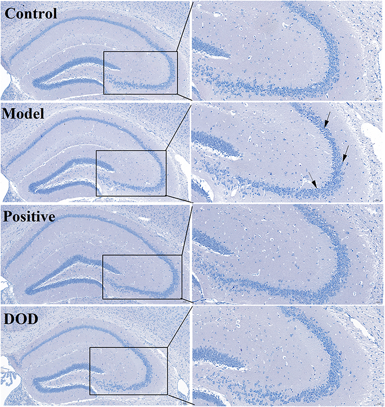 |
Figure 2 Nissl staining of brain tissue (The black arrow indicates the dissolved Nissl bodies, n=3, Scale bar = 50 um). |
OD May Produce Nanoparticles in vivo to Exert Corresponding Biological Effects
Based on a number of studies, calcium phosphate nanoparticles have good biological activity. In this research, DOD was simulated by adding artificial digestion in vitro to simulate the in vivo digestion process (Figure 3A). The particle size of the simulated digested product was measured, and the results showed that DOD produced a large number of particles smaller than 1000 nm after digestion (Figure 3B). To characterize the changes of these particles before and after digestion, the morphology of the particles after digestion was mainly fusiform (Figure 3C), OD was identified as a biological skeletal fossilized substance, and the phase was detected by OD, and after matching with the standard card, it was found that the phase before and after digestion did not change, and it was similar to the X-ray diffraction characteristic map of chlorapatite (PDF#71-0881), but the crystallinity of the powder decreased after digestion (Figure 3D), combined with infrared spectroscopy analysis (Figure 3E). The common characteristic peaks of DOD and SD-DOD are as follows: the vibrational mode at 573 cm−1 and 605 cm−1 is the asymmetric bending vibration of phosphate, the wide absorption band of 1000–1200 cm−1 is the asymmetric expansion vibration of different P-O bonds, and the absorption peak at 1456 cm−1 and 874 cm−1 is the characteristic vibration peak of carbonate ions in B-type hydroxyapatite. The only difference is that the SD-DOD lacks the contraction vibration at 631 cm−1 with an OH− shoulder peak.30,31 According to the comprehensive analysis of the above data, DOD existed as carbonaceous apatite before and after digestion, and the overall phase did not change; while hydroxyl groups in OD may have been replaced by other ions due to exposure to hydrogen ions in the acidic environment during digestion.
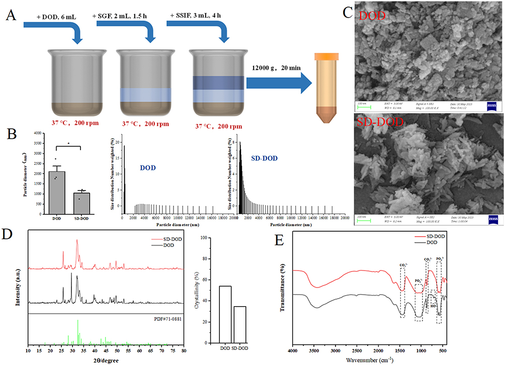 |
Figure 3 Characterization of OD-NPs. (A) Schematic diagram of DOD simulated digestion mode; (B) Particle size (n=3); (C) SEM images (Scale bar = 100 nm); (D) X-ray diffraction characteristics pattern; (E) Infrared signature pattern. Data are shown as mean ± SD, *P < 0.05 vs DOD. |
OD-NPs May Affect the Central Nervous System Through Enterochromaffin Cells, Vagus Nerve, and Nucleus Tractus Solitarius
Combined with recent studies on the gut-brain axis, ECs, as key intestinal endocrine cells, together with the intestinal vagus nerve, build an effective communication bridge between the intestine and the central nervous system, and the main physiological function of EC is to synthesize 5-HT. The results of this study showed that compared with the blank group, DOD administration could significantly increase the content of 5-HT in plasma (Figure 4A), and the NTS, as the central terminal of the vagus nerve, integrated a variety of visceral information, and the expression of c-fos protein increased after DOD administration (Figure 4B), which indirectly indicated that OD-NPs may be absorbed by ECs, increase the secretion of 5-HT, stimulate the transmission of vagus nerve signals to the nucleus of the NTS of the brain, and produce subsequent biological effects.
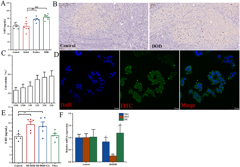 |
Figure 4 The OD-NPs pathway may be through enterochromaffin cells-vagus-nucleus solitus. (A) Plasma content of 5-HT (n=6); (B) C-fos protein expression in the nucleus of the solitary tract (n=3); (C) Effect of different concentrations on cell viability; (D) OD-NPs was uptaken by ECs (n=3); (E) Content of 5-HT in cell supernatant (n=5); (F) OD-NPs regulates the expression of genes related to 5-HT synthesis and transport (n=3). Data are shown as mean ± SD, *P < 0.05, **P < 0.01 vs control; ##P < 0.01, ###P < 0.001, vs Model. |
To continue exploring whether OD-NPs enters the cell and exert its biological effect, the relevant in vitro experiments were carried out by RIN-14B cells as a model for ECs, and the cell activity at different concentrations was detected by CCK8, and the results showed that the higher the concentration, the stronger the activity (Figure 4C), to ensure the quality of subsequent related imaging, we selected a concentration corresponding to a ratio of 1:25 as the optimal concentration. As a member of the calcium phosphate family, OD can be conjugated to substances containing hydroxyl groups, carbonyl groups, and other structures, and the results show that 12 h after administration, green fluorescence was expressed in the cytoplasm (Figure 4D), indicating that OD-NPs can be absorbed by cells. The content of 5-HT in the cell supernatant was detected, and compared with the normal group, the secretion of 5-HT was significantly increased after SD-DOD administration, but there was no effect on 5-HT secretion after the addition of Ca2+ channel blocker, and there was no increase in 5-HT secretion after ultrafiltrate administration (Figure 4E). By detecting the expression of genes and proteins related to 5-HT synthesis and transport, 24 h after administration, compared with the normal group, the SD-DOD group could significantly increase the expression of TPH1 and DdC genes and proteins, and significantly reduce the expression of SERT cause and protein, and the trend was not reversed after the addition of Ca2+ channel blocker (Figure 4F and Figure 5A and B), and the changes of Ca2+ content in the cytoplasm were detected after 12 h of administration, and the Ca2+ content in the cytoplasm increased compared with the normal group There was no change in Ca2+ content after channel blocker (Figure 5C), suggesting that Ca2+ was derived from OD-NPs rather than extracellular. In summary, after the uptake of OD-NPs, cells were decomposed into a large amount of Ca2+ in the cytoplasm, which activated 5-HT synthesis and transport-related proteins, resulting in an increase in the content of 5-HT secretion by cells.
 |
Figure 5 Regulatory mechanism of OD-NPS for 5-HT. (A and B) 5-HT synthesis and transporter protein (n=3). (C) Change in Ca2+ concentration in cytoplasm (n=3). Data are shown as mean ± SD, *P < 0.05 vs control, #P < 0.05 vs SD-DOD. |
Discussion
A multi-platform water environment method was used in this research to create an insomnia model. After 20 days of continuous gavage administration, OD was able to significantly increase the movement distance of mice in the mine, decrease the pathological damage caused by insomnia, and lower the levels of related biochemical indexes, TNF-α, and MDA when compared to the model group. These findings show that OD has relaxing and anti-convulsive properties. According to recent research, OD, a fossilized big animal skeleton, has a comparatively simple chemical makeup and virtually no organic content.32 In terms of crystal structure, it is mainly the apatite phase.4 In terms of chemical composition, the OD of different origins has slightly different element content and types.33–35 The environmental factors involved in fossilization may have anything to do with this. In short, the skeletal remains are kept in place while the biological remains undergo rapid and extensive mineralogical and chemical changes, such as the formation of biological apatite crystals and the precipitation of autogenous minerals, as biological action breaks down the majority of the body’s organic matrix during the burial process,36 because the majority of the inorganic substance in biological bones, ie, bone minerals, is amorphous calcium phosphate and crystalline hydroxyapatite, the calcium and phosphorus content of OD in diverse origins is higher, which may be a component of OD that exerts curative functions. Combined with the traditional clinical use method, OD needs to be powdered, and then decocted with water before oral administration into the body, this research simulated the digestion of DOD in vitro to detect the particle size, morphological characteristics, phase structure and other aspects of the solid products after digestion, the results showed that DOD produced a large number of particles less than 1000 nm after digestion, and the shape of the particles became more easily absorbed by cells of the fusiform.37 Comprehensive XRD and FT-IR research revealed that the primary carbon chloroapatite in DOD was not modified by the action of artificial simulated digest, with the exception of the hydroxyl group being replaced. The results show that OD can exist in the digestive tract in the form of nanoparticles without completely decomposing into different ions. It is assumed that some inorganic ions are produced after DOD digestion, but according to current studies, most cells do not actively take up these ions. For example, in resting cells, the intracellular free Ca2+ concentration is maintained at a lower level than that of extracellular fluid, and there is a 20,000-fold gradient between the outside (about 1.2 mM) and the inside (about 10–100 nM) of the cell.15,38 As a result, these ions are unable to enter the interior of the cell and perform the corresponding biological effects.
According to current research, OD can exist in the intestinal tract in the form of nanoparticles, but how OD affects the central nervous system to exert its efficacy mechanism is unknown. ECs, as key intestinal endocrine cells, together with the intestinal vagus nerve, build an effective communication bridge between the intestinal tract and the central nervous system,16,17 the main physiological function of ECs is to synthesize 5-HT, and interestingly, the content of 5-HT in the plasma of mice increased significantly after OD administration. The NTS is the place where the vagus nerve meets the central nervous system,39 the expression of c-fos protein also increased after OD administration, which indirectly indicated that the efficacy of OD may be affected by the central nervous system through intestinal pheochromaffin cells-vagus-NTS. To continue to explore how OD-NPS increases the secretion of 5-HT by intestinal chromaffin cells, in vitro cell experiments were used to explore the related mechanism of action, but because ECs account for about 1% of intestinal epithelial cells, they are widely dispersed and difficult to extract and isolate. Studies have shown that the rat pancreatic endocrine cell line RIN-14B is most similar to the physiological function of ECs.15,40 In this research, RIN-14B cells were used to simulate ECs, and the nanoparticles and ultrafiltrate in the digested product were used in vitro for cell experiments, and compared with the normal group, OD-NPs could increase the secretion of 5-HT in cells, while the ultrafiltrate did not, indicating that the substance that caused the biological effect was OD-NPs rather than ions in solution. To characterize the uptake of OD-NPs, OD-NPs were conjugated to FITC, and green fluorescence appeared in the cytoplasm, indicating that OD-NPs could be taken up by cells. Furthermore, OD-NPs treatment increased the expression of 5-HT synthesis-related proteins (TPH1, Ddc) but decreased the expression of the serotonin transporter (SERT). Combined with the intracellular uptake and intracellular processing of calcium phosphate nanoparticles,14 it is speculated that OD-NPs may be due to the decomposition of OD-NPs into Ca2+ by lysosomes and release in the cytoplasm, increasing the concentration of Ca2+ in the cytoplasm. Several studies have shown that increasing the concentration of Ca2+ in the cytoplasm may be one of the important factors triggering the increase in 5-HT secretion.41–43 In this research, the Ca2+ content in the cytoplasm was also detected by a Ca2+ probe, and the Ca2+ concentration increased significantly after SD-DOD administration. And to further confirm the source of intracellular calcium ions, the entry of exogenous Ca2+ was inhibited by adding Ca2+ channel inhibitors (Gabapentin, HC-030031), but none of the indicators reversed the trend caused by OD-NPs.
OD has been used for thousands of years and has a calming effect. However, there are few recent studies on OD, and one publication has hypothesized that the material basis of it could be Carbon Dots, but the research does not explore whether Carbon Dots can remain stable in the gastrointestinal tract, nor does it elucidate its exact mechanism of action.44 In this research, in vitro simulation and in vitro cell experiments were used to make up for this research process and elucidate the in vivo digestion process and its mechanism, but there are still some shortcomings in this research: 1) The subsequent regulatory mechanism of the intestinal vagal nerve signal generated by OD-NPs after it is transmitted to the nucleus of the solitary tract; 2) Whether OD can also affect the central nervous system through other mechanisms of action, such as regulating intestinal microbiota or short-chain fatty acid metabolism, was not explored; 3) The ions after OD-NPS are taken up and decomposed by cells are mainly calcium ions, but there are other free ions such as phosphate ions, and it is not clarified whether these ions can produce corresponding biological effects.
Conclusions
In summary, OD-NPs is a direct pharmacodynamic substance that exerts curative effect, and the mechanism of action is that after DOD is digested by the gastrointestinal tract in the human body, a large amount of OD-NPs may be produced, which is taken up by ECs after staying in the intestine and is decomposed and released, increasing the concentration of Ca2+ in the ECs cytoplasm, indirectly regulating and increasing the expression of TPH1 and Ddc, promoting the synthesis of Signals are sent to the central nervous system to alleviate insomnia symptoms. This research describes one of the mechanisms by which OD, a traditional Chinese medicine, improves insomnia symptoms. It provides vital evidence for the scientific character of OD’s clinical application, lays the groundwork for research on artificial OD products, resolves the contradiction between resource exploitation and protection, and encourages the high-quality development of traditional Chinese medicine.
Ethics Approval and Consent to Participate
No human subjects and clinical samples were involved in the reported work. Experimental animals for constructing insomnia models in multi-platform water environments were approved by the Animal Care and Use Committee of Chengdu University of Traditional Chinese Medicine (Approval No. 2025118). All experimental animals follow the “3R principle”. Before the experiment, the animals were adaptively fed for 1 week, with free water and food, maintaining a circadian rhythm, and the temperature was 25±2°C. Experimental Location: Experimental Animal Center of Chengdu University of Traditional Chinese Medicine (Animal Use Permit: SCXK(Sichuan)2024-0049).
Author Contributions
All authors made a significant contribution to the work reported, whether that is in the conception, study design, execution, acquisition of data, analysis and interpretation, or in all these areas; took part in drafting, revising or critically reviewing the article; gave final approval of the version to be published; have agreed on the journal to which the article has been submitted; and agree to be accountable for all aspects of the work.
Funding
The work was supported by the National Training Program for Inheriting Specialized Techniques of Traditional Chinese Medicine (Approval No. T20234832005) and the Collaborative Innovation Fund of Chengdu University of Traditional Chinese Medicine (Approval No. WXLH02403179, Approval No. WXLH202403046).
Disclosure
The authors report no conflicts of interest in this work.
References
1. Wu LT, Liu SJ, Wu DK, et al. Research progress on pharmacological effects and clinical application of mind-tranquilizing mineral Chinese medicine. Modern Chinese Medicine. 2015;17(09):892–898. doi:10.13313/j.issn.1673-4890.2015.9.004
2. Xu H, Wang YP, Lou ZH, et al. Study progress of common medicinal pair of traditional Chinese medicine in treatment of insomnia syndrome. China J Traditional Chin Med Pharm. 2017;32(02):693–696.
3. Chen D, Song C, Song SS, et al. Analysis of the age determination of 9 batches of genuine os draconis medicinal materials and the correlation between age and composition. Spectroscop Spectral Analysis. 2023;43(6):1900–1904.
4. Cui XH, Chen L, Liu YM, et al. Identification of rhizome based on X-ray diffraction fingerprint pattern. ChinTraditional Patent Med. 2016;38(03):624–629.
5. Ren XN, Wu P. Determination of the contents of 7 elements in the traditional Chinese medicine os draconis by ICP-MS method. Fujian Analysis Testing. 2023;32(01):34–38.
6. Wang YY, Sun Y, Liu XH, et al. Research on the quality evaluation of the hull structure. Chin Archiv Traditional Chin Med. 2025:1–14. https://link.cnki.net/urlid/21.1546.R.20250221.1746.042.
7. Cheng GY, Wu QN, Shen P, et al. Identification of dens draconis and os draconis by XRD method. J Chin Med Mater. 2012;35(4):553–557. doi:10.13863/j.issn1001-4454.2012.04.019
8. Cui XH, Chen L, Liu YM, et al. Identification of os draconis based on X-ray diffraction fingerprint. Chin Traditional Patent Med. 2016;38(03):624–629.
9. Zhou LD, Liu YK, Zhou GF. A study on modern biological apatite and fossil apatite. Acta Mineralogica Sinica. 1999;19(1):41–47. doi:10.16461/j.cnki.1000-4734.1999.01.008
10. Konofaos P, Szpalski C, Rogers G, et al. 7.21 biomaterials and their application in craniomaxillofacial surgery. Comprehensive Biomaterials II. 2017;406–428.
11. Adhikara AG, Maharani AP, Puspitasari A, et al. Bovine hydroxyapatite for bone tissue engineering: preparation, characterization, challenges, and future perspectives. Euro Polymer J. 2024;214:113171. doi:10.1016/j.eurpolymj.2024.113171
12. Safitri N, Rauf N, Tahir D. Enhancing drug loading and release with hydroxyapatite nanoparticles for efficient drug delivery: a review synthesis methods, surface ion effects, and clinical prospects. J Drug Delivery Sci Technol. 2023;90:105092. doi:10.1016/j.jddst.2023.105092
13. Qiu C, Wu Y, Guo Q, et al. Preparation and application of calcium phosphate nanocarriers in drug delivery. Materials Today Bio. 2022;17:100501. doi:10.1016/j.mtbio.2022.100501
14. Neuhaus B, Tosun B, Rotan O, et al. Nanoparticles as transfection agents: a comprehensive study with ten different cell lines. RSC Advances. 2016;6(22):18102–18112. doi:10.1039/C5RA25333K
15. Patergnani S, Danese A, Bouhamida E, et al. Various aspects of calcium signaling in the regulation of apoptosis, autophagy, cell proliferation, and cancer. Inter J Molecular Sci. 2020;21(21):8323. doi:10.3390/ijms21218323
16. Xie Z, Zhang X, Zhao M, et al. The gut-to-brain axis for toxin-induced defensive responses. Cell. 2022;185(23):4298–4316.e4221. doi:10.1016/j.cell.2022.10.001
17. Bellono NW, Bayrer JR, Leitch DB, et al. Enterochromaffin cells are gut chemosensors that couple to sensory neural pathways. Cell. 2017;170(1):185–198.e116. doi:10.1016/j.cell.2017.05.034
18. Muller PA, Schneeberger M, Matheis F, et al. Microbiota modulate sympathetic neurons via a gut-brain circuit. Nature. 2020;583(7816):441–446. doi:10.1038/s41586-020-2474-7
19. Agirman G, KB Y, Hsiao EY. Signaling inflammation across the gut-brain axis. Science. 2021;374(6571):1087–1092. doi:10.1126/science.abi6087
20. Lin HH, Kuang MC, Hossain I, et al. A nutrient-specific gut hormone arbitrates between courtship and feeding. Nature. 2022;602(7898):632–638. doi:10.1038/s41586-022-04408-7
21. Gershon MD. 5-Hydroxytryptamine (serotonin) in the gastrointestinal tract. Curr Opin Endocrinol Diabetes Obes. 2013;20(1):14–21. doi:10.1016/j.cell.2017.05.034
22. Bilgin A. Can serotonin 7 receptors be a treatment target for noncentral diseases? Eurasian J Med. 2023;55(1):S49–s54. doi:10.5152/eurasianjmed.2023.23303
23. Aaldijk E, Vermeiren Y. The role of serotonin within the microbiota-gut-brain axis in the development of Alzheimer’s disease: a narrative review. Ageing Res Rev. 2022;75:101556. doi:10.1016/j.arr.2021.101556
24. Neumann J, Hofmann B, Dhein S, et al. Cardiac roles of serotonin (5-HT) and 5-HT-receptors in health and disease. Inter J Molecular Sci. 2023;24(5). doi:10.3390/ijms24054765
25. Deng Q, Li Y, Sun Z, et al. Sleep disturbance in rodent models and its sex-specific implications. Neurosci Biobehav Rev. 2024;164:105810. doi:10.1016/j.neubiorev.2024.105810
26. Ni RJ, Wang YY, Pu WJ, et al. Differential effects of sleep deprivation on behavior and microglia in a brain-region-specific manner in young and aged male mice. Brain Behav Immun. 2024;117:12–19. doi:10.1016/j.bbi.2023.12.031
27. Zhang ZY, You LY, Liu YF, et al. Mechanism of action of the Banxia-Xiakucao herb pair in sleep deprivation: new comprehensive evidence from network pharmacology, transcriptomics and molecular biology experiments. Jo Ethnopharmacol. 2024;334:118534. doi:10.1016/j.jep.2024.118534
28. Antonello G, Marucco A, Gazzano E, et al. Changes of physico-chemical properties of nano-biomaterials by digestion fluids affect the physiological properties of epithelial intestinal cells and barrier models. Particle Fibre Toxicol. 2022;19(1):49. doi:10.1186/s12989-022-00491-w
29. Guo Y, Xu Y, Bao Q, et al. Endogenous copper for nanocatalytic oxidative damage and self-protection pathway breakage of cancer. ACS Nano. 2021;15(10):16286–16297. doi:10.1021/acsnano.1c05451
30. Copete H, López E, Baudin C. Synthesis and characterization of B-type carbonated hydroxyapatite materials: effect of carbonate content on mechanical strength and in vitro degradation. Boletín de la Sociedad Española de Cerámica y Vidrio. 2024;63(4):255–267. doi:10.1016/j.bsecv.2023.12.001
31. Han XL. The infrared absorption spectrum of carbon-fluorine-phosphate apatite. Chin J Geology. 1980;(02):156–166.
32. Chen YZ, Li S. Analysis of components in os draconis and oyster shells. J Fujian Med Univ. 1999;(04):432–434.
33. Ren XN, Wu P. Determination of 7 elements in Keel by ICP-MS. Fujian Analysis Testing. 2023;32(01):34–38.
34. Chen D, Song C, Song BB, et al. The dating of 9Batches of authentic os draconis and the correlation between the age range and the ingredients. Spectroscop Spectral Analysis. 2023;43(6):1900–1904.
35. Liu X, Chen R, Wang YQ, et al. Quality evaluation of Longgu based on character, elements and radioactive radiation. Chin J Pharmaceu Analysis. 2024;44(12):2138–2147. doi:10.16155/j.0254-1793.2024-0111
36. Trueman CNG, Behrensmeyer AK, Tuross N, et al. Mineralogical and compositional changes in bones exposed on soil surfaces in Amboseli national park, Kenya: diagenetic mechanisms and the role of sediment pore fluids. J Archaeologic Sci. 2004;31(6):721–739. doi:10.1016/j.jas.2003.11.003
37. Ma N, Ma C, Li C, et al. Influence of nanoparticle shape, size, and surface functionalization on cellular uptake. J Nanosci Nanotechnol. 2013;13(10):6485–6498. doi:10.1166/jnn.2013.7525
38. Christensen KA, Myers JT, Swanson JA. pH-dependent regulation of lysosomal calcium in macrophages. J Cell Sci. 2002;115(Pt 3):599–607. doi:10.1242/jcs.115.3.599
39. Chen J, Cheng M, Wang L, et al. A vagal-NTS neural pathway that stimulates feeding. Current Biology. 2020;30(20):3986–3998.e3985. doi:10.1016/j.cub.2020.07.084
40. Strege PR, Knutson K, Eggers SJ, et al. Sodium channel Na(V)1.3 is important for enterochromaffin cell excitability and serotonin release. Sci Rep. 2017;7(1):15650. doi:10.1038/s41598-017-15834-3
41. Racké K, Schwörer H. Regulation of serotonin release from the intestinal mucosa. Pharmacolog Res. 1991;23(1):13–25. doi:10.1016/s1043-6618(05)80101-x
42. Chen Z, Luo J, Li J, et al. Interleukin-33 promotes serotonin release from enterochromaffin cells for intestinal homeostasis. Immunity. 2021;54(1):151–163.e156. doi:10.1016/j.immuni.2020.10.014
43. Luo J, Chen Z, Castellano D, et al. Lipids regulate peripheral serotonin release via gut CD1d. Immunity. 2023;56(7):1533–1547.e1537. doi:10.1016/j.immuni.2023.06.001
44. Chen Y, Xiong W, Zhang Y, et al. Carbon dots derived from os draconis and their anxiolytic effect. Inter J Nanomed. 2022;17:4975–4988. doi:10.2147/IJN.S382112
 © 2025 The Author(s). This work is published and licensed by Dove Medical Press Limited. The
full terms of this license are available at https://www.dovepress.com/terms
and incorporate the Creative Commons Attribution
- Non Commercial (unported, 4.0) License.
By accessing the work you hereby accept the Terms. Non-commercial uses of the work are permitted
without any further permission from Dove Medical Press Limited, provided the work is properly
attributed. For permission for commercial use of this work, please see paragraphs 4.2 and 5 of our Terms.
© 2025 The Author(s). This work is published and licensed by Dove Medical Press Limited. The
full terms of this license are available at https://www.dovepress.com/terms
and incorporate the Creative Commons Attribution
- Non Commercial (unported, 4.0) License.
By accessing the work you hereby accept the Terms. Non-commercial uses of the work are permitted
without any further permission from Dove Medical Press Limited, provided the work is properly
attributed. For permission for commercial use of this work, please see paragraphs 4.2 and 5 of our Terms.