
Back to Journals » International Journal of Nanomedicine » Volume 21

Organoid-Derived Extracellular Vesicles: From Biogenesis and Cargo Mechanisms Toward Therapeutic Applications
Authors Luo L, Liu W, Li Z, Shi X, Kong R, Ma L, Tian D, Jiang L, Shao R
Received 21 March 2026
Accepted for publication 30 May 2026
Published 3 July 2026 Volume 2026:21 610960
DOI https://doi.org/10.2147/IJN.S610960
Checked for plagiarism Yes
Review by Single anonymous peer review
Peer reviewer comments 5
Editor who approved publication: Professor Eng San Thian
Li Luo,1,* Wenxiang Liu,1,* Zhengzhao Li,2 Xiang Shi,1 Ruize Kong,2,3 Lijing Ma,4,5 Dafang Tian,6 Lihong Jiang,1 Ruifei Shao,1
1Center for Translational Research in Clinical Medicine, Medical School, Kunming University of Science and Technology, Kunming, 650500, People’s Republic of China; 2Department of Vascular Surgery, The First People’s Hospital of Yunnan Province, Kunming, 650032, People’s Republic of China; 3NHC Key Laboratory of Healthy Birth and Birth Defect Prevention in Western China, The First People’s Hospital of Yunnan Province, Kunming, 650032, People’s Republic of China; 4Department of Endocrinology, The First People’s Hospital of Yunnan Province, Kunming, 650032, People’s Republic of China; 5Yunnan Key Laboratory of Innovative Application of Traditional Chinese Medicine, The First People’s Hospital of Yunnan Province, Kunming, 650032, People’s Republic of China; 6Sanqi Research Institute of Yunnan Province Co., Ltd, Kunming, 650100, People’s Republic of China
*These authors contributed equally to this work
Correspondence: Lihong Jiang; Ruifei Shao, Center for Translational Research in Clinical Medicine, Medical School, Kunming University of Science and Technology, No. 727 Jingming South Road, Chenggong District, Kunming, 650032, People’s Republic of China, Email [email protected]; [email protected]
Abstract: Organoids are three-dimensional in vitro models that mimic the anatomical and functional complexity of biological tissues, evolving into a powerful platform for studying human development and disease. Organoid-derived extracellular vesicles have been revealed as important intercellular messengers within organoid systems. Participating in the transport of functional substances such as proteins, lipids, and nucleic acids, these lipid-coated nanometer-scale carriers play a role in regulating biological responses. Unlike extracellular vesicles derived from 2D culture, organoid-derived extracellular vesicles carry tissue-specific molecular information and offer unique advantages in terms of targeting precision and biocompatibility. Strategies for engineering organoid-derived extracellular vesicles-such as loading them with specific drugs, surface-modifying them with targeting ligands, or modulating the microenvironment governing vesicle secretion-are currently being extensively explored to enhance their therapeutic potential and targeting capabilities. At the same time, the specific genetic instructions carried by organoid-derived extracellular vesicles hold great promise for early disease diagnosis and prognosis assessment. However, urgent challenges remain, including the heterogeneity of organoids and their in vivo microenvironments, low efficiency in vesicle isolation and purification, and the lack of established standardized production and quality control systems. This paper focuses on two potential applications of organoid-derived extracellular vesicles: as novel therapeutic agents and as tools for improving disease models. It also explores their application prospects in targeted drug delivery, regenerative medicine, and biomarker identification, with the aim of providing insights and guidance for research on the use of organoid-derived extracellular vesicles as multifunctional platforms and carriers in precision medicine.
Keywords: organoids, drug delivery vehicle, engineering strategies, cell-free therapy
Introduction
Organoids, which are made from stem cells and self-assemble into three-dimensional structures that resemble tiny organs, have a number of advantages when it comes to simulating the structure and functioning of actual organs. A pioneering breakthrough was achieved in the field of gut organ simulation technology in 2009.1 Subsequently, they successfully cultured a variety of organoids with important physiological structures and functions.2 Organoids are a cutting-edge technology with enormous promise for use in developmental biology, disease pathology, cell biology, regenerative processes, precision medicine, and drug toxicity and efficacy testing, among other scientific fields. During the development and maintenance of organoids, there is precise and dynamic communication and interaction among various cell types, a process that relies heavily on the mediation of extracellular vesicles (EVs).
EVs are phospholipid bilayer-structured nanoscale vesicles that range in size from 30 to 150 nm and are usually circular or elliptical in form. Exosomes, microvesicles, and apoptotic bodies are among the cell-derived membrane structures known as EVs.3,4 In previous studies, researchers have typically focused on EVs secreted by monolayer cell lines. Compared to single-cell lines, organoids contain multiple cell types that structurally and functionally resemble their corresponding organs, more closely mirroring the characteristics of the in vivo microenvironment. Vesicles secreted through multicellular coordination can more comprehensively reflect organ-specific information. Organoid-derived extracellular vesicles (OEVs) possess unparalleled potential compared to traditional EVs in the construction of disease models and the development of biomarkers and therapeutic delivery vehicles.
In this review, we outline the advantages of OEVs in therapeutic applications and disease modeling, summarize key strategies for OEV engineering, and analyze the potential challenges and limitations that may arise when integrating emerging technologies into OEV research and applications.
Biogenesis and Molecular Composition
Following their formation through endocytosis, OEVs can either be internalized by early endosomes in neighboring cells or fuse with other endosomal structures.5 Early endosomes then develop into late endosomes, whose membrane structures undergo further invagination to produce a large number of intraluminal vesicles. Together, these intraluminal vesicles form multivesicular structures inside late endosomes. Ultimately, mature multivesicular bodies fuse with the plasma membrane, releasing their intracellular vesicles as OEVs into the extracellular space.6 Released OEVs can be internalized by target cells through endocytosis, membrane fusion, or receptor-mediated mechanisms, thereby completing intercellular signal transmission (Figure 1).
 |
Figure 1 Emerging roles of OEVs in biogenesis, therapeutic delivery, and precision medicine. |
Differential centrifugation, ultracentrifugation, density gradient centrifugation, size exclusion chromatography, immunoaffinity capture, and ultrafiltration are just a few of the techniques that scientists have created over the last few decades to isolate and purify EVs. Notwithstanding the ongoing optimization efforts, ultracentrifugation remains the gold standard workhorse. Su et al7 have now adapted this approach for OEVs. Organoid culture supernatants are collected and centrifuged at 4°C and 10,000 g for 20 minutes to exclude debris and dead cells. After that, a 0.22 μm membrane and a 100 kDa limit-exclusion membrane are used to filter the supernatant. To extract OEVs, the filtrate is centrifuged at 150,000 g for 90 minutes at 4°C. While stressing the use of EV-depleted serum to avoid contamination by exogenous EVs, recent research has suggested a unique technique for isolating and purifying OEVs (Table 1).
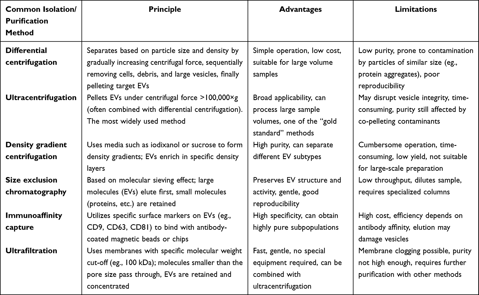 |
Table 1 Common Methods for OEVs Isolation and Purification |
The Difference Between OEVs and EVs, PDEVs, BEVs
EVs were first classified as cellular byproducts based on their role in coagulation. However, this conception was revised after electron microscopic visualization of platelet-derived EVs in 1967, which offered seminal insight into their true nature.8 For these membrane-bound vesicles, Johnstone coined the term “exosomes” in 1987.9 Ambiguous definitions and divergent isolation criteria bred nomenclatural chaos in EV research. To curb the indiscriminate use of “exosome” the International Society for Extracellular Vesicles stipulated in 2018 that EVs <200 nm should be designated “small extracellular vesicles”.10 The Minimal Information for Studies of Extracellular Vesicles (MISEV) standards were further refined in 2023, resulting in a more accurate characterization of EVs.4 Standardized characterization based on physical characteristics, biological makeup, and cellular origin was emphasized in this concept. EVs are described as particles that are expelled from cells, have a lipid bilayer, and cannot replicate themselves. Exosomes and other vesicular particles are included in this phrase. In this paper, unless otherwise specified, “EVs” refer to populations of nanoscale vesicles secreted by monolayer cells cultured in conventional adherent or suspension cultures, exhibiting a typical phospholipid bilayer structure. EVs from various biological sources, such as those released by 2D-cultured live cells, plant-derived extracellular vesicles (PDEVs), bacterial extracellular vesicles (BEVs), and OEVs, are the subject of current research (Table 2).
 |
Table 2 Comparison of the Sources, Characteristics, Applications and Limitations of Various EVs |
PDEVs come from a variety of sources. According to recent studies, several PDEVs have therapeutic value as drug delivery vehicles because of their high biocompatibility and minimal immunogenicity, as well as their antioxidant and anti-inflammatory effects.13 PDEVs have been shown to be devoid of viruses, MHC antigens, and human or animal diseases. They have been demonstrated to have low immunogenicity and little chance of causing human immunological rejection.14 Because of their extensive sourcing potential and affordable production,15 PDEVs are a topic of great interest. Strong biological qualities, such as anti-inflammatory, anti-cancer, antibacterial, antifungal, and antioxidant effects, have been shown by those produced from a variety of edible plants.16 Additionally, it has been demonstrated that PDEVs load medications well, allowing for precise administration.
The Gram-negative bacterium was originally seen in 1965 releasing outer membrane vesicles that were between 20 and 250 nanometers in size through budding.17 These vesicles serve as “decoys” to thwart phages or host-derived antimicrobial agents by carrying pathogen-associated genetic sequences. Additionally, a 2013 study found that outer-inner membrane vesicles, which are essential for pathogen stress tolerance, can be secreted by Gram-negative bacteria.18 Researchers can accurately alter the characteristics and antigen presentation mechanisms of BEVs through genetic engineering, boosting immune responses and showing great promise for vaccine development.19
It has been shown that using stereoscopic culture can improve intercellular communication, expand the area of contact between cells, and better preserve the physiological characteristics of cells.20 It has been shown that creating organoids from various cell subtypes produces extracellular vesicles with richer components and more complete architectures.21 OEVs have greater diversity and tissue specificity in proteins, RNAs, and metabolites than EVs made exclusively from single cell lines.22 Superior cellular absorption efficiency and functional activity, such as encouraging tissue regeneration, reducing inflammation, or boosting anticancer activities, are demonstrated in vitro.23 OEVs are more similar to naturally occurring vesicles in living organisms, enabling precise delivery of bioactive compounds and facilitating interactions between them. It is widely acknowledged that 3D culture systems have a significant impact on the biosynthesis and secretion characteristics of EVs. In 3D culture, the yield of EVs from mesenchymal stem cells is approximately twice that obtained from 2D monolayer culture.24 In gastric cancer cell models, 3D culture not only alters secretion flux but also results in a widespread upregulation of the miRNA profile carried by EVs, while protein components are correspondingly downregulated.25 Vesicles derived from 3D culture are more similar in molecular composition to their in vivo physiological state.26 Changes in their secretion kinetics and the abundance of key signaling molecules suggest that the flat growth pattern of 2D monolayers may deviate from the cells’ natural secretory state. Currently, significant differences remain among studies regarding culture system types, cell sources, detection time points, and isolation and purification methods, which to some extent limit the comparability of results and the generalizability of conclusions (Table 3).
 |
Table 3 Characterization of the Effects of Different Cultivation Modes on the Yield, Molecular Composition, and Function of EVs |
In addition, this organoid platform resolves the bottlenecks of low yield and significant batch-to-batch variability inherent in conventional EVs manufacturing processes, supporting the standardization of large-scale EVs production. As a result, it accelerates the pathway from laboratory research to clinical application (Figure 2).
 |
Figure 2 OEVs, PDEVs, EVs, BEVs generation diagram. |
Functional Roles in Physiology and Disease
Mediating Neuroprotection Through OEVs
Several studies have documented that mesenchymal stem cells derived from human umbilical cord mesenchymal stem cells (hucMSCs) possess neuroprotective properties.27,28 HucMSC-derived EVs have been shown to cross the blood-brain barrier, elevate substantia nigra dopamine levels, and attenuate neuronal apoptosis in Parkinson’s models,29 brain OEVs also exhibit similar neuroprotective properties.30 In particular, it has been demonstrated that these OEVs lessen apoptosis and oxidative stress in brain astrocytes. Damage to brain tissue from an ischemic stroke results in serious neurological impairments. Utilizing isolated OEVs with human induced pluripotent stem cells (hiPSCs) to generate brain organoids, a recent study further demonstrated that these OEVs conferred significant protection in an ischemic stroke mouse model.31 The observed benefits notably comprised the modulation of autophagy, clearance of reactive oxygen species, and reduction of inflammatory responses. When taken as a whole, these findings show how OEVs have neuroprotective effects and provide new information about how to treat neurodegenerative illnesses.
When studying brain OEVs, it is crucial to focus on whether the miRNA composition carried by OEVs undergoes alteration.32 For instance, miR-133b carried by mesenchymal extracellular vesicles can bind to abnormally expressed gene mRNAs and inhibit their translation, thereby mitigating neuronal damage. Notably, endogenous exosomes secreted by neurons and glial cells may also carry pathological α-synuclein, potentially exacerbating neurodegenerative processes.33 It also means that not all extracellular vesicles produce favorable impacts on disease outcomes, and their precise processes deserve additional exploration.
The Tissue Repair Function of OEVs
Researchers are investigating cell-free therapeutics for tissue repair, in which injured areas can be precisely targeted by bioactive molecules released by cells, hence increasing therapeutic efficacy.34 Compared to EVs derived from embryonic stem cells, human retinal OEVs contain a richer array of immune-regulatory and retinal development-related proteins. They actively modulate fatty acid metabolism, effectively clearing abnormal lipid deposits and exerting antioxidant functions.35 Further studies indicate that these human retinal OEVs can specifically target and inhibit the MAPK signaling pathway in retinal cells-particularly by downregulating the phosphorylation levels of p38 MAPK and JNK-thereby alleviating oxidative stress and inflammatory responses.36 This ultimately delays photoreceptor cell apoptosis and slows the progression of retinal degenerative diseases.
While Focusing on the salivary gland as an exocrine model, Ferreira et al37 employed a magnetic 3D bioassembly platform to co-culture its organoids with human dental pulp stem cells, followed by salivary gland OEVs isolation from the conditioned medium. The salivary gland OEVs demonstrated improved tissue repair capabilities by successfully stimulating neuronal growth, epithelial progenitor cell proliferation, and epithelial expansion (up to 60%) in injured salivary glands. This method has broad application potential for common epithelial injuries like burns and radiation damage, even if it is currently only seen in damaged salivary glands. Long-term radiation exposure and aging are examples of chronic injury conditions that current models based on acute injury are unable to accurately replicate. It is still unclear whether salivary gland OEVs will be stable and effective in the long run in such circumstances. Clarifying their mechanisms of action and creating individualized treatments should be the main goals of future study.
Unlike traditional anti-adhesion strategies that rely solely on physical barriers, OEV-loaded superlubricant nanoskin reshapes the cytomatrical interaction network in the postoperative microenvironment through the synergistic action of physical barriers and biological regulation, thereby preventing adhesions while promoting physiological tissue repair.38 By leveraging the natural nanoscale dimensions and surface molecular characteristics of OEVs, functional modification is achieved without compromising the mechanical properties of the superlubricant coating. Combining OEVs with nanostructured superlubricant nanoscale skin forms an innovative strategy that integrates “physical adhesion prevention with biological repair promotion.”
Exploration of OEVs in the Treatment of Diabetic Non-Healing Wounds
In the pathological state of diabetes, the hyperglycemic environment severely impedes angiogenesis by inhibiting the HIF-1α/VEGF signaling pathway and disrupting endothelial cell function, thereby becoming a major bottleneck in wound healing. VEGF and pro-angiogenic miRNAs enriched in OEVs can effectively restart the inhibited angiogenesis program.39 Furthermore, the antioxidant enzymes and anti-inflammatory factors they carry can scavenge reactive oxygen species (ROS) and reduce local inflammatory burden, thereby breaking the vicious cycle of “hyperglycemia-inflammation-hypoxia” in diabetic wounds.
Patients with diabetes frequently have poor wound healing, which can result in chronic ulcers and, in extreme situations, limb amputation.40 A safer and more economical approach to treating diabetic wound healing is being investigated by researchers. Epidermal organoids (EpiOs) have become a cutting-edge model system for studying the physiological foundations of skin function and illness. In addition to having a population of differentiable epidermal stem cells, EpiOs share structural similarities with the epidermis. It was reported that epidermal OEVs, carrying substantial quantities of miRNAs and VEGF, promoted cell migration, proliferation, and angiogenic processes.41 In individuals with diabetes, the hyperglycemic milieu inhibits angiogenesis, which prevents wound healing.
A production system using bioreactors to generate EVs derived from human adipose can reduce levels of inflammatory cytokines, stimulate collagen production, and activate the proliferation and migration of keratinocytes and fibroblasts.42 In in vivo experiments, this approach promoted wound closure in excision wound models, increased keratinization and collagen deposition, and improved overall healing rates. In a clinical case study, complete wound closure and dermal regeneration were achieved following local treatment.
Anti-Inflammatory Effects of OEVs
Intestinal homeostasis and bone metabolism are closely linked. Inflammatory bowel disease (IBD) causes a systemic inflammatory environment to develop, leading to overactivity of proinflammatory factors such as TNF-α and IL-17, which stimulate bone-resorbing cells.43 It disrupts immune homeostasis, resulting in bone loss. Existing treatments focus primarily on calcium supplementation and the administration of antiresorptive agents, often neglecting causal therapy in favor of symptom management. Su et al44 stimulated intestinal organoids with lipopolysaccharides to mimic the inflammatory microenvironment in IBD. Intestinal OEVs directly target immune cells and suppress upstream signals of osteoclast activation at the source while improving intestinal inflammation and bone loss. Intestinal OEVs therapy not only functions as a bone-targeted drug delivery system but also reconstructs the inflammatory microenvironment. By targeting the root cause of IBD inflammation, it prevents lasting skeletal damage and achieves causal treatment. Disease modeling using organoids from patients and tailored production of OEVs opens new avenues for the treatment of comorbidities.
A sustained-release technology was developed by mixing GelMA hydrogels with salivary gland OEVs.45 Experiments demonstrated that GelMA hydrogels loaded with these salivary gland OEVs significantly enhanced angiogenesis, cell migration, proliferation, and collagen synthesis, effectively promoting wound healing. This technology also modulates immune homeostasis by inducing macrophage polarization toward the M2 phenotype, thereby reducing inflammatory responses. Numerous illnesses, such as tumors,46 autoimmune disorders,47 atherosclerosis,48 and metabolic diseases,49 are believed to be associated with macrophage polarization. During macrophage polarization, 2D-EVs exhibit an inhibitory effect on the proinflammatory cytokine IL-12β, whereas 3D-EVs demonstrate an enhancing effect on the anti-inflammatory cytokine IL-10.50 Macrophage-derived EVs can directly regulate the activation status and effector functions of innate lymphoid cells, reshaping the network of immune cell interactions within the inflammatory microenvironment by delivering specific miRNAs and immunoregulatory proteins.51 Future studies should investigate whether OEVs enhance the expression of anti-inflammatory-related miRNAs to achieve anti-inflammatory effects (Figure 3 and Table 4).
 |
Table 4 Summary of Therapeutic Effects of OEVs in Different Disease Models |
 |
Figure 3 OEVs therapeutic effects and functional roles in physiology and disease. |
Applications in Disease Modeling
OEVs Promote Tumor Organoid Growth
The dynamic balance of the tumor microenvironment depends not only on the regulation by immune cells and peripheral factors, but also on the tumor’s own active remodeling of the microenvironment through the release of mediators such as EVs. As a key mechanism, this process cannot be overlooked. In the ovarian cancer microenvironment, tumor associated macrophages, regulatory T cells, and tumor cells are involved in a relationship of mutual inhibition, and the peripheral microenvironment can also attenuate the efficacy of immunotherapy through the secretion of factors.52 Neither the tumor, the immune system, nor the peripheral microenvironment exists in isolation, but rather they are in a dynamic, mutually shaping state.
One technique for producing tumor organoids is currently building in vitro tumor tissues from patient derived tumor specimens.53 An organoid library based on biopsy specimens from gastrointestinal tumor metastases was effectively established as early as 2018 by a London based research team.54 They demonstrated the clinical translational utility of patient-derived organ tissues for pharmacological efficacy assessment and individualized precision treatment by performing assessments of chemotherapy and targeted drug sensitivity.55 Meanwhile, using tumor organoid models, scientists found that these tumor OEVs can decrease immune cell activity, promote angiogenesis, and transform normal fibroblasts into cancer associated fibroblasts.56 In order to facilitate tumor growth and metastasis, this mechanism modifies the tumor microenvironment.57 Co-culturing extracellular vesicles generated from esophageal adenocarcinoma with stomach organoids increases the expression of cancer-associated miRNAs (such as miR-21 and miR-210) and triggers phenotypic alterations that impact the growth of co-cultured organoids.58 Recent studies indicate that tumor OEVs enhance organoid proliferation capacity and elevate levels of cancer associated microRNAs.59
OEVs Promote Organoid Vascularization
Building functioning vascular networks makes it easier to supply nutrition and oxygen to tumor organoids, providing a deeper comprehension of the mechanisms behind tumor spread.60 A patient-derived tumor organoid chip with vascularization and tumor microvasculature that efficiently evaluates tumor spread has been created by a research team.61 An emphasis on customized treatment strategies is replacing a tumor-type-centered strategy in cancer treatment, marking a paradigm shift.62
EVs from adipose tissue reprogram static culture environments into pro-angiogenic microenvironments by delivering key bioactive molecules.63 This microenvironmental transformation not only supports adipocyte precursor cell differentiation but also specifically activates the angiogenesis program, thereby synchronously enabling adipose tissue formation and vascular network construction within organoids.64 This discovery offers novel therapeutic strategies for chronic vascularized wounds and burn wound healing (Figure 4).
 |
Figure 4 The Role of OEVs in vitro model construction. |
Therapeutic Applications
OEVs as Highly Efficient Carriers for Targeted Drug Delivery
Through engineering modifications, drug molecules are loaded into the interior of OEVs. The integrins on the surface of OEVs specifically recognize fibronectin or vitellin ligands on the target cell membrane, triggering a cascade of adhesion signals. Transmembrane protein molecules abundant on the surface of OEVs promote the adhesion and fusion of OEVs with the target cell membrane. After the phospholipid bilayer of the OEVs fuses with the cell membrane, the drug is transported into the recipient cell via laminin-mediated endocytosis and laminin-dependent endocytosis. The membrane-fusion-mediated direct cytoplasmic delivery pathway avoids the risk of drug degradation by lysosomes, making it suitable for nucleic acid drugs and protein-based therapeutics that are susceptible to degradation in acidic environments.
Selective penetration into tumor tissues is made possible by OEVs with nanoscale diameters, which improve the enhanced permeability and Retention effect.65 OEVs are great drug delivery vehicles because they resemble cell membranes in structure and have high biocompatibility and permeability. Jia et al66 created organoids that resembled the original tumors by cultivating patient tumor tissues in vitro. They loaded medications into OEVs made from these organoids using microfluidic technology, exhibiting excellent targeting and great efficiency. Using the Folch method to treat endosomes produced from melanoma, their contents were removed, leaving only membrane components with targeting activities.67 An artificial endosome delivery system was then successfully built by assembling them onto the surface of organosilicon nanoparticles. This biomimetic system exhibited preferential enrichment of tumor tissues inside animal models. Endosomal membranes are naturally able to target the lungs. Doxorubicin-loaded nanocages efficiently accumulated at the tumor site, greatly reducing the growth of the original tumor and the spread of its metastases.
Based on a multi-site Fc-conjugation strategy, OEVs can serve as multifunctional platforms capable of simultaneously carrying chemical drugs, nucleic acid therapeutics, and photosensitizers.68 By linking different targeting molecules via Fc fragments, synergistic targeting of multiple receptors can be achieved. Fc-fused ligands can be efficiently anchored to the surface of OEVs via chemically or biologically orthogonal reactions, enabling stable, directed multi-site conjugation using native or recombinant Fc receptors. This strategy overcomes the limitations of traditional single-site modification, such as low ligand density and uneven distribution significantly enhancing tumor cell recognition and uptake, and improving targeted delivery efficiency. Building on this foundation, we further incorporated the principles of liquid-liquid phase separation to design a novel drug delivery system.69 By developing stimulus-responsive drug carriers, we achieved precise temporal and spatial control over the delivered drugs, thereby optimizing the therapeutic efficacy and safety of OEVs.
Combining vesicles from different sources to integrate their respective functional advantages is one of the key directions in the development of vesicular delivery systems. Through genetic engineering, we have engineered mixed vesicles derived from mesenchymal stem cells that can efficiently scavenge ROS and restore mitochondrial homeostasis, demonstrating significant protective effects in a model of hepatic ischemia-reperfusion injury. Delivery systems containing multiple active components enhance their synergistic therapeutic effects in complex pathological microenvironments.70
Engineering Strategies for OEVs
Natural OEVs have short half-lives and a limited capacity to carry drugs. The clinical translation of OEV-based medicines can be further advanced by molecular engineering of natural OEVs to obtain more accurate and effective treatment.71 Engineered OEVs improve targeting, maximize loading efficiency, facilitate effective nanomedicine administration, and extend circulation time in vivo.72 However, a thorough assessment is necessary for the long-term safety of modified OEVs.73
Engineering modifications of OEVs primarily fall into two categories: direct modification after isolation and indirect regulation via organoid modification. Direct engineering involves chemically or physically modifying OEVs isolated from organoid culture supernatants to enable them to carry specific functional molecules.74 For example, leveraging the transmembrane properties of membrane-penetrating peptides, small-molecule drugs or siRNA can be passively loaded into the OEV lumen through co-incubation. Click chemistry’s high efficiency and specificity enable the targeted peptide to be bound to the membrane surface of the OEV, thereby conferring functions such as targeting tumor angiogenesis.75 In contrast, electroporation technology uses intense electrical pulses to create microscopic pores in the membrane, allowing hydrophilic drugs or nucleic acids to easily penetrate into the vesicle.76 Such direct modification methods are relatively simple to implement and allow for controllable technical design. Their disadvantages, however, include unstable modification efficiency, difficulty in ensuring OEV uniformity, and the risk of damaging the vesicle surface structure, which can adversely affect its function.
Indirect engineering involves genetically or expression-regulating source cells within organoids to naturally secrete OEVs carrying specific molecules or functions.5 For instance, gene editing techniques can be used to knock out or introduce point mutations in target genes, or plasmid transfection and lentiviral packaging can be employed to overexpress fusion proteins combining transmembrane proteins with targeting peptides.77 This enables secreted OEVs to stably display targeting ligands on their surfaces, achieving precise delivery. Although these methods involve high technical barriers and extended timelines, the engineered OEVs obtained typically exhibit greater uniformity and stability, with more reliable functional performance (Figure 5).
 |
Figure 5 Engineered construction method for OEVs. |
The maturation stages of several brain areas, including the forebrain and hindbrain, can be replicated in vitro using human brain organoids. Mature tissue architectures can be produced by utilizing hiPSC to create forebrain and hindbrain organoids, respectively, and cultivating them for 30 to 45 days.31 Different brain regions produce matrix-binding vesicles (MBVs), which transport region-specific signaling chemicals and control the destiny of recipient cells.78 After ischemia-reperfusion injury, forebrain MBVs enriched with WNT3A further promote neuronal axon regeneration, while hindbrain MBVs containing SHH show more marked suppression of astrocyte proliferation. In an in vitro ischemia-reperfusion paradigm, knocking in the WNT3A-Lamp2b fusion gene in human-derived brain organoids naturally enriched WNT3A in OEVs, greatly improving neuronal survival. Nevertheless, in gene modification strategies, the overexpression of exogenous fusion genes may impose a significant metabolic and biosynthetic burden on the organoids themselves, such as increased oxidative stress, elevated endoplasmic reticulum stress, and energy metabolism dysregulation. Subsequently, this may affect the viability and proliferative capacity of the parental organoids and potentially reduce the yield and functional homogeneity of OEVs (Table 5).
 |
Table 5 Engineering Strategies for OEVs and Their Applications |
The Potential of OEVs Immunomodulatory Functions in Regenerative Medicine
Early studies have demonstrated that extracellular vesicles, owing to their low immunogenicity and immunomodulatory properties, can effectively improve graft survival rates. Concurrently, organoids, with their high physiological relevance, have been attempted for direct transplantation into patients with advanced diseases, showing tremendous potential for clinical translation.79 In a rhesus monkey model of chronic heart failure, engineered cardiac tissue derived from hiPSCs achieved six-month graft survival and significantly improved ventricular systolic function.80 The left ventricular ejection fraction of a patient with severe heart failure then rose from 35% to 39% following surgery in the first human clinical trial. Patients with retinitis pigmentosa were given retinal organoids made from hiPSCs.81 Postoperative observation revealed no adverse reactions or complications in any patient. After two years of follow-up, patients integrated safely and stably, and their sensitivity to light stimuli improved significantly. No evidence of immune rejection was observed, providing initial proof of the high safety and feasibility of temporary organ transplants.
However, the mechanism by which organoids secrete vesicles and exert their effects while “living” as grafts within recipients remains unknown.82 At the level of immune regulation, exosome-mimetic nanovesicles exert a significant regulatory effect on CD4+ T cells. These vesicles may disrupt the balance between pro-inflammatory and anti-inflammatory responses in immune cells, as evidenced by a significant increase in pro-inflammatory cytokines and a significant decrease in anti-inflammatory cytokines, providing important insights into the potential regulatory role of OEVs in the immune microenvironment.83 We hypothesize that these OEVs, generated in situ by transplanted organoids, may exert critical immunomodulatory effects locally and systemically through active intercellular communication. This could reduce rejection risks and promote long-term survival. Unraveling the post-transplant functions and mechanisms of OEVs represents a crucial frontier for advancing organoid transplantation technology and achieving breakthroughs in regenerative medicine.
Cell-Free Therapy Based on OEVs
Transplanting intact, viable cells into the body to replace damaged cells, regulate the immune system, and carry out other tasks is known as traditional cell therapy. Mesenchymal stem cell transplantation for the control of autoimmune diseases,84 hematopoietic stem cell transplantation for the treatment of hematological disorders,85 and the use of immune cells for anticancer therapy are other examples.86 However, this strategy involves the risk of unchecked proliferation that results in cancer when applied to stem cells. Additionally, the patient’s immune system may identify transplanted cells from allogeneic origins as “foreign,” leading to immunological rejection reactions.87 Furthermore, excessive immune cell activation might trigger severe inflammatory reactions that could be fatal. While eliminating the dangers of using live cells, cell-free therapies based on extracellular vesicles,88 apoptotic bodies, or released cellular substances can maintain the therapeutic advantages of conventional cell therapies.89
OEVs do not have the ability to proliferate, carry a variety of bioactive compounds released by organoids, and are not carcinogenic. They are less immunogenic than living cells after purification, which lowers the possibility of immunological rejection.90 Additionally, organoids can function as stable “production workshops” for extracellular vesicles, making it possible to produce stable and consistent vesicles in large quantities.6 It should be noted that the carcinogenic substances carried by tumor OEVs theoretically pose a potential risk of inducing malignant transformation in recipient cells, this limitation should be fully taken into account in safety assessments. Standardized organoid construction protocols, unified purification processes, systematic evaluation of long-term safety, and in vivo/in vitro tracking to comprehend pharmacokinetics and biodistribution are all necessary to move OEVs toward clinical application as therapeutics.
In vitro Antimicrobial Susceptibility Prediction Tool
Extracellular vesicles released by drug-resistant cells have been shown in cellular models to transfer membrane proteins or miRNA to sensitive cells, giving the latter resistance to chemotherapy or targeted therapy.91,92 In order to assess how hypoxia-induced extracellular vesicles containing PKM2 increase cisplatin resistance in non-small cell lung cancer (NSCLC) cells, Huang et al93 subjected NSCLC cells to hypoxic conditions. They discovered that PKM2 increases glycolysis in NSCLC cells, producing reductive metabolites to counteract reactive oxygen species caused by cisplatin and inhibiting apoptosis through the PKM2-BCL2 pathway. Simultaneously, it reprograms cancer-associated fibroblasts to produce an acidic milieu, which eventually promotes NSCLC cell proliferation and cisplatin resistance in a combinatorial manner.
Additionally, there are no reports of resistance-associated molecules (such as particular miRNAs or drug efflux pump proteins) being secreted by organoid-derived extracellular vesicles and transferred to sensitive cells, thereby inducing population-wide resistance,94 despite the fact that chemotherapy/targeted drug resistance models have been established using tumor organoids.95,96
Development of Disease Diagnostic Biomarkers
The clinical traits, genetic mutations, and heterogeneity of the original tissue are preserved in organoids made from patient-derived cells.97 Compared to those from cell lines, the extracellular vesicles released by these organoids are more clinically relevant and more closely represent the actual condition of sick cells. OEVs function not only as carriers of bioactive molecules in intercellular communication but also as potential mediators of disease pathology.98 The specific markers they carry are closely associated with the mechanisms driving disease initiation and progression. By employing a “modular structure” strategy to prepare nanoenzymes with artificial substrate binding sites and using them to construct a visual sensing platform for L-dopa, the core concepts in the fields of biosensing and diagnostics share an intrinsic connection with OEV-based biomarker discovery.99
After constructing disease-related organoids, machine learning, artificial intelligence, or multi-omics fusion models can process large volumes of OEVs data.100 By automatically extracting and ranking large datasets of membrane or intracellular proteins highly or lowly expressed in OEVs along with their significance, these approaches uncover hidden patterns and correlations within the data, enabling more accurate identification of potential disease-related biomarkers, prediction of therapeutic responses, and optimization of OEVs engineering.101 When hiPSCs with different genetic origins were differentiated into basal forebrain cholinergic neurons, different subtypes of disease-associated exosomal Aβ42 oligomers or tau proteins showed different conformations or contents. Precise patient subtyping may be made possible by these variations102,103 (Figure 6).
 |
Figure 6 Advantages and limitations of OEVs for therapeutic applications. |
Challenges and Limitations
Research on OEVs is currently in the exploratory phase, spanning from their biological mechanisms to clinical translation prospects. There are substantial research gaps concerning their impact on organoid formation as well as their therapeutic implications on illnesses. First, it’s still unknown how exactly OEVs regulate disease. How precisely do OEVs alter the immune microenvironment in tumor models? How are target cells regulated by the particular miRNAs or proteins that OEVs carry? OEVs play a key mediating role in organoid self-renewal and maturation; however, the exact molecular mechanisms and regulatory networks underlying organoid morphogenesis, cell differentiation, and functional maturation remain unclear and require further investigation.
The current literature lacks rigorous head-to-head comparisons between OEVs and conventional EVs, specifically studies that compare the therapeutic activity and batch consistency of OEVs with those derived from 2D cultures or conventional 3D spheroids under uniform isolation and quantification standards within a single controlled experiment. Most existing evidence stems from cross-sectional comparisons across different laboratories, cell lines, and isolation methods, which may introduce systematic bias.
Various organoid culture methods exist, and different approaches have varying effects on the generation and molecular composition of OEVs. Matrix-embedding techniques often involve the addition of scaffolds, such as biohydrogels, during organoid culture to provide three-dimensional support and biological signals, thereby promoting the self-organization of organoids and protecting cells from damage caused by external mechanical shear forces. However, scaffold materials such as hydrogels share similarities with OEVs in terms of particle size distribution, surface charge, and protein composition. Conventional methods—such as differential centrifugation, size-exclusion chromatography, or polymer precipitation—struggle to effectively separate OEVs from degraded gel fragments, leading to reduced extraction purity and interference from false-positive signals. Researchers have attempted to use enzymatic digestion to release embedded organoids from the gel; however, enzymatic treatment may compromise the membrane integrity of OEVs or introduce exogenous vesicular contaminants. Furthermore, additional purification steps are required after enzyme removal, resulting in a significant reduction in sample recovery. The matrix-free suspension culture method utilizes the self-aggregation properties of cells to form self-organized organoids in low-adhesion microplates. Although this method can improve the extraction efficiency and purity of OEVs, the mechanical shear forces generated during organoid culture can affect the mechanically sensitive signals of the organoids. Furthermore, the total cell count in a single culture system is lower than that in the matrix-embedded method, often resulting in lower OEV yields. To address challenges such as high-throughput screening, complex structure construction, and the preservation of cellular components, and to enable large-scale production that meets clinical needs, diverse culture strategies have been developed. Introduction of bioreactors has provided a new platform for the large-scale production of OEVs. When human vascular organoids were cultured in vertical rotary bioreactors under low shear conditions of 40 rpm, EV yield increased by 2–3 times compared to the static control group, and the particles were comparable to those from static culture in terms of particle size, marker expression, and in vitro therapeutic activity.104 Hydrodynamic bioreactor systems stimulate stem cell spheroids through fluid shear forces generated by alternating flow patterns, resulting in a significant 10–20 fold increase in EV yield compared to static culture.105 Currently, an automated organoid preparation system based on microfluidic technology has been established and has been used to prepare human hepatocellular carcinoma organoids, liver organoids, and colorectal cancer organoids.106 By reducing heterogeneity within the organoids, this method demonstrates significant advantages in terms of morphological consistency, transcriptomic fidelity, and reproducibility of cellular composition. Microfluidic chip technology integrates perfusion channels and cell culture chambers on micron-scale chips, precisely controlling liquid and fluid shear stress conditions. It enables the preparation of microspheres with uniform particle size and controllable structure, thereby influencing the number, size distribution, and content loading of OEVs, and consequently improving the consistency and functionality of OEVs in drug delivery and disease model construction. Multi-channel parallel designs enable high-throughput screening and analysis; however, the high manufacturing costs of microfluidic chips make large-scale, low-cost production challenging. 3D printing technology allows for precise control of organoid size, morphology, and internal 3D microenvironment by pre-setting cell numbers, spatial distribution, and the composition of the scaffold matrix. By printing organoids with specific surface modifications or functions, OEVs can be engineered to express specific ligands or receptors on their surfaces, thereby enhancing targeting. However, the scaffold matrix and bio-ink can affect the efficiency of OEV extraction and purification. The limitations of individual technologies have led researchers to seek solutions through the integration of multiple technologies.107 3D-printed organoids can be embedded within microfluidic chip chambers, which, by optimizing the organoid microenvironment, can significantly improve the homogeneity, yield, and functional targeting of OEVs. With innovations in materials and processes, it is expected that low-cost, high-purity, large-scale production will be achieved, propelling OEVs from laboratory research toward standardized clinical biotherapeutic products.
Although OEVs demonstrate tremendous translational potential in the field of precision medicine, their practical clinical application still faces multiple obstacles. The first challenge is the standardization of preparation processes, particularly regarding the maintenance of structural integrity and functional activity during storage and transportation. The lack of effective preservation protocols directly leads to increased batch-to-batch heterogeneity and diminished therapeutic efficacy, severely hindering their industrialization as biological products. The addition of human albumin and trehalose to PBS can significantly improve the stability of EVs during two years of storage at −80°C and increase recovery rates after repeated freeze-thaw cycles, providing a foundational protocol for OEV storage.108 Secondly, systematic in vivo safety data for OEVs remain limited.109 Current evidence indicates that, at the cellular level and in some animal models, OEVs have demonstrated good functional activity and low cytotoxicity, providing valuable references for subsequent research.110 However, to meet the safety requirements for clinical translation, more comprehensive animal studies will be needed in the future to systematically supplement and refine the evidence chain regarding the in vivo safety of OEVs.111
Conclusion and Perspectives
OEVs are derived directly from stem cells, which spontaneously assemble into organoids. Due to their multicellular nature, OEVs are better able to preserve the signaling networks of the organ microenvironment, and the biological information they convey exhibits stronger tissue polarity and organ specificity. OEVs demonstrate multifaceted functions and application potential in the biomedical field. At the fundamental mechanistic level, OEVs exhibit clear tissue repair and regenerative capabilities. By mediating neuroprotection, exerting anti-inflammatory effects, and regulating immune responses, they offer new intervention strategies for neurological diseases, chronic inflammation, and autoimmune-related damage. In disease treatment, OEVs have been explored for the management of diabetic non-healing wounds, accelerating tissue healing through synergistic anti-inflammatory and pro-angiogenic functions. Additionally, OEVs effectively promote vascularization and growth in disease models; however, it is worth noting that tumor OEVs also exert a certain promotional effect on tumor organ growth, suggesting that their bidirectional regulatory potential within the tumor microenvironment requires careful evaluation. At the translational application level, leveraging their inherent advantage of a natural multi-cellular signaling profile, OEVs have been developed as highly efficient carriers for targeted drug delivery to tumors. Combined with diverse engineering strategies, their targeting specificity, drug-loading capacity, and therapeutic efficacy can be significantly enhanced. In the diagnostic field, analyzing the contents of OEVs derived from Alzheimer’s disease organoid models, in conjunction with the expression profiles of specific neuronal markers, glial cells, and other brain cell types, can reveal complex biological signals transmitted through intercellular communication.112 As highly informative liquid biopsy biomarkers, the molecular characteristics of these pluripotent cells significantly improve the specificity and sensitivity of early-stage Alzheimer’s disease diagnosis.
As OEVs transition from basic research to clinical translation, systematic breakthroughs are urgently needed in several key areas. The primary task is to establish internationally recognized standards for the isolation and detection of OEVs, which is a fundamental prerequisite for ensuring the comparability and reproducibility of research results across different laboratories and batches. Building on this foundation, it is essential to systematically conduct OEV-based drug delivery research using animal models to comprehensively evaluate their in vivo efficacy, pharmacokinetic characteristics, and long-term safety, thereby providing robust preclinical evidence for subsequent clinical trials. Concurrently, by leveraging the deep integration of novel biomaterials and bioengineering technologies, an automated, scalable OEV production and quality control system compliant with Good Manufacturing Practice requirements must be established to fundamentally address the current industrialization bottlenecks of low yield and significant batch-to-batch variability.113 In the diagnostic field, the development of OEV-based liquid biopsy products will transform the rich multicellular signals they carry into clinically applicable early-stage, non-invasive diagnostic biomarkers, providing a novel tool for the early screening, classification, and dynamic monitoring of various major diseases. OEVs will transition from laboratory proof of concept to a new phase of standardization, automation, and clinical application, truly realizing their translational value in precision medicine.
Abbreviations
BEVs, Bacterial extracellular vesicles; EpiOs, Epidermal organoids; EVs, Extracellular vesicles; hiPSCs, Human induced pluripotent stem cells; hucMSCs, Human umbilical cord mesenchymal stem cells; IBD, Inflammatory bowel disease; MBVs, Matrix-binding vesicles; MISEV, Minimal information for studies of extracellular vesicles; NSCLC, Non-small cell lung cancer; OEVs, Organoid-derived extracellular vesicles; PDEVs, Plant-derived extracellular vesicles.
Acknowledgments
This work was supported by the National Natural Science Foundation of China (No. 82360049) (NO.82460096), NHC Key Laboratory of Healthy Birth and Birth Defect Prevention in Western China Open Project (2024XBYSKF001) (2024XBYSKF007), National Clinical Key Specialty Cultivation Project Platform for Endocrinology Open Project (2024NMKFKT-04). Thanks for biorender.
Disclosure
The authors declare that they have no known competing financial interests or personal relationships that could have appeared to influence the work reported in this paper.
References
1. Sato T, Vries RG, Snippert HJ, et al. Single Lgr5 stem cells build crypt-villus structures in vitro without a mesenchymal niche. Nature. 2009;459(7244):262–21. doi:10.1038/nature07935
2. Rauth S, Karmakar S, Batra SK, Ponnusamy MP. Recent advances in organoid development and applications in disease modeling. Biochim Biophys Acta Rev Cancer. 2021;1875(2):188527. doi:10.1016/j.bbcan.2021.188527
3. van Niel G, D’Angelo G, Raposo G. Shedding light on the cell biology of extracellular vesicles. Nat Rev Mol Cell Biol. 2018;19(4):213–228. doi:10.1038/nrm.2017.125
4. Welsh JA, Goberdhan DCI, O’Driscoll L, et al. Minimal information for studies of extracellular vesicles (MISEV2023): from basic to advanced approaches. J Extracell Vesicles. 2024;13(2):e12404. doi:10.1002/jev2.12404
5. Liu H, Su J. Organoid extracellular vesicle-based therapeutic strategies for bone therapy. Biomater Transl. 2023;4(4):199–212. doi:10.12336/biomatertransl.2023.04.002
6. Liu H, Cheng T, Li G, et al. Organoid-tissue extracellular vesicles. Interdisciplinary Medicine. 2025;3(5):6. doi:10.1002/inmd.20250006
7. Zhou G, Li R, Sheng S, et al. Organoids and organoid extracellular vesicles-based disease treatment strategies. J Nanobiotechnology. 2024;22(1):679. doi:10.1186/s12951-024-02917-3
8. Ma B, Li L, Bao Y, et al. Optical Imaging of Single Extracellular Vesicles: recent Progress and Prospects. Chem Biomed Imag. 2024;2(1):27–46. doi:10.1021/cbmi.3c00095
9. Johnstone RM, Adam M, Hammond JR, Orr L, Turbide C. Vesicle formation during reticulocyte maturation. Association of plasma membrane activities with released vesicles (exosomes). J Biol Chem. 1987;262(19):9412–9420.
10. Théry C, Witwer KW, Aikawa E, et al. Minimal information for studies of extracellular vesicles 2018 (MISEV2018): a position statement of the International Society for Extracellular Vesicles and update of the MISEV2014 guidelines. J Extracell Vesicles. 2018;7(1):1535750. doi:10.1080/20013078.2018.1535750
11. Liu X, Lou K, Zhang Y, Li C, Wei S, Feng S. Unlocking the Medicinal Potential of Plant-Derived Extracellular Vesicles: current Progress and Future Perspectives. Int J Nanomed. 2024;19:4877–4892. doi:10.2147/ijn.S463145
12. Wang Y, Luo X, Xiang X, Hao C, Ma D. Roles of bacterial extracellular vesicles in systemic diseases. Front Microbiol. 2023;14:1258860. doi:10.3389/fmicb.2023.1258860
13. Dad HA, Gu TW, Zhu AQ, Huang LQ, Peng LH. Plant Exosome-like Nanovesicles: emerging Therapeutics and Drug Delivery Nanoplatforms. Mol Ther. 2021;29(1):13–31. doi:10.1016/j.ymthe.2020.11.030
14. Zhu Y, Zhao J, Ding H, et al. Applications of plant-derived extracellular vesicles in medicine. Med Commun. 2024;5(10):e741. doi:10.1002/mco2.741
15. Xu Z, Xu Y, Zhang K, et al. Plant-derived extracellular vesicles (PDEVs) in nanomedicine for human disease and therapeutic modalities. J Nanobiotechnology. 2023;21(1):114. doi:10.1186/s12951-023-01858-7
16. Ning H, Huang X, Deng N, et al. Plant-Derived Exosome-Like Nanovesicles: a Novel Strategy for Targeted Oral Therapy in Ulcerative Colitis. Int J Nanomed. 2025;20:10595–10611. doi:10.2147/ijn.S536056
17. Bishop DG, Work E. An extracellular glycolipid produced by Escherichia coli grown under lysine-limiting conditions. Biochem J. 1965;96(2):567–576. doi:10.1042/bj0960567
18. Schetters STT, Jong WSP, Horrevorts SK, et al. Outer membrane vesicles engineered to express membrane-bound antigen program dendritic cells for cross-presentation to CD8(+) T cells. Acta Biomater. 2019;91:248–257. doi:10.1016/j.actbio.2019.04.033
19. Garling A, Auvray F, Epardaud M, Oswald É, Branchu P. Outer Membrane Vesicles as a Versatile Platform for Vaccine Development: engineering Strategies, Applications and Challenges. J Extracell Vesicles. 2025;14(9):e70150. doi:10.1002/jev2.70150
20. Zhou Y, Yu F, Guo M, Tang Y, Xu Q. Bridging the gap: the role of 3D cell cultures in mimicking tumor microenvironment for enhanced drug testing accuracy. Front Bioeng Biotechnol. 2025;13:1498141. doi:10.3389/fbioe.2025.1498141
21. Lee S, Kubota M, Park E, Heller S, Im GJ, Chang J. Analysis of miRNAs from Inner Ear Organoid-Derived Extracellular Vesicles. J Assoc Res Otolaryngol. 2025;26(4):397–408. doi:10.1007/s10162-025-00998-x
22. Lee S, Park S, Park E, Im GJ, Chang J. Identification of microRNA-Related Target Genes for the Development of Otic Organoids. Int J Mol Sci. 2025;26(21):110627. doi:10.3390/ijms262110627
23. Desai N, Katare P, Makwana V, Salave S, Vora LK, Giri J. Tumor-derived systems as novel biomedical tools-turning the enemy into an ally. Biomater Res. 2023;27(1):113. doi:10.1186/s40824-023-00445-z
24. Yuan X, Sun L, Jeske R, et al. Engineering extracellular vesicles by three-dimensional dynamic culture of human mesenchymal stem cells. J Extracell Vesicles. 2022;11(6):e12235. doi:10.1002/jev2.12235
25. Rocha S, Carvalho J, Oliveira P, et al. 3D Cellular Architecture Affects MicroRNA and Protein Cargo of Extracellular Vesicles. Adv Sci. 2019;6(4):1800948. doi:10.1002/advs.201800948
26. Chen JG, Zhang EC, Wan YY, Huang TY, Wang YC, Jiang HY. Engineered hsa-miR-455-3p-Abundant Extracellular Vesicles Derived from 3D-Cultured Adipose Mesenchymal Stem Cells for Tissue-Engineering Hyaline Cartilage Regeneration. Adv Healthc Mater. 2024;13(18):e2304194. doi:10.1002/adhm.202304194
27. Wang D, Clark K, Rouen KC, et al. Hybrid Extracellular Vesicles With Combined Functional Properties From Mesenchymal Stem Cells and Astrocytes for Targeted Neurodegenerative Disease Applications. J Extracell Vesicles. 2025;14(10):e70175. doi:10.1002/jev2.70175
28. Chen G, Tong K, Li S, et al. Extracellular vesicles released by transforming growth factor-beta 1-preconditional mesenchymal stem cells promote recovery in mice with spinal cord injury. Bioact Mater. 2024;35:135–149. doi:10.1016/j.bioactmat.2024.01.013
29. Chen HX, Liang FC, Gu P, et al. Exosomes derived from mesenchymal stem cells repair a Parkinson’s disease model by inducing autophagy. Cell Death Dis. 2020;11(4):288. doi:10.1038/s41419-020-2473-5
30. Ji X, Zhou S, Wang N, et al. Cerebral-Organoid-Derived Exosomes Alleviate Oxidative Stress and Promote LMX1A-Dependent Dopaminergic Differentiation. Int J Mol Sci. 2023;24(13):11048. doi:10.3390/ijms241311048
31. Liu C, Chen X, Ene J, et al. Engineering Extracellular Vesicles Secreted by Human Brain Organoids with Different Regional Identity. ACS Appl Mater Interfaces. 2025;17(10):15145–15162. doi:10.1021/acsami.4c22692
32. Zhu Y, Xi Q, Liu Y, Zhou Y, Liao J, Wu Q. Recent advances in exosome-based nanodelivery systems for Parkinson’s disease. Biomaterials. 2026;325:123548. doi:10.1016/j.biomaterials.2025.123548
33. de Bie RMA, Katzenschlager R, Swinnen B, et al. Update on Treatments for Parkinson’s Disease Motor Fluctuations - An International Parkinson and Movement Disorder Society Evidence-Based Medicine Review. Mov Disord. 2025;40(5):776–794. doi:10.1002/mds.30162
34. Marbán E. Deconstructing Regenerative Medicine: from Mechanistic Studies of Cell Therapy to Novel Bioinspired RNA Drugs. Circ Res. 2024;135(8):877–885. doi:10.1161/circresaha.124.323058
35. Gao H, Zeng Y, Huang X, et al. Extracellular vesicles from organoid-derived human retinal progenitor cells prevent lipid overload-induced retinal pigment epithelium injury by regulating fatty acid metabolism. J Extracell Vesicles. 2024;13(1):e12401. doi:10.1002/jev2.12401
36. Han JW, Chang HS, Yang JY, et al. Intravitreal Administration of Retinal Organoids-Derived Exosomes Alleviates Photoreceptor Degeneration in Royal College of Surgeons Rats by Targeting the Mitogen-Activated Protein Kinase Pathway. Int J Mol Sci. 2023;24(15):12068. doi:10.3390/ijms241512068
37. Chansaenroj A, Adine C, Charoenlappanit S, et al. Magnetic bioassembly platforms towards the generation of extracellular vesicles from human salivary gland functional organoids for epithelial repair. Bioact Mater. 2022;18:151–163. doi:10.1016/j.bioactmat.2022.02.007
38. Wang Y, Xu Y, Zhai W, et al. In-situ growth of robust superlubricated nano-skin on electrospun nanofibers for post-operative adhesion prevention. Nat Commun. 2022;13(1):5056. doi:10.1038/s41467-022-32804-0
39. Shi H, Han X, Lu Y, et al. Human Mesenchymal Stem Cell Derived Exosomes Endowed with miR-13474 as a Therapeutic Delivery Vehicle for Diabetic Wound Healing by Targeting the CPEB2/TWIST1 Axis. ACS Appl Bio Mater. 2025;8(11):10024–10037. doi:10.1021/acsabm.5c01352
40. Song J, Zhao T, Wang C, Sun X, Sun J, Zhang Z. Cell migration in diabetic wound healing: molecular mechanisms and therapeutic strategies (Review). Int J Mol Med. 2025;56(2):5567. doi:10.3892/ijmm.2025.5567
41. Kwak S, Song CL, Lee J, et al. Development of pluripotent stem cell-derived epidermal organoids that generate effective extracellular vesicles in skin regeneration. Biomaterials. 2024;307:122522. doi:10.1016/j.biomaterials.2024.122522
42. Gurney JP, Ludlow JW, Van Kanegan MJ, Smith RL. Modulation of ASC-derived extracellular vesicles containing cargo that specifically enhances wound healing. Front Pharmacol. 2025;16:1635151. doi:10.3389/fphar.2025.1635151
43. Wan J, Zhou J, Wang Z, et al. Epidemiology, pathogenesis, diagnosis, and treatment of inflammatory bowel disease: insights from the past two years. Chin Med J. 2025;138(7):763–776. doi:10.1097/cm9.0000000000003542
44. Wang M, Li R, Sheng S, et al. Combination therapy using intestinal organoids and their extracellular vesicles for inflammatory bowel disease complicated with osteoporosis. J Orthop Translat. 2025;53:26–36. doi:10.1016/j.jot.2025.05.008
45. Qian J, Lu E, Xiang H, et al. GelMA loaded with exosomes from human minor salivary gland organoids enhances wound healing by inducing macrophage polarization. J Nanobiotechnology. 2024;22(1):550. doi:10.1186/s12951-024-02811-y
46. Jin R, Neufeld L, McGaha TL. Linking macrophage metabolism to function in the tumor microenvironment. Nat Cancer. 2025;6(2):239–252. doi:10.1038/s43018-025-00909-2
47. Wu S, Zhao S, Hai L, et al. Macrophage polarization regulates the pathogenesis and progression of autoimmune diseases. Autoimmun Rev. 2025;24(7):103820. doi:10.1016/j.autrev.2025.103820
48. Wang X, Xie Z, Zhang J, et al. Interaction between lipid metabolism and macrophage polarization in atherosclerosis. iScience. 2025;28(4):112168. doi:10.1016/j.isci.2025.112168
49. Zhang QY, Zhang HY, Feng SG, et al. Macrophage metabolic reprogramming ameliorates diabetes-induced microvascular dysfunction. Redox Biol. 2025;79:103449. doi:10.1016/j.redox.2024.103449
50. Liu C, Chen X, Liu Y, et al. Engineering Extracellular Matrix-Bound Nanovesicles Secreted by Three-Dimensional Human Mesenchymal Stem Cells. Adv Healthc Mater. 2023;12(27):e2301112. doi:10.1002/adhm.202301112
51. Lv K, Zhang Y, Yin G, et al. Extracellular vesicles derived from lung M2 macrophages enhance group 2 innate lymphoid cells function in allergic airway inflammation. Exp Mol Med. 2025;57(6):1202–1215. doi:10.1038/s12276-025-01465-6
52. Wang ZB, Zhang X, Fang C, et al. Immunotherapy and the ovarian cancer microenvironment: exploring potential strategies for enhanced treatment efficacy. Immunology. 2024;173(1):14–32. doi:10.1111/imm.13793
53. Lee SH, Hu W, Matulay JT, et al. Tumor Evolution and Drug Response in Patient-Derived Organoid Models of Bladder Cancer. Cell. 2018;173(2):515–528.e17. doi:10.1016/j.cell.2018.03.017
54. Vlachogiannis G, Hedayat S, Vatsiou A, et al. Patient-derived organoids model treatment response of metastatic gastrointestinal cancers. Science. 2018;359(6378):920–926. doi:10.1126/science.aao2774
55. Panda SS, Sahoo RK, Patra SK, Biswal S, Biswal BK. Molecular insights to therapeutic in cancer: role of exosomes in tumor microenvironment, metastatic progression and drug resistance. Drug Discov Today. 2024;29(8):104061. doi:10.1016/j.drudis.2024.104061
56. Wang S, Shu J, Wang N, He Z. Exosomal non-coding RNAs: mediators of crosstalk between cancer and cancer stem cells. Cell Death Discov. 2025;11(1):434. doi:10.1038/s41420-025-02726-z
57. Peng T, Ma X, Hua W, et al. Individualized patient tumor organoids faithfully preserve human brain tumor ecosystems and predict patient response to therapy. Cell Stem Cell. 2025;32(4):652–669.e11. doi:10.1016/j.stem.2025.01.002
58. Yan R, Ke X, Cheng Y, Liu X, Wang Z, Meltzer SJ. Delivery of cancer cell-derived extracellular vesicles modulates the morphology and gene expression of Barrett esophagus and duodenal organoids (Article). Biochem Biophys Res Commun. 2025;769:151976. doi:10.1016/j.bbrc.2025.151976
59. Schuster M, Braun FK, Chiang DM, et al. Extracellular vesicles secreted by 3D tumor organoids are enriched for immune regulatory signaling biomolecules compared to conventional 2D glioblastoma cell systems. Front Immunol. 2024;15:1388769. doi:10.3389/fimmu.2024.1388769
60. Lee M, Yoon YJ, Lee JY, et al. Modeling the metastatic tumor microenvironment: a co-culture platform of lung cancer and blood vessel organoids for drug evaluation. Sci Rep. 2025;15(1):45252. doi:10.1038/s41598-025-29214-9
61. Du Y, Wang YR, Bao QY, et al. Personalized Vascularized Tumor Organoid-on-a-Chip for Tumor Metastasis and Therapeutic Targeting Assessment. Adv Mater. 2025;37(6):e2412815. doi:10.1002/adma.202412815
62. Kim SY, van de Wetering M, Clevers H, Sanders K. The future of tumor organoids in precision therapy. Trends Cancer. 2025;11(7):665–675. doi:10.1016/j.trecan.2025.03.005
63. Su H, Chau H, Li Q, et al. Bridging the gap: clinical translation of adipose-derived stem cells - a scoping review of clinical trials. Stem Cell Res Ther. 2025;16(1):288. doi:10.1186/s13287-025-04405-3
64. Zhu C, Huang Z, Zhou H, Han X, Li L, Yin N. Purified adipose tissue-derived extracellular vesicles facilitate adipose organoid vascularization through coordinating adipogenesis and angiogenesis. Biofabrication. 2025;17(2):e7. doi:10.1088/1758-5090/adb2e7
65. Komuro H, Aminova S, Lauro K, Woldring D, Harada M. Design and Evaluation of Engineered Extracellular Vesicle (EV)-Based Targeting for EGFR-Overexpressing Tumor Cells Using Monobody Display. Bioengineering. 2022;9(2):56. doi:10.3390/bioengineering9020056
66. Ge K, Ren Y, Hong Z, et al. Microchip Based Isolation and Drug Delivery of Patient-Derived Extracellular Vesicles Against Their Homologous Tumor. Adv Healthc Mater. 2024;13(30):e2401990. doi:10.1002/adhm.202401990
67. Sancho-Albero M, Decio A, Akpinar R, et al. Melanoma extracellular vesicles membrane coated nanoparticles as targeted delivery carriers for tumor and lungs. Mater Today Bio. 2025;30:101433. doi:10.1016/j.mtbio.2024.101433
68. Zheng Y, Cheng H, Jiang S, Tai W. Fc Multisite Conjugation and Prolonged Delivery of the Folate-Targeted Drug Conjugate EC140. Bioconjug Chem. 2025;36(4):762–769. doi:10.1021/acs.bioconjchem.5c00037
69. Xu W-X, Qu Q, Zhuang -H-H, et al. The Burgeoning Significance of Liquid-Liquid Phase Separation in the Pathogenesis and Therapeutics of Cancers. Review. Int J Bio Sci. 2024;20(5):1652–1668. doi:10.7150/ijbs.92988
70. Shen P, Huang K, Zhang X, et al. Genetically engineered MSC-derived hybrid cellular vesicles for ROS-scavenging and mitochondrial homeostasis in hepatic ischemia-reperfusion injury. Mater Today Bio. 2025;34:102215. doi:10.1016/j.mtbio.2025.102215
71. Mondal J, Pillarisetti S, Junnuthula V, et al. Hybrid exosomes, exosome-like nanovesicles and engineered exosomes for therapeutic applications. J Control Release. 2023;353:1127–1149. doi:10.1016/j.jconrel.2022.12.027
72. Zhao S, Di Y, Fan H, et al. Targeted delivery of extracellular vesicles: the mechanisms, techniques and therapeutic applications. Mol Biomed. 2024;5(1):60. doi:10.1186/s43556-024-00230-x
73. Escudé Martinez de Castilla P, Tong L, Huang C, et al. Extracellular vesicles as a drug delivery system: a systematic review of preclinical studies. Adv Drug Deliv Rev. 2021;175:113801. doi:10.1016/j.addr.2021.05.011
74. Salunkhe S, Dheeraj, Basak M, Chitkara D, Mittal A. Surface functionalization of exosomes for target-specific delivery and in vivo imaging & tracking: strategies and significance. J Control Release. 2020;326:599–614. doi:10.1016/j.jconrel.2020.07.042
75. Xing Y, Yerneni SS, Wang W, Taylor RE, Campbell PG, Ren X. Engineering pro-angiogenic biomaterials via chemoselective extracellular vesicle immobilization. Biomaterials. 2022;281:121357. doi:10.1016/j.biomaterials.2021.121357
76. Lennaárd AJ, Mamand DR, Wiklander RJ, El andaloussi S, Wiklander OPB. Optimised Electroporation for Loading of Extracellular Vesicles with Doxorubicin. Pharmaceutics. 2021;14(1):38. doi:10.3390/pharmaceutics14010038
77. Ji P, Yang Z, Li H, et al. Smart exosomes with lymph node homing and immune-amplifying capacities for enhanced immunotherapy of metastatic breast cancer. Mol Ther Nucleic Acids. 2021;26:987–996. doi:10.1016/j.omtn.2021.10.009
78. Kobayashi M, Negishi J, Ishida N, et al. Effects of the matrix-bounded nanovesicles of high-hydrostatic pressure decellularized tissues on neural regeneration. Sci Technol Adv Mater. 2024;25(1):2404380. doi:10.1080/14686996.2024.2404380
79. Xu W, Boer K, Hesselink DA, Baan CC. Extracellular Vesicles and Immune Activation in Solid Organ Transplantation: the Impact of Immunosuppression. BioDrugs. 2025;39(3):445–459. doi:10.1007/s40259-025-00713-5
80. Jebran A-F, Seidler T, Tiburcy M, et al. Engineered heart muscle allografts for heart repair in primates and humans. Nature. 2025;639(8054):503–511. doi:10.1038/s41586-024-08463-0
81. Zhang CJ, Jin ZB. Turning point of organoid transplantation: first-in-human trial of iPSC-derived retinal organoid grafting in patients with retinitis pigmentosa. Sci China Life Sci. 2024;67(5):1082–1084. doi:10.1007/s11427-023-2531-3
82. Quaglia M, Dellepiane S, Guglielmetti G, Merlotti G, Castellano G, Cantaluppi V. Extracellular Vesicles as Mediators of Cellular Crosstalk Between Immune System and Kidney Graft. Front Immunol. 2020;11:74. doi:10.3389/fimmu.2020.00074
83. Oztatlici M. Immunomodulatory Effects of MDA-MB-231-derived Exosome Mimetic Nanovesicles on CD4+ T Cell Line. Eurasian J Med Oncol. 2024;40–48. doi:10.14744/ejmo.2024.64067
84. An R, Zhu Z, Chen Y, Guan W, Wang J, Ren H. MSCs Suppress Macrophage Necroptosis and Foster Liver Regeneration by Modulating SP1/SK1 Axis in Treating Acute Severe Autoimmune Hepatitis. Adv Sci. 2025;12(12):e2408974. doi:10.1002/advs.202408974
85. Socie G. Challenges in GVHD and GVL after hematopoietic stem cell transplantation for myeloid malignancies. Blood. 2025;146(8):926–937. doi:10.1182/blood.2025028617
86. Lin NY, Fukuoka S, Koyama S, et al. Microbiota-driven antitumour immunity mediated by dendritic cell migration. Nature. 2025;644(8078):1058–1068. doi:10.1038/s41586-025-09249-8
87. Carlsson PO, Hu X, Scholz H, et al. Survival of Transplanted Allogeneic Beta Cells with No Immunosuppression. N Engl J Med. 2025;393(9):887–894. doi:10.1056/NEJMoa2503822
88. Lotfy A, AboQuella NM, Wang H. Mesenchymal stromal/stem cell (MSC)-derived exosomes in clinical trials. Stem Cell Res Ther. 2023;14(1):66. doi:10.1186/s13287-023-03287-7
89. Turturice BA, Fike A, Patel BA, et al. Disease Trajectories and Glucocorticoid Exposure in VEXAS Syndrome Treated with Cytokine-Directed Therapies. Ann Rheum Dis. 2025. doi:10.1016/j.ard.2025.05.021
90. Kalluri R, LeBleu VS. The biology, function, and biomedical applications of exosomes. Science. 2020;367(6478):6977. doi:10.1126/science.aau6977
91. Zhan Y, Zhou Z, Zhu Z, et al. Exosome-transmitted LUCAT1 promotes stemness transformation and chemoresistance in bladder cancer by binding to IGF2BP2. J Exp Clin Cancer Res. 2025;44(1):80. doi:10.1186/s13046-025-03330-w
92. Wu C, Sun X, Lu Y, et al. Cancer-associated fibroblast promotes tamoxifen resistance in estrogen receptor positive breast cancer via exosomal LncRNA PRKCQ-AS1/miR-200a-3p/MKP1 axis-mediated apoptosis suppression. J Exp Clin Cancer Res. 2025;44(1):274. doi:10.1186/s13046-025-03529-x
93. Wang D, Zhao C, Xu F, et al. Cisplatin-resistant NSCLC cells induced by hypoxia transmit resistance to sensitive cells through exosomal PKM2. Theranostics. 2021;11(6):2860–2875. doi:10.7150/thno.51797
94. Xu H, Jiao D, Liu A, Wu K. Tumor organoids: applications in cancer modeling and potentials in precision medicine. J Hematol Oncol. 2022;15(1):58. doi:10.1186/s13045-022-01278-4
95. Li K, Li W, Fu S, et al. Organoid models of drug resistant gastric adenosquamous carcinoma: recapitulating tumor features and refining precision treatment. Drug Resist Updat. 2025;79:101201. doi:10.1016/j.drup.2025.101201
96. LeSavage BL, Zhang D, Huerta-López C, et al. Engineered matrices reveal stiffness-mediated chemoresistance in patient-derived pancreatic cancer organoids. Nat Mater. 2024;23(8):1138–1149. doi:10.1038/s41563-024-01908-x
97. Abdal Dayem A, Kwak Y, Jeun H, Cho SG. Recent Insights into Organoid-Derived Extracellular Vesicles and Their Biomedical Applications. J Pers Med. 2025;15(10):492. doi:10.3390/jpm15100492
98. Pfeifer JPH, Stievani FC, Fernandes C, et al. Influence of inflammation on the expression of microRNA-140 in extracellular vesicles from 2D and 3D culture models of synovial-membrane-derived stem cells. Front Bioeng Biotechnol. 2024;12:1416694. doi:10.3389/fbioe.2024.1416694
99. Li T, Xiao L, Ling H, Yang Y, Zhong S. Preparation of artificial substrate binding sites of nanozyme with “modular structure” strategy used for the construction of visual sensing analysis platform for levodopa. Microchem J. 2025;212:113292. doi:10.1016/j.microc.2025.113292
100. Greening DW, Xu R, Rai A, Suwakulsiri W, Chen M, Simpson RJ. Clinical relevance of extracellular vesicles in cancer - therapeutic and diagnostic potential. Nat Rev Clin Oncol. 2025;22(12):924–952. doi:10.1038/s41571-025-01074-2
101. You Y, Lai X, Pan Y, et al. Artificial intelligence in cancer target identification and drug discovery. Signal Transduct Target Ther. 2022;7(1):156. doi:10.1038/s41392-022-00994-0
102. Tao R, Yue C, Guo Z, et al. Subtype-specific neurons from patient iPSCs display distinct neuropathological features of Alzheimer’s disease. Cell Regen. 2024;13(1):21. doi:10.1186/s13619-024-00204-y
103. Podvin S, Jones A, Liu Q, et al. Dysregulation of Exosome Cargo by Mutant Tau Expressed in Human-induced Pluripotent Stem Cell (iPSC) Neurons Revealed by Proteomics Analyses. Mol Cell Proteomics. 2020;19(6):1017–1034. doi:10.1074/mcp.RA120.002079
104. Ene J, Syed F, Ma S, Ma S, Joshi S, Li Y. Extracellular Vesicle Production from Human Blood Vessel Organoids in a Vertical Wheel Bioreactor. Methods Mol Biol. 2025;2025:1. doi:10.1007/7651_2025_661
105. Lenoir S, Thouvenot E, Gropplero G, et al. A Hydrodynamic Bioreactor for High-Yield Production of Extracellular Vesicles from Stem Cell Spheroids with Defined Cargo Profiling. Adv Sci. 2026;13(10):e10607. doi:10.1002/advs.202510607
106. Zhao H, Cheng Y, Li J, et al. Droplet-engineered organoids recapitulate parental tissue transcriptome with inter-organoid homogeneity and inter-tumor cell heterogeneity. Fundam Res. 2024;4(6):1506–1514. doi:10.1016/j.fmre.2022.05.018
107. Wan W, Wang X, Zhang R, et al. Construction of artificial lung tissue structure with 3D-inkjet bioprinting core for pulmonary disease evaluation. J Tissue Eng. 2025;16:20417314251328128. doi:10.1177/20417314251328128
108. Geng T, Ding L, Liu M, et al. Preservation of extracellular vesicles for drug delivery: a comparative evaluation of storage buffers. J Drug Delivery Sci Technol. 2025;107:106850. doi:10.1016/j.jddst.2025.106850
109. Hwang S, Sung SI, Kim YE, et al. Thrombin-preconditioned mesenchymal stromal cell-derived extracellular vesicles attenuate experimental necrotizing enterocolitis. Stem Cell Res Ther. 2025;16(1):101. doi:10.1186/s13287-025-04243-3
110. Saleem M, Shahzad KA, Marryum M, et al. Exosome-based therapies for inflammatory disorders: a review of recent advances. Stem Cell Res Ther. 2024;15(1):477. doi:10.1186/s13287-024-04107-2
111. Lawlor KT, Vanslambrouck JM, Higgins JW, et al. Cellular extrusion bioprinting improves kidney organoid reproducibility and conformation. Nat Mater. 2021;20(2):260–271. doi:10.1038/s41563-020-00853-9
112. Kumar A, Su Y, Sharma M, et al. MicroRNA expression in extracellular vesicles as a novel blood-based biomarker for Alzheimer’s disease. Alzheimers Dement. 2023;19(11):4952–4966. doi:10.1002/alz.13055
113. Zhong L, Zhu Q, Wang X, et al. When nanocellulose meets liquid metal: a review of the synergistic frontier for flexible electronics. Cellulose. 2025;32(17):9787–9818. doi:10.1007/s10570-025-06808-0
 © 2026 The Author(s). This work is published and licensed by Dove Medical Press Limited. The
full terms of this license are available at https://www.dovepress.com/terms
and incorporate the Creative Commons Attribution
- Non Commercial (unported, 4.0) License.
By accessing the work you hereby accept the Terms. Non-commercial uses of the work are permitted
without any further permission from Dove Medical Press Limited, provided the work is properly
attributed. For permission for commercial use of this work, please see paragraphs 4.2 and 5 of our Terms.
© 2026 The Author(s). This work is published and licensed by Dove Medical Press Limited. The
full terms of this license are available at https://www.dovepress.com/terms
and incorporate the Creative Commons Attribution
- Non Commercial (unported, 4.0) License.
By accessing the work you hereby accept the Terms. Non-commercial uses of the work are permitted
without any further permission from Dove Medical Press Limited, provided the work is properly
attributed. For permission for commercial use of this work, please see paragraphs 4.2 and 5 of our Terms.