Back to Journals » International Medical Case Reports Journal » Volume 15
Oral Albendazole as an Alternative Treatment for Moderate Crusted Scabies Along with 5% Permethrin and 5% Salicylic Acid
Authors Gunawan H ![]() , Banjarnahor ID
, Banjarnahor ID ![]() , Achdiat PA
, Achdiat PA ![]()
Received 25 January 2022
Accepted for publication 6 April 2022
Published 12 April 2022 Volume 2022:15 Pages 193—199
DOI https://doi.org/10.2147/IMCRJ.S359928
Checked for plagiarism Yes
Review by Single anonymous peer review
Peer reviewer comments 4
Editor who approved publication: Professor Ronald Prineas
Hendra Gunawan, Ivan Daniel Banjarnahor, Pati Aji Achdiat
Department of Dermatology and Venereology, Faculty of Medicine, Universitas Padjadjaran, Dr. Hasan Sadikin Hospital, Bandung, Indonesia
Correspondence: Hendra Gunawan, Department of Dermatology and Venereology, Faculty of Medicine, Universitas Padjadjaran, Dr. Hasan Sadikin Hospital, Jl. Pasteur 38, Bandung, West Java, Indonesia, 40161, Tel +62222032426 ext 3449, Fax +62222032426, Email [email protected]
Abstract: Crusted scabies (CS) is a severe variant of scabies, highly contagious, caused by numerous Sarcoptes scabiei (S. scabiei) infestation. CS is associated with immunosuppressive conditions, like systemic lupus erythematosus (SLE). Various topical and oral scabicidals are used in the treatment of CS, including topical sulfur compounds, benzyl benzoate, crotamiton, lindane, malathion, permethrin, and ivermectin. The treatment of CS does not only need scabicidals, but also keratolytic agents to remove the thick crusts. The severity of CS is classified into three levels and related to the dose of oral ivermectin treatment. When oral ivermectin is not available, oral albendazole can be used as an alternative treatment. A case of CS in a 21-year-old girl with SLE was reported. Physical examination showed multiple lesions in the form erythematous papules, plaques, scales, and hyperkeratotic crusts in almost all parts of the body. The distribution of crusting > 30% body surface area, the depth of crusting > 10 mm, and there were pyoderma. Sarcoptes scabiei, eggs, and scybala were found on skin scraping. The patient was diagnosed as a moderate CS and treated with occlusive dressings using 5% salicylic acid in vaseline until crusts fell off, 5% permethrin cream three times per week, and 800 mg/day albendazole three consecutive days per week. A clinical and microscopic cure was achieved at day 19 of observation. Albendazole is an antiprotozoal agent with larvicidal effect, therefore it can be used as an alternative treatment of CS when oral ivermectin is unavailable, along with 5% permethrin and 5% salicylic acid.
Keywords: albendazole, crusted scabies, permethrin, salicylic acid
Introduction
Scabies is a skin disease caused by the infestation of the parasite Sarcoptes scabiei (S. scabiei) var. hominis1,2 which involves all age groups, sexes, various kinds of races, and socioeconomic groups.3 Scabies is often referred to as the “great imitator” and may mimic a variety of conditions,2 including impetigo, papular urticaria, folliculitis, autoimmune vesiculobullous diseases such as bullous pemphigoid.4,6 Classic scabies is characterized by an erythematous papular eruption, serpiginous burrows, and intense pruritus. Sites of predilection include the webs of the fingers, volar wrists, lateral aspects of fingers, extensor surfaces of elbows and knees, waist, navel, abdomen, buttocks, groins, and genitals.3,4 However, atypical clinical variants of scabies, such as curvilinear track,5 nodular, bullous, hidden, incognito, and crusted mimic the morphologic features of other non-parasitic dermatoses.4,6 Crusted scabies (CS) or Norwegian scabies is a severe variant of scabies, which is rare but extremely contagious hyperinfestation.7,8 CS generally occurs in individuals with immunosuppressive conditions.1,9 However, CS can also occur in immunocompetent individuals.1 Some of the conditions that can predispose to CS include human immunodeficiency virus (HIV) infection, leukemia, malnutrition, mental retardation (Down’s Syndrome), corticosteroid use, diabetes mellitus, and systemic lupus erythematosus (SLE).1,10,11 Features of CS skin lesions include papules, plaques, scales, fissures, and hyperkeratotic crusts,7 with firmly adherent crusting lesions, gray or yellow-brown colored, and textured hard.1 CS can be localized on the scalp, face, neck, palms and soles, and nails. The treatment of CS is challenging, because of the patients’ poor immunity, large eruptions area, very large number of mites, and limited penetration of topical drugs in hyperkeratotic lesions.1 Combination therapy with several treatment modalities such as keratolytic, topical scabicidal, and systemic scabicidal is required to treat CS.1,10 As a keratolytic, 5% to 10% salicylic acid can be used to remove thick scales and crusts, also increasing the permeability of topical scabicidal. The topical scabicidal choice for CS is 5% permethrin due to its effectiveness and low toxicity.1 Ivermectin is a recommended scabicidal systemic treatment of CS.1 In a country where ivermectin is unavailable, there are a few reports about using albendazole along with 5% crotamiton and 5% salicylic acid in CS cases with good results.12,13 The aim of this case report was to evaluate the effectiveness of oral albendazole as an alternative treatment for CS along with 5% permethrin and 5% salicylic acid.
Case Presentation
A 21-year-old female with one year history of SLE, who received methylprednisolone 32mg/week was referred to our department with five months history of thick scales and crusts in almost all parts of the body (Figure 1). The complaint accompanied with pruritic erythematous macules and papules that became worse at night. Based on the history taking, two years prior to admission, the first lesion was erythematous macules and papules over the web of fingers and upper arms accompanied with itchy sensation especially at night. She went to the general practitioner and was given topical cream for suspected dermatitis, but there was no improvement. One year prior to admission, she complained of scaly patches on the malar areas, oral ulcers, arthritis, and hair loss on scalp. She was diagnosed with SLE by internist and consumed methylprednisolone every day. After five months consumed methylprednisolone, the pre-existing pruritic erythematous macules and papules spread all over her body, and some lesions became plaques and covered by thick crusts. She has a history of living in the boarding school three years before admission. From clinical findings, there were multiple erythematous macules, papules, and plaques, thick scales, hyperkeratotic crusts (depth of crusting >10mm), and pustular crusts on almost all parts of the body. In addition, there were erosions, excoriations, hypopigmented macules, surrounded by hyperpigmented macule. Burrow ink test positive for canaliculi, dermoscopy found delta wing jet sign, and skin scraping revealed S. scabiei mites, eggs, and scybala with a total of 3–5 mites per high power field (Figure 2). Gram-positive cocci were found on microscopic examination. From laboratory examination, positive antinuclear antibodies (ANA) test (speckled pattern, titer 640), positive anti-double stranded DNA antibodies (anti-dsDNA), total immunoglobulin (Ig) E were increased (1274,2 IU/mL); CD4 T cell levels in blood were 584 cell/uL (normal 350–1600); CD8 T cell was 740 cell/uL (normal 150–1000); and anti-HIV antibody was negative. The patient was consulted by an internist and advised to stop methylprednisolone therapy temporarily. Patient was diagnosed with a moderate CS with secondary infection and treated with occlusive dressing using 5% salicylic acid in vaseline album for thick scales and crusts. After the scales and crusts were removed, patient was given 5% permethrin cream that was applied three times per week and for the entire family once per week simultaneously. The patient was given 800 mg per day oral albendazole for three consecutive days per week, and until the end of the observation, the treatment had been repeated 2 times. At follow up day 19, the erythematous macules, papules, plaques and crusts had disappeared and S. scabiei mites, eggs, and scybala were not found on microscopic examination (Figure 3). There was no recurrence and no side effects occurred one year after the treatment.
 |
Figure 1 Clinical manifestation of crusted scabies before treatment. Erythematosus macules, papules, plaques, accompanied by hyperkeratotic crusts on scalp (A–C), chest, abdomen (D and E), and back (F and G). Multiple erythematous macules, papules, and scales on both palms (H) and both feet (I and J). (E) is the enlargement of a red square region in (D). (G) is the enlargement of a red square region in (F). |
 |
Figure 2 Canaliculi (red arrow), burrow ink test (A), Delta wing jet sign (blue arrow), dermoscopic examination, polarized light mode used. 40x magnification (B). Many S. scabiei mites, eggs, and scybala (C and D). |
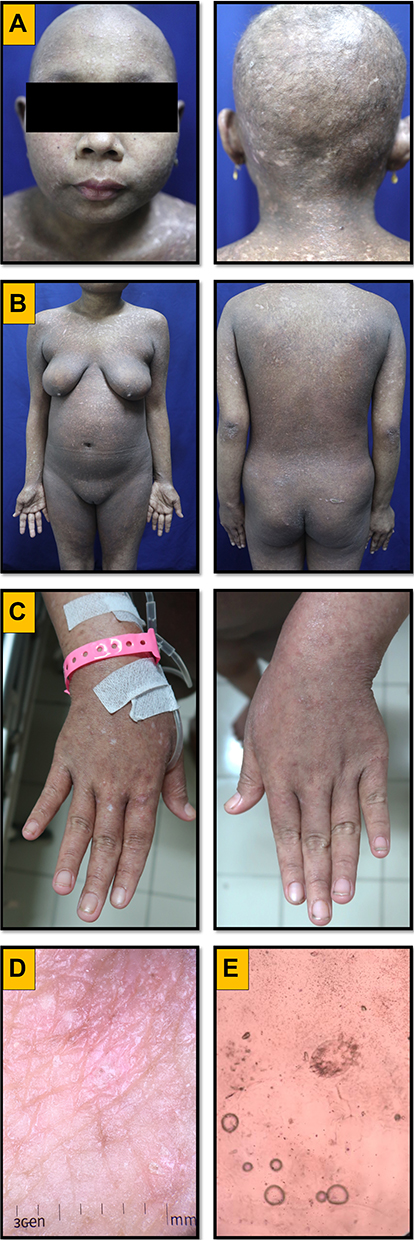 |
Figure 3 Follow up on day 19th. Erythematosus macules, papules, plaques, and crusts on scalp (A) chest, abdomen, back (B) and both hands (C) disappeared. Dermoscopic examination, delta wing jet sign not found (D). Skin scraping S. scabiei mites, eggs, and scybala were not found (E). |
Discussion
The International Alliance for the Control of Scabies (IACS) published consensus criteria in 2020 to standardize scabies diagnosis.14 It was proposed that the diagnosis can be made at one of three levels of certainty: confirmed, clinical, or suspected scabies. Confirmed scabies, diagnosed with microscopy or dermoscopy is the most specific and certain. Clinical scabies is diagnosed with typical lesions in a classic distribution with a convincing symptom and contact history. Suspected scabies is diagnosed when typical lesions present in a typical distribution with some supportive corroborative history or an atypical distribution/morphology with a convincing symptom and contact history.14 The diagnosis of CS can be made based on clinical findings and detection of scabies mites, eggs, or feces with microscopic examination.1,3 Plaques accompanied by thick hyperkeratotic crusts are a typical clinical manifestation of CS and can be observed on the head, ears, elbows, palms, feet, and toes.1 Microscopic examination is the main diagnostic modality for scabies, conducted to identify the mites, their eggs, or scybala within samples. Another useful examination is a dermoscopic examination with a delta wing jet feature being the pathognomonic sign representing the mite’s head and transparent body.3 The diagnosis of CS was established based on physical examination and results of diagnostic test. Our patient reported itchy plaques with hyperkeratotic crusts in all parts of the body, especially on the scalp, chest, and abdomen. Microscopic examination from the skin scrapings showed S. scabiei mites and eggs with a total of 3–5 mites per high power field while dermoscopic examination showed delta wing jet sign. These findings supported the diagnosis of CS.
In 2002, Davis et al15 developed an assessment of severity of CS based on four clinical aspects: the distribution and extent of crusting, depth of crusting, number of previous episode of CS, and the presence of cracking or pyoderma. Each aspect has a score of 1 to 3. A total score of 4 to 6 is categorized as mild (grade 1), a total score of 7 to 9 is categorized as moderate (grade 2), and a total score of 10 to 12 is categorized as severe (grade 3). This grading scale is a useful tool for the assessment and management of CS. In this case report, the total score was 9, the distribution of skin lesions more than 30% body surface area and affecting the scalp (score 3), depth of crusting more than 10 mm (score 3), no history of CS (score 1), and there was pyoderma (score 2), so it was categorized as a moderate CS.
Various dermatological, neurological, and immunological disorders due to immunocompromised conditions, both congenital or acquired, eg, HIV, hematological malignancies, neurological disorders, and connective tissue diseases, including SLE, can trigger CS.1,10 In SLE, the immune system is defective in many ways. The defect is due to disturbances of phagocytosis and decreased leukocyte chemotaxis. The cellular immune response to external antigens is also decreased, leading to more severe clinical manifestations of scabies in SLE patients.16 Based on clinical signs and symptoms, ANA test, as well as ANA profile, the patient was diagnosed with SLE by an internist. The patient has been taking long-term corticosteroids. Hence, SLE and immunosuppressive drugs became the risk factors for CS in this patient.
The main principle of therapy in CS is exfoliation of the hyperkeratotic layer with keratolytic agents as well as administration of topical and systemic scabicidal agents.8 Exfoliation is necessary to decrease the number of mites and increase the permeability of topical scabicidal agents. Thick scales and crusts can be removed by soaking them in warm water and application of 5% to 10% salicylic acid in vaseline album or 40% urea.1 The treatment choice for scabies is 5% permethrin due to its effectiveness and low toxicity.1 Permethrin is a pyrethroid synthetic acting on arthropod cell membranes by altering the sodium transport function to polarize the neuromembranes of arthropod motor nerves, thus causing paralysis.1 The Centers for Disease Control and Prevention (CDC) recommends administration of permethrin every 2–3 days for 1–2 weeks for the treatment of CS.17 The new anti-scabies drugs being developed for human scabies are moxidectin and afoxolaner.17,18 Both of these drugs have an antiparasitic effect and are commonly used on dogs, cats, horses, cattle, sheep, and goats by veterinarians. However, due to increasing cases of resistance to permethrin and ivermectin, this drug was developed for the treatment of human scabies and showed positive outcomes.18,19 As for CS, the treatment of choice is ivermectin.1 Only a few cases have been reported using oral albendazole instead of ivermectin for treatment of scabies and CS. Albendazole is an antihelmintic and antiprotozoal agent, which is ovicidal, larvicidal, and vermicidal. It binds to β-free tubulin, which subsequently inhibits the polymerization of tubulin into microtubulin.12 Loss of microtubulin in cytoplasm causes disruption of glucose uptake by larvae and adult parasites, leading to their death. In addition, albendazole can inhibit synapse transmission through its cholinergic effects.12,13 The side effects of albendazole are diarrhea, drowsiness, increased liver enzymes, hepatitis, blurred vision, liver failure, and rhabdomyolysis. Ayoub et al12 reported two cases of scabies treated with three doses of albendazole, 1000 mg per day given for three consecutive days in combination with 5% salicylic acid with good treatment results. Another study by Risadini et al20 compared the effectiveness of 800 mg per day oral albendazole for three consecutive days to a single application of 5% permethrin cream in 102 scabies patients, and concluded that albendazole was as effective as permethrin. Douri et al13 reported good outcomes in cases of CS treated with 800 mg per day oral albendazole for three consecutive days per week, for three cycle combined with 10% crotamiton and 5% salicylic acid. As ivermectin is unavailable in Indonesia, we gave our patient oral albendazole with a dose of 800 mg per day for three consecutive days per week combined with 5% permethrin and 5% salicylic acid.
Conclusion
Oral albendazole can be used as an alternative treatment for CS along with 5% permethrin and 5% salicylic acid, especially in countries where ivermectin is unavailable.
Ethic Statement
This study was conducted in compliance with the Declaration of Helsinki, Good Clinical Practices, local regulatory requirements, and was approved by the Medical Ethics Committee of Hasan Sadikin General Hospital Bandung (approval number: LB.02.01/X.6.5/280/2020).
Consent Statement
The patient has signed the consent forms for the use of case details, images for publication, and for scientific purposes.
Acknowledgments
Authors would like to thank all staff of the Dermatology and Venereology Department, Faculty of Medicine Universitas Padjadjaran – Hasan Sadikin General Hospital Bandung.
Disclosure
The authors report no conflicts of interest in this work.
References
1. Karthikeyan K. Crusted scabies. Indian J Dermatol Venereol Leprol. 2009;75(4):340. doi:10.4103/0378-6323.53128
2. Currier RW, Walton SF, Currie BJ. Scabies in animals and humans: history, evolutionary perspectives, and modern clinical management. Ann N Y Acad Sci. 2011;1230(1):E50–E60. doi:10.1111/j.1749-6632.2011.06364.x
3. Welch E, Romani L, Whitfeld MJ. Recent advances in understanding and treating scabies. Fac Rev. 2021;10:10. doi:10.12703/r/10-10
4. Gutte RM. Bullous scabies in an adult: a case report with review of literature. Indian Dermatol Online J. 2013;4(4):311. doi:10.4103/2229-5178.120654
5. Panigrahi A, Sil A, Bhanja DB. Pruritic curvilinear track over penis. J Paediatr Child Health. 2020;2020:27.
6. Sil A, Mondal S, Bhanja DB, Datta M, Mukherjee GS, Chakraborty S. Pruritic genital nodules. Pediatr Neonatol. 2020;61(2):243–244. doi:10.1016/j.pedneo.2019.12.004
7. Sil A, Punithakumar EJ, Chakraborty S, Bhanja DB, Panigrahi A, Das A. Crusted scabies. Postgrad Med J. 2020;96(1137):444. doi:10.1136/postgradmedj-2020-137661
8. Matsuura H, Senoo A, Saito M, Fujimoto Y. Norwegian scabies. Cleve Clin J Med. 2019;86(3):163. doi:10.3949/ccjm.86a.18081
9. Hatter AD, Soler DC, Curtis C, et al. Case report of individual with cutaneous immunodeficiency and novel 1p36 duplication. Clin Genet. 2016;9:1.
10. Roberts LJ, Huffam SE, Walton SF, et al. Crusted scabies: clinical and immunological findings in seventy-eight patients and a review of the literature. J Infect. 2005;50(5):375–381. doi:10.1016/j.jinf.2004.08.033
11. Maharani CS, Harnanti DV, Mappamasing H, et al. Crusted scabies in patients with long-term use of oral corticosteroid with different underlying diseases–case series. Dermatol Rep. 2019;11:1.
12. Ayoub N, Merhy M, Tomb R. Treatment of scabies with albendazole. Dermatol. 2009;218(2):175. doi:10.1159/000182254
13. Douri T, Shawaf AZ. Treatment of crusted scabies with albendazole: a case report. Dermatol Online J. 2009;15(2):117–126. doi:10.1016/0006-2944(75)90147-7
14. Engelman D, Yoshizumi J, Hay RJ, et al. The 2020 international alliance for the control of scabies consensus criteria for the diagnosis of scabies. Br J Dermatol. 2020;183(5):808–820. doi:10.1111/bjd.18943
15. Davis JS, McGloughlin S, Tong SY, et al. A novel clinical grading scale to guide the management of crusted scabies. PLoS Negl Trop Dis. 2013;7:e2387. doi:10.1371/journal.pntd.0002387
16. Mak A, Kow NY. The pathology of T cells in systemic lupus erythematosus. J Immunol Res. 2014;2014:1–8. doi:10.1155/2014/419029
17. Centers for disease Control and Prevention Parasites. Scabies, resources for health professionals. Available from: https://www.cdc.gov/parasites/scabies/health_professionals/meds.html.
18. Bernigaud C, Fang F, Fischer K, et al. Efficacy and pharmacokinetics evaluation of a single oral dose of afoxolaner against Sarcoptes scabiei in the porcine scabies model for human infestation. Antimicrob Agents Chemother. 2018;62(9):e02334–17. doi:10.1128/AAC.02334-17
19. Mounsey KE, Bernigaud C, Chosidow O, et al. Prospects for moxidectin as a new oral treatment for human scabies. PLoS Negl Trop Dis. 2016;10(3):e0004389. doi:10.1371/journal.pntd.0004389
20. Risadini MW, Mochtar M, Danarti R. Comparison of albendazole tablets with 5% permethrin cream for scabies treatment at AL muayyad Islamic boarding school surakarta [translated title]. Media Dermato Venereologica Indonesiana. 2017;44(3):108–112.
 © 2022 The Author(s). This work is published and licensed by Dove Medical Press Limited. The
full terms of this license are available at https://www.dovepress.com/terms
and incorporate the Creative Commons Attribution
- Non Commercial (unported, 3.0) License.
By accessing the work you hereby accept the Terms. Non-commercial uses of the work are permitted
without any further permission from Dove Medical Press Limited, provided the work is properly
attributed. For permission for commercial use of this work, please see paragraphs 4.2 and 5 of our Terms.
© 2022 The Author(s). This work is published and licensed by Dove Medical Press Limited. The
full terms of this license are available at https://www.dovepress.com/terms
and incorporate the Creative Commons Attribution
- Non Commercial (unported, 3.0) License.
By accessing the work you hereby accept the Terms. Non-commercial uses of the work are permitted
without any further permission from Dove Medical Press Limited, provided the work is properly
attributed. For permission for commercial use of this work, please see paragraphs 4.2 and 5 of our Terms.