Back to Journals » International Medical Case Reports Journal » Volume 19
Occult Fronto-Orbital Fracture Presenting as Traumatic Pseudomeningocele in a 4-Month-Old Infant: Successful Surgical Management in a Resource-Limited Setting
Authors Abukar AA, Osman AA ![]() , Mohamed Isse H
, Mohamed Isse H ![]() , Mohamed HM
, Mohamed HM ![]() , Çelik C
, Çelik C ![]() , Dahir MO
, Dahir MO ![]() , Ibrahim IG
, Ibrahim IG ![]()
Received 16 December 2025
Accepted for publication 19 March 2026
Published 25 March 2026 Volume 2026:19 589335
DOI https://doi.org/10.2147/IMCRJ.S589335
Checked for plagiarism Yes
Review by Single anonymous peer review
Peer reviewer comments 3
Editor who approved publication: Professor Thomas E Hutson
Abdirasak Ali Abukar,1 Ahmed Adam Osman,2 Hamdi Mohamed Isse,2 Hassan Muhumed Mohamed,3 Cihan Çelik,2 Mohamed Osman Dahir,2 Ismail Gedi Ibrahim2
1Department of Neurosurgery, Mogadishu Somali Türkiye Training and Research Hospital, Mogadishu, Somalia; 2Department of Radiology, Mogadishu, Somali Türkiye Training and Research Hospital, Mogadishu, Somalia; 3Department of Ophthalmology, Mogadishu Somali Türkiye Training and Research Hospital, Mogadishu, Somalia
Correspondence: Ahmed Adam Osman, Department of Radiology, Mogadishu Somali Türkiye Training and Research Hospital, Mogadishu, Somalia, Email [email protected]
Abstract: Traumatic fronto-orbital pseudomeningocele is an exceptionally rare complication of paediatric head trauma and may be overlooked because of subtle or delayed radiological findings. It results from cerebrospinal fluid (CSF) leakage through a dural defect associated with skull fractures. We report the case of a four-month-old male infant who presented with progressive right frontal and supraorbital swelling ten days after a minor fall. Cranial computed tomography revealed an occult fracture of the right frontal bone extending into the superior orbital wall with a communicating CSF collection causing inferior displacement of the globe, consistent with traumatic fronto-orbital pseudomeningocele. The patient underwent right-sided craniotomy through a bicoronal approach, during which a dural tear was identified and primarily repaired, followed by secure repositioning of the bone flap. The postoperative course was uneventful, with complete resolution of swelling and restoration of normal ocular position. At the two-week follow-up the infant remained neurologically intact with no evidence of complications. This case highlights the importance of early radiological diagnosis and timely surgical management of rare fronto-orbital pseudomeningocele in infants and demonstrates that favourable outcomes can be achieved even in resource-limited healthcare settings through prompt, multidisciplinary intervention.
Keywords: traumatic pseudomeningocele, orbital fracture, orbital roof fracture, paediatric cranial trauma, cerebrospinal fluid leak, Somalia
Introduction
Paediatric orbital trauma, though rare, can be caused by different situations like minor falls or very serious ones like road traffic accidents. Frontoorbital fractures are capable of causing various complications such as entrapment syndromes, nerve injuries, and impingement syndromes, with traumatic pseudomeningoceles being a rare result.1 Pseudo meningoceles associated with spinal and posterior fossa surgeries have been the most discussed in the literature, whereas reports on intradiploic and orbital pseudomeningoceles are very few.2 A few case report literature reviews conducted over the last few years show a few cases of traumatic skull and orbital pseudomeningoceles in children, among which only four were incidents in or near the orbit that were documented.3 Interestingly, few of these cases came back within days after the traumatic event,4 which is also the case of our patient. This is the first case by and large in Somalia, where traumatic injuries are common due to ongoing conflicts, a weak healthcare system, and bad road conditions.
Case Presentation
A four-month-old male infant from a remote area of the Lower Jubba region in Somalia was referred to the neurosurgery and ophthalmology departments at the Mogadishu Somali Türkiye Training and Research Hospital (MSTH), a tertiary care centre in Mogadishu. The patient presented with a ten-day history of progressive swelling over the right frontal and supraorbital region following a minor fall.
On clinical examination, the infant was alert and responsive. The Glasgow Coma Scale (GCS) score was 14 (E4: spontaneous eye opening; V4: irritable cry; M6: spontaneous limb movement). Inspection revealed marked swelling over the right frontal and supraorbital regions, accompanied by ipsilateral ptosis and chemosis (Figure 1B). The child had difficulty opening the affected eye, and a serous whitish discharge was observed. Palpation of the orbital region revealed increased tension without an audible bruit. No focal neurological deficits were identified, and the remainder of the systemic examination was unremarkable.
 |
Figure 1 (A) Axial CT image demonstrating a fracture of the right frontal bone extending into the superior orbital wall. (B) Clinical photograph showing right frontal and supraorbital swelling associated with ipsilateral ptosis and chemosis. |
Cranial computed tomography (CT) demonstrated mildly displaced fracture lines involving the right frontal bone extending into the superior orbital wall (Figure 1A). Axial CT images revealed a well-defined right supraorbital cystic lesion protruding anteriorly and inferiorly, resulting in downward displacement of the right globe. These findings were consistent with a cerebrospinal fluid (CSF) collection communicating with the intracranial compartment. Soft-tissue window images further illustrated the inferior displacement of the globe, while coronal and sagittal reconstructions provided improved visualisation of the lesion (blue arrows). Corresponding clinical photographs confirmed post-traumatic subcutaneous bulging in the right frontal and supraorbital regions (Figure 1B). Sagittal CT and three-dimensional volume-rendered images clearly demonstrated the right frontal bone fracture extending into the superior orbital wall, confirming fronto-orbital communication (Figure 2).
 |
Figure 2 (A) Sagittal CT reconstruction showing extension of the right frontal bone fracture into the superior orbital wall. (B) Three-dimensional volume-rendered CT image confirming the osseous defect and fronto-orbital communication. |
The differential diagnosis was between traumatic pseudomeningocele and leptomeningeal cyst (growing skull fracture). A leptomeningeal cyst was considered less likely due to the fact that the imaging did not reveal herniation of brain parenchyma or progressive enlargement of the bony defect, which are the characteristics of growing skull fractures. On the contrary, the imaging results showed a localised CSF collection which was communicating with the intracranial compartment through a dural defect without brain tissue herniation; hence, a diagnosis of traumatic fronto-orbital pseudomeningocele was made.
Due to how progressive the swelling appeared and what was shown on the imaging, a surgical procedure was performed. The surgery consisted of a bicoronal variable scalp incision with a right craniotomy. There was a known dural tear located at the wasp fracture site, confirming the diagnosis of a traumatic fronto-orbital pseudomeningocele. The dural defect was primarily repaired, and the bone flap was repositioned and secured in situ (Figure 3A–C).
 |
Figure 3 (A–C) Intraoperative photographs demonstrating the bicoronal approach, identification and repair of the dural defect, and repositioning of the bone flap. |
The postoperative course was uneventful. The child remained neurologically intact, alert, and active. Complete resolution of the right frontal and supraorbital swelling was observed with regression of periorbital oedema and restoration of normal ocular position (Figure 4). Following a brief stay as an inpatient, they obeyed the provisions related to limitations on length of stay by the patient, and the child was discharged on oral antibiotics. The two-week follow-up visit demonstrated good clinical improvement in the child, showing symmetric eye position without ptosis or significant chemosis (Figure 4). Evidence from the bicoronal incision suggested a successful healing process without any indication of infection. As the patient’s family lives far away, around 500 km from Mogadishu, further follow-up was carried out via telephone consultations arranged in advance.
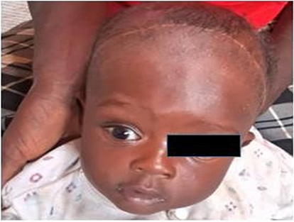 |
Figure 4 Postoperative clinical outcome. Clinical photograph demonstrating complete resolution of right frontal and supraorbital swelling with restoration of normal ocular alignment. |
Discussion
A pseudomeningocele is an abnormal extracellular collection of cerebrospinal fluid (CSF) due to a defect in the dura mater; hence, a fibrous, walled cavity without a real meningeal lining is created. It is mainly developed as a result of iatrogenic or traumatic dural injury. Iatrogenic pseudomeningoceles are very common after spinal surgical procedures, particularly lumbar ones where unintentional dural tears happen quite often. On the other hand, traumatic pseudomeningoceles occur very rarely and generally relate to skull or spinal fracture after blunt head or neck trauma. Previously documented post-traumatic intradiploic pseudomeningoceles in paediatric patients revealed the very uncommon nature of this condition following cranial trauma.5
The underlying pathophysiology involves disruption of the dura mater, allowing CSF to escape into adjacent soft tissues under physiological or elevated intracranial pressure. Over time, the leaked CSF may become encapsulated by reactive fibrous tissue or arachnoid adhesions, forming a pseudomembrane. In paediatric patients, this process may occur more rapidly because of the relative fragility of cranial bones and dura mater, combined with persistent CSF pulsations. Similar mechanisms have been described in previous reports of orbital pseudomeningocele and related cranial complications.6
Traumatic orbital pseudomeningocele is an extremely rare entity, particularly in infants and young children. Most cases reported in the literature are associated with orbital roof fractures that create a communication between the intracranial space and the orbit. Previous reports have described progressive orbital swelling or proptosis as the most common clinical presentation following trauma. Lewis et al2 explained surgical treatment of orbital pseudomeningocele via eyelid incision as a successful method and stressed the paramount need for early diagnosis and treatment. In a similar way, Mirkin et al described the case of traumatic orbital pseudomeningocele with symptoms of progressive proptosis, identifying through the CT image the fracture and the loculation of the cerebrospinal fluid (CSF) that caused the problem. These findings are consistent with other reported cases of post-traumatic orbital pseudomeningocele in children.7
The principal differential diagnosis of traumatic pseudomeningocele is a leptomeningeal cyst, also known as a growing skull fracture. Leptomeningeal cysts occur when a dural tear allows herniation of arachnoid and brain tissue through a skull fracture, leading to progressive enlargement of the bone defect. In contrast, pseudomeningocele represents an extradural collection of CSF without herniation of brain parenchyma. Imaging findings in our patient demonstrated a localised CSF collection communicating with the intracranial compartment without evidence of brain tissue herniation or progressive enlargement of the fracture defect, supporting the diagnosis of traumatic pseudomeningocele rather than a leptomeningeal cyst.8
In this case, the pseudomeningocele was a complication of a traumatic fronto-orbital fracture. This was followed by the escape of CSF leaking out from both the dural defect and the broken superior orbital wall. The CSF that had escaped gathered in the supraorbital soft tissues, thus causing progressive swelling, proptosis, and ptosis. The formation of arachnoidal adhesions around the fracture site probably enclosed the CSF, and consequently, a clinically palpable pseudomeningocele was produced. After surgical intervention, these signs fully disappeared, thereby demonstrating that the pathology was mechanically originated.
The clinical presentation of post-traumatic orbital pseudomeningoceles is mainly progressive proptosis, which may or may not be pulsatile. Patients may also have restrictions of eye movements, displacement of the eyeball, periorbital swelling, headache, seizures, or signs of raised intracranial pressure. Because these manifestations are often nonspecific, a high index of suspicion is required, particularly in infants and young children with a history of craniofacial trauma. A comprehensive neuro-ophthalmological examination, including careful palpation of the orbital rims, is therefore essential in patients presenting with exophthalmos or periorbital swelling following head injury.
Radiological examination is vital to diagnosing this type of injury. While plain x-rays can show fractures of the skull, they provide no information about soft tissue structure or the circulation of cerebrospinal fluid (CSF). Computed tomography (CT) remains the primary imaging modality for emergency evaluations. CT provides an excellent view of bony injury, such as fracture of both the skull and the orbital wall, as well as whether there is evidence of CSF leakage. Magnetic resonance imaging (MRI) can provide additional information regarding soft tissue structures and dural integrity; however, CT is often more accessible and practical in emergency settings and in resource-limited healthcare environments.9 In our patient, cranial CT imaging was sufficient to establish the diagnosis and guide surgical planning.
The definitive means to manage a traumatic pseudomeningocele involves surgical repair of a dural defect and the restoration of normal cranial contour to prevent long-term leakage of cerebrospinal fluid (CSF) and possible complications.10 Typical surgical procedures used for correction of a cranial pseudomeningocele include a craniotomy for direct dural closure and reposition/reconstruction of the bone defect. The bicoronal approach with right-sided craniotomy provided ideal exposure for identification and primary repair of the dural tear, followed by secure repositioning of the bone flap. A favourable postoperative outcome was noted in the form of complete resolution of clinical symptoms and lack of any neurological deficits; therefore, timely surgical intervention appears to have been successful.
Detecting the signs and carrying out surgery swiftly are two of the most important components when patients have traumatic fronto-orbital pseudomeningoceles, especially children. Delayed diagnosis may lead to progressive deformity, visual impairment, infection, or long-term neurological complications. The successful management of this rare condition in a resource-limited setting further demonstrates that favourable outcomes can be achieved with careful clinical assessment, appropriate radiological evaluation, and timely multidisciplinary surgical care.
Conclusion
Traumatic fronto-orbital pseudomeningocele is an exceptionally rare complication of paediatric craniofacial trauma that may present with progressive orbital swelling and proptosis. This case emphasises the importance of careful neuro-ophthalmological examination, thorough palpation of the orbital rims, and prompt radiological evaluation in infants presenting with periorbital swelling after head injury. A rapidly repaired dura defect and associated fractures can have superior clinical results while reducing the chance of complications, including vision loss, infection, or developmental changes to the skull or face, all of which are associated with these types of treatment. The success of treating uncommon neurological disorders in a low-resource environment is made possible by the collaborative approach of a variety of specialities as well as by employing the correct surgical techniques.
Consent for Publication
Written informed consent was obtained from the patient’s parent for the publication of this case report and all accompanying clinical images. Ethical approval from the Ethics Committee of the Mogadishu Somali Türkiye Training and Research Hospital was not required for publication of this single case report.
Author Contributions
Abdirasak Ali Abukar contributed to the surgical management of the patient, conceptualisation of the case report, and drafting of the manuscript. Ahmed Adam Osman contributed to radiological interpretation, image acquisition, figure preparation, and critical revision of the manuscript for important intellectual content. Hamdi Mohamed Isse contributed to clinical data collection and literature review. Hassan Muhumed Mohamed contributed to ophthalmological evaluation and postoperative follow-up of the patient. Cihan Çelik contributed to radiological supervision and manuscript revision. Mohamed Osman Dahir and Ismail Gedi Ibrahim contributed to imaging analysis, manuscript editing, and literature review. All authors made a significant contribution to the work reported, including conception, study design, execution, acquisition of data, analysis and interpretation; took part in drafting, revising, or critically reviewing the article; approved the final version to be published; agreed on the journal to which the article has been submitted; and agreed to be accountable for all aspects of the work.
Disclosure
The authors declare no conflicts of interest related to this work.
References
1. Sarfraz M, Mustansir F, Khan N, Darbar A. Acute orbital pseudomeningocele due to traumatic fracture in an infant. Neuro-Ophthalmol. 2020;44(5):339–6. doi:10.1080/01658107.2019.1611881
2. Lewis AL, Dermarkarian CR, Tao JP. Treatment of an orbital pseudomeningocele through an eyelid incision. Can J Ophthalmol. 2022;57(4):e142–e144. doi:10.1016/j.jcjo.2021.11.00.2
3. Mirkin S, Patel J, Wang W, Engel C. A rare case of traumatic orbital pseudomeningocele. Cureus. 2025;17(1):e77881. doi:10.7759/cureus.77881
4. Richardson D, Duncan C, Sinha A, Hennedige AA. Pseudomeningocele with orbital extension as a complication of fronto-orbital advancement and remodelling in craniosynostosis. J Craniofac Surg. 2015;26(7):2142–2147.
5. Mahapatra AK, Tandon PN. Post-traumatic intradiploic pseudomeningocele in children. Acta Neurochir. 1989;100(3–4):120–126. doi:10.1007/BF01403598
6. Nguyen CT, Guiney AJ, Iseli TA, King JAJ, Hardy TG. Spontaneous giant pseudomeningocele in the middle cranial fossa as a cause of pulsatile proptosis. Ophthalmic Plast Reconstr Surg. 2017;33(3 Suppl 1):S91–S99. doi:10.1097/IOP.0000000000000499
7. Kumar R, Verma A, Sharma K, Rathi B, Malik V. Post-traumatic pseudomeningocele of the orbit in a young child. J Pediatr Ophthalmol Strabismus. 2003;40(2):110–112. doi:10.3928/0191-3913-20030301-14
8. Osman AA, Karshe NA, Ibrahim IG, Tahtabaşı M. A leptomeningeal cyst in the frontal bone as a complication of childhood head trauma: a case report (Çocukluk çağı kafa travmasının bir komplikasyonu olarak frontal kemikte leptomeningeal kist: olgu sunumu). J Med Vet Invest. 2021;3(1):31. doi:10.46683/jmvi.2021.31
9. Guler I, Buyukterzi M, Oner O, Tolu I. Post-traumatic leptomeningeal cyst in a child: computed tomography and magnetic resonance imaging findings. J Emergency Med. 2015;48(5):e121.
10. Meier JD, Dublin AB, Strong EB. Leptomeningeal cyst of the orbital roof in an adult: case report and literature review. Skull Base. 2009;19(03):231–235.
 © 2026 The Author(s). This work is published and licensed by Dove Medical Press Limited. The
full terms of this license are available at https://www.dovepress.com/terms
and incorporate the Creative Commons Attribution
- Non Commercial (unported, 4.0) License.
By accessing the work you hereby accept the Terms. Non-commercial uses of the work are permitted
without any further permission from Dove Medical Press Limited, provided the work is properly
attributed. For permission for commercial use of this work, please see paragraphs 4.2 and 5 of our Terms.
© 2026 The Author(s). This work is published and licensed by Dove Medical Press Limited. The
full terms of this license are available at https://www.dovepress.com/terms
and incorporate the Creative Commons Attribution
- Non Commercial (unported, 4.0) License.
By accessing the work you hereby accept the Terms. Non-commercial uses of the work are permitted
without any further permission from Dove Medical Press Limited, provided the work is properly
attributed. For permission for commercial use of this work, please see paragraphs 4.2 and 5 of our Terms.