Back to Journals » International Journal of Nanomedicine » Volume 10 » Issue 1
Novel stable cytokine delivery system in physiological pH solution: chitosan oligosaccharide/heparin nanoparticles
Authors Wang B, Tan L, Deng D, Lu T, Zhou C, Li Z, Tang Z, Wu Z, Tang H
Received 2 February 2015
Accepted for publication 27 March 2015
Published 8 May 2015 Volume 2015:10(1) Pages 3417—3427
DOI https://doi.org/10.2147/IJN.S82091
Checked for plagiarism Yes
Review by Single anonymous peer review
Peer reviewer comments 2
Editor who approved publication: Professor Lei Yang
Bin Wang, Ling Tan, Dengpu Deng, Ting Lu, Changwei Zhou, Zhongkui Li, Zhenjie Tang, Zhongshi Wu, Hao Tang
Department of Cardiovascular Surgery, The Second Xiangya Hospital of Central South University, Changsha, People’s Republic of China
Background: Cell therapy is a promising strategy for tissue regeneration. Key to this strategy is mobilization and recruitment of exogenous or autologous stem/progenitor cells by cytokines. However, there is no effective cytokine delivery system available for clinic application, in particular for myocardial regeneration. The aim of this study was to develop a novel cytokine delivery system that is stable in solution at physiological pH.
Methods: Four groups of self-assembled chitosan oligosaccharide/heparin (CSO/H) nanoparticles were prepared with various volume ratios of chitosan oligosaccharide to heparin (5:2, 5:4, 4:15, 1:5) and characterized by laser diffraction, particle size analysis, and transmission electron microscopy. The encapsulation efficiency and loading content of two cytokines, ie, stromal cell-derived factor (SDF)-1α and vascular endothelial growth factor (VEGF) were quantified using an enzyme-linked immunosorbent assay. The biological activity of the loaded SDF-1α and VEGF was evaluated using the transwell migration assay and MTT assay. The dispersion profiles for the cytokine-loaded nanoparticles were quantified using fluorescence molecular tomography.
Results: CSO/H nanoparticles were prepared successfully in solution with physiological pH. The particle sizes in the four treatment groups were in the range of 96.2–210.5 nm and the zeta potential ranged from -29.4 mV to 24.2 mV. The loading efficiency in the CSO/H nanoparticle groups with the first three ratios was more than 90%. SDF-1α loaded into CSO/H nanoparticles retained its migration activity and VEGF loaded into CSO/H nanoparticles continued to show proliferation activity. The in vivo dispersion test showed that the CSO/H nanoparticles enabled to VEGF to accumulate locally for a longer period of time.
Conclusion: CSO/H nanoparticles have a high cytokine loading capacity and allow cytokines to maintain their bioactivity for longer, are stable in an environment with physiological pH, and may be a promising cytokine delivery system for tissue regeneration.
Keywords: cytokine delivery system, nanoparticles, chitosan oligosaccharide, physiological pH, stromal cell-derived factor-1α, vascular endothelial growth factor
Introduction
Tissue damage occurs in many diseases, including diabetes, acute graft-versus-host disease, and cardiovascular disease.1 Of these diseases, myocardial infarction, one of the leading causes of mortality, is the most common and difficult to treat in the clinic, because cardiomyocytes are not able to regenerate.2 Tissue regeneration through stem/progenitor cells has been studied extensively in a wide range of tissue repair applications, especially for regeneration of cardiomyocytes, owing to the unique pluripotency and regenerative properties of these cells.3,4 However, tissue regeneration involves a series of biological processes, including mobilization of stem cells, angiogenesis, and synthesis of extracellular matrix.5,6 Various cytokines are essential for homing, proliferation, and differentiation of stem/progenitor cells in the process of tissue regeneration.7,8 Cytokines have been widely studied in tissue regeneration, but their short half-lives and the low local concentrations render their application limited.9,10 Therefore, a reliable delivery system able to localize cytokines to specific tissues for an extended period of time and maintain their bioactivity could improve the homing, differentiation, and proliferation of stem/progenitor cells at their target tissue.
Nanoparticles are attracting an increasing amount of research attention as drug delivery systems because of their superior loading efficiency.11,12 Various techniques are available to prepare nanoparticles, including solvent evaporation, interfacial polymerization, and emulsion polymerization.13,14 Unfortunately, these methods frequently require the use of organic solvents or heat, which are undesirable steps and may affect the integrity of components and increase their biological toxicity.13,15 Among the numerous biological nanomaterials available, chitosan-based nanoparticles are one of the best studied drug delivery systems and have been widely used to load drugs due to the biodegradability and biocompatibility of chitosan.16–18 However, chitosan cannot be dissolved in solution with a pH >6.5, which limits its application in vivo, especially as a cytokine delivery system, given that the bioactivity of cytokines require a physiological pH of 7.35–7.45. Chitosan oligosaccharide (CSO), a low polymerization chitosan, has most of the advantages of chitosan, but is also soluble in solutions with physiological pH.19 Therefore, it should be possible to develop CSO-based nanoparticles that are stable in solutions with physiological pH. Many growth factors, such as stromal cell-derived factor (SDF)-1α, vascular endothelial growth factor (VEGF), placental growth factor, platelet-derived growth factor, and fibroblast growth factor,20–23 can bind to heparin through their conserved amino acid sequences. Several heparin-containing cytokine delivery systems have been developed for tissue regeneration and modified to control the release of cytokines.24–26
We have previously published a paper demonstrating that self-assembled heparin/chitosan nanoparticles can load VEGF to stimulate proliferation of endothelial cells in vitro and accelerate vascularization in a murine subcutaneous implant model in vivo.24 We hypothesized that CSO and heparin could be utilized to develop a novel nanoparticle as a delivery system to load cytokines for tissue regeneration and be stable in a solution with physiological pH.
In the present work, self-assembled chitosan oligosaccharide/heparin (CSO/H) nanoparticles were constructed. We demonstrated that these nanoparticles not only maintained a stable size in physiological pH solution, but could also load cytokines and maintain their bioactivity for tissue regeneration.
Materials and methods
Materials
CSO (molecular weight 5,000 Da, more than 90% deacetylated) was purchased from Golden-Shell Pharmaceutical Co Ltd (Zhejiang, People’s Republic of China). Low molecular weight heparin sodium (molecular weight 8,000 Da, from Fucus vesiculosus) was supplied by Sigma Chemical Company (St Louis, MO, USA). Human recombinant VEGF-165m (293-VE-010/CF, DVE00) and enzyme-linked immunosorbent assay kits for VEGF (460-SD-010/CF, DY460) and SDF-1α were purchased from R&D Systems (Minneapolis, MN, USA). Anti-VEGF antibody (Cy5) was purchased from Biorbyt LLC (San Francisco, CA, USA). MTT [3-(4,5-dimethylthiazol-2-yl)2,5-diphenyltetrazolium bromide], used for the cell proliferation assay, was sourced from Sigma-Aldrich (St Louis, MO, USA). Mouse mesenchymal stem cells (MSCs) were obtained from the Central South University Tumor Institute (Changsha, People’s Republic of China). Primary human umbilical vein endothelial cells (HUVECs) were obtained from the Central Laboratory of Xiangya Hospital (Changsha, People’s Republic of China).
Preparation and characterization of CSO/H nanoparticles
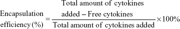
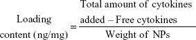
Self-assembled CSO/H nanoparticles were prepared, the composition of which is shown in Table 1. First, low molecular weight heparin sodium solution (0.25 mg/mL) and CSO solution (1.0 mg/mL) were prepared using deionized water. The CSO solution and heparin solution were then mixed in various volume ratios (5:2, 5:4, 4:15, and 1:5) at room temperature and titrated to pH 7.35–7.45 with 1% sodium hydroxide under magnetic stirring followed by ultrasonication. The prepared VEGF and SDF-1α solutions was then added at different concentrations to the CSO/H nanoparticle suspension and stirred for 5 minutes at room temperature. The prepared nanoparticles were collected by ultracentrifugation at 14,000 rpm for 20 minutes. To evaluate the encapsulation efficiency and loading content of SDF-1α and VEGF in the nanoparticles, the amounts of the free cytokines in the supernatants were assayed by enzyme-linked immunosorbent assay. Drug encapsulation efficiency and loading content were determined by the following equations:27
|
|
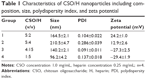 | Table 1 Characteristics of CSO/H nanoparticles including composition, size, polydispersity index, and zeta potential |
The particle size distribution and zeta potential of the CSO/H nanoparticles were measured by laser diffraction (Mastersizer, 3000HS, Malvern Instruments Ltd, Malvern, UK) and their morphology was determined by transmission electron microscopy (JEOL, Tokyo, Japan). A drop of the nanoparticle suspension was placed onto a copper grid without staining, and the air-dried samples were then observed directly by transmission electron microscopy.28
Preparation of MSCs and endothelial cells
The MSCs were cultured in Minimum Essential Medium/Earle’s Balanced Salt Solution (Thermo Fisher Scientific, Waltham, MA, USA) supplemented with 10% fetal bovine serum and 1% antibiotic-penicillin and streptomycin solution. To confirm that the cells were phenotypically MSCs, the cells were labeled for various positive and negative cell surface markers, such as CD44, CD45, CD34, and CD90, and analyzed by flow cytometry.29 MSCs were cultured up to 80% confluence for three or four passages before use in vitro.
The HUVECs were cultured with endothelial cell medium (ECM, ScienCell Research Laboratories, San Diego, CA, USA) containing 5% fetal bovine serum and 1% endothelial cell growth supplement (ECGS), which contains various growth factors, hormones, and proteins necessary for growth of endothelial cells. These cells were identified by factor VIII immunohistochemistry staining (Figure 1). HUVECs were cultured up to 80% confluence for three to five passages before use in the study.
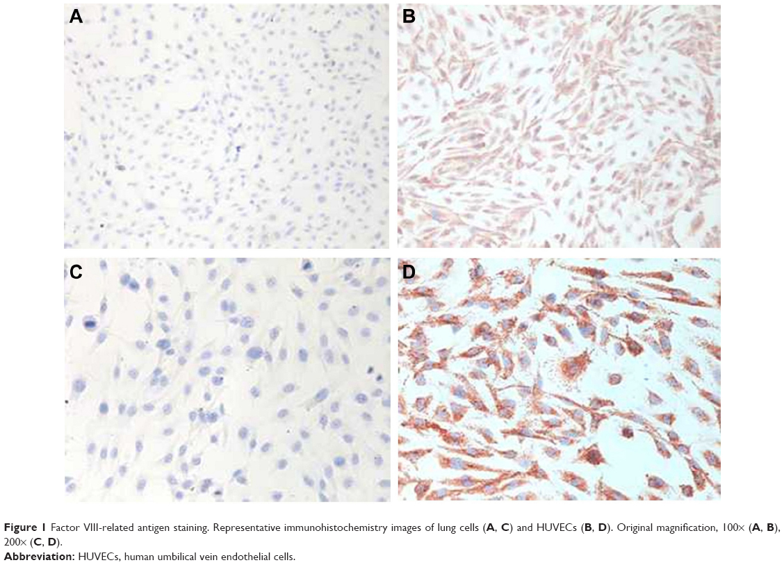 | Figure 1 Factor VIII-related antigen staining. Representative immunohistochemistry images of lung cells (A, C) and HUVECs (B, D). Original magnification, 100× (A, B), 200× (C, D). |
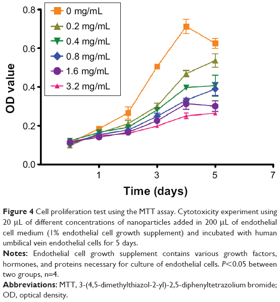
In vitro cytotoxicity model
MTT assays were applied to study the cytotoxicity of the CSO/H nanoparticles to inhibit proliferation of endothelial cells. First, 3×10³ HUVEC cells were seeded in ECM (1% ECGS, 5% fetal bovine serum) into each well of a 96-well plate. The cells were then cultured for different periods of time with 20 μL of supplement containing various concentrations of nanoparticles. Following a predetermined period of incubation, 20 μL of MTT solution was added to each well. The cells were then incubated at 37°C for 4 hours. The supernatant in each well was then discarded and replaced with 150 μL of dimethyl sulfoxide. The plates were then shaken for 10 minutes to fully dissolve the crystals. The optical density was measured at 490 nm using a Multiscan spectrometer (Varioskan Flash, Thermo Electron Corporation, Wilmington, DE, USA). The optical density is linearly proportional to the number of living cells in the sample.
In vitro model of MSC migration induced by SDF-1α-loaded CSO/H nanoparticles
A transwell migration model was used to study the ability of SDF-1α-loaded CSO/H nanoparticles to induce migration of MSCs (Figure 2A). Migration assays were carried out in a 24-well transwell using polycarbonate membranes with 8 μm pores (Corning Costar, Cambridge, MA, USA). Next, 200 μL of MSCs at a density of 4×10⁵ cells/mL in 100 μL of medium (1% fetal bovine serum) were placed in the upper chamber of the transwell assembly. The culture medium in the lower chamber contained various concentrations of SDF-1α-loaded nanoparticles, various concentrations of SDF-1α alone or nanoparticles alone (the latter serving as controls). Following a predetermined period of incubation, the upper surface of the membrane was gently swabbed to remove nonmigrating cells, then washed twice with phosphate-buffered saline. The membrane was then fixed in 5% glutaraldehyde for 30 minutes and stained with 0.5% crystal violet for 10 minutes at room temperature. The number of migrating cells was determined by counting cells in five randomly selected fields per well under the microscope at a magnification of 100×.
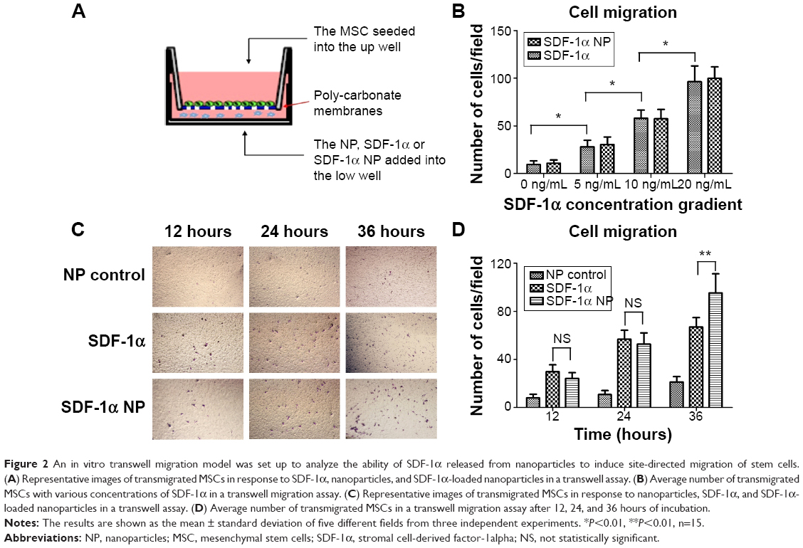 | Figure 2 An in vitro transwell migration model was set up to analyze the ability of SDF-1α released from nanoparticles to induce site-directed migration of stem cells. (A) Representative images of transmigrated MSCs in response to SDF-1α, nanoparticles, and SDF-1α-loaded nanoparticles in a transwell assay. (B) Average number of transmigrated MSCs with various concentrations of SDF-1α in a transwell migration assay. (C) Representative images of transmigrated MSCs in response to nanoparticles, SDF-1α, and SDF-1α-loaded nanoparticles in a transwell assay. (D) Average number of transmigrated MSCs in a transwell migration assay after 12, 24, and 36 hours of incubation. |
In vitro model of cell proliferation induced by VEGF-loaded CSO/H nanoparticles
First, 5×10³ HUVECs were seeded into each well of a 96-well plate. The HUVECs of ECGS group were cultured with ECM (1% ECGS, 5% fetal bovine serum), the cells of VEGF groups were cultured with ECM (5% fetal bovine serum, various concentrations of VEGF solution), the cells of VEGF NP groups were cultured with ECM (5% fetal bovine serum, various concentrations of VEGF-loaded nanoparticles), the cells of NP groups were cultured with ECM (5% fetal bovine serum, nanoparticle solution). Following a predetermined period of incubation, the cell population was estimated by MTT assay, as described earlier.
In vivo study of the cytokine dispersion profiles
Dispersion of the cytokines from the CSO/H nanoparticles was quantified in vivo using fluorescence molecular tomography (FMT4000, PerkinElmer, Wellesley, MA, USA). The VEGF was dissolved in phosphate-buffered saline solution (pH 7.4), and then mixed with Cy5 fluorescence cytokine antibody and incubated for one hour at room temperature. The Cy5 fluorescence antibody and VEGF combination was then added to the CSO/H nanoparticle suspension and stirred for 5 minutes at room temperature.
Three-week-old male ICR mice were randomly divided into two groups, with one group to receive 100 μL of the Cy5 fluorescence antibody and VEGF and the other group to receive nanoparticles containing the Cy5 fluorescence antibody and VEGF. Each study treatment was injected subcutaneously into the back of each mouse after anesthesia with sevoflurane. The mice were observed immediately by fluorescence molecular tomography to detect the dispersion of Cy5 fluorescence at predetermined time points. All procedures involving animals were approved by the animal ethics committee of Central South University, People’s Republic of China.
Statistical analysis
The study results are shown as the mean ± standard deviation. Two-way analysis of variance and Student’s t-tests were performed using GraphPad Prism version 5 software (GraphPad Software, La Jolla, CA, USA). P<0.05 was taken to be statistically significant.
Results
Characterization of CSO/H nanoparticles
Characterization of the CSO/H nanoparticles is shown in Table 1. The self-assembled nanoparticles were prepared to contain four different ratios of CSO to heparin. The particle sizes in the four groups were in the range of 96.2–210.5 nm. Nanoparticle groups 1, 3, and 4 had a good polydispersity index (less than 0.25). The zeta potentials in these three groups were also favorable (absolute values greater than 20 mV). Table 2 shows the cytokine encapsulation efficiency of the nanoparticles. The group 1, 2 and 3 nanoparticles had high encapsulation efficiency (over 90%). The optimal size of nanoparticles designed for drug delivery should be approximately 50–150 nm, to confer a high surface area-to-volume ratio. The polydispersity index less than 0.25 indicates that the size distribution of the nanoparticles was uniform and suitable for the requirements of this study. We selected the group 3 CSO/H nanoparticles for further study because of their negative charge (zeta potential approximately −27.3 mV), polydispersity index of 0.091, and particle size of about 140.2 nm. Figure 3A shows the size distribution and Figure 3B–D shows the transmission electron microscopic images for these CSO/H nanoparticles, which were spherical with a mean diameter of around 140.2 nm, suggesting an ideal size for drug delivery.
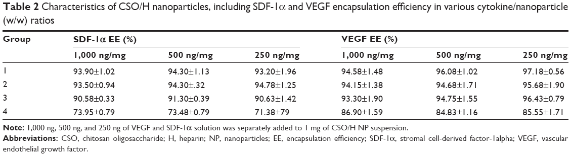 | Table 2 Characteristics of CSO/H nanoparticles, including SDF-1α and VEGF encapsulation efficiency in various cytokine/nanoparticle (w/w) ratios |
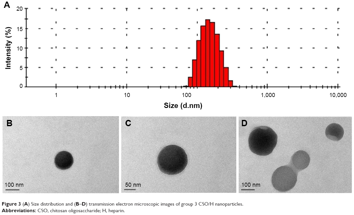 | Figure 3 (A) Size distribution and (B–D) transmission electron microscopic images of group 3 CSO/H nanoparticles. |
Effect of SDF-1α-loaded CSO/H nanoparticles on migration of MSCs
The ability of SDF-1α to induce chemotaxis in vitro was tested using a transwell system. First, MSCs were induced for 20 hours by different concentrations of SDF-1α (0, 5, 1, or 20 ng/mL) alone or loaded into CSO/H nanoparticles. Migration of MSCs across the transmembrane was then quantified by removing the cells from the seeded side and staining the underside of the membrane with crystal violet. SDF-1α induced migration of the MSCs across the transwell membrane and the effect was dependent on the SDF-1α concentration gradient (Figure 2B, P<0.05). There was no significant difference in number of migrated cells between the SDF-1α alone group and SDF-1α nanoparticle group. MSCs were then induced for 12–36 hours by CSO/H nanoparticles alone, 10 ng/mL SDF-1α alone, or 10 ng/mL SDF-1α loaded into CSO/H nanoparticles. There was no statistically significant difference in the numbers of MSCs crossing the transmembrane until 36 hours of incubation (P<0.05, Figure 2D), indicating that CSO/H nanoparticles could maintain the bioactivity of SDF-1α for a long period of time.
Cytotoxicity of CSO/H nanoparticles
An MTT assay was used to evaluate the cytotoxic effects of CSO/H nanoparticles on HUVECs, which were cultured in 200 μL of ECM (5% fetal bovine serum and 1% ECGS) with or without 20 μL of different concentrations of nanoparticles for 5 days. As shown in Figure 4, the degree of inhibition of HUVECs was dependent on the nanoparticle concentration.
 | Figure 4 Cell proliferation test using the MTT assay. Cytotoxicity experiment using 20 μL of different concentrations of nanoparticles added in 200 μL of endothelial cell medium (1% endothelial cell growth supplement) and incubated with human umbilical vein endothelial cells for 5 days. |
Activity of VEGF loaded by CSO/H NP
HUVECs were cultured in ECM (without ECGS) with various concentrations of VEGF or in 1% ECGS alone for 5 days. An MTT assay was then used to observe the proliferation of HUVECs according to their optical density. Figure 5A shows that cell proliferation increased in a VEGF concentration-dependent manner at VEGF concentrations ≤10 ng/mL (P<0.05). In contrast, the control group cultured in 1% ECGS maintained better growth because ECGS is a complete supplement for endothelial cells. However, there was no statistically significant difference in OD value between the 10 ng/mL and 50 ng/mL VEGF groups. HUVECs were then cultured with 5 ng/mL VEGF or 10 ng/mL VEGF with or without CSO/H nanoparticles for 5 days, and a comparison of the stimulating effect of VEGF alone and VEGF-loaded nanoparticles on cell proliferation is shown in Figure 5B. The only statistically significant difference in cell proliferation was between 5 ng/mL VEGF alone and VEGF-loaded nanoparticles on day 2 (P<0.05). These results show that the CSO/H nanoparticles maintained the bioactivity of VEGF.
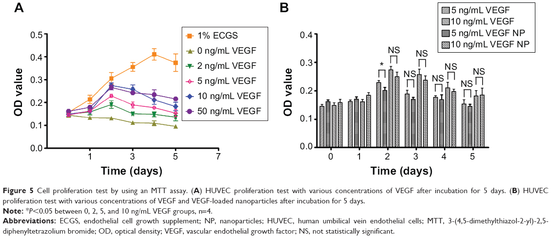 | Figure 5 Cell proliferation test by using an MTT assay. (A) HUVEC proliferation test with various concentrations of VEGF after incubation for 5 days. (B) HUVEC proliferation test with various concentrations of VEGF and VEGF-loaded nanoparticles after incubation for 5 days. |
Dispersion of cytokines from CSO/H NP in vivo
The rate of dispersion of VEGF in CSO/H nanoparticles was quantified in an vivo model using fluorescence molecular tomography (Figure 6). Figure 6A shows a representative image from fluorescence molecular tomography. The dispersion rate of VEGF loaded into CSO/H nanoparticles was significantly lower than for VEGF alone between 30 minutes and 300 minutes (Figure 6B, P<0.05 or P<0.01). These results indicate that CSO/H nanoparticles enable VEGF to be maintained for a longer period of time.
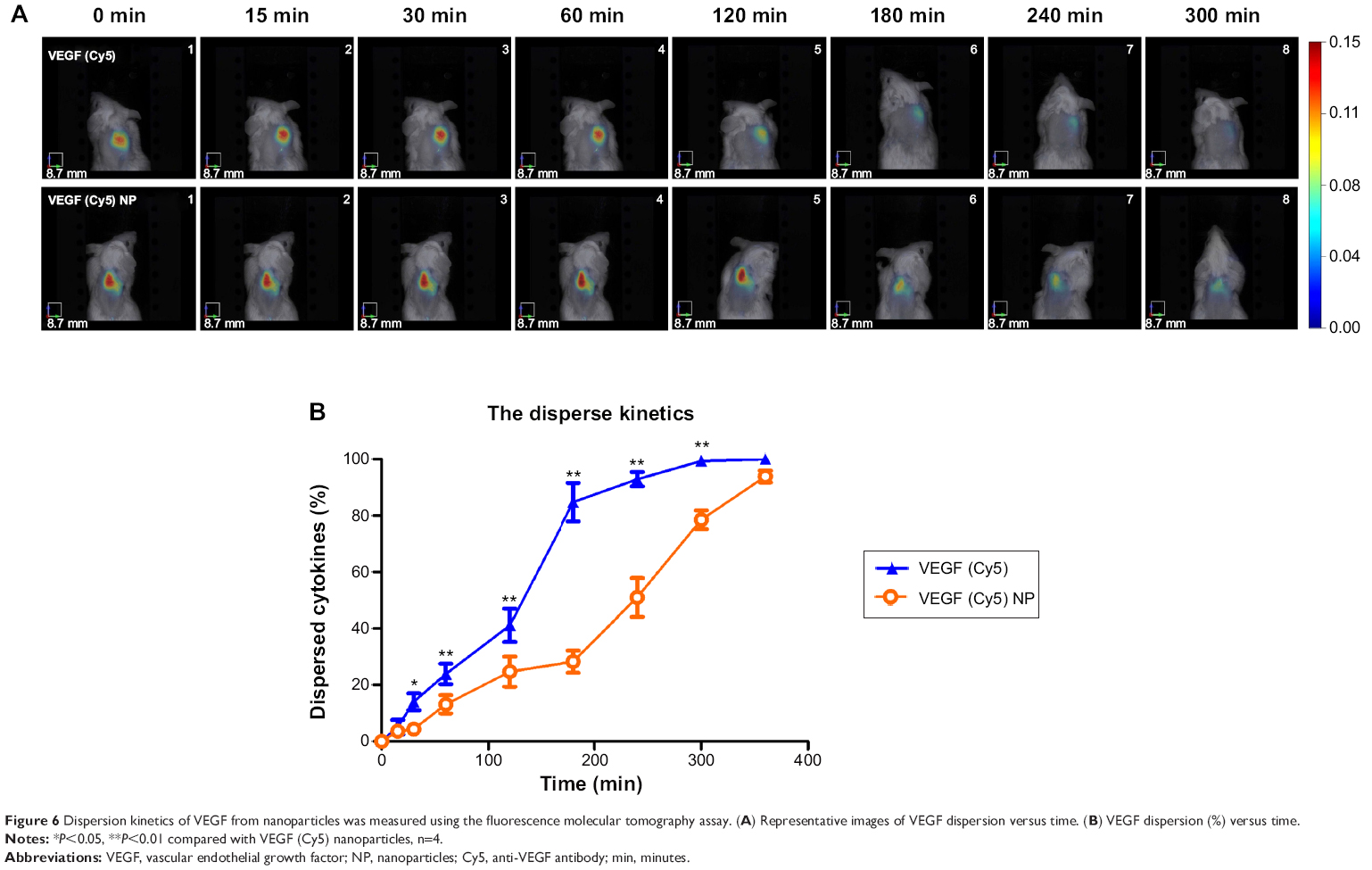 | Figure 6 Dispersion kinetics of VEGF from nanoparticles was measured using the fluorescence molecular tomography assay. (A) Representative images of VEGF dispersion versus time. (B) VEGF dispersion (%) versus time. |
Discussion
As previously mentioned, the clinical use of stem/progenitor cell therapy has many disadvantages, including a low survival rate, relative limited cell sources, and the expense involved in obtaining sufficient cells.30 Various cytokines are necessary for homing, proliferation, and differentiation of the stem/progenitor cells used in cell therapy. A delivery system targeting specific tissues is required to improve the efficiency of cytokines to promote revascularization and tissue regeneration.
Nanoparticles have been widely studied as drug delivery systems for various applications, but preparation of most nanoparticles involves organic solvents or heating, which may decrease their biodegradability and biocompatibility. For example, ferroferric oxide nanoparticles are usually prepared using an organic solvent at high temperatures.31 Liposomal nanoparticles are widely used in antibiotic drug delivery systems to enhance the activity of antibiotics.32 However, chloroform is often used to prepare liposomal nanoparticles33,34 and is neurotoxic, so may decrease the safety of liposomal nanoparticles when used as a cytokine delivery system.
Chitosan nanoparticles are widely used in drug release systems because of the excellent properties of chitosan, such as biodegradability, biocompatibility, antimicrobial activity, and low toxicity, along with ease of preparation and versatile chemical and physiological properties.35–37 However, chitosan is insoluble in physiological pH solutions, and chitosan nanoparticles are prepared in an acidified environment buffered with 1% acetic acid.16,24 A previous study by our group demonstrated that chitosan nanoparticles were only stable in acidic solutions (pH <6.5), with the optimal pH being 5.24 The low solubility of chitosan in physiological solution limits its in vivo application. Previous studies have shown that CSO, a low molecular weight chitosan (molecular weight 3,000 Da), is soluble in physiological pH solution.38 The mechanism for this is not clear. The lower degree of polymerization may lead a few branching on the molecular chain of CSO and a relative more hydrophilic non-ionic groups exposed, which may make the CSO a relative loose structure and better solubility. Use of CSO could help to overcome the limitations of chitosan. CSO (used at a molecular weight 5,000 Da in our study) nanoparticles has been confirmed to be stable in physiological pH solution, so may be more amenable for clinical application. In this study, CSO/H nanoparticles were prepared by self-assembly. Like chitosan,39 CSO is suitable for developing self-assembled polymeric nanoparticles due to the availability for crosslinking with free amino groups and the cationic nature which allows combination with multivalent anions. Heparin, with its multivalent anions and carboxylic group, can combine with CSO. The charge on the CSO/H nanoparticle is determined by the ratio of the surface molecules (CSO and heparin). The positive zeta potential for the group 1 and 2 nanoparticles suggests that the nanoparticle surface is covered by more CSO than heparin (Table 1). The negative charge on the group 3 and 4 nanoparticles meant a higher ratio of heparin to CSO on the nanoparticle surface (Table 1). At the same time, the stability and size of these nanoparticles were mainly influenced by the total charge on the particle. The relatively larger absolute zeta potential value was associated with a comparatively small size and polydispersity index (groups 1, 3, and 4, see Table 1). Therefore, we chose these CSO/H ratios because when the ratio is between 4:15 and 5:4, the size and polydispersity index will be much larger, even sediment appears in the suspension.
In general, exogenous additives affect cell growth and proliferation. As shown in Figure 4, the CSO/H nanoparticles inhibited proliferation of HUVECs to some extent. This inhibition may have been induced by endocytotic processes stimulated by the CSO/H nanoparticles during incubation. This suggests that the concentration of CSO/H nanoparticles should be taken into consideration in the following experiment. The high encapsulation efficiency and loading capacity of the CSO/H nanoparticles means that a very small amount of CSO/H nanoparticles can deliver enough cytokines to target tissues, which can reduce the inhibition effect of nanoparticles on cell proliferation. Therefore, we used CSO/H nanoparticles at a concentration of 0.2 ng/mL.
SDF-1α has been demonstrated to be important for mediating mobilization, migration, and homing of stem/progenitor cells,40 and VEGF is important for cell proliferation and angiogenesis during the process of tissue regeneration.41,42 Therefore, we used SDF-1α and VEGF to study the bioactivity of cytokine-loaded CSO/H nanoparticles. Our results show that CSO/H nanoparticles can load not only a large amount of cytokines, but also a variety of cytokines (such as SDF-1α and VEGF, Table 2), indicating that these nanoparticles could serve as a carrier for cytokines. We believe that other cytokines, such as placental growth factor, platelet-derived growth factor, and fibroblast growth factor, could also be loaded into CSO/H nanoparticles. Cytokines have a very short half-life, and are very vulnerable in a hostile environment.43,44 Maintenance of bioactivity is the most important study criterion when evaluating a cytokine delivery system. Therefore, the stability of CSO/H nanoparticles in physiological pH solution was taken into consideration. In this study, the bioactivity of SDF-1α and VEGF carried by CSO/H nanoparticles was evaluated by cell migration assay and MTT assay. Our results show that CSO/H nanoparticles maintained the bioactivity of both SDF-1α and VEGF (Figures 2D and 5B), and that this bioactivity was concentration gradient-dependent (Figures 2B and 5A). We also found that the CSO/H nanoparticles could maintain the bioactivity of SDF-1α for extended periods of time and recruit more MSCs (Figure 2D). Our in vivo dispersion test showed further that the CSO/H nanoparticles could retain VEGF for longer at the injection site (Figure 6). The above findings demonstrate that CSO/H nanoparticles can enable a large amount of cytokines to localize and retain their bioactivity for a longer period of time.
We have demonstrated that self-assembled CHO/H nanoparticles are a potential cytokine delivery system for tissue regeneration, with the advantages of stability in physiologic pH solution, a high encapsulation efficiency and loading capacity, no decrease in the bioactivity of the cytokines carried, slow/controlled cytokine dispersion, and less toxicity.
However, there are some limitations to our study. An animal model may need to be developed to investigate the availability and effectiveness of CSO/H nanoparticles for targeted delivery of cytokines in vivo. The local retention time of CHO/H nanoparticles is relatively short, so further investigations are needed to refine these nanoparticles so that they can be retained for longer at their target site.
Conclusion
CSO/H nanoparticles can be used as a cytokine delivery system. They can be prepared by self-assembly using CSO and heparin. They have a high cytokine loading capacity, are stable in physiological pH solution, and can maintain the bioactivity of cytokines. Therefore, these novel CSO/H nanoparticles may be a promising cytokine delivery system for tissue regeneration.
Acknowledgments
This work was supported by a grant from the Natural Science Foundation of Hunan Province (2015JJ4064). The authors appreciate the editorial assistance of Wenwu Zhang and Jian Hu in the preparation of this paper.
Disclosure
The authors report no conflicts of interest in this work.
References
Domian IJ, Chiravuri M, van der Meer P, et al. Generation of functional ventricular heart muscle from mouse ventricular progenitor cells. Science. 2009;326:426–429. | ||
Pasala T, Sattayaprasert P, Bhat PK, Athappan G, Gandhi S. Clinical and economic studies of eptifibatide in coronary stenting. Ther Clin Risk Manag. 2014;10:603–614. | ||
Mollmann H, Nef HM, Voss S, et al. Stem cell-mediated natural tissue engineering. J Cell Mol Med. 2011;15:52–62. | ||
Blanpain C, Fuchs E. Stem cell plasticity. Plasticity of epithelial stem cells in tissue regeneration. Science. 2014;344:1242281. | ||
Dong Q, Yang Y, Song L, Qian H, Xu Z. Atorvastatin prevents mesenchymal stem cells from hypoxia and serum-free injury through activating AMP-activated protein kinase. Int J Cardiol. 2011;153:311–316. | ||
Mehrpour M, Esclatine A, Beau I, Codogno P. Overview of macroautophagy regulation in mammalian cells. Cell Res. 2010;20:748–762. | ||
Ghadge SK, Mühlstedt S, Özcelik C, Bader M. SDF-1α as a therapeutic stem cell homing factor in myocardial infarction. Pharmacol Ther. 2011;129:97–108. | ||
Penn MS, Pastore J, Miller T, Aras R. SDF-1 in myocardial repair. Gene Ther. 2012;19:583–587. | ||
Murphy JW, Cho Y, Sachpatzidis A, Fan C, Hodsdon ME, Lolis E. Structural and functional basis of CXCL12 (stromal cell-derived factor-1 alpha) binding to heparin. J Biol Chem. 2007;282:10018–10027. | ||
Izuagie IA, Pelham CJ, Agrawal DK. Synergistic effect of angiotensin II on vascular endothelial growth factor-A-mediated differentiation of bone marrow-derived mesenchymal stem cells into endothelial cells. Stem Cell Res Ther. 2015;6:4. | ||
Saptarshi SR, Duschl A, Lopata AL. Interaction of nanoparticles with proteins: relation to bio-reactivity of the nanoparticle. J Nanobiotechnol. 2013;11:26. | ||
Yameen B, Choi WI, Vilos C, Swami A, Shi J, Farokhzad OC. Insight into nanoparticle cellular uptake and intracellular targeting. J Control Release. 2014;190:485–499. | ||
Quintanar-Guerrero D, Allemann E, Fessi H, Doelker E. Preparation techniques and mechanisms of formation of biodegradable nanoparticles from preformed polymers. Drug Dev Ind Pharm. 1998;24:1113–1128. | ||
Ragelle H, Riva R, Vandermeulen G, et al. Chitosan nanoparticles for siRNA delivery: optimizing formulation to increase stability and efficiency. J Control Release. 2014;176:54–63. | ||
Kumar A, Zhang X, Liang XJ. Gold nanoparticles: emerging paradigm for targeted drug delivery system. Biotechnol Adv. 2013;31(5):593–606. | ||
Ribeiro TG, Chavez-Fumagalli MA, Valadares DG, et al. Novel targeting using nanoparticles: an approach to the development of an effective anti-leishmanial drug-delivery system. Int J Nanomedicine. 2014;9:877–890. | ||
Yuan Q, Shah J, Hein S, Misra RD. Controlled and extended drug release behavior of chitosan-based nanoparticle carrier. Acta Biomater. 2010;6:1140–1148. | ||
Das S, Chaudhury A, Ng KY. Preparation and evaluation of zinc-pectin-chitosan composite particles for drug delivery to the colon: role of chitosan in modifying in vitro and in vivo drug release. Int J Pharm. 2011;406:11–20. | ||
Termsarasab U, Cho H, Kim DH, et al. Chitosan oligosaccharide-arachidic acid-based nanoparticles for anti-cancer drug delivery. Int J Pharm. 2013;441:373–380. | ||
Johnson NR, Wang Y. Controlled delivery of heparin-binding EGF-like growth factor yields fast and comprehensive wound healing. J Control Release. 2013;166:124–129. | ||
Fermas S, Gonnet F, Sutton A, et al. Sulfated oligosaccharides (heparin and fucoidan) binding and dimerization of stromal cell-derived factor-1 (SDF-1/CXCL 12) are coupled as evidenced by affinity CE-MS analysis. Glycobiology. 2008;18:1054–1064. | ||
Dos SC, Blanc C, Elahouel R, et al. Proliferation and migration activities of fibroblast growth factor-2 in endothelial cells are modulated by its direct interaction with heparin affin regulatory peptide. Biochimie. 2014;107 Pt B:350–357. | ||
Lee J, Yoo JJ, Atala A, Lee SJ. The effect of controlled release of PDGF-BB from heparin-conjugated electrospun PCL/gelatin scaffolds on cellular bioactivity and infiltration. Biomaterials. 2012;33: 6709–6720. | ||
Tan Q, Tang H, Hu J, et al. Controlled release of chitosan/heparin nanoparticle-delivered VEGF enhances regeneration of decellularized tissue-engineered scaffolds. Int J Nanomedicine. 2011;6:929–942. | ||
Baumann L, Prokoph S, Gabriel C, Freudenberg U, Werner C, Beck-Sickinger AG. A novel, biased-like SDF-1 derivative acts synergistically with starPEG-based heparin hydrogels and improves eEPC migration in vitro. J Control Release. 2012;162:68–75. | ||
Chiu LL, Radisic M. Scaffolds with covalently immobilized VEGF and angiopoietin-1 for vascularization of engineered tissues. Biomaterials. 2010;31:226–241. | ||
Grenha A, Seijo B, Remunan-Lopez C. Microencapsulated chitosan nanoparticles for lung protein delivery. Eur J Pharm Sci. 2005;25: 427–437. | ||
Du YZ, Ying XY, Wang L, et al. Sustained release of ATP encapsulated in chitosan oligosaccharide nanoparticles. Int J Pharm. 2010;392:164–169. | ||
Zhu H, Guo Z, Jiang X, et al. A protocol for isolation and culture of mesenchymal stem cells from mouse compact bone. Nat Protoc. 2010;5:550–560. | ||
Garbern JC, Lee RT. Cardiac stem cell therapy and the promise of heart regeneration. Cell Stem Cell. 2013;12:689–698. | ||
Wang H, Li X, Mao K, et al. Electrochemical immunosensor for alpha-fetoprotein detection using ferroferric oxide and horseradish peroxidase as signal amplification labels. Anal Biochem. 2014;465C:121–126. | ||
Craciunescu O, Moldovan L, Moisei M, Trif M. Liposomal formulation of chondroitin sulfate enhances its antioxidant and anti-inflammatory potential in L929 fibroblast cell line. J Liposome Res. 2013;23:145–153. | ||
Messiaen AS, Forier K, Nelis H, Braeckmans K, Coenye T. Transport of nanoparticles and tobramycin-loaded liposomes in Burkholderia cepacia complex biofilms. PLoS One. 2013;8:e79220. | ||
Mugabe C, Azghani AO, Omri A. Preparation and characterization of dehydration-rehydration vesicles loaded with aminoglycoside and macrolide antibiotics. Int J Pharm. 2006;307:244–250. | ||
Huang Y, Liu T. Mobilization of mesenchymal stem cells by stromal cell-derived factor-1 released from chitosan/tripolyphosphate/fucoidan nanoparticles. Acta Biomater. 2012;8:1048–1056. | ||
Cota-Arriola O, Cortez-Rocha MO, Burgos-Hernandez A, Ezquerra-Brauer JM, Plascencia-Jatomea M. Controlled release matrices and micro/nanoparticles of chitosan with antimicrobial potential: development of new strategies for microbial control in agriculture. J Sci Food Agric. 2013;93:1525–1536. | ||
Chen MC, Mi FL, Liao ZX, et al. Recent advances in chitosan-based nanoparticles for oral delivery of macromolecules. Adv Drug Deliv Rev. 2013;65:865–879. | ||
Chae SY, Son S, Lee M, Jang M, Nah J. Deoxycholic acid-conjugated chitosan oligosaccharide nanoparticles for efficient gene carrier. J Control Release. 2005;109:330–344. | ||
Lin YH, Chung CK, Chen CT, Liang HF, Chen SC, Sung HW. Preparation of nanoparticles composed of chitosan/poly-gamma-glutamic acid and evaluation of their permeability through Caco-2 cells. Biomacromolecules. 2005;6:1104–1112. | ||
Ceradini DJ, Kulkarni AR, Callaghan MJ, et al. Progenitor cell trafficking is regulated by hypoxic gradients through HIF-1 induction of SDF-1. Nat Med. 2004;10:858–864. | ||
Zhang H, Jia X, Han F, et al. Dual-delivery of VEGF and PDGF by double-layered electrospun membranes for blood vessel regeneration. Biomaterials. 2013;34:2202–2212. | ||
Frey SP, Jansen H, Raschke MJ, Meffert RH, Ochman S. VEGF improves skeletal muscle regeneration after acute trauma and reconstruction of the limb in a rabbit model. Clin Orthop Relat Res. 2012;470: 3607–3614. | ||
Vempati P, Popel AS, Mac GF. Extracellular regulation of VEGF: isoforms, proteolysis, and vascular patterning. Cytokine Growth Factor Rev. 2014;25:1–19. | ||
Nagasawa T. CXC chemokine ligand 12 (CXCL12) and its receptor CXCR4. J Mol Med (Berl). 2014;92:433–439. |
 © 2015 The Author(s). This work is published and licensed by Dove Medical Press Limited. The
full terms of this license are available at https://www.dovepress.com/terms
and incorporate the Creative Commons Attribution
- Non Commercial (unported, 3.0) License.
By accessing the work you hereby accept the Terms. Non-commercial uses of the work are permitted
without any further permission from Dove Medical Press Limited, provided the work is properly
attributed. For permission for commercial use of this work, please see paragraphs 4.2 and 5 of our Terms.
© 2015 The Author(s). This work is published and licensed by Dove Medical Press Limited. The
full terms of this license are available at https://www.dovepress.com/terms
and incorporate the Creative Commons Attribution
- Non Commercial (unported, 3.0) License.
By accessing the work you hereby accept the Terms. Non-commercial uses of the work are permitted
without any further permission from Dove Medical Press Limited, provided the work is properly
attributed. For permission for commercial use of this work, please see paragraphs 4.2 and 5 of our Terms.