Back to Journals » Medical Devices: Evidence and Research » Volume 18
Noninvasive Detection of Alpha-Amylase in Saliva Using Screen-Printed Carbon Electrodes: A Promising Biomarker for Clinical Oral Diagnostics
Authors Reviansyah FH ![]() , Ristin AD, Rauf AA, Sepirasari PD, Alim FN, Nur Y
, Ristin AD, Rauf AA, Sepirasari PD, Alim FN, Nur Y ![]() , Takarini V
, Takarini V ![]() , Yusuf M
, Yusuf M ![]() , Aripin D, Susilawati S, Komariah M
, Aripin D, Susilawati S, Komariah M ![]() , Alam BYCSSS
, Alam BYCSSS
Received 12 October 2024
Accepted for publication 20 November 2024
Published 7 January 2025 Volume 2025:18 Pages 15—27
DOI https://doi.org/10.2147/MDER.S493383
Checked for plagiarism Yes
Review by Single anonymous peer review
Peer reviewer comments 2
Editor who approved publication: Dr Scott Fraser
Faris Hernando Reviansyah,1,* Azzahra Delvyra Ristin,1,* Adil Abdul Rauf,2 Prisilia Dita Sepirasari,2 Fahmi Nur Alim,2 Yuspian Nur,3 Veni Takarini,4 Muhammad Yusuf,5 Dudi Aripin,6 Sri Susilawati,7 Maria Komariah,8 Boy Yoseph Cahya Sunan Sakti Syah Alam9
1Undergraduate Program, Faculty of Dentistry, Universitas Padjadjaran, Sumedang, 45363, Indonesia; 2Undergraduate Program, Faculty of Mathematics and Natural Sciences, Universitas Padjadjaran, Sumedang, 45363, Indonesia; 3Laboratory of Research and Development of FARMAKA TROPIS, Faculty of Pharmacy, Universitas Mulawarman, Samarinda, East Kalimantan, 75119, Indonesia; 4Department of Dental Materials and Technology, Faculty of Dentistry, Universitas Padjadjaran, Bandung, 40132, Indonesia; 5Department of Chemistry, Faculty of Mathematics and Natural Sciences, Universitas Padjadjaran, Sumedang, 45363, Indonesia; 6Department of Conservative Dentistry, Faculty of Dentistry, Universitas Padjadjaran, Bandung, 40132, Indonesia; 7Department of Dental Public Health, Faculty of Dentistry, Universitas Padjadjaran, Bandung, 40132, Indonesia; 8Department of Fundamental Nursing, Faculty of Nursing, Padjadjaran University, Bandung, 40132, Indonesia; 9Faculty of Geological Engineering, Universitas Padjadjaran, Jatinangor, Jawa Barat, 45363, Indonesia
*These authors contributed equally to this work
Correspondence: Faris Hernando Reviansyah, Email [email protected]
Background: Biomarkers are essential tools for diagnosing diseases. Saliva, as a human fluid, effectively reflects the body’s condition due to its rich composition. Analyzing saliva components allows for noninvasive, cost-effective, and time-efficient screening and diagnosis. Alpha-amylase, a key biomarker present in saliva, has been linked to oral diseases. This study introduces an innovative method for the noninvasive detection of alpha-amylase using screen-printed electrodes (SPEs), enabling easy and efficient screening and diagnosis.
Methods: The proposed method involves measuring varying concentrations of alpha-amylase using Cyclic Voltammetry (CV) and Differential Pulse Voltammetry (DPV). Saliva samples are applied directly onto electrodes pre-coated with biomarkers and a conditioning agent, allowing for precise detection and analysis.
Results: The screen-printed carbon electrode demonstrated excellent performance in detecting alpha-amylase, with clear voltammogram results, achieving a limit of detection (LOD) of 104.252 units and a limit of quantification (LOQ) of 315.915 units.
Conclusion: A gold nanoparticle-modified screen-printed electrode (SPE) was developed to measure alpha-amylase quantitatively. Despite sensitivity to external interference, notably temperature, pH, and the duration of incubation, While the sensor showed sensitivity to external factors such as pH and temperature variations, it maintained a strong linear response, reinforcing its potential for reliable diagnostics with linear regression score (R² = 0.9513) across alpha-amylase concentrations of 100– 500 units. This study underscores the sensor’s effectiveness as a non-invasive tool for early detection using saliva as a biomarker, enhancing patient comfort and compliance. However, further research is needed for medical applications.
Keywords: Biomarkers, Screen-printed electrodes, Saliva, Alpha-amylase
Introduction
Biomarkers, or biological markers, are measurable indicators that reflect biological processes, pathogenic conditions, or responses to interventions. They play a crucial role in diagnosing diseases, monitoring health, and guiding personalized medicine. The classification of biomarkers includes categories such as diagnostic, prognostic, and pharmacodynamic biomarkers, each serving distinct clinical purposes.2 Various biomarkers can be used to detect disease; nevertheless, saliva has the potential to become a biomarker. It is also a very convenient sample to acquire rather than blood in the oral category.
Human saliva is a heterogenous biofluid consisting of 99% water, 0.3% protein, and 0.2% substances.3,4 A normal person can produce saliva within the range of 0.3 to 0.4 mL per minute, with a total of 0.5 to 1.5 liters per day, this saliva is produced by three major and innumerable minor salivary glands.5 Saliva contains abundant analytes such as hormones, interleukins, MUC5B, Albumin, DNA, mRNA, and α-amylase which can represent the condition of the human body.6,7 Saliva has already been recognized as a human fluid with potential as a disease marker, with numerous studies highlighting the correlation between saliva and oral diseases by analyzing its components.8 Each of the components has its own concentrations that can be measured using various methods. However, this condition can be easily changed due to several factors, one of the simple things that can change that, is to rinse the mouth.9,10 That could affect the quality of diagnosing or screening oral disease which makes the saliva quite a sensitive biomarker.
Oral diseases are some of the most common health issues worldwide, carrying significant health concerns and economic problems, caries, periodontal disease, tooth loss, and oral cancers have a significant impact on society.11 Some of them are visible to be detected by using traditional methods for example visual and hand instruments, nonetheless for oral cancer, the limitations of conventional diagnostic methods compound the difficulty of early detection.12,13 Traditional approaches, such as tissue biopsies, computed tomography (CT) scans, magnetic resonance imaging (MRI), ultrasonography (USG), positron emission tomography (PET), and toluidine blue staining, although effective, are fraught with challenges. These methods are not only invasive and expensive but also time-consuming and often uncomfortable for patients.13 Moreover, many of these techniques involve exposure to radiation, adding another layer of risk. Given these drawbacks, there is an urgent and growing need for more accessible, patient-friendly diagnostic alternatives that can facilitate early detection and improve survival rates.
In response to these challenges, the development of non-invasive diagnostic tools for oral disease has emerged as a promising solution. One such innovative approach is the use of printed electrodes (SPEs), a technology that has the potential to revolutionize the way oral cancer is diagnosed. SPEs operate through an electrochemical immunosensor mechanism, capable of detecting specific analyte components contained in saliva. This method offers a significant advantage over traditional diagnostic techniques, primarily because it is non-invasive, cost-effective, and less time-consuming.14–16 The ability to use saliva as a sample medium further enhances the convenience and accessibility of SPEs. Saliva is not only easy to collect, but it also provides a direct interaction with oral lesions, which can increase the specificity and sensitivity of biomarker detection.6
The use of saliva as a diagnostic medium in SPE technology represents a significant breakthrough in the non-invasive detection of oral disease. The non-invasive nature of saliva collection makes it an ideal medium for regular screening and early detection, which are crucial for improving patient outcomes.1 Furthermore, the ability of SPEs to provide rapid and accurate results makes them particularly valuable in settings where access to traditional diagnostic tools is limited. Many studies have used SPEs for various purposes, for example, a study conducted by Wu et al 2022 utilized a screen-printed carbon electrode to diagnose SARS-CoV-2 during COVID-19.17 Furthermore, another study conducted by Zhao et al 2022 mentioned that the usage of SPEs for detecting the concentration of glycated hemoglobin for managing diabetes.18 SPEs performance also have been tested for diagnosing cancer, a study conducted by Ferreira et al 2020 successfully detected the target protein from undiluted human serum for breast cancer detection.19 Numerous studies have highlighted the flexibility and adaptability of SPEs across various applications, demonstrating their potential to revolutionize diagnostic methodologies.
Building on this versatility, SPEs facilitate early detection and offer significant potential for monitoring disease progression, paving the way for more tailored and effective treatment strategies.20 In addition to early detection, SPEs hold promise for predicting disease progression, thereby enabling more personalized treatment plans. The versatility of this technology also allows for the adaptation of SPEs to detect a wide range of biomarkers, potentially extending their application beyond oral cancer to other types of cancers and diseases.21,22 As research and development in this field continue to advance, SPEs could become an integral part of routine cancer screening, particularly in resource-limited settings where conventional diagnostic tools are not readily available.
Alpha-amylase has been identified to have a role in oral physiology and has the potential to reflect pathological changes associated with oral disease.23 Alpha-amylase is an enzyme primarily responsible for the breakdown of starch into sugars during digestion, and it is naturally present in high concentrations in saliva.24 Research has highlighted the potential of alpha-amylase as a biomarker for oral disease. Studies have shown that alpha-amylase levels can be significantly altered in the presence of abnormalities, with this study specifically finding elevated alpha-amylase levels in oral cancer, even from the early stages.25,26 Furthermore, the direct secretion of alpha-amylase into the oral cavity offers advantages for its use as a diagnostic marker. Unlike blood biomarkers, which may require invasive sampling techniques, alpha-amylase can be easily and non-invasively measured through saliva, making the diagnostic process more comfortable and accessible.27 This process require a saliva placement in the main component of SPE’s, notably working electrode, however due to the high sensitivity detection, it may produce an insignificant result, therefore gold nanoparticles (AuNPs) may enhances the performance of the SPE’s by making better facilitation of the electron which provide a larger active surface area for analyte interaction.28 Unfortunately, the current SPE’s still have disadvantages, specifically in environmental conditions such as humidity and temperature also biological variability may affect the performance of SPE’s.29 In this study, we aimed to develop non-invasive early-detection tools for oral disease by only using saliva as a biomarker. Furthermore, our research is to assess the performance of SPE’s ability to detect alpha-amylase concentration to be used to detect oral disease. Despite alpha-amylase promise as a diagnostic marker, non-invasive tools like SPEs for its detection in oral diseases remain underexplored. This study aims to address that gap.
Materials and Methods
Materials
About 46 SPEs (Screen Printed Electrodes) were divided into two groups, one made of PET (polyethylene terephthalate) and another group crafted of ceramic. The use of both PET and ceramic SPEs was motivated by their complementary properties: PET SPEs offer flexibility and cost-efficiency, while ceramic SPEs provide superior mechanical strength and thermal stability, essential for precise electrochemical measurements, all of the SPEs were provided by Universitas Padjadjaran. The SPEs contain a counter electrode, a working electrode, and a reference electrode, which is made from carbon graphite, the parts of the components described in Figure 1. Potentiostats bought from PalmSens, Sulfuric acid, amylum, Bovine Serum Albumin (BSA) 0.7%, alpha-amylase was prepared in five different concentrations: 100, 200, 300, 400, and 500 µg/mL, lastly, potassium hexacyanoferrate(III) were used to modify the SPEs.30,31 Incubation is done every time the SPEs are coated with temperature 25°C to simulate physiological conditions, ensuring accurate enzymatic activity measurements. Gold nanoparticles (AuNp) were also used to modify the working electrode to increase its sensitivity to analytes.32 All of the chemical materials were provided by the Department of Chemistry, Faculty of Mathematics and Natural Sciences, Universitas Padjadjaran.
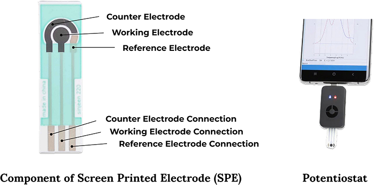 |
Figure 1 The Components of Screen Printed Electrode (SPE) and The Potentiostat as a Connector to Device (Source: Author’s Illustration). |
Experimental Optimization
The effects of alpha-amylase concentration on the electrochemical measurements were thoroughly investigated to ensure optimal conditions for accurate and reliable detection. The concentration varied from 100–500 units to assess its impact on the electrochemical response. Differential Pulse Voltammetry (DPV) is used to evaluate the variations in the current response corresponding to each concentration level.
Modification Flow and Measurement of SPE
The surface modification process began by rinsing the electrode with 45 µL of distilled water three times, allowing it to dry completely between each rinse. Next, 45 µL of 1% sulfuric acid was applied to the entire electrode as a pretreatment step, and a Cyclic Voltammetry (CV) test was conducted to check for any impurities on the electrode surface. The resulting CV voltammograms, shown in the figure below, confirmed the electrode’s cleanliness. After the pretreatment, the electrode was rinsed again with 45 µL of distilled water five times and left to dry. Subsequently, 15 µL of gold nanoparticles (AuNP) was deposited onto the working electrode to enhance its sensitivity to the analyte, followed by an incubation period until the surface was dry. The electrode was then rinsed again with 45 µL of distilled water five times and dried. Following this, 15 µL of starch was added and incubated for 1 hour and 30 minutes, after which the electrode was rinsed with 45 µL of distilled water five more times. Next, 15 µL of 0.1% Bovine Serum Albumin (BSA) was applied to the electrode and incubated for 15 minutes to block non-specific binding sites on the electrode surface. The electrode was rinsed again with 45 µL of distilled water five times. Then, 15 µL of alpha-amylase was added, and incubated for 30 minutes, and the electrode was once again rinsed with 45 µL of distilled water five times. Finally, 45 µL of potassium ferricyanide (K₃Fe(CN)₆) was applied to the electrode, which was then assessed using Differential Pulse Voltammetry (DPV). Electrochemical measurements were performed using a potentiostat, with data collection and analysis carried out through Palmsens software. The whole process can be seen in Figure 2.
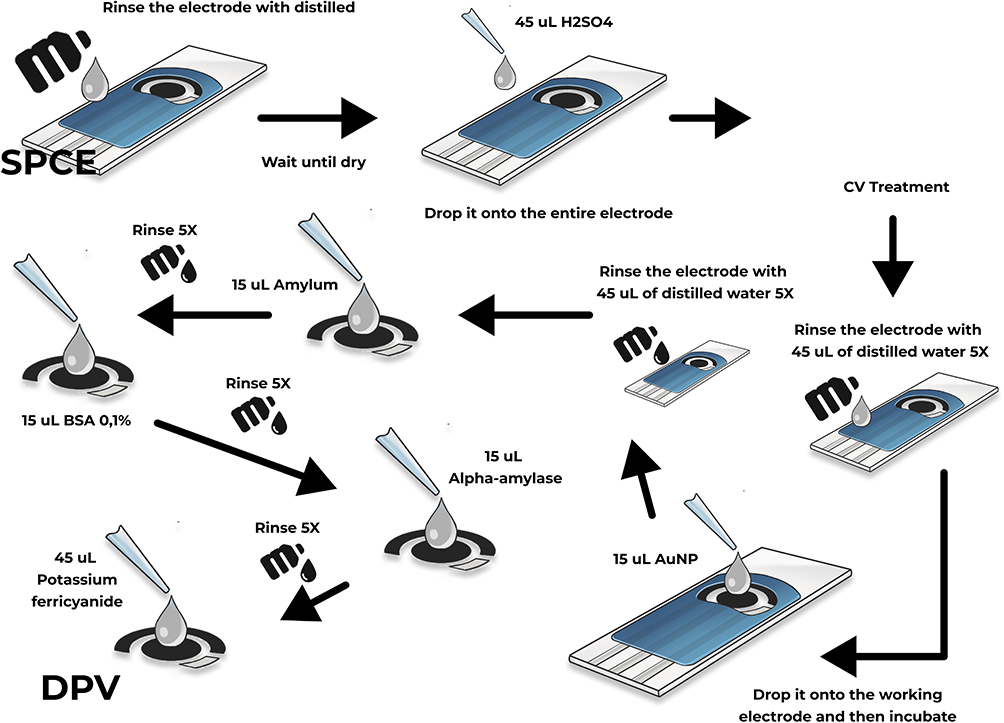 |
Figure 2 A Schematic Illustration of Modifying Screen-Printed Carbon Electrode (Source: Author’s Illustration). |
Saliva Sample Collection and Processing
Artificial saliva was acquired from Singaperbangsa Laboratory Storage Universitas Padjadjaran with an expiration date of December 2024. Samples were divided into five groups based on alpha-amylase concentration, and every group of samples contained 5 samples that will be tested, which will allow us to perform a quantitative analysis. All of the samples were collected and centrifuged at 6000 rpm for 30 minutes. This process is required to remove insoluble components, including cellular debris, bacterial contaminants, and aggregates, to ensure the purity of the sample.
Statistical Analysis
Statistical analysis was conducted using SPSS® version 27 (IBM Corporation, 1 New Orchard Road, Armonk, New York, USA), and the results were documented in a Microsoft Excel® spreadsheet. Descriptive statistics were employed to summarize and highlight the primary characteristics of the dataset, including means, standard deviations, and confidence intervals.
To evaluate the normality of the recorded data, the Kolmogorov–Smirnov and Shapiro–Wilk tests were applied. The results indicated that most datasets followed a normal distribution (p > 0.05) across the concentrations of 100 µg/mL, 300 µg/mL, 400 µg/mL, and 500 µg/mL, justifying the use of parametric tests for these groups. However, the dataset for 200 µg/mL deviated from normality (p < 0.05) as shown by both tests, necessitating a non-parametric approach for its analysis.
A one-way ANOVA was conducted to evaluate whether there were statistically significant differences in mean current values across the five concentration levels. Levene’s test was used to verify the homogeneity of variances, ensuring that the assumptions for ANOVA were met. Post hoc tests were performed to identify pairwise differences between concentration levels, while non-parametric methods were employed for datasets that did not meet normality assumptions.
Results
Electrochemical Measurement of the Concentration of Alpha-Amylase
Figures 3 and 4 represent the concentration of alpha-amylase that was quantitatively analyzed by CV and DPV. Alpha amylase with concentrations of 100 units, 200 units, 300 units, 400 units, and 500 units was used to establish the standard curve and carry out the measurement in the 45µL (K₃Fe(CN)₆) by DPV. The figure below shows the current-potential curve and the established standard curve measured by DPV, respectively. There was a linear relationship between the current (µA) and the concentration of alpha-amylase (unit) on the current-potential curve of DPV. The fitting equation was y = 0.0001x + 0.127, R2= 0.9735.
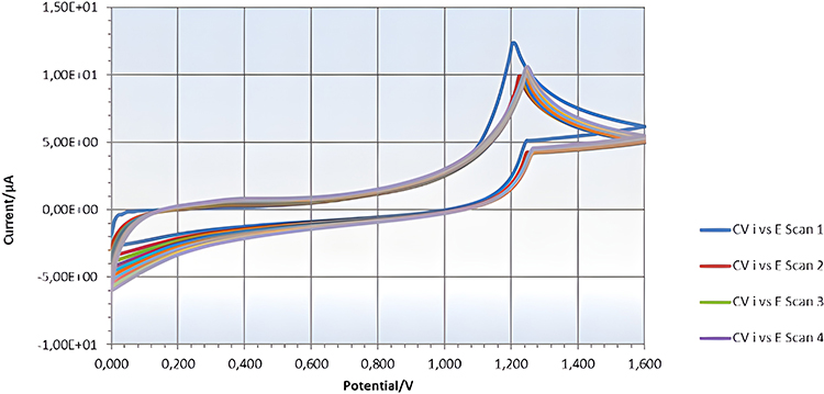 |
Figure 3 The Current Voltage (CV) Curve (Source: Author’s Illustration). |
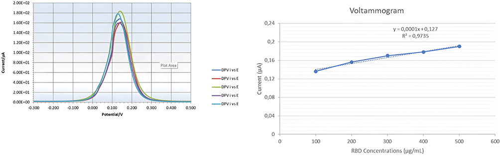 |
Figure 4 The Differential Potential Voltage (DPV) Curve and the linear regression of alpha-amylase test. (Source: Author’s Illustration). |
The limit of detection (LOD) was determined using Equation (1):

where k = 3, s represents the standard deviation, and b is the slope of the curve. From this calculation, the LOD was found to be 104.252 indicating that the smallest detectable concentration of alpha-amylase is 104.252 units. Similarly, the limit of quantitation (LOQ) was calculated using the same equation, with k = 10. The resulting LOQ was 315.915, signifying that the smallest concentration of alpha-amylase that can be both detected and accurately quantified, or classified as normal or abnormal, is 418.7488 units. Detailed information is provided in Figure 5.
 |
Figure 5 Amperometric Standard Curve Alpha-Amylase (Source: Author’s Illustration). |
Standard Curve Results
The figure below shows the relationship between α-amylase concentration and the resulting current (μA). At a concentration of 100 units of α-amylase, the resulting current was approximately 0.158 μA, indicating minimal response at low concentrations. When the concentration increases to 200 units, the current rises to about 0.16 μA, showing a small but significant increase. At a concentration of 300 units, the current increased more sharply to 0.166 μA, indicating a more pronounced increase in the electrochemical response. At a concentration of 400 units, the current continues to increase to 0.177 μA, indicating an almost linear relationship between concentration and current. At the highest concentration measured, 500 units, the current reached 0.181 μA, reflecting a strong positive correlation between increasing α-amylase concentration and the resulting current. However, the standard deviation is not included due to limitations in using screen-printed electrodes (SPE), which were successfully tested through all stages of modification and measured using a potentiostat. The test is limited to a measurement result that is stable after the electrode modification stage is complete. These limitations result in the inability to produce data for repeated measurements, so the standard deviation cannot be calculated accurately.
Statistical Analysis
Table 1 presents the descriptive statistics for the current measurements at each concentration level of RBD. The table includes the mean, standard deviation, standard error, and the 95% confidence intervals for the mean across five different concentration levels (100, 200, 300, 400, and 500 µg/mL). These values summarize the distribution and variability of the current measurements.
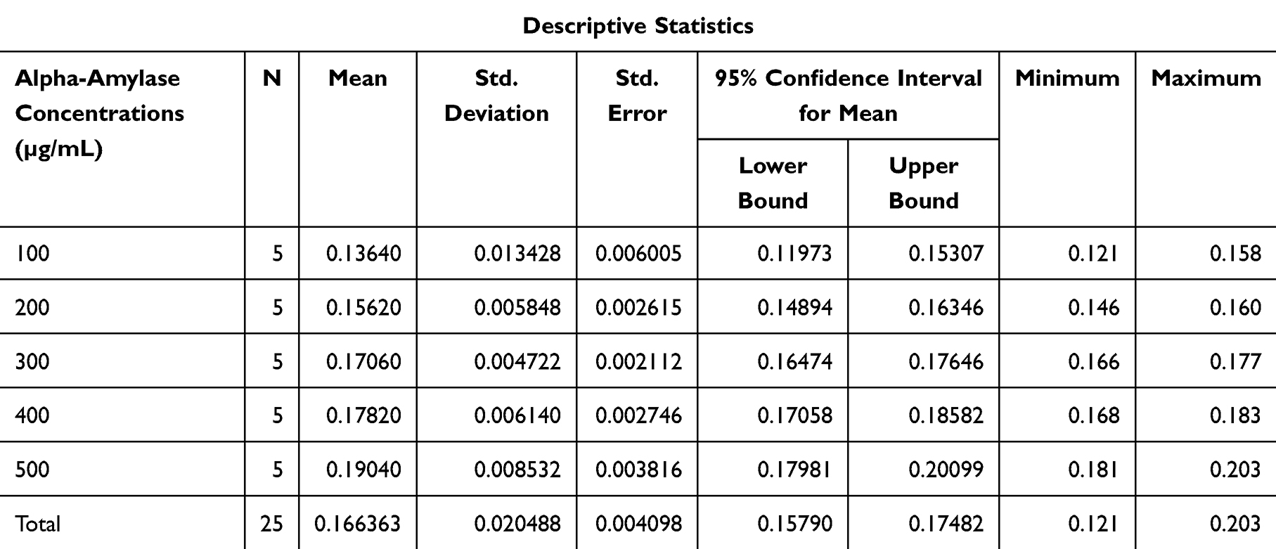 |
Table 1 Descriptive Statistics |
Tables 2 and 3 provide a detailed analysis of the current measurements recorded across the five RBD concentration levels (100, 200, 300, 400, and 500 µg/mL). According to the ANOVA analysis, significant differences were observed among the mean current values for the different concentration levels (p < 0.001) (Table 1). The effect sizes (Table 2) indicate a large impact of RBD concentration on current measurements, with η² = 0.862.
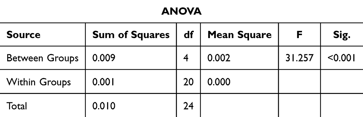 |
Table 2 One-Way ANOVA Test Result |
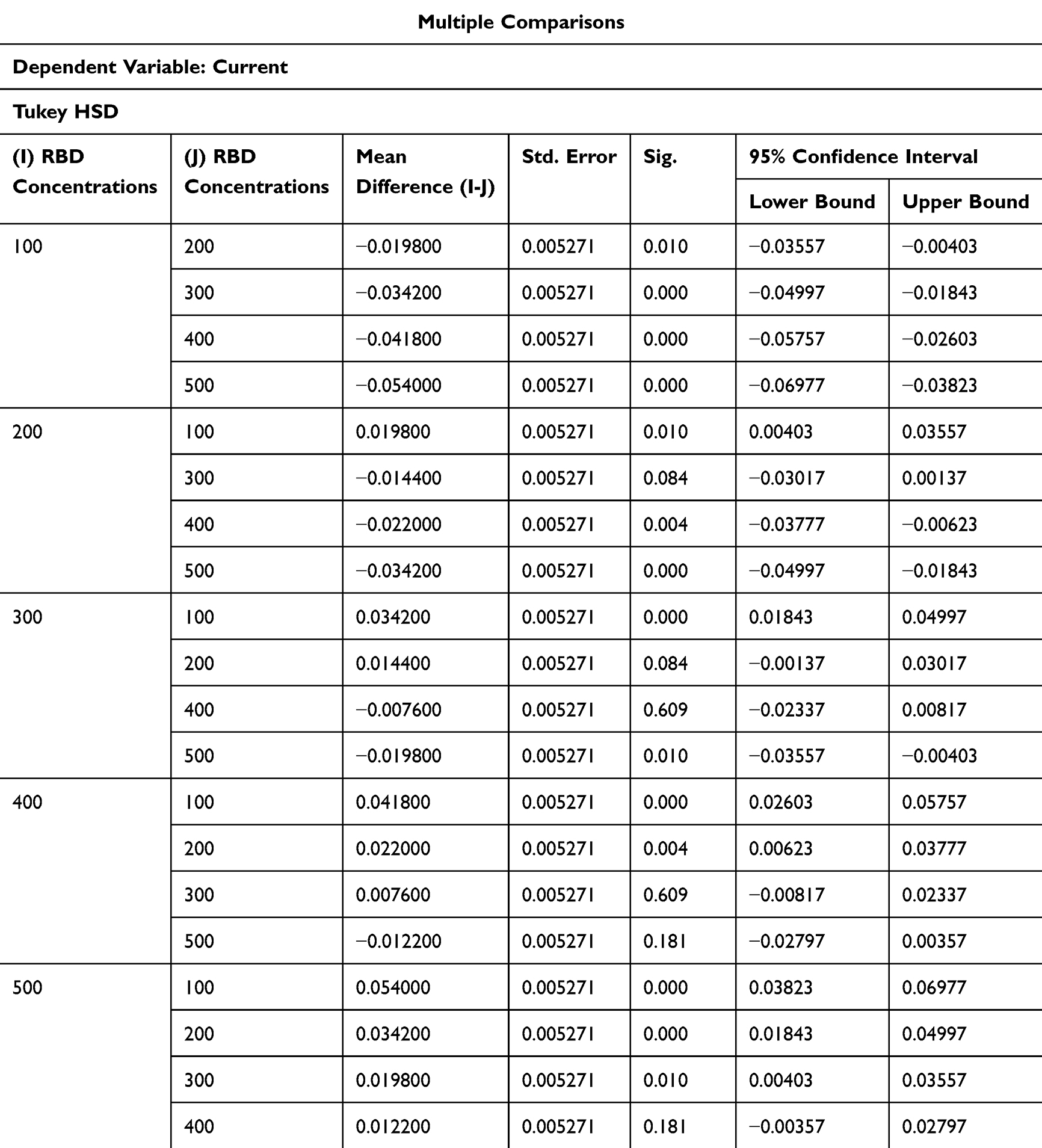 |
Table 3 Post-Hoc Tests (Tukey’s HSD) |
Post-hoc analysis was conducted using Tukey’s HSD test to explore the pairwise differences in current measurements across the five RBD concentration levels (100, 200, 300, 400, and 500 µg/mL) (the data was not shown). The results demonstrated that the mean current value at 100 µg/mL was significantly lower than those at higher concentrations, with a particularly large difference observed when compared to 500 µg/mL (mean difference = −0.054, p < 0.001). Similarly, the current values at 200 µg/mL were significantly different from those at 400 µg/mL and 500 µg/mL, with mean differences of −0.022 (p = 0.004) and −0.034 (p < 0.001), respectively.
While significant differences were observed between most concentration levels, the comparison between 300 µg/mL and 400 µg/mL did not yield a statistically significant result (p = 0.609), indicating similar current values at these concentrations. Conversely, 300 µg/mL showed significant differences compared to both lower (100 µg/mL) and higher (500 µg/mL) concentrations. These findings highlight the increasing trend in current measurements as RBD concentration increases, further supporting the robustness of the sensor’s performance in detecting variations across a broad concentration range.
Comparison to Other Sensor Tools
Salivary alpha-amylase (sAA) has been extensively utilized as a biomarker due to its ability to reflect physiological responses, particularly those related to stress and the activation of the sympathetic nervous system.33 It can be measured using various types of sensors, making it a versatile tool for both clinical and research applications. In a study conducted by Bañuelos et al, an experiment was designed to investigate the correlation between the body’s sympathetic nervous system response and salivary alpha-amylase production. The study involved 13 students, from whom saliva samples were collected following a presentation. The passive drool method was employed for saliva collection, which is a common non-invasive technique. The analysis of these samples was performed using the enzyme-linked immunosorbent assay (ELISA), a highly sensitive and widely used method for detecting specific proteins, including enzymes like alpha-amylase. The results of the study showed that the average sAA levels before the presentation were approximately 1.8 log units. After the presentation, there was a notable increase, with sAA levels rising to 2.1 log units. This significant increase in alpha-amylase production following the presentation strongly indicates that the sympathetic nervous system was activated in response to the stress of the public speaking task. Such findings align with previous research that suggests alpha-amylase levels are closely linked to acute stress responses, further validating its role as a reliable biomarker for sympathetic nervous system activity.34
In a separate study conducted by Hsiao et al, a novel colorimetry-based sensor was employed to measure the concentration of alpha-amylase in saliva. This sensor was designed to detect the enzyme by quantifying the amount of reduced sugar produced from the hydrolysis of soluble starch, a reaction catalyzed by alpha-amylase. The experiment aimed to assess the effectiveness of the colorimetric sensor in providing accurate and rapid measurements of salivary alpha-amylase (sAA) levels. The findings from the study demonstrated that the alpha-amylase concentration in the saliva samples ranged from 0.1 to 1.0 U/mL. Notably, the sensor exhibited a robust response to alpha-amylase activity, with readings reaching up to 2.0 U/mL. The limit of detection (LOD) for the sensor was determined to be 0.1 U/mL, highlighting its sensitivity to even minute concentrations of the enzyme. Additionally, the sensor’s response frequency was recorded at 1703.4 hz (log U/mL)−1, which indicates its capability to translate enzyme activity into quantifiable data. However, the linear detection range of this colorimetric sensor prototype was limited to alpha-amylase activities below 2.0 U/mL. This range is significantly narrower than the typical diurnal variation of salivary alpha-amylase, which generally falls between 10 and 300 U/mL in healthy individuals. Despite this limitation, the study underscores the potential of colorimetry-based sensors as a cost-effective and easy-to-use alternative for detecting alpha-amylase in saliva. With further optimization, particularly in expanding the detection range, these sensors could be adapted for more comprehensive diagnostic applications.35
Limitations of the Study
Out of the 46 screen-printed electrodes initially prepared for this study, 25 were successfully utilized in the final analysis. Despite being newly manufactured, a significant portion of the electrodes did not meet the rigorous standards required for accurate and reliable measurements. Some electrodes exhibited inconsistent current readings across RBD concentrations, while others failed to produce detectable signals on the potentiostat. These issues were primarily attributed to procedural challenges during the modification and preparation stages, which adversely affected the electrodes’ electrochemical performance and overall usability.
One of the primary factors influencing the performance of the electrodes was temperature. During the course of our experiment, the average ambient temperature was 26°C, which significantly exceeded the optimal temperature of 17°C required for precise measurement. This temperature discrepancy likely contributed to the suboptimal electrode performance.36 In line with our findings, a study by Rebelo et al highlights the importance of temperature control during the electrode preparation and measurement process.37 Their research demonstrated that after incubating the screen-printed electrodes with an alpha-amylase solution, the electrodes needed to be stored at 4°C, with a strict 15-minute storage window to maintain their integrity and functionality.
Furthermore, the methodology used to modify the electrodes played a significant role in the results. The type of biomarker used, its concentration, and the incubation time were all critical factors that influenced the outcomes.38,39 These procedural elements may have contributed to the observed variability in our results, particularly when compared to the work of Gulati et al, whose study demonstrated the successful use of antibodies as biomarkers under more controlled conditions.22 As shown in Figure 6 of their study, the use of antibodies provided highly reliable detection due to optimized incubation times and strict adherence to methodological protocols.
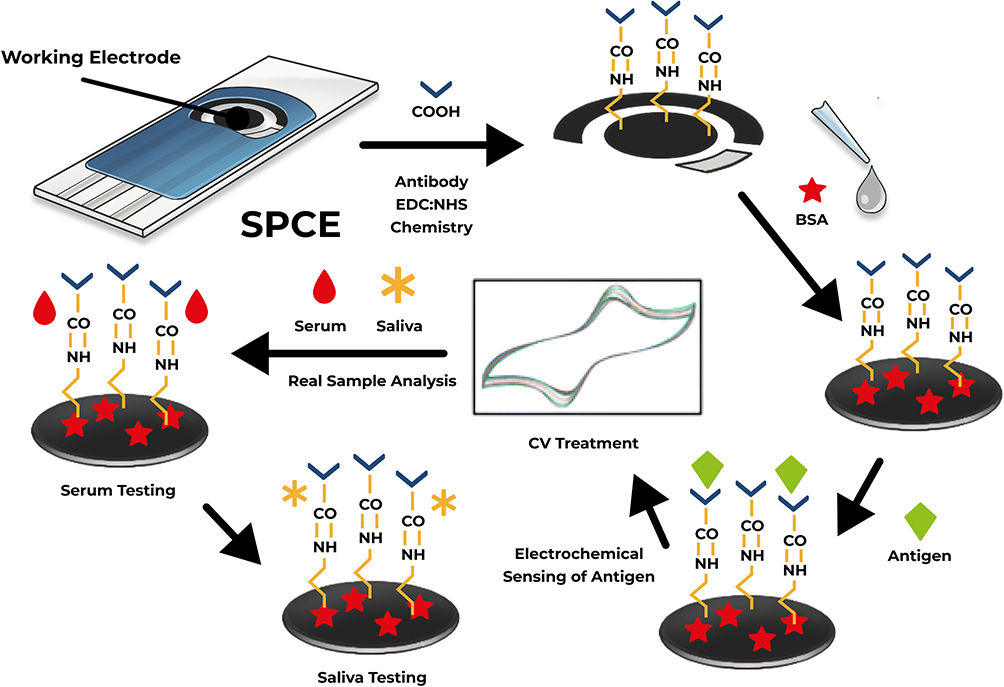 |
Figure 6 Illustration adapted from the study by Gulati et al. Schematic of the SPE-based Immunosensor Through Immobilization of Antibodies, BSA, and Antigens (Source: Author’s Illustration). |
The combination of environmental factors, such as temperature, and methodological variables, including biomarker type and procedural adjustments, underscores the complexity of achieving consistent results with screen-printed electrodes.40–42 Our findings suggest that further refinement of the experimental process is necessary, particularly in controlling external conditions and optimizing the modification procedures. This will ensure that the electrodes can consistently meet the required performance standards for reliable alpha-amylase detection. In summary, while this research demonstrates the potential of screen-printed electrodes in clinical diagnostics, it also highlights the challenges associated with achieving consistent results. Careful attention to temperature, biomarker selection, concentration, and storage conditions is essential for future studies to maximize the performance of these electrodes in detecting biomarkers like alpha-amylase.43
Discussion
Alpha-amylase has been extensively utilized as a biomarker in saliva due to several key advantages, including its high sensitivity, low cost, rapid response time, and the ability to detect small concentrations.23 These characteristics make it particularly valuable for noninvasive diagnostic applications.44 One of the most significant benefits is its small detection limit, which enables early detection of various physiological and pathological conditions.
In a study conducted by Garcia et al, a sample composed of starch and saliva with concentrations ranging from 0.5% to 2.0% was mixed to initiate a hydrolysis reaction. This reaction leads to the breakdown of starch into simpler sugars, which correlates with alpha-amylase activity. Additionally, the compound [Fe(CN)6]3− was added to the sample, serving as an electrochemical mediator. Following this preparation, the entire sample was subjected to electrochemical analysis, and the results indicated high sensitivity and accuracy. Specifically, the sensitivity was determined to be 10.7 mA/(log U mL−1), and the limit of detection (LOD) was 1.1 U mL−1. These values demonstrate the precision and efficiency of the electrochemical method in detecting alpha-amylase at very low concentrations, emphasizing its potential for clinical use.45
In another study conducted by Liu et al, the researchers measured ten sets of saliva samples to further investigate the electrochemical detection of alpha-amylase. For this experiment, 100 μL of each saliva sample was first filtered, and NAD+ (nicotinamide adenine dinucleotide) along with an acetate buffer was added to the samples. The purpose of adding NAD+ was to facilitate the reaction and enhance the accuracy of the detection process. The samples were then analyzed using a novel sensing chip integrated with an advanced electrochemical analyzer, which represents a new model of electrochemical technology aimed at improving the detection and quantification of biomarkers like alpha-amylase.46 The normal alpha-amylase activity in saliva for healthy individuals typically ranges from 2 to 900 U mL−1.47 However, the test results for the measured samples exhibited significantly higher alpha-amylase activity, with values ranging from 3,000 to 11,000 U mL−1. This marked elevation in alpha-amylase levels was considered a significant deviation from the normal range, suggesting a possible link to certain health conditions or stress-related physiological responses. The substantial increase in alpha-amylase activity detected through this advanced electrochemical method underscores its effectiveness in identifying abnormal conditions that might otherwise go undetected with traditional diagnostic tools.48
Compared to the other study, our research highlight the growing relevance of electrochemical detection methods for alpha-amylase in saliva, further solidifying its role as a promising biomarker for various health conditions. In this study, the method demonstrated high sensitivity and precision, as evidenced by statistically significant differences in current measurements across varying RBD concentrations (F(4, 20) = 31.257, p < 0.001) confirmed by one-way ANOVA. Post-hoc Tukey HSD tests revealed significant pairwise differences between most concentration levels, emphasizing the method’s capability to distinguish between subtle changes in alpha-amylase concentration, with a large effect size (η² = 0.862).
Additionally, the linear regression analysis yielded a strong calibration curve with an R² value of 0.9735, indicating excellent linearity and reliability of the method for quantitative analysis. These findings reinforce the potential of this noninvasive, rapid, and accurate approach for clinical diagnostics, particularly in early disease detection and health monitoring, even in resource-constrained settings.
Conclusion
An electrochemical sensor based on a gold nanoparticle-modified screen-printed electrode (SPE) was developed for the quantitative measurement of alpha-amylase. This sensor demonstrated a strong linear response in alpha-amylase concentrations ranging from 100 to 500 units, with a correlation coefficient of R² = 0.9735. The device also exhibited a limit of detection (LOD) of 104.252 units and a limit of quantification (LOQ) of 315.915 units, emphasizing its sensitivity and reliability for detecting low levels of alpha-amylase.
Despite being sensitive to potential external interferences, such as pH fluctuations and the presence of other biomolecules, the sensor showed promising performance. This study highlights the utility of the electrochemical sensor as a non-invasive early detection tool, leveraging saliva as a biomarker to improve patient comfort, compliance, and screening frequency.
However, further research is necessary to validate its clinical application, particularly in screening and diagnosis contexts. Future studies could focus on optimizing the sensor for better performance under varying conditions and exploring its utility in specific medical applications, such as stress monitoring or diagnostics for pancreatic function. Clinical validation in real-world scenarios will be essential to establish its robustness and efficacy in routine medical use.
Acknowledgments
We would like to express our gratitude to Universitas Padjadjaran for providing a supportive environment and resources that enabled us to conduct this research. We are grateful for the opportunities. We have had to work with esteemed faculty members and collaborate with talented peers.
Disclosure
The author(s) report no conflicts of interest in this work.
References
1. Gampa SC, Garimella SV. Marking The Cancers: importance Of Biomarkers. In: Kaur D, Prahladbhai DrPV VM, et al., eds. Futuristic Trends in Biotechnology Volume 3 Book 2. First. Iterative International Publisher, Selfypage Developers Pvt Ltd; 2024:178–191. doi:10.58532/V3BGBT2P2CH7
2. Ferreira VG, Rossini EL, Araújo LX, Almeida MB, Carrilho E. Biomarkers: an introduction. In: Biosensors in Precision Medicine. Elsevier. 2024;2024. 3–34. doi:10.1016/B978-0-443-15380-8.00001-1
3. Zalewska A, Zwierz K, Zółkowski K, Gindzieński A. Structure and biosynthesis of human salivary mucins. Acta Biochim Pol. 2000;47(4):1067–1079. doi:10.18388/abp.2000_3960
4. Humphrey SP, Williamson RT. A review of saliva: normal composition, flow, and function. J Prosthet Dent. 2001;85(2):162–169. doi:10.1067/mpr.2001.113778
5. Iorgulescu G. Saliva between normal and pathological.Important factors in determining systemic and oral health. J Med Life. 2009;2(3):303–307.
6. Zhou Y, Liu Z. Saliva biomarkers in oral disease. Clin Chim Acta. 2023;548:117503. doi:10.1016/j.cca.2023.117503
7. Lima DP, Diniz DG, Moimaz SAS, Sumida DH, Okamoto AC. Saliva: reflection of the body. Inter J Infect Dis. 2010;14(3):e184–e188. doi:10.1016/j.ijid.2009.04.022
8. Martina E, Campanati A, Diotallevi F, Offidani A. Saliva and Oral Diseases. J Clin Med. 2020;9(2):466. doi:10.3390/jcm9020466
9. Al Habobe H, Haverkort EB, Nazmi K, Van Splunter AP, Pieters RHH, Bikker FJ. The impact of saliva collection methods on measured salivary biomarker levels. Clin Chim Acta. 2024;552:117628. doi:10.1016/j.cca.2023.117628
10. Katebi K, Hassanpour S, Eslami H, Salehnia F, Hosseinifard H. Effect of Pilocarpine Mouthwash on Salivary Flow Rate in Patients with Xerostomia: a Systematic Review and Meta-Analysis. J Dent. 2023;24(1 Suppl):76–83. doi:10.30476/dentjods.2022.94335.1778
11. Peres MA, Macpherson LMD, Weyant RJ, et al. Oral diseases: a global public health challenge. Lancet. 2019;394(10194):249–260. doi:10.1016/S0140-6736(19)31146-8
12. Heitz-Mayfield LJA. Conventional diagnostic criteria for periodontal diseases (plaque-induced gingivitis and periodontitis). Periodontology 2000. 2024;2024:1. doi:10.1111/prd.12579
13. Chaurasia A, Alam SI, Singh N. Oral cancer diagnostics: an overview. Natl J Maxillofac Surg. 2021;12(3):324–332. doi:10.4103/njms.NJMS_130_20
14. Patil S, Albogami S, Hosmani J, et al. Artificial Intelligence in the Diagnosis of Oral Diseases: applications and Pitfalls. Diagnostics. 2022;12(5):1029. doi:10.3390/diagnostics12051029
15. Alhazmi A, Alhazmi Y, Makrami A, et al. Application of artificial intelligence and machine learning for prediction of oral cancer risk. J Oral Pathol Med. 2021;50(5):444–450. doi:10.1111/jop.13157
16. Ochoa-Ruiz AG, Parra G, López-Espinoza D, et al. Electrochemical Immunosensors: the Evolution from Elisa to EμPADs. Electroanalysis. 2023;35(4):e202200053. doi:10.1002/elan.202200053
17. Wu L, Wang X, Wu C, et al. Ultrasensitive SARS-CoV-2 diagnosis by CRISPR-based screen-printed carbon electrode. Anal Chim Acta. 2022;1221:340120. doi:10.1016/j.aca.2022.340120
18. Zhao Y, Zhang H, Li Y, et al. Glycated Hemoglobin Electrochemical Immunosensor Based on Screen-Printed Electrode. Biosensors. 2022;12(10):902. doi:10.3390/bios12100902
19. Ferreira DC, Batistuti MR, Bachour B, Mulato M. Aptasensor based on screen-printed electrode for breast cancer detection in undiluted human serum. Bioelectrochemistry. 2021;137:107586. doi:10.1016/j.bioelechem.2020.107586
20. Matvieiev O, Šelešovská R, Vojs M, et al. Novel Screen-Printed Sensor with Chemically Deposited Boron-Doped Diamond Electrode: preparation, Characterization, and Application. Biosensors. 2022;12(4):241. doi:10.3390/bios12040241
21. Jafari M, Hasanzadeh M. Non-invasive bioassay of Cytokeratin Fragment 21.1 (Cyfra 21.1) protein in human saliva samples using immunoreaction method: an efficient platform for early-stage diagnosis of oral cancer based on biomedicine. Biomed Pharmacother. 2020;131:110671. doi:10.1016/j.biopha.2020.110671
22. Gulati P, Singh AK, Yadav AK, et al. Nano-modified screen-printed electrode-based electrochemical immunosensors for oral cancer biomarker detection in undiluted human serum and saliva samples. Nanoscale Adv. 2024;6(2):705–721. doi:10.1039/D3NA00682D
23. Ali N, Nater UM. Salivary Alpha-Amylase as a Biomarker of Stress in Behavioral Medicine. Int J Behav Med. 2020;27(3):337–342. doi:10.1007/s12529-019-09843-x
24. Akinfemiwa O, Zubair M, Muniraj T. Amylase. In: StatPearls. StatPearls Publishing; 2024. Avaolable from: http://www.ncbi.nlm.nih.gov/books/NBK557738/.
25. Akkinepally H, Tejasvi MA, Kavitha NL, Pokala A, Reddy P, Keerthi VR. Estimation of Alpha-amylase in Smokers with and without Leukoplakia and Oral Cancer—A Comparative Study. J Indian Acad Oral Med Radiol. 2023;35(3):331. doi:10.4103/jiaomr.jiaomr_230_23
26. Bel’skaya L, Sarf E, Solomatin D, Kosenok V. Diagnostic and Prognostic Value of Salivary Biochemical Markers in Oral Squamous Cell Carcinoma. Diagnostics. 2020;10(10):818. doi:10.3390/diagnostics10100818
27. Kumar P, Gupta S, Das BC. Saliva as a potential non-invasive liquid biopsy for early and easy diagnosis/prognosis of head and neck cancer. Transl Oncol. 2024;40:101827. doi:10.1016/j.tranon.2023.101827
28. Celesti C, Giofrè SV, Espro C, Legnani L, Neri G, Iannazzo D. Modified Gold Screen-Printed Electrodes for the Determination of Heavy Metals. Sensors. 2024;24(15):4935. doi:10.3390/s24154935
29. Liang G, He Z, Zhen J, et al. Development of the screen-printed electrodes: a mini review on the application for pesticide detection. Environ. Technol. Innovation. 2022;28:102922. doi:10.1016/j.eti.2022.102922
30. Ariasena E, Putra Noerrizky IA, Althof RR, Anshori I Screen-Printed Carbon Electrode Fabrication Method for Electrochemical Biosensor Application. In: Lelono A, Akbar Bahar M, Wathon S, et al., eds.
31. Paimard G, Ghasali E, Baeza M. Screen-Printed Electrodes: fabrication, Modification, and Biosensing Applications. Chemosensors. 2023;11(2):113. doi:10.3390/chemosensors11020113
32. Matvieiev O, Šelešovská R, Marton M, et al. Effect of different modification by gold nanoparticles on the electrochemical performance of screen-printed sensors with boron-doped diamond electrode. Sci Rep. 2023;13(1):21525. doi:10.1038/s41598-023-48834-7
33. Contreras-Aguilar MD, Escribano D, Martínez-Subiela S, Martínez-Miró S, Cerón JJ, Tecles F. Changes in alpha-amylase activity, concentration and isoforms in pigs after an experimental acute stress model: an exploratory study. BMC Veterinary Res. 2018;14(1):256. doi:10.1186/s12917-018-1581-2
34. Bañuelos MS, Musleh A, Olson LE. Measuring Salivary Alpha-Amylase in the Undergraduate Neuroscience Laboratory. J Undergrad Neurosci Educ. 2017;16(1):A23–A27.
35. Hsiao HY, Chen RLC, Chou CC, Cheng TJ. Hand-held Colorimetry Sensor Platform for Determining Salivary α-Amylase Activity and Its Applications for Stress Assessment. Sensors. 2019;19(7):1571. doi:10.3390/s19071571
36. Huang Q, Yang Z, Mao J. Mechanisms of the decrease in low-temperature electrochemical performance of Li4Ti5O12-based anode materials. Sci Rep. 2017;7(1):15292. doi:10.1038/s41598-017-15504-4
37. Rebelo TSCR, Miranda IM, Brandão ATSC, et al. A Disposable Saliva Electrochemical MIP-Based Biosensor for Detection of the Stress Biomarker α-Amylase in Point-of-Care Applications. Electrochem. 2021;2(3):427–438. doi:10.3390/electrochem2030028
38. Salek Maghsoudi A, Hassani S, Rezaei Akmal M, et al. An Electrochemical Aptasensor Platform Based on Flower-Like Gold Microstructure-Modified Screen-Printed Carbon Electrode for Detection of Serpin A12 as a Type 2 Diabetes Biomarker. Int J Nanomed. 2020;15:2219–2230. doi:10.2147/IJN.S244315
39. Hosseine M, Naghib SM, Khodadadi A. Label-free electrochemical biosensor based on green-synthesized reduced graphene oxide/Fe3O4/nafion/polyaniline for ultrasensitive detection of SKBR3 cell line of HER2 breast cancer biomarker. Sci Rep. 2024;14(1):11928. doi:10.1038/s41598-024-62231-8
40. Bhatia D, Paul S, Acharjee T, Ramachairy SS. Biosensors and their widespread impact on human health. Sensors Int. 2024;5:100257. doi:10.1016/j.sintl.2023.100257
41. Nyabadza A, McCarthy É, Makhesana M, et al. A review of physical, chemical and biological synthesis methods of bimetallic nanoparticles and applications in sensing, water treatment, biomedicine, catalysis and hydrogen storage. Adv Colloid Interface Sci. 2023;321:103010. doi:10.1016/j.cis.2023.103010
42. Habboush S, Rojas S, Rodríguez N, Rivadeneyra A. The Role of Interdigitated Electrodes in Printed and Flexible Electronics. Sensors. 2024;24(9):2717. doi:10.3390/s24092717
43. Liu R, Ye X, Cui T. Recent Progress of Biomarker Detection Sensors. Research. 2020;2020:7949037. doi:10.34133/2020/7949037
44. Nater UM, Rohleder N. Salivary alpha-amylase as a non-invasive biomarker for the sympathetic nervous system: current state of research. Psychoneuroendocrinol. 2009;34(4):486–496. doi:10.1016/j.psyneuen.2009.01.014
45. Garcia PT, Guimarães LN, Dias AA, Ulhoa CJ, Coltro WKT. Amperometric detection of salivary α-amylase on screen-printed carbon electrodes as a simple and inexpensive alternative for point-of-care testing. Sensors and Actuat B Chem. 2018;258:342–348. doi:10.1016/j.snb.2017.11.068
46. Liu C, Gong X, Yang X, et al. Development of enzyme–inorganic hybrid nanoflower-modified electrodes and a smartphone-controlled electrochemical analyzer for point-of-care testing of salivary amylase in saliva. Nanoscale. 2024;16(1):212–222. doi:10.1039/D3NR04388F
47. Mandel AL, Peyrot Des Gachons C, Plank KL, Alarcon S, Breslin PAS. Individual Differences in AMY1 Gene Copy Number, Salivary α-Amylase Levels, and the Perception of Oral Starch. PLoS One. 2010;5(10):e13352. doi:10.1371/journal.pone.0013352
48. Zhang J, Cui J, Liu Y, Chen Y, Li G. A novel electrochemical method to determine α-amylase activity. Analyst. 2014;139(13):3429–3433. doi:10.1039/C3AN01839C
 © 2025 The Author(s). This work is published and licensed by Dove Medical Press Limited. The
full terms of this license are available at https://www.dovepress.com/terms
and incorporate the Creative Commons Attribution
- Non Commercial (unported, 3.0) License.
By accessing the work you hereby accept the Terms. Non-commercial uses of the work are permitted
without any further permission from Dove Medical Press Limited, provided the work is properly
attributed. For permission for commercial use of this work, please see paragraphs 4.2 and 5 of our Terms.
© 2025 The Author(s). This work is published and licensed by Dove Medical Press Limited. The
full terms of this license are available at https://www.dovepress.com/terms
and incorporate the Creative Commons Attribution
- Non Commercial (unported, 3.0) License.
By accessing the work you hereby accept the Terms. Non-commercial uses of the work are permitted
without any further permission from Dove Medical Press Limited, provided the work is properly
attributed. For permission for commercial use of this work, please see paragraphs 4.2 and 5 of our Terms.