Back to Journals » International Journal of Women's Health » Volume 17
Neutrophil Extracellular Traps and Endometriosis: Insights from a Case-Control Study
Authors Xu Z, Ji R, Wang M, Li T, Hu H
Received 2 November 2024
Accepted for publication 8 June 2025
Published 16 July 2025 Volume 2025:17 Pages 2121—2131
DOI https://doi.org/10.2147/IJWH.S504327
Checked for plagiarism Yes
Review by Single anonymous peer review
Peer reviewer comments 2
Editor who approved publication: Dr Vinay Kumar
Zailong Xu,1,* Rui Ji,2,* Mei Wang,1 Tiantian Li,1 Hui Hu1
1Department of Obstetrics and Gynecology, Pudong New Area People’s Hospital in Shanghai, Shanghai, People’s Republic of China; 2Department of Obstetrics and Gynecology, Shanghai Sixth People’s Hospital, Shanghai, People’s Republic of China
*These authors contributed equally to this work
Correspondence: Hui Hu, Department of Obstetrics and Gynecology, Pudong New Area People’s Hospital in Shanghai, NO. 490 Chuanhuan South Road, Pudong New Area, Shanghai, 201200, People’s Republic of China, Email [email protected]
Background: Endometriosis (EMS) is characterized by the occurrence, growth, infiltration, and recurrent bleeding of endometrial tissue outside the uterus. Recent studies have revealed a close association between Neutrophil Extracellular Traps (NETs) and the proliferation and metastasis of tumors. As EMS and tumours have similar biological behaviours, we hypothesised that key factors in tumourigenesis may also significantly influence the pathogenesis of EMS. Therefore, this study aimed to explore the potential link that exists between NETs and endometriosis.
Methods: A total of 190 patients were selected and divided into control and endometriosis groups (95 patients each) based on inclusion and exclusion criteria. Samples from ectopic and eutopic endometrium in the experimental group, eutopic endometrium in the control group, and peripheral blood serum and neutrophils (PMN) were collected and analyzed. ROC curve analysis was used to evaluate the diagnostic ability of NETs-associated markers to differentiate endometriosis patients from controls.
Results: The analysis of both patient groups revealed that levels of circulating free DNA (cf-DNA), nucleosomes, and neutrophil elastase (NE) were significantly higher in the experimental group compared to the controls. Further analysis indicated that the levels of NETs-related factors were elevated in patients with infertility and there was a positive correlation between pain severity and NETs markers in patients experiencing pain. Additionally, the ROC curve analysis demonstrated significant differences in cf-DNA, nucleosomes, and NE levels between the experimental and control groups (P < 0.05).
Conclusion: NETs-associated markers may be linked to endometriosis, likely due to their involvement in tumor proliferation and metastasis, which share similarities with endometriosis. Further studies are needed to explore the mechanisms connecting endometriosis and NETs markers.
Keywords: NETs, markers, endometriosis, infertility, case-control study
Introduction
Endometriosis (EMS) is characterized by the occurrence, growth, infiltration, and recurrent bleeding of endometrial tissue outside the uterus. The pathogenesis of EMS involves a complex interplay of genetic, environmental, infectious, and immunological factors.1 Despite numerous proposed models, the exact mechanisms underlying endometriosis remain unclear. Studies suggest that hormonal imbalances, inflammatory mediators, immune dysfunction, and genetic and epigenetic factors, along with environmental influences, significantly contribute to EMS development.2 Neutrophil Extracellular Traps (NETs) are web-like structures composed of DNA and substances such as circulating free DNA (cf-DNA), calprotectin, nucleosomes, DNase I, neutrophil elastase (NE), and myeloperoxidase (MPO). Released by neutrophils, NETs have been shown to promote the growth and metastasis of several cancers, including breast, liver, and ovarian cancers.3 NETosis is closely related to hormones such as estrogen and progesterone, which are able to regulate the shedding of the endometrium independent of pregnancy status.4 Previous research has highlighted an increase in NETs in the peritoneal fluid and plasma of EMS patients, and in animal models, NETs have also been implicated in the progression of endometrial fibrosis.5 However, the precise correlation between NETs and EMS has yet to be firmly established.
Recent findings suggest an increase in NETosis in peripheral blood, with indirect evidence pointing to a potential relationship between NETosis and endometriosis. NETs, which can capable of capturing, neutralizing, and killing bacteria,6 have also been found to play a significant role in the proliferation, adhesion, and metastasis of tumors. NETs promote cancer cell adhesion and affect metastasis by releasing pro-inflammatory cytokines, such as TNFα and IL-1β. These cytokines enhance tumor cell adhesion to endothelial cells, thereby promoting systemic inflammation and cell invasion.7 Additionally, NET-induced disruption of endothelial cell junctions increases vascular permeability, facilitating cell metastasis. cfDNA, as fragmented DNA released by necrotic or apoptotic cells, has been shown to promote inflammation and immune dysregulation in the cancer microenvironment. Similarly, nucleosomes - the basic units of chromatin formed by DNA wrapped around histone proteins - have been found to be elevated in the circulation of patients with a variety of malignancies, including EMS, suggesting their potential as diagnostic biomarkers. In addition, NETs have emerged as key players in the regulation of immune response and inflammation as reticulocytes released by neutrophils during inflammation and infection. The formation and release of NETs are part of the inflammatory response, and the onset of EMS is closely related to peritoneal inflammation. In an inflammatory environment, neutrophils are activated and release NETs, which may further exacerbate peritoneal inflammation and thereby promote the development of EMS. NETs can affect vascular function and increase vascular permeability. This may lead to more inflammatory cells and factors penetrating into the peritoneal cavity, further intensifying the inflammatory response and pathological changes of EMS. NETs may play a role in the pathogenesis of EMS through multiple mechanisms, such as promoting inflammation, cell proliferation and invasion, increasing vascular permeability, disrupting immune regulation balance, and promoting fibrosis.
Toll-like receptors (TLRs) act as protective immunological sentinels that detect pathogen-associated molecular patterns (PAMPs), such as unmethylated double-stranded DNA (CpG), single-stranded RNA (ssRNA), lipoproteins, lipopolysaccharides (LPS), and flagellin.8 In myeloid cells of the innate immune system, TLRs trigger the secretion of inflammatory cytokines, which attract lymphocytes to initiate an adaptive, antigen-specific immune response. Numerous studies have demonstrated that the function of NETs depends on these receptors and that NETs can facilitate changes in tumor cell proliferation and invasion.
Infertility and pain are two common clinical manifestations in patients with endometriosis (EMS) and often place a great physical and mental burden on patients. NETs play an important role in inflammatory response, tissue damage and repair. We noted that during the occurrence and development of EMS, the local inflammatory response was significantly enhanced, accompanied by the destruction and remodeling of tissue structure. Therefore, we speculate that NETs may be involved in this process and contribute to the appearance of symptoms such as infertility and pain. Specifically, the release of NETs may promote the persistence and intensification of local inflammatory responses, which in turn have adverse effects on the reproductive system and pain receptors.
Ectopic endometrial tissue within the pelvic cavity can form lesions that undergo adhesion, invasion, and angiogenesis, which are processes closely resembling the metastatic seeding of tumors. Mechanisms such as the overexpression of non-coding RNAs (miRNA, lncRNA), abnormal activation or silencing of DNA methylation, gene mismatches, and chromosomal deletions can lead to dysfunction in cellular processes. These conditions are crucial in promoting abnormal angiogenesis, cell proliferation, invasion, and metastasis, which are also prevalent in endometriosis. Importantly, these mechanisms, which facilitate tumor growth, also contribute to the formation of endometriotic lesions in patients with retrograde menstruation. Furthermore, endometriosis itself can cause pelvic inflammation, with repeated inflammatory responses leading to an abnormal increase in inflammatory cytokines. This, in turn, mediates the adhesion, proliferation, differentiation, and invasion of EMS lesions. Literature has shown an increase in NETs9 in the peritoneal lavage fluid of patients with endometriosis. However, the relevance and mechanisms of action of NETs in endometriosis remain unclear. At present, although some studies on the relationship between NETs and gynecological diseases have been reported, in-depth studies on the specific mechanism of action of NETs in EMS and its relationship with symptoms such as infertility and pain are relatively lacking. Therefore, this study focuses on exploring the potential connection between NETs and endometriosis by employing experimental methods such as Enzyme-Linked Immunosorbent Assay (ELISA) and fluorescent staining to quantitatively analyze cf-DNA, calprotectin, nucleosomes, DNase I, MPO, and NE in clinical peripheral blood samples. This case-control study further confirms the association between NETs and EMS, paving the way for future research into the mechanisms linking NETs with endometriosis. By exploring the connection between NETs and EMS, the medical community has deepened its understanding of the pathogenesis of EMS, providing a new theoretical basis for the prevention and treatment of the disease. By revealing the role of NETs in the progression of EMS, researchers may provide clues for the development of new treatment strategies targeting NETs, thereby slowing down or halting the progression of EMS.
Methods
Study Design
This study adhered strictly to the inclusion and exclusion criteria, selecting patients who underwent surgery for endometriosis at our institution between January 2022 to November 2023. All research protocols were approved by the ethics committee, and informed consent was obtained from all participants prior to sample collection. This study is in line with the Declaration of Helsinki. The control group were patients who had undergone surgery for similar symptoms, such as pelvic pain or infertility, but had no endometriosis confirmed by laparoscopic evaluation or histology. Patients in the control group were free of endometriosis and the following inclusion and exclusion criteria were also followed.
Inclusion Criteria
(1) patients who underwent surgical treatment for endometriosis in our hospital between January 2022 and November 2023 were selected; (2) the age range of the included patients was set to be between 18 and 45 years old; (3) all participants voluntarily signed an informed consent form to participate in the study and allowed the collection of their relevant samples and clinical data; (4) Included patients were required to undergo laparoscopic or open surgery for removal of endometriosis lesions or related treatment.
Exclusion Criteria
(1) Patients with autoimmune diseases; (2) Patients diagnosed with malignant tumors; (3) Patients who have undergone hormone therapy in the past three months; (4) Patients who have experienced acute inflammation or severe infections within the past month, or chronic inflammation within the past three months.
Patient Data
Patient characteristics, including age, BMI, marital status, smoking history, number of pregnancies, family history of endometriosis, and the number of symptoms, were recorded using case report forms. There were no significant differences between the two groups in terms of age, BMI, marital status, smoking history, number of pregnancies, lymphocytes, neutrophils, and platelets, ensuring comparability. Compared to the control group, the experimental group exhibited significant increases in endometriosis, family history of the condition, and number of symptoms, all with statistical significance (P<0.05).
Biomarker Assays
Peripheral blood was collected using anticoagulant tubes, then centrifuged at 3000 rpm for 5 minutes. The supernatant was transferred to EP tubes in 100μL aliquots and stored at −80°C. MPO levels in peripheral blood were measured using a test kit from Shanghai Shuangying Biotechnology Co., Ltd. cf-DNA levels were determined using a fluorescent dye method with kits purchased from Roche, Germany, and results were obtained with the Multiskan FC microplate reader from Thermo Fisher Scientific, USA. Serum levels of neutrophil elastase (NE) and calprotectin were quantified using ELISA kits from Shanghai Runyu Biotechnology Co., Ltd. and Hangzhou Zhenyoupin Biotechnology Co., Ltd., respectively.
Statistical Methods
Statistical analyses were conducted using GraphPad 8.0 software. Quantitative data are presented as mean ± standard deviation. Comparisons between two independent samples following anormal distribution were performed using the t-test, while comparisons for non-normally distributed data utilized the non-parametric Mann–Whitney U-test. One-way ANOVA was used for normally distributed data involving multiple groups, with pairwise comparisons conducted using the LSD-t test. Categorical data were analyzed with the chi-square test, and a P-value of <0.05 was considered statistically significant.
Results
Demographics and Clinical Characteristics of Study Participants
Sample size estimation was carried out through statistical software taking into account the purpose of this study, expected effect size, significance level and statistical efficacy. A total of 190 patients were finally enrolled in this study (Figure 1). No significant differences were found between the experimental and control groups with respect to age, BMI, marital status, smoking history, number of pregnancies, lymphocytes, neutrophils, or platelet levels. However, significant differences were observed in parameters such as the presence of endometriosis, family history, and the number of symptoms, with these differences being statistically significant (P<0.05). The average age of the control group was 33.5 and that of the experimental group was 34.1. The average age of the control group was 24.3 and that of the experimental group was 23.9. The demographic and clinical characteristics of the study participants are presented in Table 1.
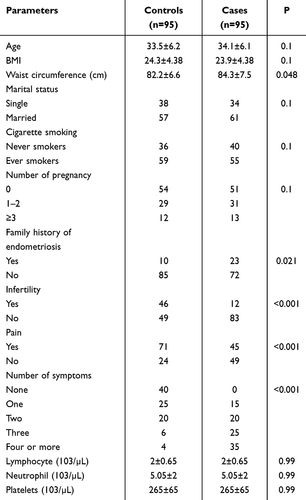 |
Table 1 Demographics and Clinical Characteristics of Study Participants |
 |
Figure 1 Patient selection flow chart. |
When patients were divided into experimental and control groups, 23 patients (24.2%) in the experimental group had a family history of endometriosis, compared to 10 patients (10.5%) in the control group. Infertility was reported in 12 patients (12.6%) in the experimental group, in contrast to 46 patients (48.4%) in the control group. Pain was reported by 45 patients (47.4%) in the experimental group, whereas 71 patients (74.7%) in the control group experienced pain. In the control group, 40 patients (42.1%) had no symptoms, 25 patients (26.3%) had one symptom, 20 patients (21.1%) had two symptoms, 6 patients (6.3%) had three symptoms, and 4 patients (4.2%) had four or more symptoms. In contrast, in the experimental group, no patients had no symptoms, 15 patients (15.8%) had one symptom, 20 patients (21.1%) had two symptoms, 25 patients (26.3%) had three symptoms, and 35 patients (36.8%) had four or more symptoms (Table 1). Clinical studies also indicate that symptoms related to endometriosis include dysmenorrhea, chronic pelvic pain, painful intercourse, cyclic bowel and urinary disorders, and infertility.10
The NETs-Associated Markers in Endometriosis and Controls
Fluorescent dye methods and ELISA were employed to measure the levels of NETs-associated markers (cf-DNA, calprotectin, nucleosomes, DNase I, MPO, NE) (Table 2). The results indicated that patients with endometriosis had significantly higher levels of cf-DNA (1349.53±100.41), nucleosomes (0.21±0.26), and NE (24.84±6.89) compared to the control group, with the differences being statistically significant (p<0.05). Conversely, levels of calprotectin, MPO, and DNase I did not show significant differences between the two groups (p>0.05). Detailed results are presented in Table 2.
 |
Table 2 The NETs-Associated Markers in Endometriosis and Controls |
The concentrations of cf-DNA, nucleosomes, and NE, as well as a composite biomarker panel, effectively distinguish between patients with endometriosis and those in the control group. Detailed results are presented in Table 3.
 |
Table 3 The Concentrations of Cf-DNA, Nucleosomes, and NE in Endometriosis with and Without Pian |
ROC analysis revealed significant differences in the levels of NETs-associated markers (cf-DNA, nucleosomes, NE) between patients with endometriosis and the control group, with areas under the curve (AUC) of 0.805, 0.748, and 0.832, respectively (Figure 2).
 |
Figure 2 The ROC curve analysis. The ROC curves of plasma cf-DNA, nucleosomes, and NE and the composite biomarker combination to distinguish 95 endometriosis cases from 95 controls. |
The Concentrations of Cf-DNA, Nucleosomes, and NE in Endometriosis Vary with Disease Severity
The 95 patients with endometriosis were classified into stages I, II, III, and IV based on disease severity. Analysis of cf-DNA, nucleosomes, and NE levels across these stages revealed a gradual increase in NETs-associated markers (cf-DNA, nucleosomes, NE) as the disease stage advanced (Figure 3).
 |
Figure 3 The concentrations of cf-DNA, nucleosomes, and NE in endometriosis with different severity. |
The Concentrations of Cf-DNA, Nucleosomes, and NE in Endometriosis with and without Infertility
The 95 patients with endometriosis were divided into groups with and without infertility to analyze the concentrations of cf-DNA, nucleosomes, and NE. The analysis indicated that the levels of NETs-associated markers (cf-DNA, nucleosomes, NE) were higher in the infertility group, with the difference being statistically significant (p<0.05) (Figure 4). The NE level of the infertile group increased the most than that of the non-infertile group.
 |
Figure 4 The concentrations of cf-DNA, nucleosomes, and NE in endometriosis with and without infertility. P<0.05: *, P<0.01: **; P<0.001: ***. |
The Concentrations of Cf-DNA, Nucleosomes, and NE in Endometriosis with and without Pain
The 95 patients with endometriosis were categorized into those experiencing pain and those not experiencing pain. The concentrations of cf-DNA, nucleosomes, and NE were qualitatively analyzed in both groups. The analysis demonstrated that patients experiencing pain had higher levels of NETs-associated markers (cf-DNA, nucleosomes, NE).
The Correlation Between NETs-Associated Markers with Visual Analogue Scale (VAS)
The Visual Analogue Scale (VAS) was employed for pain assessment, a widely used method in clinical practice in China. The VAS consists of a 10cm sliding scale marked with ten divisions, ranging from “0” (no pain) to “10” (unbearable, most severe pain). Pain severity in the 95 patients with endometriosis was assessed using the VAS, and the correlation between NETs-associated markers and VAS scores was quantitatively analyzed. The analysis revealed a positive correlation between VAS scores and NETs-associated markers (cf-DNA, nucleosomes, NE), indicating that the levels of these markers increase with rising VAS scores (Figure 5).
 |
Figure 5 The correlation between NETs related markers with VAS. P<0.05: *, P<0.01: **; P<0.001: ***. |
Discussion
Endometriosis is prevalent among reproductive-age women, especially those aged 25 to 45, with a higher incidence in Asian women and a rising trend in recent years.11 The retrograde menstruation hypothesis is widely accepted, but it alone is insufficient to cause endometriosis.12 The disease’s development is closely linked to the peritoneal cavity’s local microenvironment, where neutrophils (6% to 15% of endometrial cells during menstruation) are recruited by chemokines and cytokines.10,13 These factors play key roles in endometriosis,14,15 with recent studies showing that neutrophils release neutrophil extracellular traps (NETs) along with cytokines, enhancing inflammation, increasing vascular permeability, and promoting cell proliferation and invasion.7 This study investigates the relationship between endometriosis and NETs.
NETs represent a previously unknown mechanism of bactericidal action,16 distinct in morphology from apoptosis and necrosis. Upon stimulation, neutrophils release NETs. These NETs are web-like structures with DNA as the scaffold and are composed of various protein molecules, including core histones (H2A, H2B, H3 and H4) and granule proteins such as MPO, cathepsin G, NE, high mobility group box 1 (HMGB1), and proteinase 3, along with chromatin decondensation.17 NETs can release MPO, which then forms a stable complex with decondensed DNA, thereby protecting the DNA. HMGB1, a highly conserved nuclear protein with unique functions, plays a crucial role in the formation of DNA-based complexes in NETs. This protein is involved in DNA binding and in the regulation and modification of transcriptional processes, which is indispensable for NET formation.9,18 Furthermore, extracellular HMGB1 stimulates the production of chemokines, promoting inflammation.19 Previous studies have shown a significant increase in MPO levels in EMS.20 Although research on the role of NETs in endometriosis is limited, there is evidence linking increased MPO levels to endometriosis. Studies indicate that at least 95% of MPO in blood originates from PMNs, and the level of MPO activity reflects PMN function and activity.6 Additionally, existing literature suggests that extracellular trap proteases from neutrophils can facilitate the production of type I collagen by the endometrium in vitro, and endogenous ovarian steroids may inhibit the anti-fibrotic PGE2 pathway.21 Importantly, reports have shown elevated NETs in the peritoneal lavage fluid and peripheral blood of patients with endometriosis compared to the control group.22 Beside these direct findings, numerous indirect pieces of evidence suggest a complex relationship between endometriosis and NETs.
Our study further validates and reinforces the significant relationship between endometriosis and NETs. By comparing peripheral blood NETs-associated markers (cf-DNA, calprotectin, nucleosomes, DNase I, MPO, NE) between patients with endometriosis and healthy controls, we found that levels of cf-DNA, nucleosomes, and NE were higher in patients with endometriosis. ROC curve analysis revealed that cf-DNA, nucleosomes, NE, and a composite of biomarkers effectively distinguished endometriosis cases from the control group. The clinical applicability of ROC curve analysis results may be affected by a number of factors. For example, sample size, patient selection criteria, severity of disease, and accuracy of laboratory testing methods can all affect the accuracy and reliability of the ROC curve. Therefore, when applying NETs related markers to clinical diagnosis, we need to carefully consider these factors and conduct a comprehensive assessment in conjunction with the specific circumstances of the patient. Further analysis revealed that in patients with endometriosis, the concentrations of cf-DNA, nucleosomes, and NE increased with disease severity. When we examined the concentrations of these markers among endometriosis patients with and without infertility, we discovered elevated levels of NETs-associated markers in those with infertility. Additionally, qualitative analysis of cf-DNA, nucleosomes, and NE concentrations in patients with and without pain within the endometriosis group indicated higher levels of NETs-associated markers in those experiencing pain. Lastly, using the VAS to quantitatively assess pain, we found a positive correlation between VAS and NETs-associated markers. This means that as VAS score increased, the levels of NETs-associated markers (cf-DNA, nucleosomes, NE) also increased.
In summary, our study reveals a close connection between NETs-associated markers and endometriosis. However, the specific mechanisms through which NETs-associated markers contribute to the development and progression of endometriosis remain to be elucidated and warrant further investigation.
Our study has several limitations. The sample size was relatively small, which may affect the generalizability of our findings. Although we utilized advanced methodologies, we did not fully explore the underlying mechanisms by which NETs-associated markers influence endometriosis, leaving our research exploratory in nature. In order to rule out the potential influence that control group selection may have on the interpretation of NETS-related markers, we plan to expand the sample size to include more patients with different backgrounds and characteristics as controls in the future to more comprehensively evaluate the specific changes in NETs-related markers in endometriosis. Although patients were selected according to strict inclusion and exclusion criteria, confounding factors (such as unmeasured covariates, duration of disease, prior treatment history, etc.) may have influenced our results. These factors, although not directly involved in this study, may interact with NETs to jointly promote the occurrence and development of endometriosis. Therefore, the specific mechanism of action of NETs related markers on EMS remains to be fully elucidated. Despite these limitations, this case-control study establishes a potential link between NETs and endometriosis. It provides a valuable foundation for further research aimed at uncovering the mechanisms by which NETs impact endometriosis and for identifying new biomarkers and therapeutic targets.
Conclusions
NETs-associated markers can effectively differentiate endometriosis from control subjects. ROC analysis indicates that these markers significantly distinguish between patients with endometriosis and those without.
There is a strong correlation between NETs-associated markers and the symptoms associated with endometriosis, such as pain and infertility. As the severity of the disease increases, the levels of NETs-associated markers also rise progressively. Specifically, patients with infertility exhibited elevated levels of these markers, and similarly, those experiencing pain also showed higher levels of NETs-associated markers.
Abbreviations
EMS, Endometriosis; NE, neutrophil elastase; NETs, Neutrophil Extracellular Traps; PMN, peripheral blood serum and neutrophils; ROC, receiver operating characteristic; TLRs, Toll-like receptors; PAMPs, pathogen-associated molecular patterns; ssRNA, single-stranded RNA; ELISA, Enzyme-Linked Immunosorbent Assay; cf-DNA, Circulating free DNA; VAS, Visual Analogue Scale; MPO, myeloperoxidase.
Ethics Approval
This research was approved by Shanghai Pudong New Area People’s Hospital Medical Ethics Committee (2024-LW-04).
Acknowledgments
This work was supported by Key Discipline Project of Shanghai Pudong New Area Health Commission (PWZxk2022-28).
Disclosure
The authors declare no conflicts of interest that pertain to this work.
References
1. Barnado A, Crofford LJ, Oates JC. At the bedside: neutrophil extracellular traps (NETs) as targets for biomarkers and therapies in autoimmune diseases. J Leukoc Biol. 2016;99(2):265–278. doi:10.1189/jlb.5BT0615-234R
2. Cerecedo D, Martínez-Vieyra I, López-Villegas EO, et al. Heterogeneity of neutrophils in arterial hypertension. Exp Cell Res. 2021;402(2):112577. doi:10.1016/j.yexcr.2021.112577
3. Volkman R, Ben-Zur T, Kahana A, et al. Myeloperoxidase deficiency inhibits cognitive decline in the 5XFAD mouse model of Alzheimer’s disease. Front Neurosci. 2019;13:990. doi:10.3389/fnins.2019.00990
4. Giaglis S, Lu C, Dai Z, et al. Multimodal regulation of NET formation in pregnancy: progesterone antagonizes the Pro-NETotic effect of estrogen and G-CSF. Front Immunol. 2016;7:7. doi:10.3389/fimmu.2016.00007
5. Dinallo V, Marafini I, Di Fusco D, et al. Neutrophil extracellular traps sustain inflammatory signals in ulcerative colitis. J Crohns Colitis. 2019;13(6):772–784. doi:10.1093/ecco-jcc/jjy215
6. Manfredi AA, Ramirez GA, Rovere-Querini P, et al. The neutrophil’s choice: phagocytose vs make neutrophil extracellular traps. Front Immunol. 2018;9:288. doi:10.3389/fimmu.2018.00288
7. Holmes CL, Shim D, Kernien J, et al. Insight into neutrophil extracellular traps through systematic evaluation of citrullination and peptidylarginine deiminases. J Immunol Res. 2019;2019:2160192. doi:10.1155/2019/2160192
8. Song C, Li H, Mao Z, et al. Delayed neutrophil apoptosis may enhance NET formation in ARDS. Respir Res. 2022;23(1):155. doi:10.1186/s12931-022-02065-y
9. Zhou J, Liu T, Wang W. Prognostic significance of matrix metalloproteinase 9 expression in osteosarcoma: a meta-analysis of 16 studies. Medicine. 2018;97(44):e13051. doi:10.1097/MD.0000000000013051
10. Hamam HJ, Khan MA, Palaniyar N. Histone acetylation promotes neutrophil extracellular trap formation. Biomolecules. 2019;9(1):32. doi:10.3390/biom9010032
11. Eghbalzadeh K, Georgi L, Louis T, et al. Compromised anti-inflammatory action of neutrophil extracellular traps in PAD4-Deficient mice contributes to aggravated acute inflammation after myocardial infarction. Front Immunol. 2019;10:2313. doi:10.3389/fimmu.2019.02313
12. Ghasemisedaghat S, Eslamian G, Kazemi SN, et al. Association of fertility diet score with endometriosis: a case-control study. Front Nutr. 2023;10:1222018. doi:10.3389/fnut.2023.1222018
13. Herranz R, Oto J, Hueso M, et al. Bladder cancer patients have increased NETosis and impaired DNaseI-mediated NET degradation that can be therapeutically restored in vitro. Front Immunol. 2023;14:1171065. doi:10.3389/fimmu.2023.1171065
14. Saitoh T, Komano J, Saitoh Y, et al. Neutrophil extracellular traps mediate a host defense response to human immunodeficiency virus-1. Cell Host Microbe. 2012;12(1):109–116. doi:10.1016/j.chom.2012.05.015
15. Simila-Maarala J, Soovares P, Pasanen A, et al. TCGA molecular classification in endometriosis-associated ovarian carcinomas: novel data on clear cell carcinoma. Gynecol Oncol. 2022;165(3):577–584. doi:10.1016/j.ygyno.2022.03.016
16. Kolaczkowska E, Jenne CN, Surewaard BGJ, et al. Molecular mechanisms of NET formation and degradation revealed by intravital imaging in the liver vasculature. Nat Commun. 2015;6(1):6673. doi:10.1038/ncomms7673
17. Smolarz B, Szyllo K, Romanowicz H. Endometriosis: epidemiology, classification, pathogenesis, treatment and genetics (Review of Literature). Int J Mol Sci. 2021;22(19):10554. doi:10.3390/ijms221910554
18. Lachowicz-Scroggins ME, Dunican EM, Charbit AR, et al. Extracellular DNA, neutrophil extracellular traps, and inflammasome activation in severe asthma. Am J Respir Crit Care Med. 2019;199(9):1076–1085. doi:10.1164/rccm.201810-1869OC
19. Wang J, Shi Q, Yuan T-X, et al. Matrix metalloproteinase 9 (MMP-9) in osteosarcoma: review and meta-analysis. Clin Chim Acta. 2014;433:225–231. doi:10.1016/j.cca.2014.03.023
20. Liu Y, Wang Y, Teng Z, et al. Matrix metalloproteinase 9 expression and survival of patients with osteosarcoma: a meta-analysis. Eur J Cancer Care. 2017;26(1):e12364. doi:10.1111/ecc.12364
21. Ren J, He J, Zhang H, et al. Platelet TLR4-ERK5 axis facilitates NET-mediated capturing of circulating tumor cells and distant metastasis after surgical stress. Cancer Res. 2021;81(9):2373–2385. doi:10.1158/0008-5472.CAN-20-3222
22. McDonald B, Spicer J, Giannais B, et al. Systemic inflammation increases cancer cell adhesion to hepatic sinusoids by neutrophil mediated mechanisms. Int J Cancer. 2009;125(6):1298–1305. doi:10.1002/ijc.24409
 © 2025 The Author(s). This work is published and licensed by Dove Medical Press Limited. The
full terms of this license are available at https://www.dovepress.com/terms
and incorporate the Creative Commons Attribution
- Non Commercial (unported, 4.0) License.
By accessing the work you hereby accept the Terms. Non-commercial uses of the work are permitted
without any further permission from Dove Medical Press Limited, provided the work is properly
attributed. For permission for commercial use of this work, please see paragraphs 4.2 and 5 of our Terms.
© 2025 The Author(s). This work is published and licensed by Dove Medical Press Limited. The
full terms of this license are available at https://www.dovepress.com/terms
and incorporate the Creative Commons Attribution
- Non Commercial (unported, 4.0) License.
By accessing the work you hereby accept the Terms. Non-commercial uses of the work are permitted
without any further permission from Dove Medical Press Limited, provided the work is properly
attributed. For permission for commercial use of this work, please see paragraphs 4.2 and 5 of our Terms.