Back to Journals » Drug Design, Development and Therapy » Volume 18
Network Pharmacology and Experimental Verification: SanQi-DanShen Treats Coronary Heart Disease by Inhibiting the PI3K/AKT Signaling Pathway
Received 26 July 2024
Accepted for publication 11 September 2024
Published 9 October 2024 Volume 2024:18 Pages 4529—4550
DOI https://doi.org/10.2147/DDDT.S480248
Checked for plagiarism Yes
Review by Single anonymous peer review
Peer reviewer comments 3
Editor who approved publication: Professor Anastasios Lymperopoulos
Min Zhao,1 Liuxiang Feng,2 Wenhua Li3
1School of Medicine, Lijiang University of Culture and Tourism, Lijiang, Yunnan, 674100, People’s Republic of China; 2People’s Hospital of Yulong Naxi Autonomous County of Lijiang City, Lijiang, Yunnan, 674112, People’s Republic of China; 3School of Medicine, Xizang Minzu University, Xianyang Shaanxi, 712082, People’s Republic of China
Correspondence: Min Zhao, School of Medicine, Lijiang University of Culture and Tourism, Lijiang, Yunnan, 674100, People’s Republic of China, Email [email protected]
Objective: To employee network pharmacology to predict the components and pathways of SanQi-DanShen (SQDS) in treating coronary heart disease, followed by in vitro experiments to validate the molecular mechanism of SQDS in treating coronary heart disease.
Methods: We sourced the active ingredients and targets of Panax notoginseng and Danshen from the Traditional Chinese Medicine Systems Pharmacology database. Coronary heart disease related genes were retrieved from the OMIM, Genecards, and Therapeutic Target databases. Using Cytoscape 3.7.2 software, we constructed a network diagram illustrating the components and targets of SQDS. The associated targets were then imported into the STRING database to build a protein-protein interaction network. The Metascape database and WeChat software were utilized for Gene Ontology and Kyoto Encyclopedia of Genes and Genomes enrichment analyses. Lastly, we performed molecular docking between the key components and related targets using AutoDock Vina. To validate the potential mechanism of SQDS in treating coronary heart disease, we established an acute coronary heart disease rat model via tail vein injection of pituitrin.
Results: Network pharmacology analysis revealed that 65 active ingredients and 167 targets of SQDS are implicated in the treatment of coronary heart disease. The key targets identified include AKT1, TNF, TP53, IL6, and VEGFA. Notably, the PI3K/AKT signaling pathway emerged as the primary pathway. Furthermore, animal experiments showed that, compared to the model group, SQDS significantly reduced levels of TNF-α, IL-6, Bax, and cardiac troponin I, while increasing Bcl-2 content. It also notably suppressed the expression of p-PI3K and p-AKT, thereby offering protection to myocardial tissue.
Conclusion: Through the integrated approach of network pharmacology and molecular docking, we have established that SQDS exerts a multi-component, multi-target, and multi-pathway synergistic therapeutic effect on coronary heart disease. Its mechanism may involve the inhibition of the PI3K/AKT signaling pathway and the reduction of inflammatory factor expression.
Keywords: SQDS, cardiac troponin I, PI3K/AKT signaling pathway, coronary heart disease, experiment validation
Introduction
Currently, coronary heart disease remains a devastating illness posing a global threat to human health, particularly in low- and middle-income countries.1 The exorbitant cost of treatment imposes a heavy burden on individuals and economies alike. Recent studies reveal that between 1990 and 2019,2 cardiovascular disease was the primary cause of death in China. In clinical practice, there are currently two main types of drugs used to treat coronary heart disease. The first type is drugs that prevent ischemia and alleviate symptoms, including nitrate esters, metabolic drugs, potassium channel blockers, etc; The second type of drugs used to prevent myocardial infarction and improve prognosis includes antiplatelet drugs, statins, beta blockers, etc. (see Table 1 for details), but these drugs can cause numerous adverse reactions, particularly in the digestive and skin systems.3 Clinical practice and related studies have confirmed that plant medicine has certain advantages in the treatment of coronary heart disease, including relieving angina pectoris, interfering with restenosis after percutaneous coronary intervention, preventing and treating no-reflow after reperfusion, improving quality of life, increasing exercise tolerance, and reducing the incidence of cardiovascular events and adverse reactions. And the side effects are small and economical.4,5 Therefore, the development and utilization of plant medicine is a promising option. Notably, traditional Chinese medicine has demonstrated remarkable efficacy in clinical trials for treating coronary heart disease,6 thanks to its multi-target approach. SQDS stands out as a popular medication for replenishing qi and enhancing blood circulation. Modern pharmacological research has uncovered that SanQi can decrease blood viscosity, regulate blood flow, prevent thrombosis, thereby enhancing cardiac function and effectively intervening in cardiovascular disease.7,8 Meanwhile, DanShen exhibits several beneficial effects, including myocardial protection, reducing lipid accumulation in arterial intima, inhibiting myocardial calcification, and anti-thrombosis properties.9 Studies indicated that DanShen improves blood circulation between arterioles and venules, inhibits platelet adhesion and thromboxane production, and relaxes coronary vessels, making it extensively used in treating coronary heart disease and other cardiovascular ailments.10,11 The combination of SQDS proves more effective in activating blood circulation and eliminating blood stasis than SanQi and DanShen administered separately.12
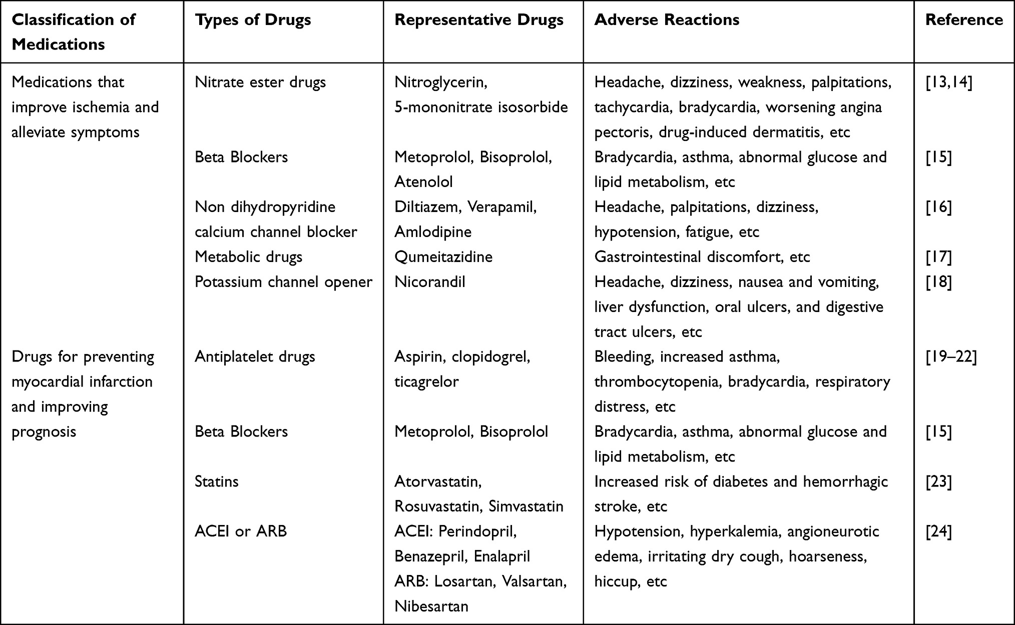 |
Table 1 Common Clinical Drugs and Adverse Reactions for Coronary Heart Disease |
Network Pharmacology offers innovative concepts and approaches for the integrative research between artificial intelligence and both traditional Chinese and Western medicinal practices. It facilitates the analysis of vast biomedical datasets and enables the conversion of raw data into valuable knowledge. This methodology offers a fresh perspective to elucidate the mechanisms of action inherent in traditional Chinese medicine.25 Furthermore, it underscores the versatility of treatment modalities for various ailments.26 In parallel, molecular docking stands as a technique to assess the binding affinity of small molecules with their designated receptors, paving the way for the utilization of SQDS in coronary heart disease prevention. Through the application of both network pharmacology and molecular docking, this study delved into the components, targets, and signaling pathways associated with SQDS in addressing coronary heart disease.
The purpose of this study is to explore the main components, targets, and pathways of SanQi DanShen, two traditional Chinese medicines, in the treatment of coronary heart disease through network pharmacology; Secondly, the mechanism of action of the traditional Chinese medicine compound SanQi DanShen tablets in treating coronary heart disease was validated through in vitro experiments. Provide theoretical basis for the development and utilization of traditional Chinese medicine. The novelty of this study lies in combining network pharmacology with in vitro experiments to validate the mechanism of action of the traditional Chinese medicine compound SanQi-DanShen tablets in treating coronary heart disease.
Materials and Methods
Network Pharmacology Analysis
Collection of SQDS Active Ingredients and Target Genes
The components of SQDS are derived from the systematic pharmacology of traditional Chinese Medicine (https://tcmspw.com/tcmsp.php) based on pharmacokinetic principles, encompassing absorption, distribution, metabolism, and excretion (ADME).27 To obtain the corresponding target, we set the oral bioavailability threshold at ≥ 30% and the drug similarity threshold at ≤ 0.18.28,29 Furthermore, we utilized UniProt (https://www.uniprot.org/) for verification, selecting “Homo sapiens” for accurate correction.
Prediction and Acquisition of Disease Targets
We searched for CHD-related targets in the OMIM (http://OMIM.org/), Genecards (http://www.gengcards.org/), and TTD (http://db.idrblab.net/ttd/) databases, and subsequently confirmed them using the UniProt database with the keyword “coronary artery disease”.
Construction of the Target Network
To ascertain potential interactions between active ingredients and targets, the ingredients of SQDS and their associated targets were inputted into Cytoscape 3.7.2, and subsequently, the ingredient-target network was constructed.
Collection of Genes with Overlapping Drug and Disease Associations
The overlapping genes of sqds and CHD were obtained using the online software Venny 2.1 (https://bioinfogp.cnb.csic.es/tools/Venny/).
Construction of PPI Network and Screening of Core Targets
Drug disease overlapping genes were inputted into the STRING database (https://string-db.org), with species restricted to “Homo sapiens” and a minimum required interaction score of ≥ 0.400. Subsequently, the obtained results were imported into Cytoscape 3.7.2 for further analysis.30 By utilizing the CytoHubba plugin, we assigned values to each gene via topological network algorithms, enabling us to sort and identify the core genes. Furthermore, bar graphs representing these core genes were also generated.
Conduct Comprehensive Go and KEGG Pathway Enrichment Analysis
To delve deeper into the mechanism of SQDS in treating CHD, we imported drug-disease overlapping genes into the Metascape database (https://metascape.org)31 for gene ontology functional enrichment analysis and Kyoto Encyclopedia of Genes and Genomes pathway analysis. Simultaneously, we utilized Cytoscape 3.7.2 to create the component target pathway network diagram.
Molecular Docking
We downloaded the two-dimensional structures of small molecules from the PubChem database (https://pubchem.ncbi.nlm.nih.gov/) and converted them into three-dimensional structures, saving them in mol format. We then processed these structures using the AutoDock 1.5.6 tool and saved them as pdbqt files. Additionally, we retrieved crystal structures of AKT1, TNF, TP53, IL6, and VEGFA (with PDB IDs: 1unq, 5uui, 1aie, 1alu, 1mkk) from the PDB database (http://www.rcsb.org). The target protein molecules underwent processing with both PyMOL and AutoDock software, subsequently saving them in pdbqt format. Lastly, we employed AutoDock Vina for molecular docking, with the results visualized in Discovery Studio 4.5 and PyMOL 1.7.x.
Animal Experimental Validation
Reagents
The posterior pituitary injection (lot: H34022977) was procured from Anhui Hongye Pharmaceutical Co., Ltd. Sanqidancanpian (lot: Z20050013) was obtained from Hunan Patron Saint Pharmaceutical Co., Ltd. ELISA kits for Tumor Necrosis Factor-α (TNF-α) (lot: ml002859) and Interleukin-6 (IL-6) (lot: ml064292) were sourced from Shanghai Enzyme Linked Biotechnology Co., Ltd. Rat B-cell Lymphoma Factor 2 (Bcl-2) (lot: D731031-0048) and Rat Bcl-2-related X Protein (Bax) (lot: D731032-0048) ELISA kits were supplied by Shanghai SANGON Biotech Co., Ltd. Rat phosphorylated phosphatidylinositol 3-kinase (p-PI3K) (lot: JL50027) and rat phosphorylated AKT protein (p-AKT) (lot: JL21143) were purchased from Shanghai Jianglai Biotechnology Co., Ltd. Rat Cardiac Troponin I (cTnI) (lot: D731143) was provided by Shanghai SANGON Biotech Co., Ltd. Primary antibodies for Rat p-PI3K (lot: [EPR25156-60] ab302958) and p-AKT (lot: [EP2109Y] ab81283) were acquired from Shanghai Abcam Biological Company.
Animal and Drug Handling
Male SD rats (n=72, weighing 190 ± 10 g, and aged 6–8 weeks) were procured from the Guangdong Medical Laboratory Animal Center in Changzhou, bearing the laboratory animal license number: scxk (Yue) 2022–0002. The conducted animal experiments adhered to the guidelines set forth by the National Institutes of Health for the care and use of laboratory animals.
After a 1-week adaptation period, all rats were randomly assigned to 6 groups, with 12 rats in each group: (1) PBS control (control); (2) The model group received 15 u/kg pituitrin via tail vein injection; (3) Positive control group, which received 10 mg/kg rosuvastatin calcium tablets plus 15 u/kg pituitrin (Rst); (4) Sanqidancanpian at 900 mg/kg plus 15 u/kg pituitrin (H-SQDS); (5) Sanqidancanpian at 300 mg/kg plus 15 u/kg pituitrin (M-SQDS); (6) Sanqidancanpian at 100 mg/kg plus 15 u/kg pituitrin (L-SQDS). Except for the control group, all other rats were injected with 15 u/kg pituitrin through the tail vein following the literature method (5-second constant speed injection).32 Upon successful modeling, the rats were administered intragastrically: the H-SQDS group received 900 mg/kg/day, the M-SQDS group received 300 mg/kg/day, and the L-SQDS group received 100 mg/kg/day. The Rst group received 10 mg/kg/day, while the control and model groups were given the equivalent volume of normal saline for 14 days.
Histological Analysis
After obtaining samples from rats, the hearts were washed in pre cooled PBS, dried with clean filter paper, and fixed in 4% paraformaldehyde for at least 24 hours. Then, gradient dehydration is performed, after dehydration, the heart tissue is embedded in paraffin and soaked in a mixture of ice and water for a period of time, it is sliced into 5um thick tissue using a slicer, and then dewaxed, stained, dehydrated, permeabilized, and sealed. Furthermore, a light microscope (Nikon) was employed for the meticulous examination of pathological alterations in these tissue sections.
Preparation of Myocardial Tissue Homogenate
After the model of acute coronary heart disease was successfully established, about 0.1 g of the ischemic tissue was quickly cut out and rinsed in ice-cold normal saline, the blood was removed, blotted dry by filter paper, put into a 1.5 mL plastic centrifuge tube, frozen in liquid nitrogen, and stored in a −70 °C refrigerator. At the end of the experiment, all the specimens were removed, precooled saline (the volume weight of saline was 9 times the weight of the tissue block) was taken by pipette, added into the plastic centrifuge tube containing the tissue, and the tissue block was cut as soon as possible with ophthalmic scissors. Then the hearts were centrifuged at 10000–15000 r/min for 14 min, and 10% normal saline was used to make myocardial tissue homogenate. 0.5 mL of the supernatant was used for determination of TNF-α, IL-6, Bcl-2, Bax, and cTnI. The assay was performed according to the kit instructions.
Measurement of Inflammatory Factors
The supernatant from the homogenized myocardial tissue of each group was collected, and the levels of TNF-α and IL-6 were measured using ELISA kits,(following the instructions provided by Shanghai Enzyme Linked Biotechnology Co., Ltd. The detection range of TNF-α is 10 pg/mL to 320 pg/mL, with a sensitivity of less than 1.0 pg/mL; The detection range of IL-6 is 5 pg/mL to 160 pg/mL, with a sensitivity of less than 1.0 pg/mL).
Detection of Apoptosis Factors, Bcl-2, Bax, and cTnI Levels
Supernatant from homogenized myocardial tissue was collected for each group. The concentrations of Bcl-2, Bax and cTnI were ascertained through the utilization of ELISA kits, adhering to the guidelines provided by Shanghai SANGON Biotech Co., Ltd. (The detection range of Bcl-2 is 0.16–10 ng/mL, with a sensitivity of 0.1 ng/mL; The detection range of Bax is 0.16–10 ng/mL, with a sensitivity of 0.1 ng/mL. The detection range of cTnI is 31.25–2000 pg/mL, with a sensitivity of 18.75 pg/mL).
Determination of p-AKT and p-PI3K Content
The supernatant of myocardial tissue homogenate in each group was collected, and the contents of p-AKT and p-PI3K were determined using ELISA kits (according to the instructions of Shanghai Jianglai Biotechnology Co., Ltd. The detection range of p-AKT is 0.156–10 ng/mL, with a sensitivity of 0.076 ng/mL; The detection range of p-PI3K is 15.6–1000 pg/mL, with a sensitivity of 7.92 pg/mL.)
Western Blot Analysis
The myocardial tissue from each group underwent lysis with protein lysis buffer on ice, followed by protein concentration determination using the BCA protein assay kit (PC0020, Solarbio, China). Equal protein quantities were then separated via SDS polyacrylamide gel and transferred to a PVDF membrane. These membranes were blocked with 5% skim milk in TBST for 1 hour at room temperature, followed by incubation with specific primary antibodies overnight at 4°C. Antibodies targeting AKT, p-AKT, PI3K, p-PI3K, and GAPDH were procured from Shanghai Abcam biological company. Following this, the membrane underwent incubation with the secondary antibody for one hour at room temperature. Western blots were then analyzed and imaged using a Tanon 4800 imaging system (Tanon, China) in conjunction with the BeyoECL plus Kit (P0018S, Beyotime, China). Among them, primary antibody: PI3 Kinase p85 Alpha Monoclonal antibody (lot: 60225-1-Ig, proteintech (China), dilution ratio 1:5000); Phospho-PI3 Kinase p85 (Tyr458)/p55 (Tyr199) (E3U1H) (lot: 17366, CST (USA), dilution ratio 1:1000); Anti-AKT (phospho S473) antibody [EP2109Y] (lot: ab81283, abcam (UK), dilution ratio 1:5000); Phospho-AKT (Ser473) (D7F10) XP (lot: 9018, CST (USA), dilution ratio 1:1000); GAPDH (lot: ab8245, abcam (UK), dilution ratio 1:2000).
Secondary antibody: Goat Anti Mouse IgG/HRP (lot: SE131, Soleibao (China), dilution ratio 1:2000); Goat Anti Rabbit IgG H&L (HRP) (lot: ab6721, abcam (UK), dilution ratio 1:2000). The study’s technical roadmap is illustrated in Figure 1.
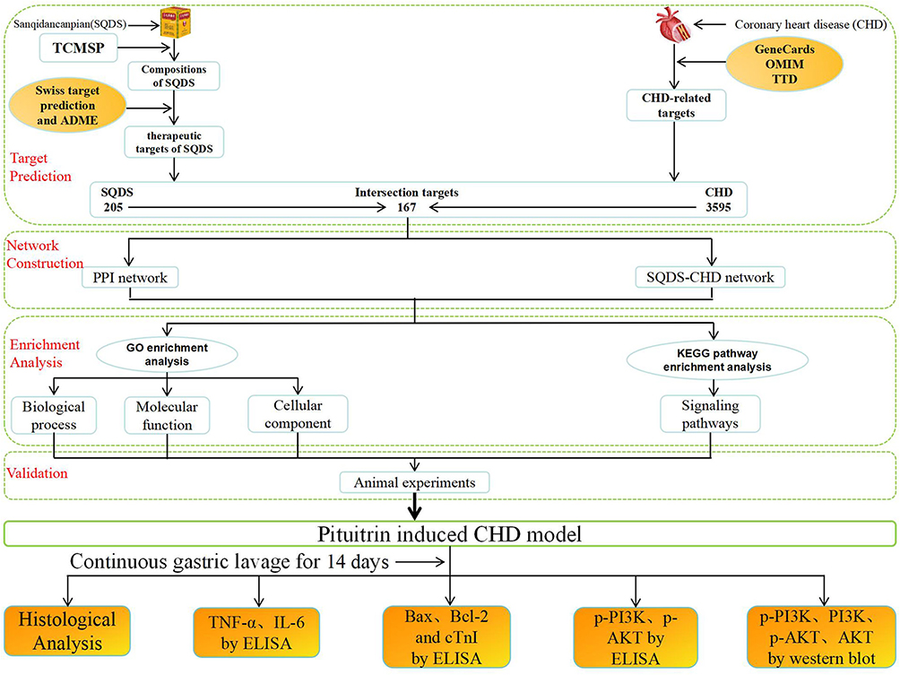 |
Figure 1 Technical roadmap of this study. |
Statistical Method
Network pharmacology data analysis was conducted utilizing Cytoscape version 3.7.2. The GO and KEGG analyses were accomplished through the use of Metascape and Wechat software. Molecular docking was facilitated by Autodock Vina version 1.5.6 and PyMOL version 1.7.x software. Animal experiments were carried out and analyzed with one-way ANOVA using graphpadprism version 7.0 (Graphpad Software Inc, San Diego, CA, USA) for comparing the two groups. The data is presented in the form of means ± SD, with statistical significance denoted as P < 0.05.
Results
Network Pharmacology and Molecular Docking results
The Active Ingredient in SQDS
Based on the OB screening criteria of ≥ 30% and DL ≥ 0.18, we obtained seven active ingredients from Panax notoginseng and 58 active ingredients from Salvia miltiorrhiza through the TCMSP database, and further confirmed them using the UniProt database, as presented in Table 2. Additionally, we gathered and identified 205 target genes.
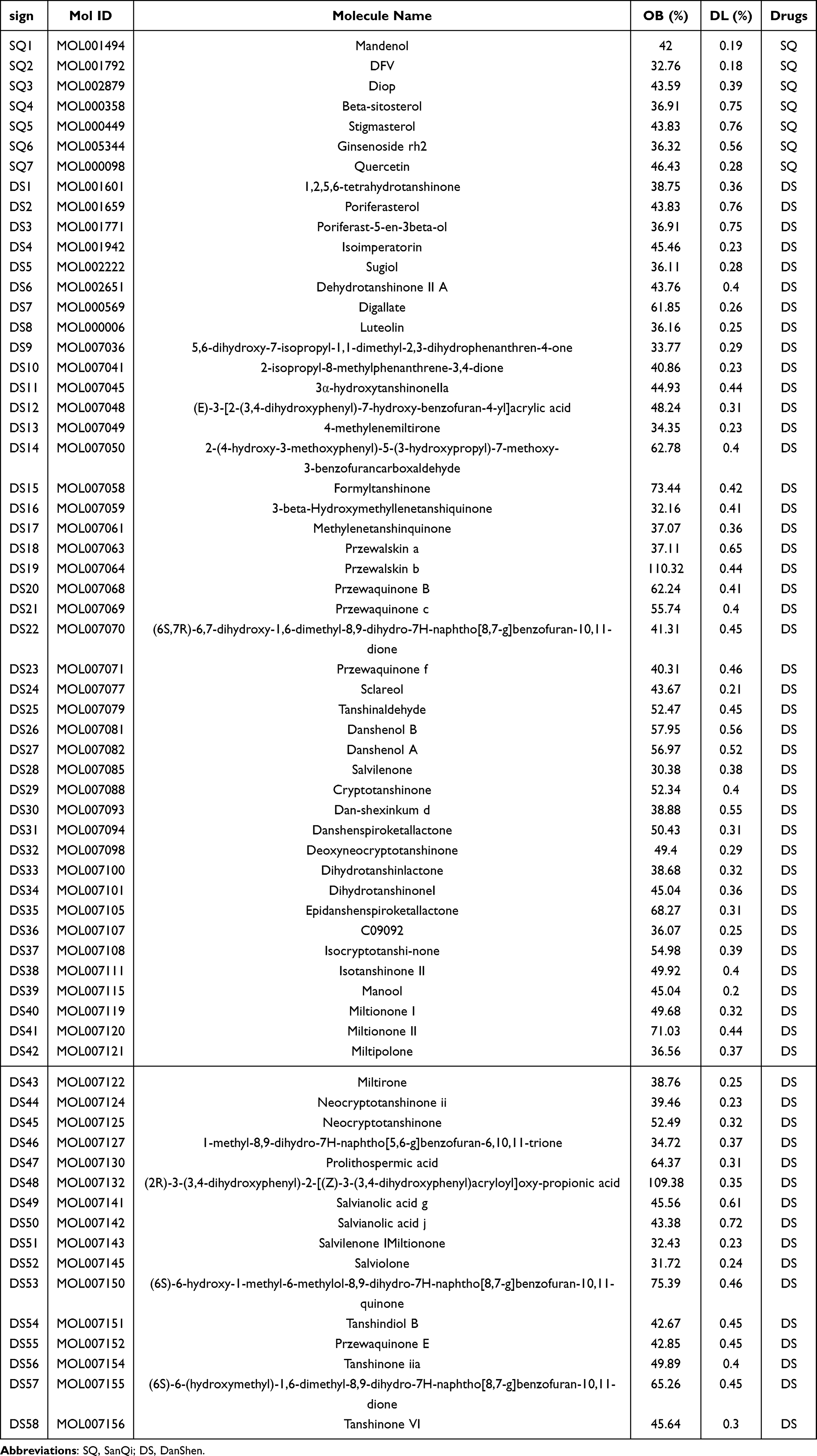 |
Table 2 Active Ingredients of SanQi-DanShen |
CHD Related Targets
Using genecards, TTD and OMIM databases, we identified 8244, 9, and 557 targets related to CHD, respectively. After screening, a total of 3595 genes were obtained. Of these, 167 genes were found to overlap with drug targets and are presented in the Venn diagram (refer to Figure 2). These genes were then inputted into Cytoscape 3.7.2 software to generate a component target network map (see Figure 3). The top 10 components are listed in Table 3.
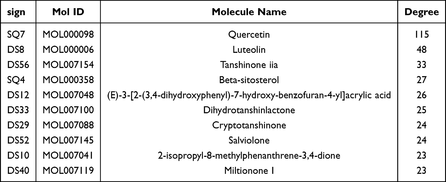 |
Table 3 Top 10 Active Ingredients of SQDS |
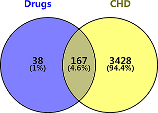 |
Figure 2 Venn diagram of SQDS–CHD intersection targets. |
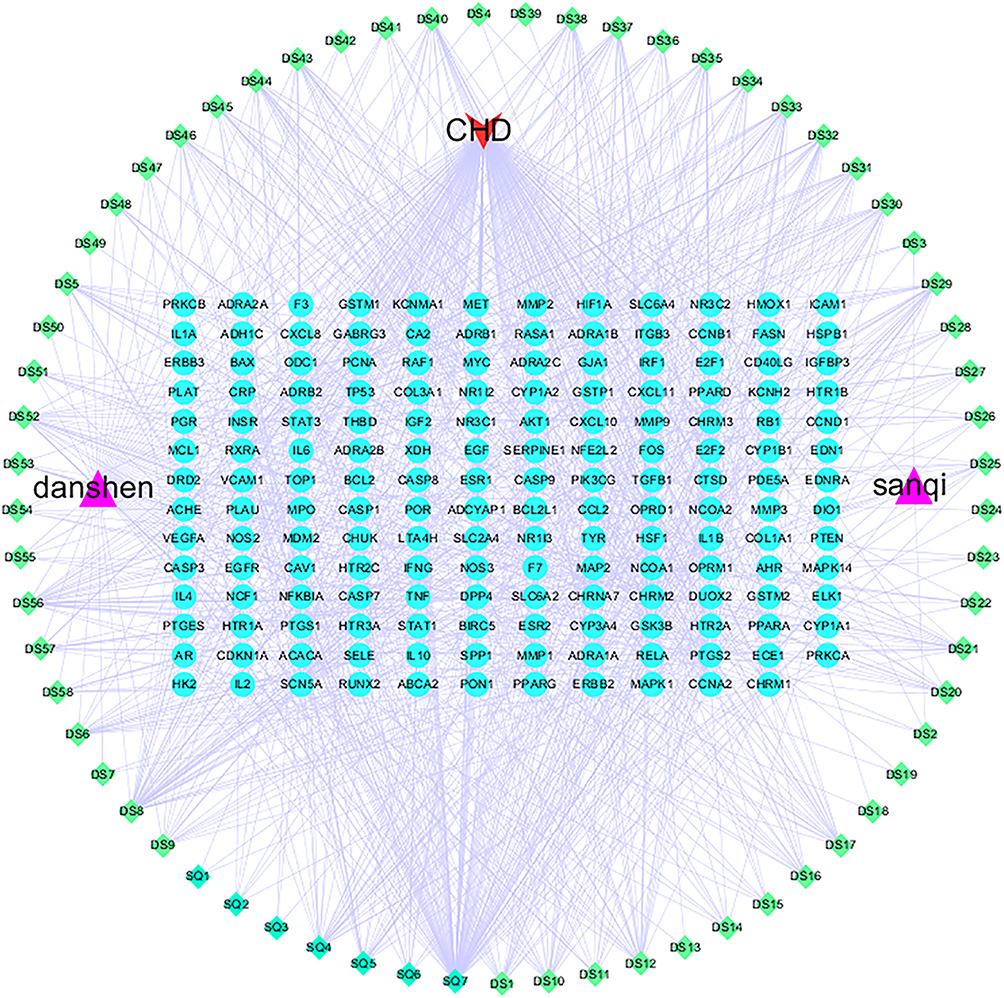 |
Figure 3 Network graph analysis of the interaction between active ingredients of SQDS and intersection targets in CHD. |
Construction of PPI Network and Screening of Core Genes
We entered the 167 overlapping genes of SQDS and CHD into the string platform, selecting “Homo sapiens” with an interaction score minimum of ≥ 0.400 to generate the PPI network diagram (refer to Figure 4A). This diagram was then imported into Cytoscape 3.7.2 to illustrate the protein interaction network (see Figure 4B, where node values, colors, and degree parameters are proportional). Lastly, employing the cytohubba plug-in, we utilized a topological network algorithm to assign values to each gene, sorting and identifying the core genes to determine the primary target genes of SQDS in treating coronary heart disease, namely: AKT1, TNF, TP53, IL6, and VEGFA (as shown in Figure 4C).
 |
Figure 4 (A): The PPI network of SQDS and CHD. (B): Protein-protein interaction network.(The larger the green circle and font, the more important the target). (C): top 10 core target molecular bar graph.(The x-coordinate represents the degree of the target, and the y-coordinate represents the target). |
GO Enrichment Analysis of SQDS for CHD
The overlapping genes of drugs and diseases were imported into the Metascape database for GO enrichment analysis. This analysis uncovered 1912 biological processes, 111 cellular components, and 202 molecular functions. Specifically, the biological process related to CHD involves responses to hormones, cellular responses to organic cyclic compounds, and cellular responses to lipids (refer to Figure 5A). The molecular function results indicated that Sqds could potentially impact DNA transcription factor binding, G protein-coupled amine receptor activity, protein kinase binding, and cytokine receptor binding (see Figure 5B). In terms of cellular composition, the findings revealed that the efficacy of CHD involves membrane rafts, transcriptional regulatory complexes, and receptor complexes (refer to Figure 5C).
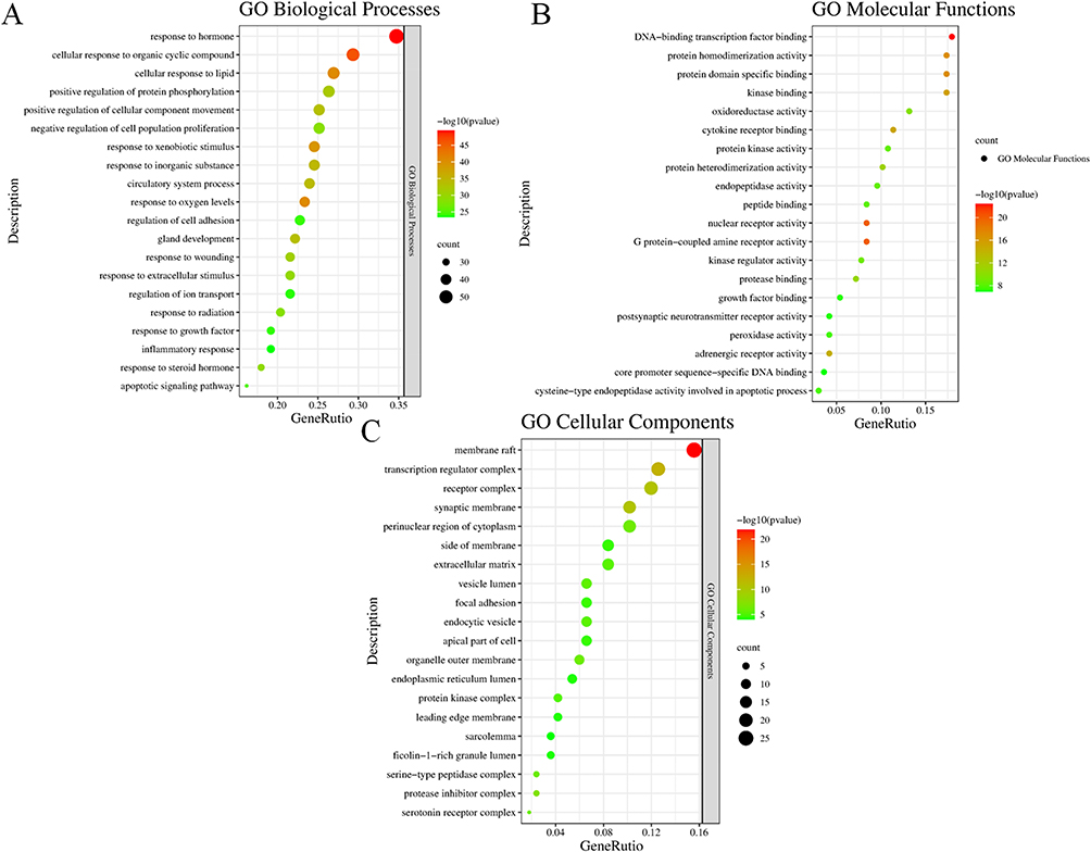 |
Figure 5 Bubble diagram of GO enrichment analysis; (A): Biological processes; (B): Molecular Functions; (C): Cellular Components. |
KEGG Enrichment Analysis of SQDS in the Treatment of CHD
KEGG pathway enrichment analysis has uncovered that factors associated with CHD are linked to the PI3K-AKT signaling pathway, lipid metabolism and atherosclerosis, as well as the AGE-RAGE signaling pathway involved in diabetic complications. The top 20 results of this analysis are presented in Table 4 and Figure 6. Additionally, the top 20 pathways have been integrated with their respective targets to create a “component-target-pathway” network diagram, as illustrated in Figure 7.
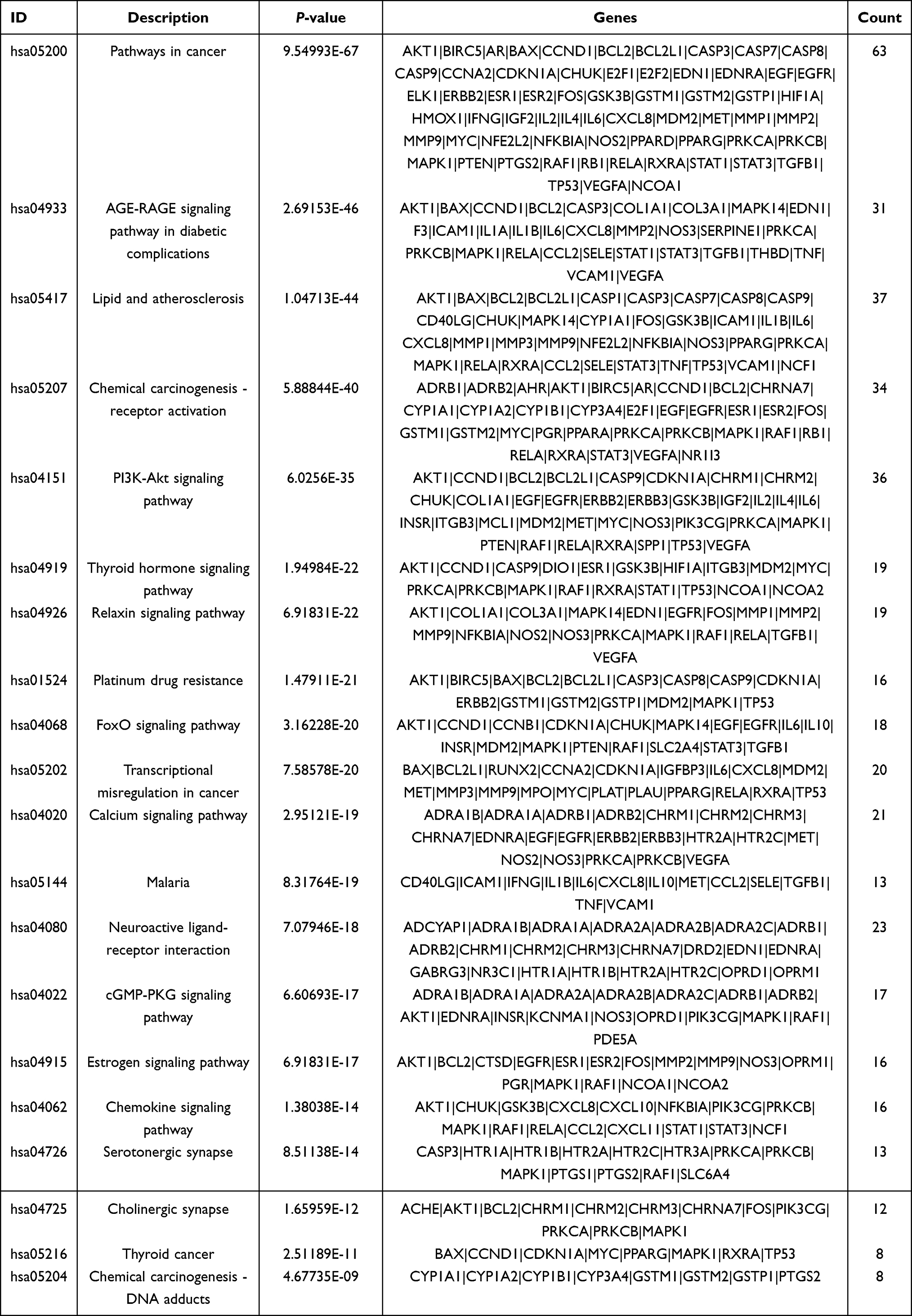 |
Table 4 KEGG Enrichment Analysis of SanQi-DanShen for CHD Treatment (TOP 20) |
 |
Figure 6 Bubble plots of KEGG pathway enrichment analysis. |
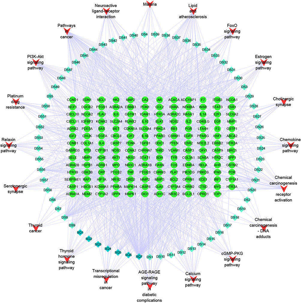 |
Figure 7 SanQi-DanShen and CHD interaction network diagram of ingredients-Targets-Pathways. |
Molecular Dockin
Based on the network diagram illustrating components and targets, we chose four components: Quercetin, luteolin, tanshinone, and cryptotanshinone. We then selected AKT1, TNF, TP53, IL6, and VEGFA. For molecular docking, we utilized AutoDock Vina. The outcomes were visualized using Discovery Studio and PyMOL. Our findings indicated that the chosen components could form stable bonds with the target, exhibiting a binding energy of less than <-5kj/mol (refer to Table 5 and Figure 8).
 |
Table 5 Verification Results of Molecular Docking |
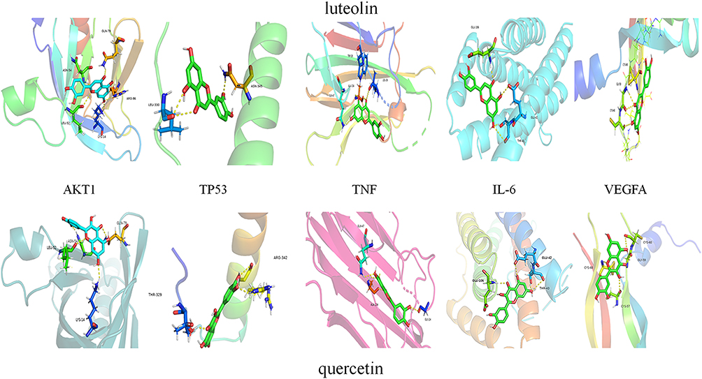 |
Figure 8 Molecular docking of main active components of SQDS with key targets. |
Validation of Animal-Level Research
Histological Analysis
The results of H&E staining revealed evident pathological alterations in the myocardial tissue. In comparison to the control group, the cardiomyocytes in the model group exhibited a disordered arrangement, accompanied by an increase in intercellular gaps. However, following SQDS treatment, a concentration-dependent improvement was observed in the rat myocardium when compared to the model group. Notably, the therapeutic efficacy of Rst was found to be comparable to that of H-SQDS (Figure 9).
 |
Figure 9 Histochemistry analysis, magnification: 200×; scale=50 µm. (n=6). |
Measurement of Inflammatory Factors, TNF-α and IL-6 Levels
In the supernatant of myocardial tissue homogenate from each experimental group, significant changes were observed in IL-6 (Figure 10A) and TNF-α (Figure 10B) levels. Specifically, the model group exhibited a notable increase in both IL-6 and TNF-α compared to the control group (P<0.001). It is important to highlight that various SQDS treatment groups demonstrated significant concentration-dependent attenuation of IL-6 and TNF-α (P<0.001) when compared to the model group.
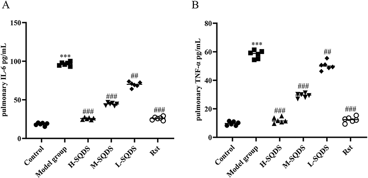 |
Figure 10 Detection of IL-6 and TNF-α by ELISA; (A): IL-6; (B): TNF-α. (n=6). ***P=0.001, ##P=0.01, ###P=0.001. (*the model group was compared with the control group; #the model group was compared with the medication group). |
Apoptosis Factor Detection, Bcl-2, Bax and cTnI Levels
Bcl-2, Bax and cTnI were significantly changed in the myocardial tissue homogenate supernatant of each experimental group. Compared with the control group, the content of Bcl-2 (P<0.001) in the model group decreased, and the content of Bcl-2 (P<0.001) (Figure 11A) increased significantly after different SQDS treatment. While Bax (P<0.001) (Figure 11B) and cTnI (Figure 11C) were significantly increased in the model group, compared with the model group, different SQDS treatment groups showed a significant decrease in Bax (P<0.001) and cTnI (P<0.001) in a concentration-dependent manner.
 |
Figure 11 Detection of Bcl-2, Bax and cTnI by ELISA; (A): Bcl-2; (B): Bax. (C): cTnI. (n=6). ***P=0.001, ##P=0.01, ###P=0.001. (*the model group was compared with the control group; #the model group was compared with the medication group). |
ELISA Was Used to Detect the Content of p-AKT and p-PI3K
There were significant changes of p-AKT and p-PI3K in myocardial tissue homogenate supernatant in all experimental groups. Compared with the control group, p-AKT (P<0.001) and p-PI3K (P<0.001) were significantly increased in the model group. Compared with the model group, different SQDS treatment groups showed a significant decrease in p-AKT (P<0.001) (Figure 12A) and p-PI3K (P<0.001) (Figure 12B) in a concentration-dependent manner.
 |
Figure 12 Detection of p-PI3K and p-AKT by ELISA; (A): p-AKT; (B): p-PI3K. (n=6). ***P=0.001, ##P=0.01, ###P=0.001. (*the model group was compared with the control group; #the model group was compared with the medication group). |
Western Blot Analysis
The expression of AKT, p-AKT, PI3K and p-PI3K was significantly changed in the experimental group. Compared with the control group, the expressions of AKT, p-AKT, PI3K and p-PI3K in the model group were significantly increased. Compared with the model group, the levels of AKT, p-AKT, PI3K and p-PI3K in different SQDS treatment groups showed a dose-dependent decrease (Figure 13A). The gray value of the strip is shown in the figure (Figure 13B).
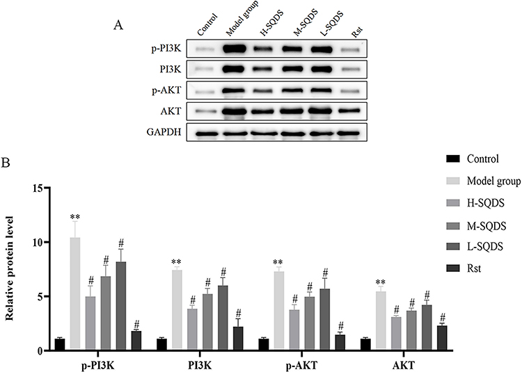 |
Figure 13 (A): The protein expression of p-PI3K, PI3K, p-AKT, and AKT in myocardium tissue myocardial homogenate supernatant by Western blot. (n=3). (B): The relative ratio of p-PI3K, PI3K, p-AKT, and AKT in myocardium tissue myocardial homogenate supernatant by Western blot. (n=6). **P=0.0078, #P=0.0150. (*the model group was compared with the control group; #the model group was compared with the medication group). |
Discusssion
An important factor contributing to cardiovascular disease is the impairment of vascular endothelial function, which disrupts tissue blood supply. Consequently, promoting angiogenesis and restoring vascular endothelial function stand as crucial strategies to minimize plaque ruptures and detachments from the vascular wall—key steps in preventing and treating cardiovascular diseases.33 According to the 《China Cardiovascular Disease Report 2021》, the number of coronary heart disease patients in China is on the rise, with projections indicating a steep increase in prevalence over the next decade, posing a significant public health challenge in the country.34 In the traditional Chinese medical framework, coronary heart disease falls under the umbrella of “chest obstruction”. Its etiology and pathogenesis are primarily attributed to visceral Qi deficiency, manifesting in syndromes of blood stasis and phlegm—a combination of deficiency and excess origin syndromes.35 In clinical practice, it is essential to consider both symptoms and signs holistically. Currently, primary treatment methods for cardiovascular diseases include drug therapies such as angiotensin receptor antagonists, angiotensin converting enzyme inhibitors, beta-blockers, aspirin, and statins,36 as well as percutaneous coronary intervention (PCI) and coronary artery bypass grafting (CABG). While these approaches can delay disease progression to some extent, they remain unable to fundamentally alter the pathological nature of cardiovascular diseases. Moreover, interventional surgeries are not without their limitations, including postoperative restenosis and vascular occlusion.37 Therefore, the primary task remains to continue exploring effective treatment methods with minimal side effects.8,38 Modern studies have demonstrated that both Panax notoginseng and Salvia miltiorrhiza can enhance the physiological and pathological processes of cardiovascular disease23,24, although the underlying mechanism remains unclear. In our study, we employed network pharmacology and molecular docking techniques to uncover the active ingredients and the mechanism of action of sqds in treating coronary heart disease. Our findings were subsequently validated through animal experiments. Our results indicate that (1) SQDS can address coronary heart disease via multiple targets and signaling pathways, and (2) SQDS effectively suppresses the expression of TNF-α and IL-6, Bax, and cardiac troponin I, while increasing Bcl-2 content and significantly reducing p-AKT and p-PI3K levels.
Through a search of the tcmsp database, we obtained 65 active components of SQDS. These comprise 58 active components from DS and 7 from SQ. Additionally, we screened 205 related target genes. Simultaneously, utilizing the GeneCards, TTD, and OMIM databases, we identified 3595 CHD-related target genes. By overlapping these with our initial set of target genes, we discovered 167 common genes. Using Cycloscape 3.7.2, we constructed a target component network diagram. Degree value analysis highlighted quercetin (115) and luteolin (48) as the primary components. Previous studies have shown that quercetin significantly inhibits mitochondrial oxidative stress, cardiac fibrosis, inflammatory responses, and apoptosis. It also regulates autophagy, improves myocardial ischemia/reperfusion injury, and modulates intestinal microbiota, thereby effectively treating cardiovascular diseases.39 Meanwhile, quercetin has been found to regulate the silent information regulator 1/nuclear factor-κB (SIRT1/NF-κB) pathway. This regulation can lead to a reduction in myocardial hypertrophy and an improvement in cardiac function.40 In patients suffering from coronary heart disease, quercetin demonstrates anti-inflammatory properties. It significantly lowers the levels of IL-1β and TNF-α, along with reducing the transcriptional activity of NF-κB.41 A randomized controlled trial, in which patients with coronary heart disease were administered 120 mg of quercetin per day for 2 months, revealed a positive impact on chronic systemic inflammation.41 Furthermore, a study involving a cohort of 5133 healthy adults indicated that lower dietary intake of flavonoids, including quercetin, is associated with a higher risk of developing CHD.42 Luteolin is known to reduce Ischemia reperfusion injury (Ischemia/ReperfusionI/R) and protect the heart. The mechanism is related to the activation of the Phosphatidylinositol 3-Kinase/Protein Kinase B (PI3K/AKT) signaling pathway.43 Luteolin inhibits oxidative stress and regulates the Wnt/ β-catechin pathway to protect endothelial cells from I/R damage.44 Luteolin may protect the heart by activating the nuclear factor E2-related factor 2 (Nrf2) -mediated antioxidant response. In addition, it can significantly reduce cisplatin-induced apoptosis and oxidative stress by modulating the Kelch-like chlorohydrin-associated protein 1 (Keap1) /Nrf2 pathway.45
Using Cytoscape 3.7.2 software, 167 cross-cutting targets were identified in the network of potential targets of SQDS active ingredients associated with coronary heart disease. The core target genes with the highest Degree were AKT1, TNF, TP53, IL6, VEGFA, CASP3 and so on. AKT1 is a core component of the PI3K/AKT signaling pathway and plays a role in regulating metabolism, apoptosis, and vascular endotheliogenesis.46 TNF mediates immune and inflammatory responses and regulates apoptosis and necrosis of cardiomyocytes and damage to vascular endothelial cells.47,48 SQ can play a role in the treatment of cardiovascular diseases through TNF pathway and NF-kB signaling pathway. Therefore, TNF is an important target for the treatment of cardiovascular diseases.49 The expression of TP53 gene marks the beginning of apoptosis. It has been shown that upregulation of TP53 gene expression in cardiomyocytes will reduce cell activity.50 As an important pro-inflammatory cytokine, IL-6 is involved in the inflammatory response and aggravates the development of atherosclerosis.51 Multiple studies have found that levels of IL-6 in atherosclerotic coronary plaques are ten or even hundreds of times higher than in normal tissue.52–54 IL-6 is an important risk factor for coronary heart disease, and its level can be used as a predictor of coronary heart disease risk.55,56 Studies have shown that down-regulating CASP-3 expression can inhibit cardiomyocyte apoptosis and improve cardiac function.57 Vascular endothelial growth factor (VEGF) is a key mediator of angiogenesis. It is responsible for a variety of physiological/pathological processes, including cardiovascular disease (CVD)58. A large number of animal experiments have proved that VEGF can promote the formation of new blood vessels and the establishment of collateral circulation, and improve the blood supply to ischemic tissues. VEGF and its gene therapy have shown initial results in animal experiments and clinical practice, can improve myocardial ischemia, prevent and treat restenosis, and even play an important role in heart and blood vessel graft.59 In the context of atherosclerosis related to cardiovascular disease, VEGFA exhibits dual functions.60 Firstly, it is capable of inducing beneficial effects and protecting endothelial cells by elevating the expression levels of anti-apoptotic proteins and inhibiting nitric oxide synthesis.61 Secondly, VEGFA preserves blood vessels through re-endothelialization62 and by preventing or repairing endothelial damage that could potentially lead to atherosclerosis.63 Hence, SQDS might play a significant role in the treatment of coronary heart disease by regulating these key target genes, ultimately aiming to reduce apoptosis and suppress the inflammatory response.
The results of go functional enrichment analysis indicated that SQDS plays a key role in assessing BP, MF, and CC, which act on CHD. The potential targets for CHD treatment primarily involve cellular responses to organic cyclic compounds, lipids, exogenous stimuli, and oxygen levels. These also encompass DNA binding, translation factor binding, G protein-coupled amine receptor activity, protein domain-specific binding, protein kinase binding, cytokine receptor binding, transmission regulatory complex, receptor complex, and more. According to the results of KEGG pathway enrichment analysis, SQDS primarily enhances CHD treatment by regulating 20 crucial signaling pathways. These notably include the AGE-RAGE signaling pathway in diabetic complications, lipid and atherosclerosis pathways, and the PI3K-AKT signaling pathway, among others. Studies have revealed that, within the AGE-RAGE signaling pathway, Sqds can directly or indirectly promote endothelial dysfunction in type 2 diabetes by modulating tnf-α, ultimately leading to complex cardiovascular diseases.64 Studies have demonstrated that age/rage signaling modifies protein expression and reactive oxygen species generation in cardiac fibroblasts, ultimately impacting cellular functions like migration and contraction.65 The PI3K/AKT signaling pathway, on the other hand, is integral to regulating cell activity, energy metabolism, and the development of vascular endothelium.46 According to Zhang et al, upon cardiomyocyte damage, the pi3k/akt pathway can enhance cell viability and myocardial protection by elevating the expression of mitochondrial tcx43 and pcx43.66 Furthermore, this pathway holds significant influence over the growth and survival of cells, particularly cardiomyocytes.67
The results of molecular interconnection showed that four components with high degree value were screened from notoginseng salvia miltiorrhiza according to the core genes (AKT1, TNF, TP53, IL6 and VEGFA), namely quercetin, luteotin, tanshinone iia and cryptotanshinone. Their binding energy is basically less than <-5KJ/mol, indicating that the active components of Notoginseng and salvia miltiorrhiza have high bioaffinity with potential target molecules, and have high pharmacodynamic activity. Studies have confirmed that AKT1, TNF, TP53, IL-6 and VEGFA play an important role in the treatment of cardiovascular diseases.
The results of network pharmacology confirm that the key pathways involved in the treatment of coronary heart disease by SanQi DanShen may be the PI3K/AKT signaling pathway and AKT1, TNF, TP53, IL6, and VEGFA related targets. Animal experiments have found that SanQi DanShen tablets may achieve the goal of treating coronary heart disease by inhibiting the PI3K/AKT signaling pathway and regulating inflammatory factors, which is consistent with the two. In addition The in vitro experiment used an acute coronary heart disease model induced by posterior pituitary hormone tail vein injection. The results showed that SanQi DanShen tablets could inhibit the PI3K/AKT signaling pathway, reduce inflammatory factors, and regulate the expression of apoptosis related genes, thereby exerting a therapeutic effect on coronary heart disease; One study has shown that after ligating the left anterior descending coronary artery to prepare a rat model of coronary heart disease, Luhong Formula may treat coronary heart disease by inhibiting the PI3K/AKT signaling pathway and cell apoptosis;68 Wang et al69 research shows that Schisandrin B can exert myocardial protection by increasing endogenous hydrogen sulfide content, inhibiting TXNIP expression, alleviating oxidative stress damage during acute myocardial ischemia, promoting the recovery of autophagic flow, and inhibiting apoptosis; Lv Lei70 research also found that after ligating the left anterior descending coronary artery to create a model, ligustrazine can reduce myocardial cell apoptosis and inflammatory response caused by myocardial ischemia in rats, thereby protecting myocardial tissue; In addition, Qidantongmai tablets can inhibit myocardial cell injury caused by ischemia-reperfusion and regulate the activation levels of survival signaling pathways PI3K/Akt and Erk1/2;71 Duan et al72 have confirmed that Gualou Xiebai Banxia Tang can upregulate the protein expression of JAK2 and STAT3, increase Ado and BK content, reduce myocardial infarction area, and have a protective effect on ischemia-reperfusion induced by ligation of the left anterior descending coronary artery. Although the modeling method of coronary heart disease in this article is different from the commonly used method of ligating the left anterior descending coronary artery, the mechanism of action of drug therapy for coronary heart disease is basically the same, which is related to regulating signaling pathways and inflammatory factors.
However, there are certain limitations to this paper: (1) Animal studies may yield incomplete results, and data from animal models may lack reliability and may not be applicable to other species. (2) Animal experiments can sometimes be misleading, as abnormalities observed in animals may actually be normal biological characteristics of the species under study or may arise from unnatural conditions or stress induced by the laboratory setting. (3) The study has not been validated at the cellular level.
Conclusion
In this study, we found through network pharmacology that the key components of SQDS in treating coronary heart disease may be quercetin and luteolin, and mainly involve the PI3K/AKT signaling pathway to regulate the expression of related genes. In vitro experiments showed that SQDS may treat coronary heart disease by inhibiting the PI3K/AKT signaling pathway and regulating the expression of apoptosis related genes.
Abbreviations
SQDS, SanQi-DanShen/Sanqidancanpian; CHD, Coronary heart diseas; TCMSP, Traditional Chinese Medicine Systems Pharmacology; IL-6, Interleukin-6; PI3K/AKT, Phosphatidylinositol 3 kinase/protein kinase B; GO, Gene Ontology; KEGG, Kyoto Encyclopedia of Genes and Genomes; VEGFA, Vascular endothelial growth factor A; Bcl2, B-cell lymphoma-2; Bax, B-cell lymphoma-2-associated X protein; cTnI, Cardiac troponin I; TP53, Cellular tumor antigen p53; TNF-α, Tumor necrosis factor α; NF-κB, nuclear factor kappa-B.
Statement of Human and Animal Rights
All experimental procedures involving animals were carried out in accordance with the guidelines of the Helsinki Declaration and approved by the Ethics Committee of the Medical Department of Xizang University for Nationalities.
Ethical Approval
All plans have been approved by the Ethics Committee of the Medical Department of Xizang University for Nationalities. Ethics Approval No. 2022.
Statement of Informed Consent
There are no human subjects in this article and informed consent is not applicable.
Author Contributions
All authors made a significant contribution to the work reported, whether that is in the conception, study design, execution, acquisition of data, analysis and interpretation, or in all these areas; took part in drafting, revising or critically reviewing the article; gave final approval of the version to be published; have agreed on the journal to which the article has been submitted; and agree to be accountable for all aspects of the work.
Funding
The author(s) have disclosed that they received financial support for their research, authorship, and/or publication of this article. Study received support from National Natural Science Foundation of China project (No. 81760332), the Yunnan Provincial Department of Education research project (No.2023J1460), the key discipline of Lijiang University of Culture and Tourism - Pharmacy (No.2021xk04), the third batch of young and middle-aged academic and technical reserve talents at Lijiang Culture and Tourism College (No. 2024xshb03), and the commissioned by the Key Laboratory of Molecular Mechanism and Intervention Research of High Altitude Related Diseases in Tibet (No. KF202008).
Disclosure
The authors declared no potential conflicts of interest with respect to the research, authorship, and/or publication of this article.
References
1. Safiri S, Karamzad N, Singh K. et al. Burden of ischemic heart disease and its attributable risk factors in 204 countries and territories, 1990-2019. Eur J Prev Cardiol. 2022;29(2):420–431. doi:10.1093/eurjpc/zwab213
2. Cieza A, Causey K, Kamenov K, et al. Global estimates of the need for rehabilitation based on the global burden of disease study 2019: a systematic analysis for the global burden of disease study 2019. Lancet. 2021;396(10267):2006–2017. doi:10.1016/S0140-6736(20)32340-0
3. Fuqin D, Sha K, Hongke C, Qiuhuan J. Analysis of Common Therapeutic Drugs for Coronary Heart Disease and their Adverse Reactions. ChiN J Subst Abuse Treat. 2022;28(8):1031–1034.
4. Jingyuan M, Yongjian W, Dazhuo S. Guidelines for clinical application of traditional Chinese patent medicines and simple preparations in the treatment of coronary heart disease (2020). J Integrated Trad Chin Wes Med Cardiocerebral Vas Dis. 2021;19(9):1409–1435.
5. Chenggeng W. Research progress on traditional Chinese medicine treatment of coronary heart disease. Adv Clin Med. 2023;13(8):12765–12769. doi:10.12677/ACM.2023.1381789
6. Wei Y, Ding QY, Yeung C, et al. Evidence and potential mechanisms of traditional chinese medicine for the adjuvant treatment of coronary heart disease in patients with diabetes mellitus: a systematic review and meta-analysis with trial sequential analysis. J Diabetes Res. 2022;2022:2545476. doi:10.1155/2022/2545476
7. Lu S, Yan, Gao J, et al. Recent Advances in Clinical Application of Panax Notoginseng Saponins in Cardiovascular Diseases. Chinese General Practice Medicine. 2021;24(5):539–545.
8. Shuli L, Yan F, Jie G, et al. Research progress on the clinical application of sanqi extract in cardiovascular diseases. Chin Gen Pract Med. 2021;24(5):7.
9. Yunlong G. Current research status on the chemical composition, pharmacological effects, and clinical applications of danshen. Medl Front. 2022;12(8):19–21.
10. Xinhuan W, Yuliang W, Changzheng Z, et al. Research progress on the chemical components and pharmacological effects of Salvia miltiorrhiza. Chin Herb Med. 2020;51(3):788–798.
11. Bing G. Pharmacological effects and clinical application analysis of Danshen. Mod Drug Applications Chin. 2018;12(1):2.
12. Siyun Z, Min S, Jungang L, Taijun H. The effect of the combination of danshen and sanqi on the pharmacokinetic behavior of the main active ingredients. J Pharm. 2010;45(11):1433–1439.
13. Association CM, Association CPBO, Association JOTC, et al. Guidelines for rational drug use at the grassroots level in ST segment elevation myocardial infarction. Chin J Gen Practitioners. 2021;20(4):13.
14. Chunyan L, Changchuan H, Weihong G. Adverse reactions of sustained-release isosorbide 5-mononitrate tablets. HER MED. 2011;30(12):2.
15. Zhenci L. Common adverse reactions and management strategies of beta blockers. Chin J Hypertens. 2012;20(5):2.
16. Shige Z, Guohui M, Jing Z. Research progress and rational application of dihydropyridine calcium channel blockers. Chin Gen Pract Med. 2006;9(4):3.
17. Li L, Linsong F, Guofang W. Pharmacological research progress of trimetazidine in cardiovascular diseases. Pract Clin Med. 2016; 17(2):99–107.
18. Yongquan L, Ning L. Clinical application progress of nicorandil in the treatment of coronary heart disease. J Clin Exp Medi. 2009;8(9):2.
19. Kejun Y. Chinese expert consensus on antiplatelet therapy. Chin J Cardiovasc Dis. 2013;41(014):4.
20. Meijden PEJV, Heemskerk JWM. Platelet biology and functions: new concepts and clinical perspectives. Nat Rev Cardiol. 2019;16(3):166–179. doi:10.1038/s41569-018-0110-0
21. Yaling H. Consensus of Chinese experts on the clinical application of ticagrelor. Clin Med J. 2016;44(5): 444–453.
22. Association CPBO, Association CS, Branch IVUC. Expert consensus on the diagnosis and treatment of intolerant and low responsive populations to commonly used oral antiplatelet drugs. Chin J Interv Cardiol. 2021;29(5):241–250.
23. Adhyaru BB, Jacobson TA. Safety and efficacy of statin therapy. Nat Rev Cardiol. 2018;15(12):757–769. doi:10.1038/s41569-018-0098-5
24. Cuifen H. Current status and prospects of clinical application of ACEI/ARB drugs. Mod Healthcare. 2007;4(26):136–137.
25. Ziyi W, Xin W, Daiyan Z, Yuanjia H, Shao L. Traditional chinese medicine network pharmacology: new era development led by the guidelines. Chin J Trad Chin Med. 2022;47(1):11.
26. Congwei Z, Li L, Du X, Yanni L, Xiaolong L. Exploring the molecular mechanism of guchang zhixie pill in the treatment of ulcerative colitis based on network pharmacology and molecular docking technology. Liaoning J Tradit Chin Med. 2023;43(6):1168–1175. doi:10.19852/j.cnki.jtcm.20230814.002
27. Ru J, Li P, Wang J, et al. TCMSP: a database of systems pharmacology for drug discovery from herbal medicines. J Cheminform. 2014;6(1):13. doi:10.1186/1758-2946-6-13
28. Walters JR. New advances in the molecular and cellular biology of the small intestine. Curr Opin Gastroenterol. 2002;18(2):161–167. doi:10.1097/00001574-200203000-00002
29. Xu X, Zhang W, Huang C, et al. A novel chemometric method for the prediction of human oral bioavailability. Int J Mol Sci. 2012;13(6):6964–6982. doi:10.3390/ijms13066964
30. Lima DB, Zhu Y, Liu F. XlinkCyNET: a cytoscape application for visualization of protein interaction networks based on cross-linking mass spectrometry identifications. J Proteome Res. 2021;20(4):1943–1950. doi:10.1021/acs.jproteome.0c00957
31. Zhou Y, Zhou B, Pache L, et al. Metascape provides a biologist-oriented resource for the analysis of systems-level datasets. Nat Commun. 2019;10(1):1523. doi:10.1038/s41467-019-09234-6
32. Xiaoqing Z, Qiong W, Tian L, et al. Study on the effects of different injection methods of posterior pituitary hormone on rat myocardial ischemia model. J Hunan Univ Tradl Chin Med. 2022 42;(7):1087–1090.
33. Yanping Y, Zhijuan Z, Yunlun L. Research progress on the improvement of endothelial mechanism in cardiovascular disease by astragaloside IV. Chin J Trad Chin Med. 2021;39(8):120–124.
34. Wang Z, Hu S. Interpretation of key points in the chinese cardiovascular health and disease report 2022. Chinese J Cardiol. 2023;28(4):297–312.
35. Na Z, Lanjun K, Shuyan L, Dongyang L. The application of qi and blood theory in cardiovascular diseases. J Beijing Univ Trad Chin Med. 2021;28(1):64–68.
36. Min Z, Yanyan Z, Shaofang T, et al. Risk stratification of atherosclerotic cardiovascular disease in Chinese patients with acute myocardial infarction before onset. Chin J Circ. 2021;36(9):6.
37. Ferrari R, Ford I, Fox K, et al. Efficacy and safety of trimetazidine after percutaneous coronary intervention (ATPCI): a randomised, double-blind, placebo-controlled trial. Lancet. 2020;396(10254):830–838. doi:10.1016/S0140-6736(20)31790-6
38. Zijun W, Mei Z, Jumin J. Research on the molecular mechanism of traditional Chinese medicine danshen in treating cardiovascular diseases. J Hubei Univ Techn. 2023;39(5):44–48.
39. Zhou Y, Suo W, Zhang X, et al. Roles and mechanisms of quercetin on cardiac arrhythmia: a review. Biomed Pharmacother. 2022;153:113447. doi:10.1016/j.biopha.2022.113447
40. Yongfeng C, Xin T, Pinfang K, Hongju W. Research progress of quercetin in cardiovascular disease. J Qiqihar Med College. 2021;42(10):5.
41. Chekalina N, Burmak Y, Petrov Y, et al. Quercetin reduces the transcriptional activity of NF-kB in stable coronary artery disease. Indian Heart J. 2018;70(5):593–597. doi:10.1016/j.ihj.2018.04.006
42. Knekt P, Jarvinen R, Reunanen A, Maatela J. Flavonoid intake and coronary mortality in Finland: a cohort study. BMJ. 1996;312(7029):478–481. doi:10.1136/bmj.312.7029.478
43. Qingcen W, Xin Y, Luohui Z, Hong W. Research progress on the cardiovascular protective effect of luteolin. Chin J Modern Med. 2020;30(11):4.
44. Qin X, Qin H, Li Z, et al. Luteolin alleviates ischemia/reperfusion injury-induced no-reflow by regulating Wnt/beta-catenin signaling in rats. Microvasc Res. 2022;139:104266. doi:10.1016/j.mvr.2021.104266
45. Qi Y, Fu S, Pei D, et al. Luteolin attenuated cisplatin-induced cardiac dysfunction and oxidative stress via modulation of Keap1/Nrf2 signaling pathway. Free Radic Res. 2022;56(2):209–221. doi:10.1080/10715762.2022.2067042
46. Ye P, Jia Y, Xuemeng C, et al. Research progress on the treatment of coronary heart disease with traditional Chinese medicine based on the PI3K/Akt signaling pathway. Chinese Herbal Medicines. 2017;48(019):4100–4104.
47. Fischer R, Kontermann RE, Pfizenmaier K. Selective targeting of tnf receptors as a novel therapeutic approach. Front Cell Dev Biol. 2020;8:401. doi:10.3389/fcell.2020.00401
48. Chenxiao W. The combination of xinmaitong capsules and perindopril has an effect on serum IL-6 in patients with coronary heart disease, the influence of MMP - and TNF - α levels. Med Inf. 2021;034(007):150–152,156.
49. Shuo C, Xiang P, Na L, et al. Exploring the molecular mechanisms of danshen and sanqi drugs in intervention of angiogenesis based on network pharmacology. J Guizhou Univ Tradit Chin Med. 2022;44(3):60–65.
50. Long X, Boluyt MO, Hipolito ML, et al. p53 and the hypoxia-induced apoptosis of cultured neonatal rat cardiac myocytes. J Clin Invest. 1997;99(11):2635–2643. doi:10.1172/JCI119452
51. Min T, Kangchao Z, Miaomiao X, et al. Research progress on the correlation between IL-6 and coronary heart disease. J Huazhong Univ Sci Technolog Med Sci. 2016;45(5):3.
52. Seino Y, Ikeda U, Ikeda M, et al. Interleukin 6 gene transcripts are expressed in human atherosclerotic lesions. Cytokine. 1994;6(1):87–91. doi:10.1016/1043-4666(94)90013-2
53. Chen Y, Huang H, Liu S, et al. IL-16 rs11556218 gene polymorphism is associated with coronary artery disease in the Chinese Han population. Clin Biochem. 2011;44(13):1041–1044. doi:10.1016/j.clinbiochem.2011.06.010
54. Chiappelli M, Tampieri C, Tumini E, et al. Interleukin-6 gene polymorphism is an age-dependent risk factor for myocardial infarction in men. Int J Immunogenet. 2005;32(6):349–353. doi:10.1111/j.1744-313X.2005.00537.x
55. Koenig W, Khuseyinova N. Biomarkers of atherosclerotic plaque instability and rupture. Arterioscler Thromb Vasc Biol. 2007;27(1):15–26. doi:10.1161/01.ATV.0000251503.35795.4f
56. Gigante B, Strawbridge RJ, Velasquez IM, et al. Analysis of the role of interleukin 6 receptor haplotypes in the regulation of circulating levels of inflammatory biomarkers and risk of coronary heart disease. PLoS One. 2015;10(3):e0119980. doi:10.1371/journal.pone.0119980
57. Binxiang S, Xiaojiang L, Qiliang C, Yingjie J. Xin jia sheng mai yin’s effect on doxorubicin induced myocardial cell apoptosis and bax the influence of Bcl-2 and Caspase-3 protein expression. Chin Herb Med. 2020;51(2):433–438.
58. Braile M, Marcella S, Cristinziano L, et al. VEGF-A in cardiomyocytes and heart diseases. Int J Mol Sci. 2020;21(15):5294. doi:10.3390/ijms21155294
59. Xiaoxian Q. Vascular endothelial growth factor and cardiovascular disease. Fore Med. 1999;26(4):3.
60. Holm PW, Slart RH, Zeebregts CJ, Hillebrands JL, Tio RA. Atherosclerotic plaque development and instability: a dual role for VEGF. Ann Med. 2009;41(4):257–264. doi:10.1080/07853890802516507
61. Ferrara N, Gerber HP, Lecouter J. The biology of VEGF and its receptors. Nat Med. 2003;9(6):669–676. doi:10.1038/nm0603-669
62. Asahara T, Chen D, Tsurumi Y, et al. Accelerated restitution of endothelial integrity and endothelium-dependent function after phVEGF165 gene transfer. Circulation. 1996;94(12):3291–3302. doi:10.1161/01.CIR.94.12.3291
63. Camare C, Pucelle M, Negre-Salvayre A, Salvayre R. Angiogenesis in the atherosclerotic plaque. Redox Biol. 2017;12:18–34. doi:10.1016/j.redox.2017.01.007
64. Gao X, Zhang H, Schmidt AM, Zhang C. AGE/RAGE produces endothelial dysfunction in coronary arterioles in Type 2 diabetic mice. Ajp Heart Circ Physiology. 2008;295(2):H491–H498. doi:10.1152/ajpheart.00464.2008
65. Burr SD, Stewart JJ. Rap1a regulates cardiac fibroblast contraction of 3D diabetic collagen matrices by increased activation of the AGE/RAGE cascade. Cells-Basel. 2021;10(6):1286.
66. Zhang KX, Mao HY, Meng X, Hui X, Ji Y. Cardioprotective effect of connexin 43 expression regulated by PI3K/Akt on hydrogen sulfide postconditioning in isolated ischemic and reperfused rat hearts. Chin Pharmacological Bull. 2013;29(2):248–253.
67. Walkowski B, Kleibert M, Majka M, Wojciechowska M. Insight into the role of the PI3K/Akt pathway in ischemic injury and post-infarct left ventricular remodeling in normal and diabetic heart. Cells-Basel. 2022;11(9):1553.
68. Dandan Z, Huiyan Q, Tao Y, et al. Mechanism of luhong fang regulating PI3K/AKT pathway and inhibiting myocardial cell apoptosis in heart failure rats. Chin J Tradit Chin Med Inform. 2022;007:029.
69. Yuan W, Shennan L, Jiaojiao M, et al. Protective effect of schisandrin B on acute myocardial ischemia injury in rats. HEBEI MED J. 2022;44(21):3216–3220.
70. Lei L. The role of PI3K/Akt signaling pathway in the protective effect of ligustrazine on myocardial ischemia-reperfusion injury in rats; 2012.
71. Junchang L, Zongren W, Yuemin W, et al. The effect of qidantongmai tablets on the PI3K/Akt and Erk1/2 signaling transduction systems in acute ischemia-reperfusion rats. J Xi’an Jiaotong Univ. 2007;28(3): 255–258.
72. Xuetao D, Hongbin J, Yaqiao Y, Bingfill Z. Study on the modulation of JAK-STAT cell signaling in rats with myocardial ischemia-reperfusion injury by pretreatment with Gualou Xiebai Banxia decoction. Chin J Exp Traditional Med Formulae. 2011;17(24):4.
 © 2024 The Author(s). This work is published and licensed by Dove Medical Press Limited. The
full terms of this license are available at https://www.dovepress.com/terms
and incorporate the Creative Commons Attribution
- Non Commercial (unported, 3.0) License.
By accessing the work you hereby accept the Terms. Non-commercial uses of the work are permitted
without any further permission from Dove Medical Press Limited, provided the work is properly
attributed. For permission for commercial use of this work, please see paragraphs 4.2 and 5 of our Terms.
© 2024 The Author(s). This work is published and licensed by Dove Medical Press Limited. The
full terms of this license are available at https://www.dovepress.com/terms
and incorporate the Creative Commons Attribution
- Non Commercial (unported, 3.0) License.
By accessing the work you hereby accept the Terms. Non-commercial uses of the work are permitted
without any further permission from Dove Medical Press Limited, provided the work is properly
attributed. For permission for commercial use of this work, please see paragraphs 4.2 and 5 of our Terms.