Back to Journals » International Journal of Nanomedicine » Volume 21
Nanozymes in Therapeutic Prospects and Challenges for Autoimmune Diseases
Authors Hu J ![]() , Tang X, He Q
, Tang X, He Q ![]() , Fan X, Su H, Zhang L, Ma D
, Fan X, Su H, Zhang L, Ma D
Received 8 August 2025
Accepted for publication 30 December 2025
Published 12 January 2026 Volume 2026:21 556762
DOI https://doi.org/10.2147/IJN.S556762
Checked for plagiarism Yes
Review by Single anonymous peer review
Peer reviewer comments 2
Editor who approved publication: Prof. Dr. RDK Misra
Jingjin Hu,1,2 Xiaoyu Tang,1,2 Qian He,1,2 Xinying Fan,1,2 Haodong Su,1,2 Liyun Zhang,1,2 Dan Ma1,2
1Department of Rheumatology, Shanxi Bethune Hospital, Shanxi Academy of Medical Sciences, Tongji Shanxi Hospital, Taiyuan, Shanxi Province, People’s Republic of China; 2Shanxi Province Clinical Research Center for Dermatologic and Immunologic Diseases (Rheumatic Diseases) and Shanxi Province Clinical Theranostics Technology Innovation Center for Immunologic and Rheumatic Diseases, Third Hospital of Shanxi Medical University, Taiyuan, Shanxi province, People’s Republic of China
Correspondence: Dan Ma, Department of Rheumatology, Shanxi Bethune Hospital, Shanxi Academy of Medical Sciences, Tongji Shanxi Hospital, No.99 Longcheng Street, Taiyuan, Shanxi province, 030032, People’s Republic of China, Email [email protected]
Abstract: Nanozymes are a class of nanomaterial-based catalysts with enzyme-like functionalities. They exhibit excellent physicochemical properties and stable catalytic activity in both in vivo and in vitro environments, demonstrating immense potential for biomedical applications. Autoimmune diseases arise from the immune system’s erroneous attack on self-tissues or cells, affecting individuals across all age groups. Current therapies primarily rely on immunosuppressive drugs, which may control disease progression or alleviate symptoms but often fail to achieve a cure. Long-term use of these drugs is associated with significant side effects, imposing substantial health burdens on patients. Oxidative stress, driven by excessive reactive oxygen species (ROS) production or dysfunctional antioxidant defense systems, is a key mechanism underlying many autoimmune diseases. Excessive ROS accumulation exacerbates cellular damage and inflammatory responses, accelerating disease progression. Nanozymes, with their enzyme-mimicking catalytic capabilities, are ideal tools for modulating ROS levels, offering promising applications in the prevention and treatment of autoimmune diseases. Furthermore, by regulating the ROS microenvironment, nanozymes may enhance the proliferation, differentiation, and regenerative capacity of stem cells, further amplifying their therapeutic potential. This review comprehensively explores recent advancements in nanozymes for biomedical applications, focusing on their roles in oxidative stress modulation and mesenchymal stem cell (MSC)-based therapies. It aims to provide innovative insights and solutions for future clinical strategies.
Keywords: nanozymes, autoimmune diseases, mesenchymal stem cell, reactive oxygen species, antibacterial
Introduction
Natural enzymes are a class of highly efficient biocatalysts renowned for their specificity, versatility, and catalytic superiority over conventional catalysts.1 They are extensively utilized in disease diagnosis and therapy, light industry, energy development, and food processing.2 However, inherent limitations—such as low stability, high production costs, and poor recyclability—significantly restrict their broader application.3 Consequently, the development of artificial enzymes with enhanced catalytic stability, cost-effectiveness, and reusability has become a critical research frontier. A groundbreaking milestone was achieved in 2007 when Professor Yan Xiyun’s team discovered intrinsic peroxidase-like activity in Fe3O4 nanoparticles (NPs), marking the advent of “nanozymes”.4 Since then, numerous nanomaterials have been identified to exhibit dual or multi-enzyme-mimicking activities. Notably, Robert et al recently highlighted that most “peroxidase-like nanozymes” generate hydroxyl radicals through non-catalytic pathways, diverging fundamentally from the two-electron oxidation mechanism of natural peroxidases.5 This discrepancy underscores the need for further refinement in defining nanozymes. For this review, nanozymes are defined as catalytic nanomaterials with enzyme-like characteristics. Over the past decade, rapid advancements in nanozyme research have garnered widespread attention, with growing interdisciplinary efforts focused on their development.6 As an emerging field, nanozymology holds immense promise for revolutionizing disease diagnosis and therapeutic strategies.
Autoimmune diseases, including rheumatoid arthritis (RA), systemic lupus erythematosus (SLE), Sjögren’s syndrome, multiple sclerosis (MS), and inflammatory bowel disease (IBD), arise from disrupted immune tolerance or dysregulated immune cell activity. Their etiology involves complex interactions among genetic predisposition, environmental triggers, hormonal influences, and immune system dysfunction.7,8 Current therapeutic goals prioritize immune response suppression, symptom alleviation, disease progression delay, and quality-of-life improvement.9 However, treatment outcomes vary significantly among patients, and long-term reliance on immunosuppressants carries risks of severe side effects, such as systemic immunosuppression, increased infection susceptibility, and osteoporosis.10 Additionally, biologics often entail high costs and unpredictable adverse effects. While existing therapies have partially succeeded in symptom management, challenges persist, including incomplete efficacy and difficulties in personalized treatment.11 These limitations underscore the urgent need for innovative therapeutic approaches.
Oxidative stress, driven by excessive reactive oxygen species (ROS) production or impaired antioxidant defenses, is a pivotal mechanism underlying many autoimmune diseases. ROS, including superoxide anion (O2-), hydrogen peroxide (H2O2), hydroxyl radicals (·OH), and singlet oxygen (1O2), are byproducts of mitochondrial electron transport chain activity.12,13 While physiologically essential for signaling and homeostasis, ROS overaccumulation disrupts redox balance, exacerbating cellular damage, perpetuating inflammation, and accelerating autoimmune progression.14 Nanozymes, with their enzyme-mimicking catalytic properties, offer a promising solution by modulating ROS generation and clearance.15,16 Their ability to rebalance oxidative stress positions them as ideal tools for diagnosis and therapy. Nevertheless, potential long-term toxicity remains a critical barrier to clinical translation. Enhancing catalytic efficiency to minimize dosage and optimizing biocompatibility are widely recognized strategies to mitigate toxicity risks. Advances in synthetic methodologies and theoretical modeling continue to refine nanozyme performance, enabling safer and more effective biomedical applications. This review systematically outlines the evolution of nanozymes, their ROS-targeting mechanisms, and their multifaceted roles in autoimmune diseases—from early diagnosis to therapeutic intervention—providing novel insights for future clinical strategies (Scheme 1).
 |
Scheme 1 This image shows the classification, design, applications and advantages of nanozymes. |
Classification and Applications of Nanozymes
Nanozymes can be categorized based on their compositional materials as follows:①Carbon-Based Nanozymes: Composed primarily of carbon, these include carbon nanotubes (CNTs), graphene, and fullerenes. Their unique structural arrangements—ranging from one-dimensional (eg, CNTs) and two-dimensional (eg, graphene) to zero-dimensional architectures (eg, fullerenes)—endow them with exceptional physicochemical properties, making them highly attractive for diverse applications.②Metal-Based Nanozymes: This category encompasses metallic nanoparticles and bimetallic/multimetallic nanocrystals with core-shell structures. Their catalytic activity is driven by redox-active metal centers.③Metal Oxide and Sulfide Nanozymes: These materials, such as Mn3O4, CeO2, and Fe3O4, exhibit redox enzyme-like activities, leveraging their intrinsic electronic and geometric configurations.17–19④Organic Frameworks: Including covalent organic frameworks (COFs) and metal-organic frameworks (MOFs), these hybrid materials combine organic linkers with metal nodes, offering tunable catalytic sites.
Based on their regulatory effects on ROS, nanozymes can be classified into two categories. The first category consists of nanozymes that scavenge ROS, capable of mimicking enzymes such as catalase (CAT), superoxide dismutase (SOD), and glutathione peroxidase (GPx). The second category includes nanozymes that generate ROS, which can simulate enzymes like peroxidase (POD) and oxidase (OXD). The catalytic performance of nanozymes is influenced by intrinsic factors (eg, morphology, size, composition) and extrinsic factors (eg, surface charge, coatings, dopants, external fields). This review focuses on widely studied nanozymes—carbon-based, metal-based, cerium-based, and manganese-based systems—analyzing their catalytic mechanisms, electronic/geometric structures, and biocompatibility.
Carbon-Based Nanozymes
Carbon nanomaterials are considered ideal candidates for developing nanozymes in biomedical applications due to their physical similarities to extracellular matrix components. Carbon atoms can bond in various ways, forming distinct allotropes such as graphene, diamond, CNTs, fullerenes, and amorphous carbon.20–22 Except for diamonds, these carbon allotropes exhibit enzyme-like activities, including POD, CAT, SOD, and OXD activities. These nanozymes have garnered significant attention owing to their exceptional physicochemical properties. Furthermore, inspired by the metal-nitrogen (M-N) coordination structures found in the active centers of natural enzymes, M-N-C structures can be synthesized by coordinating metal atoms (eg, Zn, Fe, Co, Cu) with various precursors.23 These structures not only enhance catalytic performance but also serve as models for the biomimetic design of carbon-based nanozymes. The inherent electronic properties and broad-spectrum absorption capabilities of carbon materials demonstrate immense potential, particularly in a wide range of biomedical applications such as bioanalysis, disease diagnosis, and therapy.
Metal-Based Nanozymes
The catalytic mechanism of metal-based nanozymes primarily relies on the redox reactions of metal ion centers. Metal nanoparticles, such as those composed of gold, platinum, or copper, possess tunable electronic structures in their metal ions, enabling them to provide abundant active sites during catalytic reactions.24 These metal ions facilitate the activation of reactants through electron transfer processes, thereby accelerating the chemical reaction.25 The catalytic activity of metal-based nanozymes is typically influenced by the surface structure of the metal, including factors such as the exposure of specific crystal facets and their interactions with reactants.26 In terms of electronic structure, the metal ions in these nanozymes often exhibit high electron affinity or low ionization energy, allowing them to readily accept or donate electrons during catalysis.27 Geometrically, metal nanozymes can adopt various morphologies—such as spherical, rod-like, or sheet-like forms—which significantly impact the efficiency and selectivity of catalytic reactions.28 For instance, the surface of metal nanoparticles can be modified to fine-tune their catalytic activity, while their nanoscale size enhances their specific surface area, further boosting catalytic performance. However, a notable drawback of metal-based nanozymes is their potential to induce biological toxicity. Certain metals, such as silver and copper, may cause cytotoxicity or trigger immune responses at high concentrations, necessitating careful consideration of their biocompatibility and toxicity in medical applications.
Cerium-Based Nanozymes
Cerium (Ce), a rare, earth element with a distinctive electronic structure, exhibits unique physical and chemical properties.29 Cerium dioxide (CeO2), a key metal oxide, is widely utilized in industrial applications—particularly in catalysis—due to its exceptional catalytic capabilities. Furthermore, the use of CeO2 in biomedical fields is rapidly expanding, driven by its remarkable antioxidant, antibacterial, low-toxicity, and biocompatible properties, demonstrating significant potential in treating ROS-related conditions such as radiation protection, bone tissue repair, angiogenesis, and the management of tumors and inflammation.30 CeO2 displays a variety of enzyme-mimetic activities, including POD, OXD, CAT, and SOD-like activity. Its catalytic performance is influenced by its nanoscale morphology and particle size. Studies reveal that as particle size decreases, the concentration of Ce3⁺ ions and oxygen vacancies increases, enhancing the ROS-scavenging ability of smaller particles. For instance, CeO2 nanoparticles with a diameter of 5 nm exhibit the highest SOD-like activity, while those with a diameter of 7.8 nm demonstrate optimal CAT-like activity. Moreover, the nanostructural morphology of cerium significantly affects its catalytic activity. Research indicates that porous nanorods with elevated Ce3⁺ proportions and oxygen vacancy concentrations show superior POD-like activity.31 Additionally, CeO2 nanoparticles with different exposed crystal facets exhibit varying enzyme-mimetic behaviors: large cubic particles exposing the {100} facet display high POD-like activity, whereas nanorods exposing the {110} facet exhibit reduced SOD-like activity.32,33
Manganese-Based Nanozymes
Manganese (Mn), an essential trace element in the human body, is a biocompatible element with physiological significance, making Mn-based materials highly promising for critical biomedical applications.34 These include their use as contrast agents in magnetic resonance imaging (MRI) and for detecting intracellular glutathione (GSH).35 More notably, the ability of manganese to undergo multivalent state transitions endows it with exceptional catalytic activity, enabling the development of various Mn-based nanozymes.36 Common methods for preparing Mn-based nanozymes include hydrothermal/solvothermal synthesis, chemical co-precipitation, sol-gel techniques, and other approaches such as microwave-assisted methods and ultrasonic precipitation. Variations in materials, preparation techniques, and reaction conditions lead to significant differences in the morphology, particle size, properties, and application scopes of Mn-based nanozymes. These nanozymes exhibit enzyme-mimetic activities, including OXD, CAT,SOD, and POD activities, with the OXD-like activity further giving rise to GOx and GPx behaviors—attributes arising from manganese’s multivalent nature. Additionally, different preparation methods result in Mn-based nanozymes with diverse morphologies and sizes, which in turn influence their physical and chemical properties.37 Together, these characteristics highlight the potential multifunctionality of Mn-based nanozymes, positioning them for significant advancements in biomedical applications, particularly in tumor therapy, biosensing, inflammation mitigation, and bacterial suppression, where they play a pivotal role.
Nanozyme Applications in the Management of Autoimmune Diseases
Biological Detection
Nanozymes offer a powerful approach to detecting biomolecules, including antigens, antibodies, and proteins. By specifically binding to target biomolecules, nanozymes can produce signals—such as color changes or luminescence—facilitating quantitative analysis.38,39 After discovering the peroxidase-like activity of Fe3O4 NPs, Professor Yan Xiyun and her team developed a nanozyme-based immunoassay. This method employs Fe3O4 magnetic NPs coated with capture antibodies to enable target capture, magnetic separation, and nanozyme-driven colorimetric detection. Their groundbreaking work culminated in the development of a clinical nanozyme detection kit, which gained approval from the China Food and Drug Administration in 2018.
Chemiluminescence immunoassays (CLIA) are a sophisticated and well-established technology for detecting ultra-trace bioactive substances. Known for their wide linear range, rapid response, high specificity, and robust stability, CLIAs outperform traditional enzyme-linked immunosorbent assays (ELISA) and other labeled immunoassay techniques.40 The label-free CLIA method stands out for its simplicity, speed, and cost-efficiency. Li and his team advanced this field by creating a highly effective label-free chemiluminescence immunosensor using bifunctional copper oxide nanorods (CuONRs), synthesized through a simple hydrothermal process.41 By mimicking peroxidase activity to catalyze chemiluminescent (CL) reactions, this sensor achieved ultrasensitive detection of carcinoembryonic antigen (CEA).
While research on nanozymes for biomolecular detection in autoimmune diseases remains scarce, their exceptional sensitivity, ease of use, and multiplexing potential make them promising candidates for future diagnostic tools. Nanozymes could significantly impact early diagnosis, treatment monitoring, and personalized medicine. If further studies confirm their efficacy and safety, nanozymes have the potential to drive revolutionary changes in this domain.
In vivo Imaging
Nanozymes have demonstrated broad application prospects in in vivo imaging, particularly in techniques such as MRI. The research group led by Yan Xiyun utilized the peroxidase-mimicking activity of ferritin nanozymes to successfully achieve ex vivo staining of tumor tissues.42 By leveraging the unique physicochemical properties of nanozymes, effective visualization of diseased tissues has become possible. In recent years, multimodal imaging probe technologies based on nanozymes have undergone rapid development, largely due to deeper exploration of the multifunctional properties of nanomaterials. For instance, Tian and colleagues developed iridium oxide nanoparticles that exhibit catalase-like activity, exceptional photothermal conversion efficiency, and a high X-ray absorption coefficient.43 These nanoparticles enable simultaneous tumor phototherapy, photothermal imaging, and computed tomography (CT) scanning.
Photoacoustic imaging (PAI), as a cutting-edge molecular imaging technique, offers excellent tissue penetration and high spatial resolution.44 To this end, Wu and collaborators developed a ROS-responsive, nanozyme-enhanced photoacoustic nanoprobe (RSPN) for the early diagnosis and treatment of acute liver failure (ALF).45 This novel nanoprobe, RSPN, is composed of zinc phthalocyanine (ZnPc) and a nanozyme (CeNZ). By fine-tuning the ratio of CeNZ to ZnPc, the performance of the nanoprobe was optimized. Experimental results revealed that in the presence of H2O2, the photoacoustic signal of RSPN was significantly enhanced, demonstrating high sensitivity to ROS. Beyond this, RSPN converts toxic ROS into oxygen, creating a regenerative microenvironment that markedly reduces hepatocyte necrosis, suppresses inflammatory responses, and promotes liver regeneration. Furthermore, RSPN exhibits excellent biocompatibility, positioning it as a promising theranostic nanoplatform for the early diagnosis and timely nanocatalytic treatment of ALF.
Therapeutic Effect
Antibacterial Effect
Due to the widespread misuse of antibiotics in clinical practice, bacterial resistance has become a pervasive issue. Without the development of new, highly effective antibacterial drugs, bacterial resistance will pose a severe threat to human health and impose an even greater economic burden.46 Patients with autoimmune diseases, who often rely on long-term use of steroids and immunosuppressive drugs, face a significantly heightened risk of infection. Addressing the challenges of drug resistance and infection has thus become an urgent priority. In this context, nanotechnology-based antibacterial agents and nanozymes have emerged as promising new research directions. Compared to traditional antibiotics, nanozymes—through rational engineering such as size modulation, surface modification, crystal structure alteration, and responsive functionalization—can achieve unique interactions with bacteria. ROS play a central role in the body’s defense mechanisms against pathogenic invasion, and nanozymes exhibit remarkable capabilities in regulating ROS.47,48 Once high-intensity free radicals generated by nanozymes enter bacterial cells, they can efficiently disrupt nucleic acid structures, inactivate proteins, and compromise cell membrane integrity. Additionally, within biofilm matrices, nanozymes can degrade various molecules, including polysaccharides, proteins, extracellular DNA, and lipids, enabling them to effectively kill multidrug-resistant bacteria and eliminate biofilms.
The Wang research team developed an eco-friendly method to synthesize carbon oxide nanotubes that mimic natural peroxidase activity.49 Under physiological conditions, these nanotubes effectively catalyze H2O2 to produce hydroxyl radicals (·OH). By combining trace amounts of H2O2 with carbon oxide nanotubes, the team successfully treated wound infections caused by bacteria, avoiding the multiple side effects associated with excessive H2O2 use for bacterial clearance. Similarly, the Qu research group designed a safe antibacterial system based on carbon nanomaterials—graphene quantum dots (GQDs)—paired with low-concentration H2O2.50 Their studies demonstrated that GQDs exhibit peroxidase-like activity, catalyzing the decomposition of H2O2 into ·OH radicals, which possess potent antibacterial effects. Converting H2O2 into ·OH enhances antibacterial performance while mitigating the toxicity risks associated with high H2O2 concentrations during wound disinfection. This system exhibited broad-spectrum antibacterial activity against both Gram-negative and Gram-positive bacteria. The team also prepared GQD-modified bandages and tested them in a wound disinfection model using experimental mice.51 The results showed that, even with low H2O2 concentrations, GQD bandages achieved excellent bactericidal effects, highlighting their vast potential for practical applications.
Antioxidant Effect
We have summarized the research on the antioxidant effects of nanozymes in autoimmune diseases and listed representative studies among them (Table 1). Having established the ROS-scavenging capacity of nanozymes (Figure 1), we next integrated their immunoregulatory function into a coherent therapeutic model. Figure 1 mechanistically depicts how catalytic ROS decomposition disrupts the NF-κB inflammation cascade and reprograms macrophage polarization, thereby reversing autoimmune pathology.
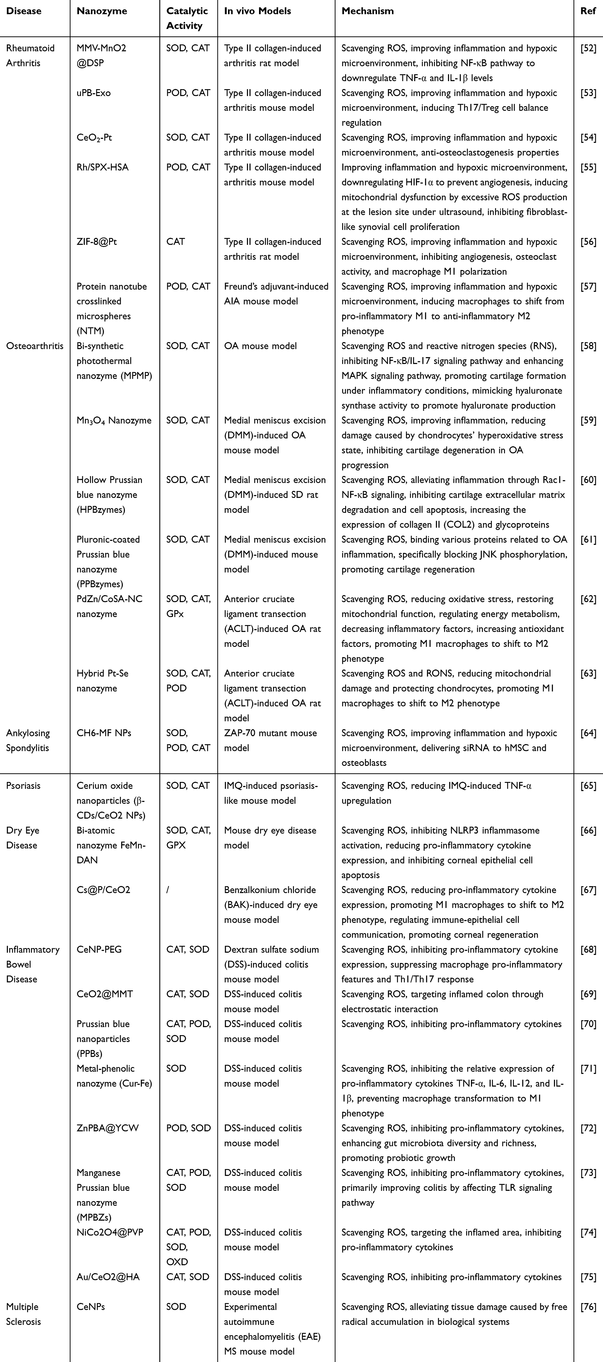 |
Table 1 The Therapeutic Application of Nanozymes in Autoimmune Diseases |
 |
Figure 1 Therapeutic mechanism of nanozymes in autoimmune disease via ROS scavenging and immunomodulation. Schematic model contrasting the pathological state (left panel) and the restored state following nanozyme intervention (right panel). Under pathological conditions, pro-inflammatory M1 macrophages drive pathological accumulation of ROS in autoimmune tissue, causing cellular damage and activating NF-κB signaling (nuclear translocation → pro-inflammatory gene expression). Nanozymes intervene by catalytically decomposing ROS through multi-enzyme mimetic activities: SOD-like, CAT-like and GPx-like. This efficient ROS clearance inhibits NF-κB activation and promotes polarization of M1 macrophages towards the reparative M2 phenotype, collectively resolving inflammation and promoting tissue repair. By Figdraw. |
Rheumatoid arthritis (RA) is an autoimmune disease characterized primarily by synovial inflammation and progressive joint destruction.77,78 Current treatment options for RA include glucocorticoids, disease-modifying antirheumatic drugs (DMARDs), and biologics, all of which focus on controlling inflammation. While these therapies have significantly improved RA prognosis, a subset of patients still experiences suboptimal responses, and these treatments fail to fully ameliorate the local joint microenvironment in RA.79,80 Oxidative stress, a critical component of chronic inflammation pathogenesis, is closely linked to RA progression.81,82 To address this, Zhang et al developed a novel neutrophil-derived exosome platform functionalized with ultra-small Prussian blue nanoparticles (<5 nm, termed uPB-Exo) using click chemistry.53 This design leverages the inherent targeting ability of neutrophils toward biomolecules, enabling uPB-Exo to selectively neutralize pro-inflammatory cytokines and alleviate oxidative stress in activated fibroblast-like synoviocytes (FLS), macrophages, and chondrocytes.83 In a late-stage RA mouse model (collagen-induced arthritis, CIA), uPB-Exo significantly mitigated joint damage and reduced overall arthritis severity by modulating the Th17/Treg cell balance, offering a promising new therapeutic approach for RA.
In a separate study, Liu et al engineered a Janus-CPS nanoplatform featuring a dual-component structure: one side incorporates a Pt nanozyme subunit paired with CeO2, which scavenges ROS and exerts anti-inflammatory effects, while the other side consists of periodic mesoporous organosilica (PMO) with a high surface area, ideal for loading micheliolide (MCL), an anti-bone-resorption drug.54 Upon integration with the near-infrared dye indocyanine green (ICG), the resulting Janus-CPS-MI system gained near-infrared-II fluorescence imaging capabilities, enabling early RA diagnosis and treatment monitoring. This research highlights the rational design of the Janus nanoplatform, underscoring its potential in integrated diagnostics and therapy, and broadening the application of Janus-structured biomaterials in biomedical fields. The therapeutic efficacy of these nanozymes is underpinned by their multifaceted intervention in RA-specific pathogenic circuits. Primarily, their ROS-scavenging capability directly quenches intracellular oxidative stress, leading to the inhibition of the NF-κB signaling pathway. This inhibition subsequently suppresses the production of pivotal pro-inflammatory cytokines such as TNF-α, IL-1β, and IL-6. Concurrently, by reshaping the oxidative microenvironment, nanozymes facilitate the repolarization of pro-inflammatory M1 macrophages towards the anti-inflammatory M2 phenotype. Furthermore, as demonstrated by uPB-Exo, the restoration of the Th17/Treg cell balance emerges as a critical immunomodulatory mechanism, effectively breaking the cycle of chronic inflammation and autoimmunity in RA.
Systemic lupus erythematosus (SLE) is an autoimmune disease characterized by multi-organ damage, with lupus nephritis (LN) affecting approximately 30% to 60% of adult SLE patients and up to 70% of pediatric cases.84 LN represents one of the most severe manifestations of SLE and is a critical determinant of overall morbidity and mortality.85 Current strategies to prevent the progression of LN-related chronic kidney disease (CKD) primarily involve two approaches: suppressing autoimmune SLE activity and managing non-immune risk factors that influence CKD progression. However, the diagnosis and treatment of LN remain fraught with challenges, as current methods often suffer from low efficacy or severe side effects at this stage. In contrast, nanomedicine has shown immense potential in imaging diagnostics and controlled drug release.86 To address these issues, Li et al developed a polydopamine (PDA)-based nanocarrier functionalized with Fe3O4 and Pt nanoparticles and loaded with necrostatin-1 (Nec-1), resulting in the Nec-1/PDA@Pt-Fe3O4 nanocarrier.87 This nanocarrier exhibits excellent biocompatibility and effectively reduces neutrophil extracellular trap formation, thereby suppressing inflammation progression. PDA, enriched with phenolic groups, serves as an efficient free radical scavenger, effectively neutralizing ROS generated during the inflammatory response.88,89 Meanwhile, Pt, with its high catalase-like activity, significantly ameliorates the hypoxic microenvironment of LN, alleviating symptoms to a certain extent. Experimental results further revealed that this nanocarrier produces strong photoacoustic signals and a negative enhancement effect on T2-weighted imaging, highlighting its potential for dual-modality imaging to accurately and noninvasively monitor LN progression. Nevertheless, additional effects of this approach in mouse models, including optimal dosage and safety concerns such as side effects, warrant further investigation. Regardless, this study undoubtedly paves a valuable new path for the diagnosis and treatment of lupus nephritis.
Osteoarthritis (OA) is the most common joint disease, often affecting cartilage and surrounding tissues. In its advanced stages, it leads to cartilage damage and loss, making it one of the leading causes of disability in the elderly.90,91 Clinically, the treatment of OA typically involves non-steroidal anti-inflammatory drugs (NSAIDs) or intra-articular injections of hyaluronic acid and platelet-rich plasma (PRP). Although NSAIDs can alleviate symptoms to some extent, they do not halt disease progression, and long-term use can lead to various adverse effects, such as nausea, gastric or duodenal ulcers, and even acute cardiovascular events.92 Joint replacement surgery is generally reserved for advanced OA patients, making it crucial to find effective interventions for early- to mid-stage OA, which is currently a research focus.93,94 Articular cartilage consists of extracellular matrix (ECM) and chondrocytes, and the primary pathological changes in OA are characterized by chondrocyte apoptosis and ECM degradation.95 ROS play a critical role in the pathogenesis of OA. ROS not only regulate chondrocyte apoptosis but also participate in the synthesis and degradation of ECM, influencing the production of various cytokines.96 The excessive generation of ROS is one of the key factors contributing to cartilage degeneration and joint damage.97,98 Therefore, regulating the balance between ROS and antioxidants is a key approach in OA treatment.
Researchers have designed and evaluated a hollow Prussian blue nanozyme (HPBzymes) to explore its impact on OA progression and assess its biological activity in vitro and in vivo.60 HPBzymes can mimic the activity of SOD and CAT, suppressing ROS production in a rat model and modulating the Rac1-NF-κB signaling pathway to slow the development of OA. This nanozyme effectively reduces ECM degradation and cell apoptosis while increasing the expression of type II collagen (COL2) and aggrecan. Additionally, the hollow structure and size of HPBzymes can be precisely controlled, showing promise as a drug carrier with greater therapeutic potential in the future.99
Li et al successfully constructed a bionic photothermal nanzyme (MPMP) for OA treatment by modifying magnesium ions (Mg2+) doped with dopamine (polydopamine) on molybdenum disulfide (MoS2) and coating them with polysulfobetaines.58 This nanzyme mimics the functions of antioxidant enzymes and hyaluronic acid synthase (HAS). Upon near-infrared radiation, the MPMP nanzyme triggers heat generation and magnesium ion release, promoting cartilage regeneration. It reduces inflammation by inhibiting the NF-κB/IL-17 signaling pathway and enhancing the MAPK signaling pathway. The MPMP nanzyme, which mimics the functions of antioxidant enzymes SOD and CAT, effectively scavenges harmful free radicals (ROS/RNS) in the body. Additionally, the nanzyme can deliver oxygen to relieve hypoxic conditions. It upregulates HSP70 expression, promoting the synthesis of hyaluronic acid (HA), and further improving cartilage repair and joint function. This study highlights the tremendous potential of dual-bionic nanzymes in OA and other inflammatory treatments, offering a new direction for future research. Nanozymes like hollow Prussian blue and Mn3O4 primarily exert their chondroprotective effects by scavenging excessive ROS within the joint cavity. This ROS elimination inhibits key pathways like Rac1-NF-κB, leading to reduced chondrocyte apoptosis and inflammatory factor release. Additionally, these nanozymes suppress the activity of matrix metalloproteinases (MMPs), thereby preserving crucial ECM components—including type II collagen and aggrecan—and slowing cartilage breakdown. MPMP offer further regenerative potential by catalyzing oxygen production under NIR irradiation and upregulating HSP70 expression, which enhances hyaluronic acid synthesis to collaboratively create a pro-regenerative microenvironment.
Psoriasis is a chronic inflammatory skin disorder, clinically characterized by red patches covered with white or silver scales.100 It is also associated with various comorbidities, including psoriatic arthritis, metabolic syndrome, and cardiovascular diseases. The etiology of psoriasis is complex, involving genetic susceptibility, environmental triggers, and immune dysregulation.101 Current conventional treatments include corticosteroids, vitamin D analogs, calcineurin inhibitors, phototherapy, methotrexate, cyclosporine, and apremilast. WU and colleagues developed β-cyclodextrin-modified cerium oxide nanoparticles (β-CDs/CeO2NPs) for the treatment of psoriasis.65 These nanoparticles demonstrated significant SOD and CAT activity in vitro and were able to significantly reduce intracellular ROS levels. Furthermore, the researchers loaded dithranol (DIT), a therapeutic agent for psoriasis, onto these nanoparticles. In the imiquimod (IMQ)-induced psoriasis model, the treatment resulted in a reduction of both the affected area and the Psoriasis Area and Severity Index (PASI) score, showing promising therapeutic effects. Although the application of nanozymes in the treatment of psoriasis and psoriatic arthritis is still in the early stages of exploration, the potential benefits and efficacy of combining nanozymes with therapeutic drugs hold great promise for future treatments.
Ankylosing spondylitis (AS) is a common chronic inflammatory joint disease, and its treatment remains challenging, with conventional therapeutic approaches having various limitations.102 Researchers have developed a novel delivery system based on manganese ferrite nanoparticles (MnFe2O4 nanoparticles, MF NPs) that targets ROS and specifically delivers therapeutic agents to osteoblasts.64 This system effectively eliminates ROS, and analysis of trabecular bone parameters (such as BV/TV, BA/BV, etc.) demonstrated that it can significantly alleviate bone damage in AS mice and partially restore bone mass. When loaded with BMP2 siRNA, CH6-MF-Si NPs exhibited good therapeutic effects on ectopic ossification in AS and could inhibit the abnormal osteogenic differentiation of hMSCs in vitro. This highlights the potential of CH6-MF NPs/BMP2 siRNA dual therapy in addressing chronic inflammation and ectopic ossification in AS.
Sjögren’s Syndrome (SS) is a chronic autoimmune disease that primarily affects the exocrine glands, particularly the salivary and lacrimal glands.103–105 Dry Eye Syndrome (DES) is one of the main clinical manifestations of Sjögren’s Syndrome, characterized by insufficient tear secretion or excessive tear evaporation, leading to symptoms such as dryness, a foreign body sensation, burning, and blurred vision.106 Dry eye disease not only represents a common symptom in Sjögren’s patients but also significantly contributes to a decrease in their quality of life. Dry Eye Disease (DED) is the most prevalent ocular surface disorder, affecting approximately one-third of the global population.107 As technological advancements increase the use of electronic devices, the incidence of DED continues to rise. The pathogenesis of DED is complex and involves oxidative stress, tear film instability, and ocular inflammation. Existing anti-inflammatory treatments show varying degrees of efficacy and risk. Conventional therapies, including artificial tears and immunosuppressants, provide only temporary symptom relief and may have potential side effects.108 Nanzyme-based drugs, integrating both antioxidant and anti-inflammatory functions, show great promise in treating DED.109,110 Cai and colleagues developed an innovative eye drop formulation based on dual-atom nanzymes (DAN).66 This formulation incorporates Fe and Mn dual-metal single atoms into nitrogen-doped carbon materials, which are further modified with hydrophilic polymers. In vitro and in vivo studies demonstrated that DAN exhibited exceptional biological activity in scavenging reactive oxygen species (ROS), inhibiting NLRP3 inflammasome activation, reducing pro-inflammatory cytokine expression, and preventing cell apoptosis. DAN effectively alleviates ocular inflammation, promotes corneal epithelial repair, restores goblet cell density, and enhances tear secretion, thus breaking the vicious cycle of DED.
Cui’s research team developed a nanzyme-based eye drop formulation—cyclosporine A-loaded nanoceria (Cs@P/CeO2).67 The drug was synthesized by first preparing hydrophobic oleylamine-capped nanoceria (OA/CeO2), followed by PEGylation and cyclosporine A loading, resulting in Cs@P/CeO2. In vitro studies revealed that Cs@P/CeO2 exhibited significant antioxidant and anti-inflammatory properties, inhibiting hydrogen peroxide-induced corneal epithelial cell death, reducing reactive oxygen species (ROS) levels, restoring mitochondrial function, and significantly reducing inflammatory cytokine secretion while suppressing NLRP3 protein levels. In vivo, results showed that in a mouse model of DED, Cs@P/CeO2 effectively improved corneal epithelial damage, accelerated corneal repair, reduced inflammation, and restored tear secretion as well as goblet cell quantity and structure in the conjunctiva. Single-cell sequencing analysis revealed that the eye drops could remodel the corneal microenvironment, suppress the inflammatory response of immune cells, and promote corneal epithelial cell regeneration. Dual-atom nanozymes (FeMn-DAN) and drug-loaded nanoceria (Cs@P/CeO2) mitigate the pathology of dry eye disease by leveraging their potent reactive oxygen species (ROS)-scavenging capacity to suppress the activation of the NLRP3 inflammasome and subsequent release of pro-inflammatory cytokines, such as IL-1β. This action not only alleviates ocular surface inflammation but also modulates immune-epithelial crosstalk, thereby inhibiting corneal epithelial cell apoptosis and promoting its repair and regeneration. Concurrently, these nanozymes contribute to restoring goblet cell density and tear secretion, effectively disrupting the vicious cycle of dry eye disease. These newly developed nanzyme-based eye drops hold great potential, and we eagerly anticipate the completion of in vivo safety evaluations, paving the way for their application in DED and Sjögren’s Syndrome patients.
Multiple sclerosis (MS) is a chronic inflammatory autoimmune disease of the central nervous system (CNS), primarily affecting young individuals, with the incidence in women being twice that of men.111–113 MS is currently considered a CD4+ Th1-mediated autoimmune disorder.114 Reactive oxygen species (ROS) are believed to play a pathogenic role in the inflammation associated with MS, leading to the loss of oligodendrocytes, axonal damage, and subsequent neuronal degeneration. Heckman and colleagues reported findings regarding cerium oxide nanoparticles (CeNPs) in an experimental autoimmune encephalomyelitis (EAE) mouse model of MS.76 The CeNPs reduced ROS levels and alleviated clinical symptoms and motor deficits in the mice. In cerebellar brain slices treated with CeNPs, ROS levels were significantly reduced, indicating a potential therapeutic effect on MS. The development and application of nanzymes in MS remain limited and are still in the early stages of research, requiring further experimental studies.
Inflammatory bowel disease (IBD), encompassing ulcerative colitis (UC) and Crohn’s disease (CD), is a chronic, immune-mediated inflammatory condition of the gastrointestinal tract that is challenging to treat.115,116 The pathogenesis of IBD is complex, with oxidative stress emerging as a central contributor.117,118 Excessive production of ROS triggers the sustained release of pro-inflammatory mediators, causing irreversible damage to proteins, lipids, and DNA, which may drive the onset and progression of IBD.119–121 To address this, Zeng et al developed biocompatible, drug-free cerium oxide nanoparticles (CeNP-PEG) with renewable capacity to scavenge multiple ROS types.68 CeNP-PEG ameliorates the pro-inflammatory microenvironment by continuously neutralizing ROS, downregulating pro-inflammatory cytokines, and suppressing the pro-inflammatory characteristics of macrophages and Th1/Th17 responses. This mechanism is likely linked to the co-inhibition of NF-κB and JAK2/STAT3 signaling pathways.122
In IBD, elevated ROS levels promote anaerobic respiration and proliferation of facultative anaerobes, enabling pathogens like Escherichia coli to dominate the gut ecological niche.123–125 However, conventional broad-spectrum antibiotics, while targeting these pathogens, can disrupt the gut microbiome and foster the emergence of antibiotic-resistant bacteria.126,127 Thus, designing strategies to selectively eliminate pathogenic E. coli is critical. Shi et al developed an enzyme-mimetic oral agent, ZnPBA@YCW, which encapsulates ZnPBA nanozymes within YCW to modulate the gut microbiota and restore redox balance for IBD treatment.72 ZnPBA@YCW specifically recognizes and adheres to harmful bacteria like E. coli, promotes the growth of beneficial microbiota through YCW degradation, and scavenges ROS to alleviate intestinal inflammation. Notably, other ROS-mediated diseases, such as rheumatoid arthritis and gout, are also closely associated with gut microbiota dysbiosis, suggesting that this therapeutic approach could offer novel insights for treating such conditions.
Combination of Nanozymes and Mesenchymal Stem Cells for the Treatment of Autoimmune Diseases
Stem cells possess the remarkable ability to self-renew and differentiate into multiple cell lineages. They are broadly categorized into embryonic stem cells (ESCs), induced pluripotent stem cells (iPSCs), and mesenchymal stem cells (MSCs).128 Among these, MSCs have emerged as the most widely utilized in cell-based therapies due to their extensive therapeutic potential, particularly in treating autoimmune diseases. MSCs exert their effects through mechanisms such as promoting tissue repair, modulating immune responses, and exhibiting anti-inflammatory properties.129–132 These versatile cells can be sourced from various tissues, including adipose tissue, amniotic fluid, bone marrow, dental pulp, peripheral blood, umbilical cord, and umbilical cord blood.133–135
In 1966, Friedenstein first described a unique population of cells found in rat bone marrow.136 These cells exhibited fibroblast-like morphology, adhered to surfaces, formed colonies, served as precursors to bone cells, and demonstrated tissue repair potential. In 1991, Arnold Caplan coined the term “mesenchymal stem cells” for these cells.137 However, subsequent research revealed that the primary mechanism of MSC-based therapies is their paracrine effect rather than differentiation into new cell types.138 MSCs secrete a range of reparative mediators, growth factors, cytokines, and other molecules that stimulate host cells to initiate repair responses. Reflecting this understanding, Caplan proposed renaming these cells “medicinal signaling cells” in 2017.139 Despite this, the term has not gained widespread traction, and nearly all registered clinical trials continue to use “mesenchymal stem cells” or “mesenchymal stromal cells.”
MSCs demonstrate significant potential in modulating both adaptive and innate immune responses.140 They suppress T-cell proliferation and activity, promote the generation of regulatory T cells (Tregs), and regulate the function of dendritic cells (DCs) and M2 macrophages, thereby exerting potent immunomodulatory effects. Concurrently, nanozymes—nanomaterials with enzyme-like catalytic properties—have garnered substantial attention as a cutting-edge research area. The integration of nanozymes with MSCs has shown promising therapeutic applications. For instance, Karakoti and colleagues found that cerium oxide nanoparticles (CeONPs) significantly enhance collagen production by human mesenchymal stem cells (hMSCs) cultured on porous bioactive glass scaffolds.141 This effect is likely due to CeONPs acting as oxygen buffers, modulating hMSC differentiation. Further studies by Wei and others demonstrated that CeONPs exhibit low toxicity in macrophages and MSCs, offering protective effects under both acute and chronic inflammatory conditions.142 Notably, at a concentration of 1 μg/mL, CeONPs promote MSC proliferation, osteogenic differentiation, and mineralization.
MSCs have emerged as a promising therapeutic strategy for RA. However, their efficacy is often hindered by the accumulation of ROS and hypoxia in the joint microenvironment.143–146 To address these challenges, Mao and colleagues developed copper sulfide@manganese dioxide nanoparticles (CuS@MnO2 NPs) with SOD and CAT-like activities to scavenge ROS in the RA oxidative stress microenvironment.147 These nanoparticles were surface-modified with an MSC-targeting peptide, VTAMEPGQ (abbreviated as VQ), engineered using phage display technology, significantly enhancing the MSC uptake of the NPs. Additionally, the core-shell structure of the NPs was loaded with metformin (MET), forming VQ-CuS@MnO2/MET NPs. MSCs internalized these NPs to create engineered VCMM-MSCs. In a simulated RA oxidative stress environment (supplemented with H2O2), VCMM-MSCs exhibited approximately 80% cell viability, markedly higher than the 55% observed in unmodified MSCs (Figure 2). In collagen-induced arthritis (CIA) and adjuvant-induced arthritis (AIA) rat models, the VCMM-MSC group outperformed unmodified MSCs and other treatment groups in clinical scores and hind paw thickness, effectively suppressing synovial invasion and cartilage destruction.
 |
Figure 2 This image shows the therapeutic effect of mesenchymal stem cells combined with nanoenzymes on rheumatoid arthritis. By Figdraw. |
In parallel, Zhao and colleagues designed a nanozyme-enhanced hydrogel (ε-PLE@MnCoO/Gel) that functions as an H2O2-driven oxygen generator to improve bone integration in RA treatment.148 This hydrogel significantly mitigated the detrimental effects of H2O2 on the osteogenic differentiation of bone marrow MSCs (BMSCs). By decomposing ROS and generating oxygen, the hydrogel promoted BMSC osteogenic differentiation, enabling transplanted BMSCs to achieve superior therapeutic outcomes in localized bone defects. BMSC-loaded nanozyme-enhanced hydrogels reduced prosthesis loosening, displacement, and periprosthetic fractures following RA prosthesis implantation, while demonstrating excellent biocompatibility in vivo. These findings highlight a promising avenue for MSC-based therapies in RA and other autoimmune diseases.
Tregs play a central role in immune tolerance, making them a critical therapeutic target for autoimmune diseases. The balance between T-helper 17 (Th17) cells and Tregs is pivotal in the pathogenesis of autoimmune and inflammatory conditions, particularly in RA.149–151 To address this, Hyeon’s team developed a hybrid system combining cerium dioxide (CeO2) nanoparticles (Ce NPs) with mesenchymal stem cell-derived nanovesicles (MSCNVs), integrating anti-inflammatory and immune-modulatory functions to alleviate joint inflammation and restore immune homeostasis.152 Using an improved reverse micelle method, the team synthesized uniform Ce NPs, which exhibit antioxidant properties by cycling between Ce3+ and Ce4+ oxidation states to scavenge ROS. These NPs effectively clear excessive ROS in RA-affected knee joints and promote the polarization of pro-inflammatory M1 macrophages toward anti-inflammatory M2 macrophages. MSCNVs were generated by continuously extruding MSCs through porous membranes. Subsequently, maleimide-functionalized Ce NPs were conjugated to thiol-functionalized MSCNVs via a thiol-maleimide reaction, forming Ce-MSCNVs. This hybrid system demonstrated exceptional SOD and CAT-mimetic activities. The MSCNV component protects chondrocytes, preventing bone and cartilage degradation. The two key components of Ce-MSCNVs—Ce NPs and MSCNVs—function independently or synergistically to enable rapid joint repair and rebalance the Th17/Treg cell ratio. As a comprehensive and innovative immunomodulatory nanohybrid therapy, Ce-MSCNVs outperformed single-modality treatments, effectively disrupting the vicious cycle of multifactorial RA and offering a highly efficient approach to its management.
Many autoimmune diseases, such as systemic lupus erythematosus, rheumatoid arthritis, and scleroderma, are frequently associated with the development of pulmonary fibrosis.153 Pulmonary fibrosis is characterized by the fibrotic transformation of lung tissue due to inflammation or other pathological factors, resulting in impaired lung function and potentially leading to respiratory failure. In its early stages, pulmonary fibrosis often presents with no apparent symptoms, making early diagnosis challenging. By the time it is detected, significant lung damage may have already occurred, limiting the effectiveness of treatment. Traditional pharmacological interventions, including immunosuppressants and anti-fibrotic agents, can partially slow the progression of the disease but are unable to reverse established fibrosis. Consequently, improvements in lung function are modest, and the disease exhibits a high recurrence rate.
MSCs have emerged as a promising therapeutic option due to their ability to promote tissue repair.154,155 In the context of pulmonary fibrosis, MSCs possess the potential to differentiate into alveolar type II epithelial cells (AEC IIs) under specific conditions.156 Moreover, MSCs can exert anti-inflammatory and anti-fibrotic effects by secreting various cytokines, such as vascular endothelial growth factor (VEGF), hepatocyte growth factor (HGF), and interleukin-10 (IL-10).157,158 Through paracrine mechanisms, MSCs modulate the immune microenvironment and suppress fibroblast activation, thereby potentially mitigating or reversing the fibrotic process in the lungs. The synergistic use of nanozymes, which exhibit potent antioxidant and anti-inflammatory properties, with MSCs can enhance drug delivery efficiency and amplify the reparative capabilities of MSCs. A notable study by Jiang et al introduced a nano-engineered platform, MSCs-Lip@NCAF, created by biologically conjugating MSCs with liposomes modified with type I collagenase (Lip@NCAF).159 This platform leverages the targeting capabilities of MSCs to migrate to fibrotic lung tissue and release Lip@NCAF in a sensitive manner. Lip@NCAF facilitates the degradation of collagen fibers, delivers nintedanib specifically to fibroblasts, and inhibits their excessive activation.160 Additionally, MSCs can differentiate into AEC IIs, aiding in the repair of alveolar structures and promoting lung regeneration in aged mice with pulmonary fibrosis.
The pathogenesis of pulmonary fibrosis is intricately linked to dysregulated immune responses, particularly the imbalance between M1 and M2 macrophage phenotypes. Future research should delve into strategies for modulating immune responses, especially within the framework of mesenchymal stem cell therapy. By precisely controlling the activation states of immune cells, it may be possible to further enhance therapeutic outcomes and minimize adverse effects.
Challenges in Clinical Translation
Nanozymology, as an emerging discipline, has experienced vigorous development over the past decade or more, but future development and applications still face considerable challenges. Although extensive research has demonstrated many nanozymes with effective therapeutic effects in in vitro and in vivo experimental models, these are basically experiments conducted in animal models, most of which are still in the preclinical stage. How to translate these studies into clinical applications that actually benefit patients is worthy of our deep consideration (Table 2).
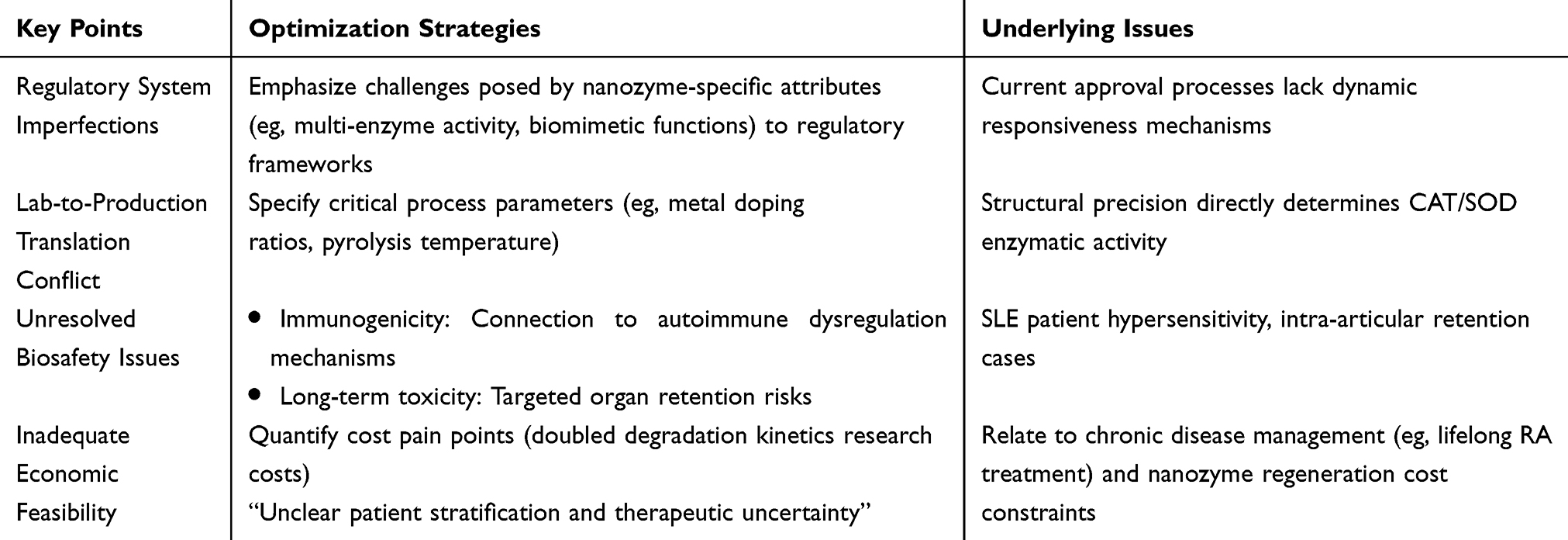 |
Table 2 Potential Issues and Optimization Strategies in Clinical Translation |
Nanozymes need to precisely target immune cells or pathways while avoiding interference with normal immune function. Additionally, ensuring the stability and biocompatibility of nanozymes in vivo is crucial. A significant knowledge gap, however, persists regarding their comprehensive long-term toxicity and immunogenicity profile. A thorough safety assessment for nanozymes must therefore investigate their dynamic interactions with the immune system, which can range from complement activation and macrophage uptake to the induction of specific antibody responses. Specific concerns revolve around their sequestration by the mononuclear phagocyte system (MPS) and subsequent long-term accumulation in organs, which could lead to unforeseen inflammatory or fibrotic reactions over time. While a reduction in particle size below 200 nm enhances capillary circulation and tissue penetration, it concurrently elevates nanotoxicological risks due to the increased specific surface area.161 Therefore, achieving an optimal particle size is a critical design parameter to ensure efficacious delivery while mitigating adverse effects. Surface coatings engineered with hydrophilic character, low charge density and flexible mechanical properties have been demonstrated to mitigate immunogenic responses and enhance systemic tolerance.162,163
The challenges of early diagnosis also affect treatment timing and effectiveness, as current diagnostic methods often struggle to detect problems in the early stages of disease. Clinical trial regulatory barriers, large-scale production requirements, and the need to demonstrate efficacy and safety across different patient populations further hinder the clinical application of nanozymes. While the vision of personalized nanozyme medicine is undoubtedly promising, its clinical realization is inherently tied to overcoming the broader translational challenges discussed above. The path toward personalization hinges on a more profound understanding of the disease-specific microenvironment. For instance, nanozymes could be engineered to target unique pathological signatures, such as a specific pH, overexpressed enzymes, or distinctive redox gradients. However, this level of precision requires advancements in several key areas: the development of robust biomarkers to guide patient stratification, the design of modular nanozyme platforms whose activities and surface properties can be finely tuned, and comprehensive assessments of individual variations in immune response and nanozyme biodistribution. Therefore, current research efforts, while perhaps not yet delivering immediate personalized therapies, are laying the essential groundwork by addressing fundamental issues of safety, targeting efficiency, and manufacturability. The journey toward personalized nanozyme-based treatment is parallel to the effort of maturing the entire field; both are concerted toward the ultimate goal of achieving safe, effective, and patient-specific nanotherapeutic interventions.
Overcoming these challenges requires a deeper understanding of disease mechanisms and nanozyme behavior in biological systems, as well as strengthened collaboration among researchers, clinicians, and regulatory agencies. Currently, AI prediction models based on multimodal data (patient immunomics, nanozyme catalytic kinetics) can simulate the efficacy-toxicity balance in real-time, providing quantitative evidence for regulatory agencies to establish dynamic risk assessment frameworks.164,165 Machine learning analyzes the correlation between physical parameters (such as nanozyme surface charge distribution and oxygen vacancy concentration) and immunomodulatory effects, guiding the rational design of materials with “high catalytic activity-low immunogenicity.” Combined with high-throughput screening using microfluidic chips, nanozyme synthesis processes can be parametrically optimized (such as metal doping gradient control). AI can also mine multi-omics data (single-cell transcriptomics and serum antibody profiles) to precisely identify populations with nanozyme response advantages, significantly reducing trial scale and costs.
Conclusions and Outlook
We provide a systematic review of the application of nanozymes in the treatment of autoimmune diseases, with a focus on conditions such as rheumatoid arthritis, osteoarthritis, ankylosing spondylitis, psoriasis, dry eye syndrome, inflammatory bowel disease, and multiple sclerosis. Nanozymes, as a novel class of biological catalysts, exhibit significant therapeutic potential due to their high catalytic activity, excellent stability, and tunable catalytic properties. We discuss in detail the catalytic mechanisms of carbon-based nanozymes, metal-based nanozymes, and several other types of nanozymes, emphasizing their key roles in scavenging ROS, improving inflammatory and hypoxic microenvironments, and regulating immune responses.
The convergence of nanozyme technology and stem cell therapeutics represents a transformative strategy for autoimmune disease management, where nanozymes engineered with multi-enzyme activities (eg, SOD/CAT/GPx mimicry) protect transplanted mesenchymal stem cells (MSCs) from inflammatory microenvironment assaults by scavenging pathological ROS cascades, while simultaneously enhancing MSC viability and immunomodulatory functions through valence transition-mediated metabolic reprogramming. This synergistic approach not only overcomes the critical limitation of conventional MSC therapy—where >70% cell death occurs within hostile autoimmune microenvironments—but also leverages surface-functionalized nanozymes to direct stem cell homing to affected tissues (eg, arthritic joints or inflamed intestinal mucosa), ultimately establishing a self-amplifying cycle of inflammation resolution and tissue regeneration through bystander effects of nanozyme-armed stem cells.
Nanozymes can mimic the activities of natural enzymes, including SOD, CAT, and POD, thereby effectively eliminating ROS, alleviating oxidative stress, and mitigating inflammation and tissue damage. For example, in the treatment of rheumatoid arthritis, nanozymes such as MMV-MnO2@DSP and CeO2-Pt significantly reduce inflammatory factor levels by clearing ROS and inhibiting the NF-κB signaling pathway. In osteoarthritis, nanozymes like Mn3O4 and HPBzymes exert their effects by protecting chondrocytes and promoting cartilage regeneration. Additionally, nanozymes show diverse therapeutic potentials, including regulating the Th17/Treg cell balance, suppressing angiogenesis, and promoting the transformation of M1 macrophages to the M2 phenotype.
This review makes a comprehensive summary of the current applications and therapeutic mechanisms of nanozymes across various autoimmune diseases, as well as its organization of the design and functions of nanozymes. This work offers theoretical foundations and practical guidance for the further development of nanozymes in biomedicine. Through the analysis of existing research, the paper highlights the broad prospects of nanozymes in enhancing the inflammatory microenvironment, promoting tissue repair, and regulating the immune system.
Although nanozymes have demonstrated significant advantages in autoimmune disease treatment, their development and application still face several challenges. Future research should focus on the following areas:
- Design and optimization of nanozymes: Developing nanozymes with higher catalytic activity and selectivity to enhance therapeutic efficacy and reduce potential side effects. This includes exploring new materials (eg, M-N-C structures) and optimizing the electronic structure of nanozymes to improve their performance.
- Targeting and biocompatibility: Enhancing the targeting ability of nanozymes to ensure precise action at diseased sites while minimizing interference with healthy tissues. Additionally, ensuring their biocompatibility in vivo to avoid adverse immune reactions.
- Long-term safety and stability: Conduct systematic evaluations of the long-term safety and stability of nanozymes in vivo, investigating their metabolic pathways and potential degradation products to ensure treatment safety. This requires additional in vivo experiments and preclinical validations.
- Development of multifunctional nanozymes: Designing nanozymes with multiple functions, such as simultaneous diagnosis and treatment, or stimulus-responsive drug release (eg, ultrasound, photothermal), to meet the needs of personalized therapy.
- In-depth mechanism research: Further elucidating the mechanisms of nanozymes in autoimmune disease treatment, particularly the molecular details of immune regulation, inflammatory signaling pathways, and microenvironment improvement, to provide scientific support for optimizing treatment strategies.
In summary, nanozymes, as an emerging therapeutic tool, have shown tremendous potential in the field of autoimmune diseases. With advancements in materials science and continued research, nanozymes are expected to become a vital pillar of biomedicine, offering patients more efficient and safer treatment options and significantly improving their quality of life.
Data Sharing Statement
No primary research results, software or code have been included and no new data were generated or analysed as part of this review.
Author Contributions
All authors made a significant contribution to the work reported, whether that is in the conception, study design, execution, acquisition of data, analysis and interpretation, or in all these areas; took part in drafting, revising or critically reviewing the article; gave final approval of the version to be published; have agreed on the journal to which the article has been submitted; and agree to be accountable for all aspects of the work.
Funding
This work was supported by the Shanxi Provincial Department of Science and Technology Basic Research Program General Project (202403021211113), the Research and Innovation Team Project for Scientific Breakthroughs at Shanxi Bethune Hospital (2024AOXIANG02), the Climbing Plan of Shanxi Bethune Hospital (PF202505) and Shanxi Province Clinical Theranostics Technology Innovation Center for Immunologic and Rheumatic Diseases (CXZX-202301).
Disclosure
The authors declare that the current review work was conducted in the absence of any commercial or financial relationships that could be construed as a potential conflict of interest.
References
1. Garcia-Viloca M, Gao J, Karplus M, Truhlar DG. How enzymes work: analysis by modern rate theory and computer simulations. Science. 2004;303(5655):186–22. doi:10.1126/science.1088172
2. Natalio F, André R, Hartog AF, et al. Vanadium pentoxide nanoparticles mimic vanadium haloperoxidases and thwart biofilm formation. Nature Nanotechnol. 2012;7(8):530–535. doi:10.1038/nnano.2012.91
3. Lin Y, Ren J, Qu X. Catalytically active nanomaterials: a promising candidate for artificial enzymes. Acc Chem Res. 2014;47(4):1097–1105. doi:10.1021/ar400250z
4. Gao L, Zhuang J, Nie L, et al. Intrinsic peroxidase-like activity of ferromagnetic nanoparticles. Nat Nanotechnol. 2007;2(9):577–583. doi:10.1038/nnano.2007.260
5. Robert A, Meunier B. How to define a nanozyme. Acs Nano. 2022;16(5):6956–6959. doi:10.1021/acsnano.2c02966
6. Zhang L, Wang H, Qu X. Biosystem-inspired engineering of nanozymes for biomedical applications. Adv Mater. 2024;36(10):2211147. doi:10.1002/adma.202211147
7. Lincoln MR, Connally N, Axisa -P-P, et al. Genetic mapping across autoimmune diseases reveals shared associations and mechanisms. Nature Genet. 2024;56(5):838–845. doi:10.1038/s41588-024-01732-8
8. Sumida TS, Cheru NT, Hafler DA. The regulation and differentiation of regulatory T cells and their dysfunction in autoimmune diseases. Nat Rev Immunol. 2024;24(7):503–517. doi:10.1038/s41577-024-00994-x
9. Balogh L, Oláh K, Sánta S, Majerhoffer N, Németh T. Novel and potential future therapeutic options in systemic autoimmune diseases. Front Immunol. 2024;15:1249500. doi:10.3389/fimmu.2024.1249500
10. Song Y, Li J, Wu Y. Evolving understanding of autoimmune mechanisms and new therapeutic strategies of autoimmune disorders. Signal Transduct Targeted Therapy. 2024;9(1):263. doi:10.1038/s41392-024-01952-8
11. Miller FW. The increasing prevalence of autoimmunity and autoimmune diseases: an urgent call to action for improved understanding, diagnosis, treatment, and prevention. Curr Opinion Immunol. 2023;80:102266. doi:10.1016/j.coi.2022.102266
12. Nakazawa MS, Keith B, Simon MC. Oxygen availability and metabolic adaptations. Nat Rev Cancer. 2016;16(10):663–673. doi:10.1038/nrc.2016.84
13. Niethammer P, Grabher C, Look AT, Mitchison TJ. A tissue-scale gradient of hydrogen peroxide mediates rapid wound detection in zebrafish. Nature. 2009;459(7249):996–999. doi:10.1038/nature08119
14. Lin W, Shen P, Song Y, Huang Y, Tu S. Reactive oxygen species in autoimmune cells: function, differentiation, and metabolism. Front Immunol. 2021;12:635021. doi:10.3389/fimmu.2021.635021
15. Wu G, Berka V, Derry PJ, Mendoza K, Kakadiaris E, Roy T. Critical comparison of the superoxide dismutase-like activity of carbon antioxidant nanozymes by direct superoxide consumption kinetic measurements. ACS Nano. 2019;13(10):11203–11213. doi:10.1021/acsnano.9b04229
16. Cao S, Zhao Z, Zheng Y. A library of ROS-catalytic metalloenzyme mimics with atomic metal centers. Adv Mater. 2022;34(16):e2200255. doi:10.1002/adma.202200255
17. Singh N, Savanur MA, Srivastava S, D’Silva P, Mugesh G. A redox modulatory Mn 3 O 4 nanozyme with multi-enzyme activity provides efficient cytoprotection to human cells in a Parkinson’s disease model. Angew Chem. 2017;129(45):14455–14459. doi:10.1002/ange.201708573
18. Yan R, Sun S, Yang J, Long W, Wang J, Mu X, Ming D. Nanozyme-based bandage with single-atom catalysis for brain trauma. ACS nano. 2019;13(10):11552–11560. doi:10.1021/acsnano.9b05075
19. Huo M, Wang L, Chen Y, Shi J. Tumor-selective catalytic nanomedicine by nanocatalyst delivery. Nat Commun. 2017;8(1):357. doi:10.1038/s41467-017-00424-8
20. Zhang Q, He X, Han A, et al. Artificial hydrolase based on carbon nanotubes conjugated with peptides. Nanoscale. 2016;8(38):16851–16856. doi:10.1039/C6NR05015H
21. Hu Y, Gao X, Zhu Y, et al. Nitrogen-doped carbon nanomaterials as highly active and specific peroxidase mimics. Chem Mater. 2018;30(18):6431–6439. doi:10.1021/acs.chemmater.8b02726
22. Xu Z, Lan J, Jin J, Dong P, Jiang F, Liu Y. Highly photoluminescent nitrogen-doped carbon nanodots and their protective effects against oxidative stress on cells. ACS Appl Mater Interfaces. 2015;7(51):28346–28352. doi:10.1021/acsami.5b08945
23. He J, Hou Y, Zhang Z, et al. Carbon-based nanozymes: how structure affects performance. Nano Biomed Eng. 2024;16(1):28. doi:10.26599/NBE.2024.9290053
24. Zhao H, Zhang R, Yan X, Fan K. Superoxide dismutase nanozymes: an emerging star for anti-oxidation. J Mat Chem B. 2021;9(35):6939–6957. doi:10.1039/D1TB00720C
25. Meng X, Fan H, Chen L, et al. Ultrasmall metal alloy nanozymes mimicking neutrophil enzymatic cascades for tumor catalytic therapy. Nat Commun. 2024;15(1):1626. doi:10.1038/s41467-024-45668-3
26. Liang M, Wang Y, Ma K, et al. Engineering inorganic nanoflares with elaborate enzymatic specificity and efficiency for versatile biofilm eradication. Small. 2020;16(41):2002348. doi:10.1002/smll.202002348
27. Li S, Shang L, Xu B, et al. A nanozyme with photo-enhanced dual enzyme-like activities for deep pancreatic cancer therapy. Angew Chem. 2019;131(36):12754–12761. doi:10.1002/ange.201904751
28. Liang S, Deng X, Chang Y, et al. Intelligent hollow Pt-CuS janus architecture for synergistic catalysis-enhanced sonodynamic and photothermal cancer therapy. Nano Lett. 2019;19(6):4134–4145. doi:10.1021/acs.nanolett.9b01595
29. Montini T, Melchionna M, Monai M, Fornasiero P. Fundamentals and catalytic applications of CeO 2-based materials. Chem Rev. 2016;116(10):5987–6041. doi:10.1021/acs.chemrev.5b00603
30. Weng Q, Sun H, Fang C, et al. Catalytic activity tunable ceria nanoparticles prevent chemotherapy-induced acute kidney injury without interference with chemotherapeutics. Nat Commun. 2021;12(1):1436. doi:10.1038/s41467-021-21714-2
31. Trenque I, Magnano GC, Bolzinger MA, et al. Shape-selective synthesis of nanoceria for degradation of paraoxon as a chemical warfare simulant. Phys Chem Chem Phys. 2019;21(10):5455–5465. doi:10.1039/C9CP00179D
32. Fisher TJ, Zhou Y, Wu T-S, Wang M, Soo Y-L, Cheung CL. Structure–activity relationship of nanostructured ceria for the catalytic generation of hydroxyl radicals. Nanoscale. 2019;11(10):4552–4561. doi:10.1039/C8NR09393H
33. Xu B, Zhang Q, Yuan S, Zhang M, Ohno T. Morphology control and characterization of broom-like porous CeO2. Chem Eng J. 2015;260:126–132. doi:10.1016/j.cej.2014.09.001
34. Huang G, Zang J, He L, et al. Bioactive nanoenzyme reverses oxidative damage and endoplasmic reticulum stress in neurons under ischemic stroke. ACS nano. 2022;16(1):431–452. doi:10.1021/acsnano.1c07205
35. Shen J, Chen A, Cai Z, et al. Exhausted local lactate accumulation via injectable nanozyme-functionalized hydrogel microsphere for inflammation relief and tissue regeneration. Bioact Mater. 2022;12:153–168. doi:10.1016/j.bioactmat.2021.10.013
36. Tang M, Zhang Z, Sun T, Li B, Wu Z. Manganese-based nanozymes: preparation, catalytic mechanisms, and biomedical applications. Adv Healthcare Mater. 2022;11(21):2201733. doi:10.1002/adhm.202201733
37. Wang X, Wen F, He L, Su J, Jiang P, He D. Engineering porous Co–Mn oxide nanosheets with abundant oxygen vacancy as an efficient oxidase-like mimic for heparin colorimetric sensing. Anal Chim Acta. 2022;1198:339564. doi:10.1016/j.aca.2022.339564
38. Jiang B, Duan D, Gao L, Zhou M, Fan K. Standardized assays for determining the catalytic activity and kinetics of peroxidase-like nanozymes. Nat Protoc. 2018;13(7):1506–1520. doi:10.1038/s41596-018-0001-1
39. Li F, Guo L, Li Z, He J, Cui H. Temporal-spatial-color multiresolved chemiluminescence imaging for multiplex immunoassays using a smartphone coupled with microfluidic chip. Anal Chem. 2020;92(10):6827–6831. doi:10.1021/acs.analchem.0c01405
40. Zeng X, Liu H, Wu K, Deng A, Li J. Ultra-sensitive detection of florfenicol by flow injection chemiluminescence immunoassay based on Nickel/Cobalt bimetallic metal–organic framework nanozymes. Analyst. 2022;147(7):1321–1328. doi:10.1039/D2AN00126H
41. Li J, Cao Y, Hinman SS, et al. Efficient label-free chemiluminescent immunosensor based on dual functional cupric oxide nanorods as peroxidase mimics. Biosens Bioelectro. 2018;100:304–311. doi:10.1016/j.bios.2017.09.011
42. Fan K, Cao C, Pan Y, et al. Magnetoferritin nanoparticles for targeting and visualizing tumour tissues. Nat Nanotechnol. 2012;7(12):459–464. doi:10.1038/nnano.2012.235
43. Zhen W, Liu Y, Lin L, et al. BSA-IrO 2: catalase-like nanoparticles with high photothermal conversion efficiency and a High X-ray absorption coefficient for anti-inflammation and antitumor theranostics. Angew Chem. 2018;130(32):10466–10470. doi:10.1002/ange.201804466
44. Fu Q, Zhu R, Song J, Yang H, Chen X. Photoacoustic imaging: contrast agents and their biomedical applications. Adv Mater. 2019;31(6):1805875. doi:10.1002/adma.201805875
45. Wu H, Xia F, Zhang L, et al. A ROS-sensitive nanozyme-augmented photoacoustic nanoprobe for early diagnosis and therapy of acute liver failure. Adv Mater. 2022;34(7):2108348. doi:10.1002/adma.202108348
46. Moja L. Antibacterial agents in clinical development: an analysis of the antibacterial clinical development pipeline, including tuberculosis. World Health Organization. 2017
47. Wang Y, Yang Y, Shi Y, Song H, Yu C. Antibiotic-free antibacterial strategies enabled by nanomaterials: progress and perspectives. Adv Mater. 2020;32(18):e1904106. doi:10.1002/adma.201904106
48. Zhang Y, Zhang X, Song J, Jin L, Wang X, Quan C. Ag/H-ZIF-8 nanocomposite as an effective antibacterial agent against pathogenic bacteria. Nanomaterials. 2019;9(11):1579. doi:10.3390/nano9111579
49. Wang H, Li P, Yu D, et al. Unraveling the enzymatic activity of oxygenated carbon nanotubes and their application in the treatment of bacterial infections. Nano Lett. 2018;18(6):3344–3351. doi:10.1021/acs.nanolett.7b05095
50. Sun HJ, Gao N, Dong K, Ren J, Qu X. Dong K.Graphene quantum dots-band-aids used for wound disinfection. ACS Nano. 2014;8(6):6202–6210. doi:10.1021/nn501640q
51. Wang Z, Dong K, Liu Z. Activation of biologically relevant levels of reactive oxygen species by Au/g-C3N4 hybrid nanozyme for bacteria killing and wound disinfection. Biomaterials. 2017;113:145–157. doi:10.1016/j.biomaterials.2016.10.041
52. Jia M, Ren W, Liu Y, et al. Messenger nanozyme for reprogramming the microenvironment of rheumatoid arthritis. ACS Appl Mater Interfaces. 2023;15(1):338–353. doi:10.1021/acsami.2c16458
53. Zhang L, Qin Z, Sun H, et al. Nanoenzyme engineered neutrophil-derived exosomes attenuate joint injury in advanced rheumatoid arthritis via regulating inflammatory environment. Bioact Mater. 2022;18:1–14. doi:10.1016/j.bioactmat.2022.02.017
54. Liu Y, Chen L, Chen Z, et al. Multifunctional Janus nanoplatform for efficiently synergistic theranostics of rheumatoid arthritis. ACS Nano. 2023;17(9):8167–8182. doi:10.1021/acsnano.2c11777
55. Li W, Song Y, Liang X. Mutual-reinforcing sonodynamic therapy against rheumatoid arthritis based on sparfloxacin sonosensitizer doped concave-cubic rhodium nanozyme. Biomaterials. 2021;276:121063. doi:10.1016/j.biomaterials.2021.121063
56. Xuelan LEI, Fangxue DU. ZIF-8@ Pt nanozyme used for scavenging reactive oxygen species in the treatment of rheumatoid arthritis. J Sichuan Univ. 2024;55:826–837.
57. Hou G, Chen S, Ngai T, et al. The nanozymes of protein nanotubes-constructed microspheres with dual peroxidase- and catalase-like properties for M1-to-M2 macrophages repolarization and the synergistic anti-rheumatoid arthritis effect with loaded capsaicin. Nano Today. 2024;56:102290. doi:10.1016/j.nantod.2024.102290
58. Yu P, Li Y, Sun H, et al. Mimicking antioxidases and hyaluronan synthase: a zwitterionic nanozyme for photothermal therapy of osteoarthritis. Adv Mater. 2023;35(44):2303299. doi:10.1002/adma.202303299
59. Wang W, Duan J, Ma W, et al. Trimanganese tetroxide nanozyme protects cartilage against degeneration by reducing oxidative stress in osteoarthritis. Adv Sci. 2023;10(17):2205859. doi:10.1002/advs.202205859
60. Hou W, Ye C, Chen M, et al. Excavating bioactivities of nanozyme to remodel microenvironment for protecting chondrocytes and delaying osteoarthritis. Bioact Mater. 2021;6(8):2439–2451. doi:10.1016/j.bioactmat.2021.01.016
61. Cho C, Oh H, Lee JS, et al. Prussian blue nanozymes coated with pluronic attenuate inflammatory osteoarthritis by blocking c-Jun N-terminal kinase phosphorylation. Biomaterials. 2023;297:122131. doi:10.1016/j.biomaterials.2023.122131
62. Yang X, Tan M, Guo J, et al. PdZn/Co SA -NC nanozymes with highly efficient SOD/CAT activities for treatment of osteoarthritis via regulating immune microenvironment. Adv Funct Mater. 2024;34(41):2401963. doi:10.1002/adfm.202401963
63. Wei H, Huang H, He H, et al. Pt–Se hybrid nanozymes with potent catalytic activities to scavenge ROS/RONS and regulate macrophage polarization for osteoarthritis therapy. Research. 2024;7:0310. doi:10.34133/research.0310
64. Zheng G, Peng X, Zhang Y, et al. A novel Anti-ROS osteoblast-specific delivery system for ankylosing spondylitis treatment via suppression of both inflammation and pathological new bone formation. J Nanobiotechnol. 2023;21(1):168. doi:10.1186/s12951-023-01906-2
65. Wu L, Liu G, Wang W, et al. Cyclodextrin-modified CeO2 nanoparticles as a multifunctional nanozyme for combinational therapy of psoriasis. Int J Nanomed. 2020;15:2515–2527. doi:10.2147/IJN.S246783
66. Cui W, Chen S, Hu T, et al. Nanoceria-mediated cyclosporin a delivery for dry eye disease management through modulating immune–epithelial crosstalk. ACS nano. 2024;18(17):11084–11102. doi:10.1021/acsnano.3c11514
67. Chu D, Zhao M, Rong S, et al. Dual-atom nanozyme eye drops attenuate inflammation and break the vicious cycle in dry eye disease. Nano-Micro Lett. 2024;16(1):120. doi:10.1007/s40820-024-01322-7
68. Zeng F, Shi Y, Wu C, et al. A drug-free nanozyme for mitigating oxidative stress and inflammatory bowel disease. J Nanobiotechnol. 2022;20(1):107. doi:10.1186/s12951-022-01319-7
69. Zhao S, Li Y, Liu Q, et al. An orally administered CeO 2 @montmorillonite nanozyme targets inflammation for inflammatory bowel disease therapy. Adv Funct Mater. 2020;30(45):2004692. doi:10.1002/adfm.202004692
70. Zhao J, Cai X, Gao W, et al. Prussian blue nanozyme with multienzyme activity reduces colitis in mice. ACS Appl Mater Interfaces. 2018;10(31):26108–26117. doi:10.1021/acsami.8b10345
71. Zhu Y, Huang X, Deng Z, et al. Orally biomimetic metal-phenolic nanozyme with quadruple safeguards for intestinal homeostasis to ameliorate ulcerative colitis. J Nanobiotechnol. 2024;22(1):545. doi:10.1186/s12951-024-02802-z
72. Shi Z, Li X, Chen J, et al. Enzyme-like biomimetic oral-agent enabling modulating gut microbiota and restoring redox homeostasis to treat inflammatory bowel disease. Bioact Mater. 2024;35:167–180. doi:10.1016/j.bioactmat.2024.01.016
73. Zhao J, Gao W, Cai X, et al. Nanozyme-mediated catalytic nanotherapy for inflammatory bowel disease. Theranostics. 2019;9(10):2843. doi:10.7150/thno.33727
74. Ma Y, Zhao J, Cheng L, et al. Versatile carbon dots with superoxide dismutase-like nanozyme activity and red fluorescence for inflammatory bowel disease therapeutics. Carbon. 2023;204:526–537. doi:10.1016/j.carbon.2023.01.006
75. Chen Q, Zhang X. Research advances on nanozyme-guided therapy of inflammatory bowel diseases ★. Acta Chim Sinica. 2023;81(8):1043. doi:10.6023/A23040144
76. Heckman KL, DeCoteau W, Estevez A, et al. Custom cerium oxide nanoparticles protect against a free radical mediated autoimmune degenerative disease in the brain. ACS nano. 2013;7(12):10582–10596. doi:10.1021/nn403743b
77. Aletaha D, Smolen JS. Diagnosis and management of rheumatoid arthritis: a review. JAMA. 2018;320(13):1360–1372. doi:10.1001/jama.2018.13103
78. McInnes IB, Schett G. The pathogenesis of rheumatoid arthritis. N Engl J Med. 2011;365(23):2205–2219. doi:10.1056/NEJMra1004965
79. Favalli EG, Raimondo MG, Becciolini A, Crotti C, Biggioggero M, Caporali R. The management of first-line biologic therapy failures in rheumatoid arthritis: current practice and future perspectives. Autoimmunity Rev. 2017;16(12):1185–1195. doi:10.1016/j.autrev.2017.10.002
80. Friedman B, Cronstein B. Methotrexate mechanism in treatment of rheumatoid arthritis. Joint Bone Spine. 2019;86(3):301–307. doi:10.1016/j.jbspin.2018.07.004
81. Wruck CJ, Fragoulis A, Gurzynski A, et al. Role of oxidative stress in rheumatoid arthritis: insights from the Nrf2-knockout mice. Ann Rheumatic Dis. 2011;70(5):844–850. doi:10.1136/ard.2010.132720
82. Stamp LK, Khalilova I, Tarr JM, et al. Myeloperoxidase and oxidative stress in rheumatoid arthritis. Rheumatology. 2012;51(10):1796–1803. doi:10.1093/rheumatology/kes193
83. Kalluri R, LeBleu VS. The biology, function, and biomedical applications of exosomes. Science. 2020;367(6478):eaau6977. doi:10.1126/science.aau6977
84. Lichtnekert J, Anders H-J. Lupus nephritis-related chronic kidney disease. Nat Rev Rheumatol. 2024;20(11):699–711. doi:10.1038/s41584-024-01158-w
85. Mifflin L, Ofengeim D, Yuan J. Receptor-interacting protein kinase 1 (RIPK1) as a therapeutic target. Nat Rev Drug Discov. 2020;19(8):553–571. doi:10.1038/s41573-020-0071-y
86. Siddique S, Chow JCL. Recent advances in functionalized nanoparticles in cancer theranostics. Nanomaterials. 2022;12(16):2826. doi:10.3390/nano12162826
87. Li M, Wang Y, Han X, Liu Y, Ma M, Zhang L. Multifunctional polydopamine-based nanoparticles for dual-mode imaging guided targeted therapy of lupus nephritis. Pharmaceutics. 2022;14(10):1988. doi:10.3390/pharmaceutics14101988
88. Xu H, Zhang Y, Zhang H, et al. Smart polydopamine-based nanoplatforms for biomedical applications: state-of-art and further perspectives. Coord Chem Rev. 2023;488:215153. doi:10.1016/j.ccr.2023.215153
89. Battaglini M, Emanet M, Carmignani A, Ciofani G. Polydopamine-based nanostructures: a new generation of versatile, multi-tasking, and smart theranostic tools. Nano Today. 2024;55:102151. doi:10.1016/j.nantod.2024.102151
90. Moseng T, Vlieland TPM, Battista S, et al. EULAR recommendations for the non-pharmacological core management of Hip and knee osteoarthritis: 2023 update. Ann Rheumatic Dis. 2024;83(6):730–740. doi:10.1136/ard-2023-225041
91. Yao Q, Wu X, Tao C, et al. Osteoarthritis: pathogenic signaling pathways and therapeutic targets. Signal transduction and targeted therapy. Signal Transduct Targeted Therapy. 2023;8(1):56. doi:10.1038/s41392-023-01330-w
92. Gregori D, Giacovelli G, Minto C, et al. Association of pharmacological treatments with long-term pain control in patients with knee osteoarthritis: a systematic review and meta-analysis. JAMA. 2018;320(24):2564–2579. doi:10.1001/jama.2018.19319
93. Grosser T, Ricciotti E, FitzGerald GA. The cardiovascular pharmacology of nonsteroidal anti-inflammatory drugs. Trends Pharmacol Sci. 2017;38(8):733–748. doi:10.1016/j.tips.2017.05.008
94. Castorina S, Guglielmino C, Castrogiovanni P, et al. Clinical evidence of traditional vs fast track recovery methodologies after total arthroplasty for osteoarthritic knee treatment. A retrospective observational study. Muscle Ligaments Tendons J. 2019;7(03):504. doi:10.32098/mltj.03.2017.14
95. Hwang HS, Kim HA. Chondrocyte apoptosis in the pathogenesis of osteoarthritis. Int J Mol Sci. 2015;16(11):26035–26054. doi:10.3390/ijms161125943
96. Ahmad N, Ansari MY, Haqqi TM. Role of iNOS in osteoarthritis: pathological and therapeutic aspects. J Cell Physiol. 2020;235(10):6366–6376. doi:10.1002/jcp.29607
97. Lepetsos P, Papavassiliou AG. ROS/oxidative stress signaling in osteoarthritis. Biochimica et Biophysica Acta. 2016;1862(4):576–591. doi:10.1016/j.bbadis.2016.01.003
98. Lepetsos P, Papavassiliou KA, Papavassiliou AG. Redox and NF-κB signaling in osteoarthritis. Free Radic Biol Med. 2019;132:90–100. doi:10.1016/j.freeradbiomed.2018.09.025
99. Zhang W, Hu S, Yin -J-J, et al. Prussian blue nanoparticles as multienzyme mimetics and reactive oxygen species scavengers. J Am Chem Soc. 2016;138(18):5860–5865. doi:10.1021/jacs.5b12070
100. Kharouf F, Gladman DD. Advances in the management of psoriatic arthritis in adults. BMJ. 2024;387:e081860. doi:10.1136/bmj-2024-081860
101. Najm A, Goodyear CS, McInnes IB, Siebert S. Phenotypic heterogeneity in psoriatic arthritis: towards tissue pathology-based therapy. Nat Rev Rheumatol. 2023;19(3):153–165. doi:10.1038/s41584-022-00874-5
102. Braun J, Sieper J. Fifty years after the discovery of the association of HLA B27 with ankylosing spondylitis. RMD Open. 2023;9(3):e003102. doi:10.1136/rmdopen-2023-003102
103. Beydon M, McCoy S, Nguyen Y, Sumida T, Mariette X, Seror R. Epidemiology of Sjögren syndrome. Nat Rev Rheumatol. 2024;20(3):158–169. doi:10.1038/s41584-023-01057-6
104. Baldini C, Fulvio G, La Rocca G, Ferro F. Update on the pathophysiology and treatment of primary Sjögren syndrome. Nat Rev Rheumatol. 2024;20(8):473–491. doi:10.1038/s41584-024-01135-3
105. Thorlacius GE, Björk A, Wahren-Herlenius M. Genetics and epigenetics of primary Sjögren syndrome: implications for future therapies. Nat Rev Rheumatol. 2023;19(5):288–306. doi:10.1038/s41584-023-00932-6
106. Hakim FE, Farooq AV. Dry eye disease: an update in 2022. JAMA. 2022;327(5):478–479. doi:10.1001/jama.2021.19963
107. Kitazawa K, Inomata T, Shih K, et al. Impact of aging on the pathophysiology of dry eye disease: a systematic review and meta-analysis. Ocular Surf. 2022;25:108–118. doi:10.1016/j.jtos.2022.06.004
108. Mohamed HB, Abd El-Hamid BN, Fathalla D, Fouad EA. Current trends in pharmaceutical treatment of dry eye disease: a review. Eur J Pharm Sci. 2022;175:106206. doi:10.1016/j.ejps.2022.106206
109. Qu S, Zheng S, Muhammad S, Huang L, Guo B. An exploration of the ocular mysteries linking nanoparticles to the patho-therapeutic effects against keratitis. J Nanobiotechnol. 2025;23(1):184. doi:10.1186/s12951-025-03230-3
110. Yu H, Yu X, Huang Y, et al. Engineering biocompatible carbon dots nano-enzymes hydrogel for efficient antioxidative and anti-inflammatory treatment of dry eye disease. J Control Release. 2025;381:113490. doi:10.1016/j.jconrel.2025.01.081
111. Attfield KE, Jensen LT, Kaufmann M, Friese MA, Fugger L. The immunology of multiple sclerosis. Nat Rev Immunol. 2022;22(12):734–750. doi:10.1038/s41577-022-00718-z
112. Graves JS, Krysko KM, Hua LH, Absinta M, Franklin RJ, Segal BM. Ageing and multiple sclerosis. Lancet Neurol. 2023;22(1):66–77. doi:10.1016/S1474-4422(22)00184-3
113. Kuhlmann T, Moccia M, Coetzee T, et al. Multiple sclerosis progression: time for a new mechanism-driven framework. Lancet Neurol. 2023;22(1):78–88. doi:10.1016/S1474-4422(22)00289-7
114. Lublin FD, Häring DA, Ganjgahi H, et al. How patients with multiple sclerosis acquire disability. Brain. 2022;145(9):3147–3161. doi:10.1093/brain/awac016
115. Rudbaek JJ, Agrawal M, Torres J, Mehandru S, Colombel JF, Jess T. Deciphering the different phases of preclinical inflammatory bowel disease. Nat Rev Gastroenterol Hepatol. 2024;21(2):86–100. doi:10.1038/s41575-023-00854-4
116. Zheng J, Sun Q, Zhang M, et al. Noninvasive, microbiome-based diagnosis of inflammatory bowel disease. Nature Med. 2024;30(12):3555–3567. doi:10.1038/s41591-024-03280-4
117. Gilliland A, Chan JJ, De Wolfe TJ, Yang H, Vallance BA. Pathobionts in inflammatory bowel disease: origins, underlying mechanisms, and implications for clinical care. Gastroenterology. 2024;166(1):44–58. doi:10.1053/j.gastro.2023.09.019
118. Kudelka MR, Stowell SR, Cummings RD, Neish AS. Intestinal epithelial glycosylation in homeostasis and gut microbiota interactions in IBD. Nat Rev Gastroenterol Hepatol. 2020;17:597–617.
119. De Souza HS, Fiocchi C. Immunopathogenesis of IBD: current state of the art. Nat Rev Gastroenterol Hepatol. 2016;13(1):13–27. doi:10.1038/nrgastro.2015.186
120. Hamilton JA. Colony-stimulating factors in inflammation and autoimmunity. Nat Rev Immunol. 2008;8(7):533–544. doi:10.1038/nri2356
121. Piechota-Polanczyk A, Fichna J. The role of oxidative stress in pathogenesis and treatment of inflammatory bowel diseases. Naunyn-Schmiedeberg’s Arch Pharmacol. 2014;387(7):605–620. doi:10.1007/s00210-014-0985-1
122. Tao JH, Duan JA, Zhang W, Jiang S, Guo JM, Wei DD. Polysaccharides from Chrysanthemum morifolium Ramat ameliorate colitis rats via regulation of the metabolic profiling and NF-κ B/TLR4 and IL-6/JAK2/STAT3 signaling pathways. Front Pharmacol. 2018;9:746. doi:10.3389/fphar.2018.00746
123. Pereira C, Grácio D, Teixeira JP, Magro F. Oxidative stress and DNA damage: implications in inflammatory bowel disease. Inflammatory Bowel Diseases. 2015;21(10):2403–2417. doi:10.1097/MIB.0000000000000506
124. Cao F, Jin L, Gao Y, et al. Artificial-enzymes-armed Bifidobacterium longum probiotics for alleviating intestinal inflammation and microbiota dysbiosis. Nature Nanotechnol. 2023;18(6):617–627. doi:10.1038/s41565-023-01346-x
125. Liu S, Zhao W, Lan P, Mou X. The microbiome in inflammatory bowel diseases: from pathogenesis to therapy. Protein and Cell. 2021;12(5):331–345. doi:10.1007/s13238-020-00745-3
126. Glassner KL, Abraham BP, Quigley EM. The microbiome and inflammatory bowel disease. J Allergy Clin Immunol. 2020;145:16–27. doi:10.1016/j.jaci.2019.11.003
127. Caruso R, Lo BC, Núñez G. Host–microbiota interactions in inflammatory bowel disease. Nat Rev Immunol. 2020;20(7):411–426. doi:10.1038/s41577-019-0268-7
128. Zuk PA, Zhu MIN, Mizuno H, et al. Multilineage cells from human adipose tissue: implications for cell-based therapies. Tissue Eng. 2001;7(2):211–228. doi:10.1089/107632701300062859
129. Wagner J, Kean T, Young R, Dennis JE, Caplan AI. Optimizing mesenchymal stem cell-based therapeutics. Curr Opin Biotechnol. 2009;20(5):531–536. doi:10.1016/j.copbio.2009.08.009
130. Ha DH, Kim HK, Lee J, et al. Mesenchymal stem/stromal cell-derived exosomes for immunomodulatory therapeutics and skin regeneration. Cells. 2020;9(5):1157. doi:10.3390/cells9051157
131. Zhang L, Jiao G, Ren S, et al. Exosomes from bone marrow mesenchymal stem cells enhance fracture healing through the promotion of osteogenesis and angiogenesis in a rat model of nonunion. Stem Cell Res Ther. 2020;11:1–15.
132. Abbaszadeh H, Ghorbani F, Derakhshani M, Movassaghpour A, Yousefi M. Human umbilical cord mesenchymal stem cell‐derived extracellular vesicles: a novel therapeutic paradigm. J Cell Physiol. 2020;235(2):706–717. doi:10.1002/jcp.29004
133. Fei X, Jiang S, Zhang S, et al. Isolation, culture, and identification of amniotic fluid-derived mesenchymal stem cells. Cell Biochem Biophys. 2013;67(2):689–694. doi:10.1007/s12013-013-9558-z
134. Peng Y, Ke M, Xu LU, et al. Donor-derived mesenchymal stem cells combined with low-dose tacrolimus prevent acute rejection after renal transplantation: a clinical pilot study. Transplantation. 2013;95(1):161–168. doi:10.1097/TP.0b013e3182754c53
135. Romanov YA, Svintsitskaya VA, Smirnov VN. Searching for alternative sources of postnatal human mesenchymal stem cells: candidate MSC-like cells from umbilical cord. Stem Cells. 2003;21(1):105–110. doi:10.1634/stemcells.21-1-105
136. Friedenstein AJ, Piatetzky-Shapiro II, Petrakova KV. Osteogenesis in transplants of bone marrow cells. Development. 1966;16(3):381–390. doi:10.1242/dev.16.3.381
137. Caplan AI. Mesenchymal stem cells. J Orthop Res. 1991;9(5):641–650. doi:10.1002/jor.1100090504
138. Gnecchi M, Danieli P, Malpasso G, Ciuffreda MC. Paracrine mechanisms of mesenchymal stem cells in tissue repair. Mesenchymal Stem Cells. 2016;1461:123–146.
139. Caplan AI. Mesenchymal stem cells: time to change the name! Stem Cells Transl Med. 2017;6(6):1445–1451. doi:10.1002/sctm.17-0051
140. Maldonado VV, Patel NH, Smith EE, et al. Clinical utility of mesenchymal stem/stromal cells in regenerative medicine and cellular therapy. J Biol Eng. 2023;17(1):44. doi:10.1186/s13036-023-00361-9
141. Karakoti AS, Tsigkou O, Yue S, et al. Rare earth oxides as nanoadditives in 3-D nanocomposite scaffolds for bone regeneration. J Mater Chem. 2010;20(40):891219. doi:10.1039/c0jm01072c
142. Wei F, Neal CJ, Sakthivel TS, Kean T, Seal S, Coathup MJ. Multi-functional cerium oxide nanoparticles regulate inflammation and enhance osteogenesis. Mater Sci Eng C. 2021;124:112041.
143. Yang Y, He X, Zhao R, et al. Serum IFN-γ levels predict the therapeutic effect of mesenchymal stem cell transplantation in active rheumatoid arthritis. J Transl Med. 2018;16(1):1–9. doi:10.1186/s12967-018-1541-4
144. He X, Yang Y, Yao M, et al. Combination of human umbilical cord mesenchymal stem (stromal) cell transplantation with IFN-γ treatment synergistically improves the clinical outcomes of patients with rheumatoid arthritis. Ann Rheumatic Dis. 2020;79(10):1298–1304. doi:10.1136/annrheumdis-2020-217798
145. Djouad F, Fritz V, Apparailly F, et al. Reversal of the immunosuppressive properties of mesenchymal stem cells by tumor necrosis factor α in collagen-induced arthritis. Arthritis Rheum. 2005;52(5):1595–1603. doi:10.1002/art.21012
146. Munir H, McGettrick HM. Mesenchymal stem cell therapy for autoimmune disease: risks and rewards. Stem Cells Develop. 2015;24(18):2091–2100. doi:10.1089/scd.2015.0008
147. Lu Y, Li Z, Li L, et al. Highly effective rheumatoid arthritis therapy by peptide-promoted nanomodification of mesenchymal stem cells. Biomaterials. 2022;283:121474. doi:10.1016/j.biomaterials.2022.121474
148. Zhao Y, Song S, Wang D, et al. Nanozyme-reinforced hydrogel as a H2O2-driven oxygenerator for enhancing prosthetic interface osseointegration in rheumatoid arthritis therapy. Nat Commun. 2022;13(1):6758. doi:10.1038/s41467-022-34481-5
149. Dominguez-Villar M, Hafler DA. Regulatory T cells in autoimmune disease. Nat Immunol. 2018;19(7):665–673. doi:10.1038/s41590-018-0120-4
150. Komatsu N, Okamoto K, Sawa S, et al. Pathogenic conversion of Foxp3+ T cells into TH17 cells in autoimmune arthritis. Nature Med. 2014;20(1):62–68. doi:10.1038/nm.3432
151. Esensten JH, Wofsy D, Bluestone JA. Regulatory T cells as therapeutic targets in rheumatoid arthritis. Nat Rev Rheumatol. 2009;5(10):560–565. doi:10.1038/nrrheum.2009.183
152. Koo S, Sohn HS, Kim TH, et al. Ceria-vesicle nanohybrid therapeutic for modulation of innate and adaptive immunity in a collagen-induced arthritis model. Nature Nanotechnol. 2023;18(12):1502–1514. doi:10.1038/s41565-023-01523-y
153. Cojocaru M, Cojocaru IM, Silosi I, Vrabie CD. Pulmonary manifestations of systemic autoimmune diseases. Maedica. 2011;6(3):224.
154. Wijsenbeek M, Swigris JJ, Inoue Y, et al. Effects of nintedanib on symptoms in patients with progressive pulmonary fibrosis. Eur Respir J. 2024;63(2):2300752. doi:10.1183/13993003.00752-2023
155. Cheng W, Zeng Y, Wang D. Stem cell-based therapy for pulmonary fibrosis. Stem Cell Res Ther. 2022;13(1):492. doi:10.1186/s13287-022-03181-8
156. Wu J, Song D, Li Z, et al. Immunity-and-matrix-regulatory cells derived from human embryonic stem cells safely and effectively treat mouse lung injury and fibrosis. Cell Res. 2020;30(9):794–809. doi:10.1038/s41422-020-0354-1
157. Toonkel RL, Hare JM, Matthay MA, Glassberg MK. Mesenchymal stem cells and idiopathic pulmonary fibrosis. Potential for clinical testing. Am J Respir Crit Care Med. 2013;188(2):133–140. doi:10.1164/rccm.201207-1204PP
158. El Agha E, Kramann R, Schneider RK, et al. Mesenchymal stem cells in fibrotic disease. Cell Stem Cell. 2017;21(2):166–177. doi:10.1016/j.stem.2017.07.011
159. Han -M-M, He X-Y, Tang L, et al. Nanoengineered mesenchymal stem cell therapy for pulmonary fibrosis in young and aged mice. Sci Adv. 2023;9(29):eadg5358. doi:10.1126/sciadv.adg5358
160. Ma H, Wu X, Li Y, Xia Y. Research progress in the molecular mechanisms, therapeutic targets, and drug development of idiopathic pulmonary fibrosis. Front Pharmacol. 2022;13:963054. doi:10.3389/fphar.2022.963054
161. Yu Z, Lepoitevin M, Serre C. Iron-MOFs for biomedical applications. Adv Healthcare Mater. 2025;14(8):2402630. doi:10.1002/adhm.202402630
162. Chakraborty D, Yurdusen A, Mouchaham G, Nouar F, Serre C. Large-scale production of metal–organic frameworks. Adv Funct Mater. 2024;34(43):2309089. doi:10.1002/adfm.202309089
163. Mamidi N, De Silva FF, Mahmoudsalehi AO. Advanced disease therapeutics using engineered living drug delivery systems. Nanoscale. 2025;17(13):7673–7696. doi:10.1039/D4NR05298F
164. Wang N, Dong G, Qiao R, Yin X, Lin S. Bringing artificial intelligence (AI) into environmental toxicology studies: a perspective of AI-enabled Zebrafish high-throughput screening. Environ Sci Technol. 2024;58(22):9487–9499. doi:10.1021/acs.est.4c00480
165. Serov N, Vinogradov V. Artificial intelligence to bring nanomedicine to life. Adv Drug Delivery Rev. 2022;184:114194. doi:10.1016/j.addr.2022.114194
 © 2026 The Author(s). This work is published and licensed by Dove Medical Press Limited. The
full terms of this license are available at https://www.dovepress.com/terms
and incorporate the Creative Commons Attribution
- Non Commercial (unported, 4.0) License.
By accessing the work you hereby accept the Terms. Non-commercial uses of the work are permitted
without any further permission from Dove Medical Press Limited, provided the work is properly
attributed. For permission for commercial use of this work, please see paragraphs 4.2 and 5 of our Terms.
© 2026 The Author(s). This work is published and licensed by Dove Medical Press Limited. The
full terms of this license are available at https://www.dovepress.com/terms
and incorporate the Creative Commons Attribution
- Non Commercial (unported, 4.0) License.
By accessing the work you hereby accept the Terms. Non-commercial uses of the work are permitted
without any further permission from Dove Medical Press Limited, provided the work is properly
attributed. For permission for commercial use of this work, please see paragraphs 4.2 and 5 of our Terms.