
Back to Journals » International Journal of Nanomedicine » Volume 21
Nanoreagents Against the Blood-Testis Barrier: A Strategy for Diagnosing and Treating Male Reproductive System Diseases
Authors Zhang G, Zhao Y, Du X, Li N, Jiang M, Zhi W, Zeng Q, Yan H  , Liu J, Zeng J, Miao Y, Liu W
Received 7 August 2025
Accepted for publication 11 December 2025
Published 15 January 2026 Volume 2026:21 559115
DOI https://doi.org/10.2147/IJN.S559115
Checked for plagiarism Yes
Review by Single anonymous peer review
Peer reviewer comments 3
Editor who approved publication: Dr Sachin Mali
Guohui Zhang,1– 3,* Yuhong Zhao,1– 3,* Xinrong Du,4,* Ningjing Li,4 Min Jiang,4 Weiwei Zhi,5 Qin Zeng,5 Heqiu Yan,6 Jun Liu,4 Jiuzhi Zeng,5 Yangbao Miao,7 Weixin Liu1– 3
1Center of Women’s and Children’s Health Medical Research and Technology Innovation, Sichuan Provincial Women’s and Children’s Hospital/ The Affiliated Women’s and Children’s Hospital of Chengdu Medical College, Chengdu, 610045, People’s Republic of China; 2Key Laboratory of Fertility Preservation and Promotion, Sichuan Provincial Women’s and Children’s Hospital/ The Affiliated Women’s and Children’s Hospital of Chengdu Medical College, Chengdu, 610045, People’s Republic of China; 3Key Laboratory for Minimally Invasive/Non-invasive Diagnosis and Treatment of Female Reproductive System Diseases and Lifelong Reproductive Health Protection, Sichuan Provincial Women’s and Children’s Hospital/ The Affiliated Women’s and Children’s Hospital of Chengdu Medical College, Chengdu, 610045, People’s Republic of China; 4School of Medicine and Life sciences, Chengdu University of Traditional Chinese Medicine, Chengdu, Sichuan, 611137, People’s Republic of China; 5Key Laboratory of Reproductive Medicine, Sichuan Provincial Women’s and Children’s Hospital/ The Affiliated Women’s and Children’s Hospital of Chengdu Medical College, Chengdu, 610045, People’s Republic of China; 6School of Clinical Laboratory Medicine, Chengdu Medical College, Chengdu, 610500, People’s Republic of China; 7Department of Haematology, Sichuan Academy of Medical Sciences & Sichuan Provincial People’s Hospital, School of Medicine of University of Electronic Science and Technology of China, Chengdu, 610072, People’s Republic of China
*These authors contributed equally to this work
Correspondence: Weixin Liu, Email [email protected] Yangbao Miao, Email [email protected]
Abstract: Male reproductive health represents a critical yet challenging area of medical science, where conventional diagnostics and therapies are often limited by physiological barriers—particularly the blood-testis barrier (BTB)—and the complex etiology of disorders such as infertility, prostate cancer, and erectile dysfunction. In recent years, nanoreagents has emerged as a transformative tool, enabling unprecedented advances in drug delivery, imaging, and therapeutic precision. This review systematically examines the application of diverse nanomaterials—including metal nanoparticles, lipid-based systems, polymeric carriers, and natural derivatives such as nanobodies, extracellular vesicles, and cell membrane-coated nanoparticles—in the context of male reproductive medicine. These platforms facilitate targeted drug delivery across the BTB, enhance local drug bioavailability, minimize systemic toxicity, and support sustained release profiles. In diagnostic imaging, nanotechnology augments modalities such as MRI, PET, and photoacoustic imaging. Furthermore, natural biomaterials offer biocompatible and immunologically inert strategies for drug encapsulation and targeted transport, improving both efficacy and safety. Despite these promising developments, challenges remain in standardization, long-term biosafety, and scalable synthesis. Nevertheless, the integration of nanotechnology in male reproductive health is poised to redefine treatment paradigms, offering personalized, efficient, and minimally invasive solutions. As research progresses, nano-based approaches hold significant potential to address unmet clinical needs and improve reproductive outcomes worldwide.
Keywords: nanoparticle, blood-testis barrier, reproductive system diseases, drug delivery, biomaterials
Introduction
Reproduction is a critical priority for all human populations, with men playing an essential role. According to global surveys conducted by the World Health Organization, one in seven couples of reproductive age worldwide experiences infertility,1 and male factors account for approximately 50% of these cases. The rising incidence of male infertility has attracted increasing attention worldwide, prompting extensive research into the physiological functions and pathological mechanisms of the male reproductive system. Male reproductive system diseases pose serious threats to men’s health. Conditions such as sperm abnormalities, sexual dysfunction, reproductive tract infections, and tumors not only impair physical and psychological well-being but are also major contributors to male infertility. Male reproductive disorders constitute a complex spectrum of conditions with multifactorial etiology. A prominent example is asthenozoospermia, characterized by reduced sperm motility and associated with impaired fertilization potential.2,3 The pathophysiology of this condition—linked to genetic predisposition, oxidative stress, inflammation, and immune dysregulation—remains incompletely elucidated.4 Current management includes oral hormone therapy, antioxidant regimens, and traditional Chinese medicine.5 Oxidative stress represents another major contributor to male infertility, inducing sperm membrane lipid peroxidation and DNA damage.6,7 These alterations have been associated not only with subfertility, but also with adverse outcomes such as miscarriage, genetic disorders, and childhood malignancies.8 Clinically, oral antioxidants including vitamin C, vitamin E, carnitine, and zinc are commonly used to mitigate such reproductive damage.9 Erectile dysfunction (ED), another prevalent condition, arises from diverse risk factors such as diabetes, obesity, psychological disturbances, and age-related testosterone decline.10–13 Its pathogenesis frequently involves oxidative stress pathways,14 making phosphodiesterase-5 (PDE-5) inhibitors a first-line treatment. However, their efficacy can be limited by side effects and food interactions.12,15 A common challenge across these disorders is the inability of conventional oral drugs to achieve site-specific delivery within the reproductive system. This often results in prolonged therapy, suboptimal efficacy, and systemic side effects. There is a clear clinical need for more precise and targeted treatment strategies to overcome these limitations.
Oral drug administration remains a primary treatment strategy for these conditions. However, its efficacy is limited by issues related to dosing regimen, timing, and bioavailability, and is further compromised by first-pass metabolism.16 Moreover, many therapeutic agents are unable to effectively cross the blood-testis barrier due to their molecular weight or solubility, hindering their action on the germinal epithelium. In the case of chronic bacterial prostatitis (CBP), prolonged antibiotic therapy is required, and its success largely depends on achieving adequate drug concentrations at the infection site within the prostate.17 For prostate cancer (PCa), localized treatment has emerged as a promising strategy to minimize damage to healthy glandular tissue. These challenges highlight the urgent need for drug delivery systems that enable precise targeting, sustained release, and enhanced penetration of physiological barriers. To address these complex issues, this article proposes an innovative strategy: the protective use of nanoreagents, which offers a novel and promising direction for the treatment of male reproductive system diseases. Experimental evidence clearly demonstrates the impediment of the blood-testis barrier to drug delivery: analysis of biological samples from gossypol-treated rats revealed that the drug concentration in the seminiferous tubule fluid was significantly lower than that in plasma.18 However, when gossypol was encapsulated in liposomes, this nano-formulation exhibited a enhanced ability to penetrate the blood-testis barrier without altering its systemic pharmacokinetics, thereby providing a robust strategy for achieving precise drug delivery to the testicular site.19
After years of advances, nanotechnology has achieved breakthroughs and found applications across multiple disciplines. It has become a vital area of development in biomedicine, particularly in early tumor diagnosis, drug encapsulation and delivery, and tissue engineering.20–22 In the field of reproductive health, nanotechnology is increasingly recognized by physicians and biologists. For example, in 2014, Barkalina et al investigated mesoporous silica nanoparticles as targeted carriers for reproductive medicine and evaluated their effects on in vitro sperm function.23,24 In 2018, Lloyd-Parry et al reviewed the applications of nanomedicine in women’s health, spanning reproductive medicine, mental health, and sexual health.25 That same year, Remião et al summarized potential uses of nanotechnology in assisted reproductive technology (ART) and outlined emerging prospects for its application.26 More recently, in 2021, Silva et al discussed the role of nanotechnology in ART—particularly for delivering hormones and antioxidants to embryos and oocytes—while also highlighting the challenges in the application of nanotechnology.27
Given this context, the emergence of nanomaterials and nanotechnology in healthcare is highly promising. Programmable nanomodulators offer new opportunities to address challenges once considered insurmountable The unique physiological structure of the male reproductive system provides an ideal platform for the application and development of nanotechnology. A growing body of research has documented the use of nanotechnology in managing various male reproductive disorders, including ED, prostatitis, reproductive tract damage, oxidative stress, asthenospermia, and reproductive tumors, as well as in sperm cryopreservation and artificial insemination. Nonetheless, a systematic review that offers detailed and comprehensive guidance for researchers in this field remains lacking. Therefore, this article aims to explore the convergence of nanomedicine with the diagnosis and treatment of male reproductive system diseases, focusing on advances in various nanoparticle-based strategies—including metal nanoparticles. We also evaluate the beneficial effects and significant potential of programmable nanoreagents constructed from polymer-based, metal-based, and lipid-based nanocarriers in treating these conditions. The emergence of naturally derived nanomaterials with better biocompatibility, enhanced cellular penetration, and higher bioavailability indicates that the vision of a widespread intersection between nanomedicine and reproductive system disease diagnosis and treatment is on the verge of becoming a reality. Although the full clinical translation of nanotechnology has yet to be realized, this work will provide valuable insights and support to advance its biomedical applications.
Physiological Architecture and Importance of the Male Reproductive System
The male reproductive system comprises both external and internal organs. The external structures include the penis and scrotum, while the internal components consist of the testes, epididymis, vas deferens, ejaculatory ducts, seminal vesicles, and prostate, among others.28 A central component of this system is the testes—ellipsoidal organs located bilaterally within the scrotum. Within the testicular interstitial tissue, Leydig cells are responsible for the secretion of androgens, primarily testosterone (T).29,30
Androgens, which include several types present in the human body, play a central role in regulating male reproductive health. Governed by the hypothalamic-pituitary-gonadal axis (HPG), the secretion of androgens is initiated by gonadotropin-releasing hormone (GnRH), stimulating the anterior pituitary to release luteinizing hormone (LH) and follicle-stimulating hormone (FSH). These hormones act on the testes, promoting testosterone production and stimulating spermatogenesis.31
Androgens also play important roles beyond reproduction, participating in key metabolic processes such as protein synthesis, nitrogen balance regulation, calcium deposition in bone, skeletal muscle development, and erythrocyte production.32,33 As central endocrine organs, the testes not only produce hormones but also provide essential structural and biochemical support for spermatogenesis—including specialized physiological barriers, nutrients, and growth factors.34
Spermatogenesis is a complex physiological process that takes place in the seminiferous tubules of the testes. It is tightly regulated by the HPG axis and key signaling pathways—including TGF-β/Smad, AMPK, and MAPK—and represents a fundamental aspect of male reproductive health,35,36 A critical structure supporting this process is the BTB, a dynamic ultrastructure formed by Sertoli cells that maintains a stable microenvironment essential for spermatogenesis. However, under inflammatory or pathological conditions, the BTB impedes the passage of immune mediators and therapeutic agents, thereby complicating treatment approaches (Figure 1).37 Furthermore, drug efflux transporters such as P-glycoprotein (P-gp) and breast cancer resistance protein, expressed at the BTB, limit the penetration of exogenous compounds and help protect developing germ cells from damage.38
 |
Figure 1 Traversing the blood-testis barrier poses a considerable challenge for common drugs. Red colored upward arrows and cross symbols indicate that common drugs cannot pass through BTB. |
Overcoming the BTB is essential for effective treatment of male reproductive system diseases. Conventional oral drug administration is substantially limited by such physiological obstacles, highlighting the urgent need for advanced delivery strategies or engineered carriers.
The prostate gland, a chestnut-shaped organ situated below the bladder, plays multiple essential roles in male reproductive physiology. Its functions encompass the secretion of prostatic fluid, containing enzymes vital for semen liquefaction and antimicrobial properties due to its high zinc concentration. Additionally, the prostate facilitates the conversion of testosterone to dihydrotestosterone (DHT), supporting the maturation of male reproductive organs. It is further involved in penile erection, ejaculation, urinary control, and overall reproductive function.39 A detailed understanding of these physiological mechanisms provides the foundation for developing effective treatments for prostate-related disorders and advancing therapeutic interventions.
Nanotechnology - A New Dawn in the Diagnosis and Treatment of Male Reproductive System Diseases
The Enormous Potential of Nanotechnology for the Diagnosis and Treatment of Male Reproductive System Diseases
Nanotechnology is an interdisciplinary field focused on the study, fabrication, and application of materials at the nanometer scale (1–100 nm). It has been widely adopted across diverse disciplines—including materials science, engineering, chemistry, computer science, physics, and biology—unlocking a wide range of scientific and practical possibilities.40 Nanomaterials, comprising nanoscale particles, can either exist naturally, like antibodies, cell membranes, and exosomes, or be synthetically engineered through nanotechnology, as seen with metal nanoparticles, quantum dots, and organic polymers. The ongoing advancement of nanotechnology has not only spurred the growth of high-tech industrial sectors but has also enabled transformative integration with conventional industries.
Nanomedicine represents a rapidly evolving frontier that is catalyzing transformative advances in medical science. This discipline utilizes materials and techniques at the nanoscale to significantly improve the diagnosis, treatment, and prevention of diseases. In biomedicine, nanotechnology plays a pivotal role in drug delivery, with liposomes and polymer nanoparticles among the most widely studied systems.41 It also contributes substantially to oncology, enabling enhanced diagnostic and therapeutic outcomes through tools such as nanocoatings, nanosensors, quantum dots, metal nanoparticles, liposomes, dendrimers, DNA-based nanostructures, and extracellular vesicles.20,42 These innovations improve accuracy and efficiency across multiple modalities—including endoscopy, tumor marker assays, medical imaging, intraoperative navigation, chemotherapy, phototherapy, and targeted drug delivery. To overcome limitations of conventional vaccines, nanoscale materials—including liposomes, polymers, and inorganic nanoparticles—have been employed as antigen carriers and adjuvants. Their tailored designs enhance immunogenicity, stability, and specificity through targeted delivery and controlled release.43 Owing to their unique properties, such as targeting ability, radical scavenging, antioxidant activity, high biocompatibility, and anticancer or antibacterial effects, nanomaterials hold substantial promise in addressing male reproductive disorders. Nanomedicine has thus begun to integrate into the diagnosis and treatment of male reproductive system conditions—covering testicular and prostate diseases, fertility impairments, and other related disorders—which profoundly impact physical and psychological well-being. Faced with the constraints of conventional methods, there is a clear need for more innovative, precise, and personalized therapeutic strategies. Nanotechnology offers such an approach, providing improved diagnostic accuracy and refined drug delivery platforms that enhance treatment efficacy and patient quality of life. Currently, nanotechnology and nanomaterials are being applied across a spectrum of areas in male reproductive health, including disease treatment, drug delivery, tumor imaging, radiotherapy, sperm cryopreservation, and assisted reproductive technologies (Figure 2).
 |
Figure 2 The utilization of nanoparticles in the diagnosis and treatment of male reproductive system diseases. The intricate process of spermatogenesis is intricately regulated by the Hypothalamus-Pituitary-Gonadal (HPG) axis. Red colored arrows: the GnRH, originating from the hypothalamus, stimulates the secretion of LH and FSH from the anterior pituitary gland. These hormones, in turn, act upon the testes, initiating the production of T and fostering spermatogenesis. Silver nanoparticles (AgNP), ATP-loaded h-ferritin nanoparticles (HFn@ATP), and solid lipid nanoparticles (Lipid NPs) exhibit the remarkable ability to penetrate the BTB and exert their effects on male reproductive system organs, including the testicles. These nanoparticles present promising prospects for the treatment of various male reproductive system diseases such as asthenospermia, ED, reproductive damage, prostatitis, reproductive tumors, and oxidative stress of the reproductive system. Moreover, their applications extend to diverse fields, including sperm cryopreservation and artificial insemination. |
Within this comprehensive review, we embark on an exploration of how nanomedicine is transforming the diagnosis, treatment, and pioneering research surrounding male reproductive system disorders. We outline the core principles of nanomedicine, survey its current applications in male reproductive health, evaluate recent research advances, and identify promising future directions. The integration of nanomedicine not only offers new prospects for clinical management in this field but also establishes a foundation for transformative medical progress, with far-reaching implications for improving reproductive health outcomes.
Precision Treatment of Male Reproductive System Diseases - Nanosolutions
The rapid evolution of precision medicine has brought personalized treatment strategies to the forefront of modern healthcare. By tailoring therapies to both disease characteristics and individual patient profiles, precision medicine enables more customized interventions, improved clinical outcomes, reduced side effects, and enhanced quality of life.44 This approach has been increasingly applied in the management of male reproductive disorders, including infertility, hypogonadism, erectile dysfunction, prostate cancer, and benign prostatic hyperplasia.45 The emergence of nanomaterials and nanotechnology is now reshaping the paradigm of precision treatment for male reproductive system diseases. By harnessing the passive or active targeting abilities of nanoparticles and nano-delivery systems, nanomedicine allows drugs to penetrate physiological barriers such as the blood-testis barrier with improved accuracy. This targeted approach enhances therapeutic efficacy while minimizing off-target effects and damage to healthy tissues.
The exceptional designability of nanoparticles enables their application in precision medicine, a principle powerfully demonstrated in subsequent chapters. For instance, gold-silica nanoshells (GSNs) integrate magnetic resonance imaging guidance with near-infrared photothermal ablation, achieving precise control and minimally invasive therapy for prostate cancer lesions.46 Similarly, to address the challenge of poor targeting in stem cell therapy for erectile dysfunction, superparamagnetic iron oxide nanoparticles (SPIONs) facilitate the magnetic field-guided precise accumulation of adipose-derived stem cells (ADSCs) at target sites, substantially improving therapeutic outcomes.47 These examples collectively underscore that tailoring nanomaterials based on specific clinical challenges and etiological insights is pivotal to advancing precision medicine in male reproductive health.
Nanomedicine in the Treatment of Male Reproductive System Disorders
Nanomedicine, characterized by the application of nanoscale materials and technologies, is emerging as a transformative approach for diagnosing and treating male reproductive system disorders. This comprehensive exploration into nanomedicine’s role in treating male reproductive system disorders underscores its potential to usher in a new era of precision, effectiveness, and reduced side effects.
Fusion of Nanoparticles in the Treatment of Male Reproductive System Disorders
The integration of nanoparticles offers a novel therapeutic pathway for addressing male reproductive conditions. This exploration into the fusion of nanoparticles in treating male reproductive system disorders unveils a transformative path forward. As we navigate this intersection of nanotechnology and male reproductive health, the synergy of nanoparticles offers unprecedented potential for tailored and effective therapeutic strategies, heralding a new era in advancing patient outcomes and overall quality of life.
Inorganic Nanoparticles
Metal and metal compound nanoparticles—including common metals (eg, zinc, iron), noble metals (eg, gold, silver), and rare metals—play an essential role in biomedical applications.48 These nanoparticles often exhibit intrinsic anticancer activity, making them valuable in targeted drug delivery systems for chemotherapeutic agents. They can also enhance the efficacy of tumor radiotherapy and enable localized ablation of advanced tumors.49 Furthermore, when employed as contrast agents, metal-based nanoparticles improve the diagnostic performance of in vivo imaging techniques (Figure 3).50
 |
Figure 3 The utilization of metal nanoparticles in biomedical applications. NPs represent a sophis ticated tool in the realm of biomedicine. Their versatile applications extend to targeted drug delivery for anticancer agents, tumor radiotherapy, and the localized ablation of tumors. Additionally, they play a pivotal role in advancing in vivo imaging techniques and antiviral therapy, contributing to the forefront of biomedical innovation. |
Beyond their established roles in cancer therapy, metal nanoparticles also demonstrate significant antiviral activity. They inhibit viral attachment and uncoating by directly binding to viral particles or by inducing oxidative denaturation of viral nucleic acids and proteins via reactive oxygen species (ROS) generation. Furthermore, they can disrupt disulfide bonds within viral structural proteins.51
In the context of the male reproductive system, metal nanoparticles offer broad applicability. In addition to conventional benefits such as targeted drug delivery, extended tissue retention, and enhanced therapeutic efficacy, they provide distinctive functional advantages tailored to reproductive medicine.
Zinc Nanoparticles (Zn-NPs) and Zinc Oxide Nanoparticles (ZnO-NPs)
Zinc nanoparticles52 and zinc oxide nanoparticles exhibit notable antioxidant properties. ZnO-NPs, in particular, are characterized by excellent biocompatibility, cost-effectiveness, and low toxicity, rendering them highly promising for biomedical applications. ZnO-NPs are employed in diabetes management by preserving insulin activity, and their intrinsic toxicity is leveraged in anticancer and antibacterial therapies. Additionally, their luminescent properties make them suitable for bioimaging, while their use as drug carriers significantly enhances bioavailability.53
In the context of male reproductive health, zinc nanoparticles improve semen quality for in vitro fertilization and cryopreservation. Studies show that nano-zinc supplementation reduces lipid peroxidation damage without adversely affecting sperm concentration, viability, or fertilization capacity. This protective effect on sperm membrane integrity increases the number of functional mitochondria in bovine sperm. Furthermore, the inclusion of zinc nanoparticles in in vitro embryo culture systems enhances embryonic development rates.54 Co-administration of 100 nm Zn-NPs effectively mitigated the reproductive toxicity induced by Ag-NPs. This protective effect was evidenced by increased sperm concentration, motility, and viability, elevated androgen levels, as well as a reduction in the number of sperm with abnormal morphology.52
ZnO-NPs have also been shown to alleviate cisplatin-induced reproductive toxicity in male rats by reducing oxidative stress and improving sperm antioxidant capacity and DNA integrity.55 In streptozotocin (STZ)-induced diabetic male rats, ZnO-NPs mitigate testicular damage, supporting the recovery of stromal and seminiferous epithelial structure, steroidogenesis, and spermatogenesis.56 Similarly, intraperitoneal injection of ZnO-NPs alleviates AlCl3-induced reproductive toxicity in rats through antioxidative, anti-apoptotic, and anti-inflammatory mechanisms, along with the restoration of normal androgen levels.57
In summary, Zn NPs and ZnO-NPs demonstrate multifaceted protective effects in various models of male reproductive toxicity, predominantly attributed to their potent antioxidant properties. They not only significantly improve sperm quality and function under conditions of cryopreservation and stress, but also effectively protect testicular tissue and support embryonic development across diverse toxicity models (such as those induced by Ag NPs, cisplatin, and diabetes), revealing broad application prospects. Future research should focus on precisely defining their therapeutic window and elucidating their in vivo metabolic pathways to facilitate clinical translation.
Silver Nanoparticles (AgNPs) and Calcium Oxide Nanoparticles (CaO NPs)
Silver nanoparticles are among the most widely used nanomaterials in biomedicine, valued for their scalable production and potent antimicrobial activity.58 They are employed in treating wound infections—particularly burns59—and serve as targeted carriers for retinal therapeutics.59 When synthesized using borohydride and starch, AgNPs exhibit dual effects in zebrafish fertilized eggs, promoting growth at low concentrations.60 Their antimicrobial mechanism involves disruption of membrane electron transport, induction of oxidative damage, and release of heavy metal ions.61,62 In assisted reproduction, AgNPs help maintain semen sample sterility by inhibiting bacterial and fungal contamination without compromising sperm membrane integrity, morphology, or acrosome function. For instance, 10–20 nm AgNPs effectively suppress Staphylococcus aureus proliferation in boar semen.63 However, AgNPs also demonstrate reproductive toxicity, making them suitable for modeling the biosafety of reproductive medical devices.58 It is therefore essential to strictly control their dosage to minimize potential adverse effects.
Calcium oxide nanoparticles have been found to influence testosterone levels and testicular mass. Oral administration of low to moderate CaO NP doses significantly increases testicular weight and serum testosterone in male rats. However, high doses may cause testicular histopathological alterations and reduce testosterone production.64
The roles of AgNPs and CaO NPs in male reproductive health highlight the “double-edged sword” nature of nanomaterials. AgNPs exhibit a delicate balance between antibacterial efficacy and reproductive toxicity, while CaO NPs demonstrate typical dual dose-dependent effects: beneficial at low doses yet harmful at high doses. This clearly demonstrates that the biological outcomes are fundamentally determined by precise dosage and specific nanomaterial properties. Future research must prioritize defining the safe therapeutic window for each material to enable the harnessing of their benefits while mitigating potential risks.
Selenium Nanoparticles (Se-NPs) and Cerium Oxide Nanoparticles (CeO2 NPs)
Beyond conventional metal nanoparticles, rare metal nanoparticles such as selenium nanoparticles and cerium oxide nanoparticles play an important role in protecting sperm morphology and function. Research has demonstrated selenium’s ability to alleviate testicular oxidative stress and germ cell apoptosis induced by heat stress in mice.65 Compared to elemental selenium, Se-NPs exhibit higher bioavailability, greater surface activity, and lower toxicity. They protect testicular structure and function through multiple pathways, ultimately improving reproductive outcomes in male rats.66 Notably, Se-NPs counteract aflatoxin B1 (AFB1)-induced reproductive toxicity in male mice by restoring spermatogenesis, reducing testicular damage, improving in vitro fertilization rates, and modulating testicular hormone synthesis. They also support normal embryonic development, reducing AFB1-induced cleavage abnormalities and embryonic arrest.67 Through free radical scavenging, Se-NPs alleviate deltamethrin (DLM)-induced testicular oxidative stress in rats, enhance glutathione peroxidase activity and total antioxidant capacity (TAC), reduce sperm oxidative damage and death, and improve overall sperm quality.68–71 Similarly, Se-NPs mitigate cisplatin (CIS)-induced reproductive toxicity in male rats, counteracting sperm and DNA damage, decreased peroxynitrite levels, and elevated serum testosterone.72 In sexually maturing male goats, adequate Se-NPs supplementation increases testicular selenium levels, enhances glutathione peroxidase and ATPase activity in testicular tissue and semen, preserves sperm membrane integrity and mitochondrial architecture, and prevent the increase in abnormal sperm rates and abnormal sperm mitochondria associated with direct selenium addition.73
Cerium oxide nanoparticles, often referred to as nanoceria, are widely used in semiconductors and automotive applications. In biomedicine, their antioxidant and anti-inflammatory properties show promise in treating ocular diseases and protecting retinal neurons from damage.74,75 Known for low synthesis cost, straightforward preparation, and stable catalytic activity under physiological conditions,76 CeO2 NPs also help maintain testicular function. Diabetes can damage the blood-testis barrier—essential for normal spermatogenesis—leading to impaired sperm quality and male infertility.77 It also induces oxidative stress in germ cells by downregulating Nrf2, a central transcription factor in antioxidant defense.78 Antioxidant therapies such as CeO2 NPs, which scavenge free radicals and inhibit oxidative stress,79 can alleviate diabetic symptoms and complications.74 In diabetic male rats with reproductive injury, CeO2 NPs upregulate Nrf2 expression, ameliorate testicular and sperm damage, and reduce sperm DNA fragmentation.80 Furthermore, in pregnant mice treated with CeO2 NPs, pro-apoptotic gene expression is downregulated in the testicular cells of male offspring, thereby enhancing their anti-apoptotic capacity.81
Owing to their exceptional antioxidant and antibacterial properties, metal nanoparticles are widely applicable across various aspects of male reproduction. Their favorable biocompatibility and cost-effectiveness further broaden their utility, representing a significant advance at the intersection of nanotechnology, energy, and biomedicine. As medical technology evolves and demand grows for precise, targeted therapies, metal nanoparticles are poised to play an increasingly important role in male reproductive medicine.
Lipid Nanoparticles
Beyond metal-based nanoparticles, a variety of other nanomaterial compositions are increasingly being applied in male reproductive medicine. One notable example is the use of nano-micelles, prepared from glycerophospholipids (GPL) and cholesterol-loaded cyclodextrin (CLC), through a technique termed Membrane Lipid Replacement (MLR). This approach has been successfully introduced into cryopreserved human semen samples, demonstrating considerable potential for improving sperm preservation.82
The MLR method, facilitated by these nano-micelles, has been shown to significantly improve post-thaw sperm motility and viability. It effectively inhibits sperm apoptosis, preserves acrosome integrity, and enhances mitochondrial function. The underlying mechanism involves the integration of nano-micelles into the mitochondrial membrane, where they facilitate the replacement of oxidized lipids. This process helps counteract the damaging effects of excessive ROS generated during sperm cryopreservation. By reducing mitochondrial membrane potential, nano-micelles contribute to the recovery of mitochondrial structure and activity, thereby supporting overall sperm function.
This application underscores the potential of lipid-based nanotechnology to address key challenges in male reproductive health, pointing toward a promising direction for further research and clinical translation.
Other Nanoparticles
Hydrated C60 fullerene, encapsulated in chitosan–silicate nanoparticles, exhibits strong antioxidant activity. In a streptozotocin-induced diabetic model of male reproductive dysfunction, this formulation was shown to normalize testosterone levels, alleviate testicular structural damage, reduce germ cell apoptosis, and significantly recover sperm quality.83
For the treatment of multifocal and localized prostate cancer, focal laser ablation has emerged as an effective strategy.84,85 Clinical studies combining magnetic resonance-guided focused ultrasound with GSNs have demonstrated precise lesion control without serious complications or harmful changes in urogenital system function, particularly in patients with low- to intermediate-risk disease. GSNs absorb near-infrared light and convert it into heat, enabling localized thermal ablation of tumor cells while sparing adjacent healthy tissues.46
A range of nanoparticle types—each leveraging distinct optical, thermal, and biological properties—plays an increasingly important role in diagnosing and treating male reproductive diseases (see Table 1). This is especially evident in imaging and targeted drug delivery. Beyond their intrinsic physicochemical features, nanoparticle-based carrier systems have substantially advanced drug delivery strategies for male reproductive conditions, a topic explored in detail in the following subsection.
 |
Table 1 Integration of Nanoparticles with the Treatment of Male Reproductive System Diseases |
Nanoparticle-Based Drug Delivery in the Male Reproductive System
The male reproductive system exhibits a complex physiological architecture, characterized by intricate tissue organization and the presence of the blood-testis barrier. These features often hinder effective drug delivery using conventional methods. Nanoparticle-based systems can overcome these limitations by enabling targeted transport of therapeutic agents to specific reproductive tissues. This strategy enhances local drug concentrations, minimizes systemic exposure, and reduces off-target effects.
Furthermore, nanoparticle platforms offer versatile applications across multiple domains of male reproductive health, including disease treatment, diagnostic imaging, and fertility preservation. Nanocarriers such as liposomes, polymeric nanoparticles, and metal-based particles serve as key platforms for improving drug stability, bioavailability, and controlled release profiles.
This section examines the diverse applications of nanoparticle-mediated drug delivery in the male reproductive system. It addresses both the challenge of crossing the blood-testis barrier and the development of targeted therapies for specific reproductive disorders. The integration of nanotechnology with pharmaceutical delivery is offering novel opportunities for precision medicine in male reproductive health. By examining fundamental principles, current applications, and recent advances, this review highlights the transformative potential of nanoparticle-based strategies for treating male reproductive system diseases.
Precise in Organ-Targeted Drug Delivery
Nanomaterials used as drug carriers are broadly classified into three categories: polymeric, lipid-based, and metal/metallic compound nanoparticles.86 Therapeutic agents can be loaded onto or encapsulated within these nanoparticles through noncovalent or covalent interactions.87 Thanks to their tunable physicochemical properties, nanoparticle-based delivery systems have been widely adopted across multiple biomedical fields (Figure 4).
 |
Figure 4 Application of nanocarrier drug delivery in various fields of life medicine. Polymer nanomaterials, metal and compound nanomaterials, lipid nanomaterials, and magnetic nanomaterials can be extensively employed as drug delivery carriers in various fields of life medicine, including brain diseases, eye diseases, coronary heart disease, osteoporosis, dental diseases, cancer, infectious diseases, rheumatoid arthritis, and diseases related to the female reproductive system. |
For example, in the central nervous system, certain nanoparticles can cross the blood–brain barrier to enable targeted therapy for neurological disorders.88 Liposomes, dendrimers, and solid lipid nanoparticles are commonly used in ophthalmic drug delivery to improve bioavailability and prolong residence time.89 In cardiovascular disease, nanoparticle-formulated statins offer potential for managing conditions such as coronary heart disease.90 Similarly, nanoparticles serve as effective carriers for bone-targeting agents in orthopedic and dental applications, including osteoporosis treatment.91
In oncology, various nanoparticles are extensively used for targeted drug delivery in lung, breast, and brain cancers.92 For infectious diseases, nanocarriers enhance the efficacy of antimicrobial agents and serve as platforms for vaccine development.93 In autoimmune disorders such as rheumatoid arthritis, magnetic nanoparticles enable targeted drug delivery with reduced systemic side effects.94 Within the female reproductive system, nanomedicine has been applied in pregnancy monitoring, diagnosis and treatment of vaginal and reproductive tract infections, and fetal congenital disease screening.95 The targeting precision of nanoparticle delivery systems has thus demonstrated considerable value across clinical specialties. In the male reproductive system, the unique physiological structure of the blood-testis barrier presents both a challenge and an opportunity for nanoparticle-based strategies. The following sections detail recent advances in nanocarrier applications within this specific biological context.
Nanoparticle-Based Nanoreagents in the Male Reproductive System
Nanoparticles represent a transformative platform for advancing male reproductive health. A diverse range of nanomaterials—including polymeric, metallic, and lipid-based nanoparticles—forms the foundation of these innovative strategies.86 Through rational design, therapeutic agents can be attached to the surface or encapsulated within nanoparticles, enabling precise drug delivery via covalent or non-covalent interactions.87 As summarized in Figure 4, nanoparticle-based delivery systems are being applied across numerous biomedical fields. Their targeting precision, tunable properties, and biocompatibility make them particularly promising for disease diagnosis and treatment in various clinical contexts. In the male reproductive system, the unique physiological structure of the blood-testis barrier further underscores the importance and potential of nanocarrier systems. In the subsequent discussion, we will delve into the current applications and recent breakthroughs of nanoreagents in the male reproductive system.
Polymer-Based Nanomaterials
Common polymeric nanoparticles include dendrimers, polylactic-co-glycolic acid (PLGA), polyethylene glycol (PEG), and chitosan. Among these, dendrimers offer multiple advantages for targeted drug delivery and have already entered clinical use.96 PLGA and PEG—the most widely employed synthetic polymers—are extensively utilized for tumor-targeted drug delivery.97 Chitosan, a natural polysaccharide, is also frequently used in nanoparticle preparation due to the favorable stability, permeability, and bioactivity of chitosan-based nanocarriers.98 Studies indicate that polymer nanoparticles play a protective role in sperm cryopreservation and can enhance drug bioavailability and release kinetics in the treatment of male reproductive disorders, thereby improving therapeutic outcomes.
Antrodia camphorata (AC), a medicinal mushroom native to Taiwan, exhibits anti-inflammatory and antioxidant properties with potential application in diabetes treatment. However, its bioactive components suffer from poor water solubility and low oral bioavailabilityt.99 Recent research shows that chitosan–silicate nanoparticles loaded with ethanol extract of AC mycelium (nano-SAC) exert therapeutic effects in a diabetic rat model of reproductive dysfunction. Nano-SAC not only alleviates diabetic symptoms but also elevates superoxide dismutase (SOD) and glutathione peroxidase (GPx) activities, thereby reducing oxidative stress, restoring sperm morphology and function, and normalizing testosterone levels. Compared with conventional SAC, nano-SAC demonstrates higher bioavailability and more effectively ameliorates diabetes-induced testicular abnormalities.100 In another example, nanoparticles prepared from biodegradable chitosan and dextran sulfate preserve rabbit sperm quality and acrosome integrity during in vitro fertilization, while improving the bioavailability of loaded GnRH.101 In ED treatment, topical application of nanoparticles encapsulating nitric oxide, sialorphin, or tadalafil improved symptoms in aged rats by enhancing drug efficacy and enabling controlled release, suggesting a safer and more efficient delivery strategy.102 For CBP, antibiotic efficacy is limited by drug properties, prostate epithelium penetration, intraluminal accumulation, and bacterial resistance.103 Since prostate inflammation involves macrophages expressing high levels of folate receptors (FRs),104 researchers developed folate-modified, ROS-responsive nanoparticles loaded with cefpodoxime proxetil. In a CBP mouse model, this nanocarrier system successfully crossed the prostate epithelium, accumulated in glandular lumina, eliminated bacteria, reduced ROS levels, and alleviated inflammatory symptoms.105
In summary, polymer-based nanoparticles represent a highly versatile and promising platform for advancing targeted therapies in male reproductive medicine. Their utility extends beyond merely protective roles in sperm cryopreservation to enabling sophisticated drug delivery strategies. These systems overcome fundamental pharmacological barriers that plague conventional treatments, such as poor solubility, limited bioavailability, and inadequate tissue penetration. By ensuring precise, controlled delivery of active agents—whether antioxidants, hormones, or antibiotics—directly to pathological sites, polymer nanoparticles significantly enhance therapeutic efficacy while minimizing off-target effects. The future of this field lies in the rational design of next-generation “smart” polymers that demonstrate greater specificity and responsiveness to the unique pathophysiological microenvironment of reproductive organs, thereby translating this immense potential into tangible clinical solutions.
Metal and Its Compound Nanomaterials
Sperm motility is closely linked to cellular ATP levels, and external ATP supplementation has been shown to significantly enhance sperm vitality.106,107 Human H-ferritin (HFn), an iron-storage protein, has been exploited not only for cancer diagnosis but also for its ability to cross physiological barriers such as the blood–brain and blood–testis barriers.108–112 In a mouse model of gossypol-induced asthenozoospermia, ATP-loaded HFn nanoparticles traversed the blood–testis barrier and accumulated in the epididymal ducts via binding to HFn receptors on supporting cells. This enabled targeted ATP release into the acrosomal region of elongated sperm, resulting in a marked improvement in sperm motility (Figure 5). Exogenous ATP administration was also shown to enhance sperm function without adverse reproductive effects, supporting the potential of HFn@ATP as a therapeutic strategy, though the precise mechanism warrants further investigation.112
 |
Figure 5 Enhancing sperm motility in asthenozoospermic mice through BTB with HFn@ATP.112 (A) The preparation and characterization of HFn@ATP. (i) Schematic illustration of the preparation process of HFn@ATP. Panels (ii-v) highlight the optimal formula (HFn: ATP: Fe2+ = 40:10:4) and the stability of HFn@ATP for up to 48 hours. (vi-vii) The Transmission Electron Microscopy (TEM) image of HFn and HFn@ATP showcases the successful formulation. Scale bar = 50 nm. Values were presented as means ± SEM, *P < 0.05, ns: no significance. Moving to the in vitro BTB model, panels (B) vividly depict the journey of HFn as it traverses the BTB and selectively accumulates in the heads of elongated sperm. (i-ii) The time-dependent signal of HFn in the testicles, reaching its peak within 12 hours, further substantiates its efficient traversal through the BTB. (iii) HFn traversed the BTB and was specifically enriched in the heads of the elongated sperms. Scale bar = 20 μm. White arrow: elongated sperms. (iv) Illustration of the in vitro BTB model. SC: Sertoli cell. Notably, fluorescence intensity measurements in the medium from the lower chamber affirm the specific enrichment of HFn in elongated sperms’ heads. The lower HFn protein levels in the medium from the lower chamber, compared to the upper chamber, and staining results indicating HFn’s entry into Sertoli cells are presented in panels (v-vii). Scale bar = 20 μm. Values were presented as means ± SEM, **P < 0.01. This insightful illustration, reprinted with permission from ref.112 published by the American Chemical Society in 2022, provides a comprehensive visual representation of the successful application and effectiveness of HFn@ATP in sperm motility enhancement through BTB. |
Curcumin, known for its anti-inflammatory and antioxidant properties, has shown promise in alleviating heat stress–induced testicular cell damage and apoptosis.113,114 However, its clinical translation is hindered by poor aqueous solubility, low bioavailability, and structural instability.115 A promising approach involves combining curcumin with SPIONs, which in one study substantially attenuated heat stress–induced testicular injury. This formulation reduced sperm oxidative stress, decreased sperm DNA fragmentation, and inhibited apoptosis, collectively improving sperm quality.116 In a related study, curcumin combined with iron oxide nanoparticles (IONPs) ameliorated heat-induced sperm damage. This treatment significantly improved testicular volume, seminiferous tubule length, sperm parameters, and stereological parameters. The underlying mechanism involves the upregulation of key spermatogenic genes such as C-kit, Stra8, and Pcna, which promotes spermatogonial proliferation and leads to increased serum testosterone levels.117
ADSCs have shown potential for treating type II diabetes–induced erectile dysfunction, but intracavernosal injection—the primary delivery route—is limited by short local retention and low engraftment rates.118,119 To address this, ADSCs were combined with SPIONs, enabling magnetic field–guided targeted delivery. This approach extended local retention while maintaining stem cell viability, significantly improving therapeutic outcomes.47
From a broader perspective, these cases reveal that the true value of metal-based nanomaterials lies not merely in their material properties, but in their versatility as adaptable platforms that can be tailored to diverse therapeutic requirements—whether for small molecule delivery (ATP, curcumin) or cellular therapy (ADSCs). The consistent themes of enhanced targeting, improved pharmacokinetics, and multimodal therapeutic effects position these nanomaterials as foundational tools for precision medicine in male reproductive health.
Lipid Nanomaterials
Lipid nanoparticles (LNPs) offer stable drug loading, prolonged circulation half-life, reduced off-target effects, and efficient targeted delivery across biological barriers. These advantages have led to their widespread use in pharmaceuticals, drug delivery, hyperthermia, phototherapy, and bioimaging.120,121 Within the male reproductive system, LNPs demonstrate significant antioxidant activity via multiple signaling pathways in models of human asthenozoospermia. They also alleviate reproductive toxicity induced by certain chemotherapeutic agents and herbal medicines, such as tripterygium glycosides (TG).
Asthenozoospermia, a major cause of male infertility, has been treated using progesterone-derived solid lipid nanoparticles (SLNs) prepared by solvent emulsification-evaporation. These SLNs upregulate genes such as P38MAPK, SPACA1, PKA, and PTK, enhance TAC in semen, improve sperm membrane integrity, and restore sperm motility, capacitation rate, and acrosome reaction rate.122 Ifosfamide (IFO), a common chemotherapeutic for pediatric osteosarcoma, can damage male germ cells and impair fertility, with potential transgenerational effects.123–125 Studies show that Span 80 nanovesicles co-loaded with IFO and caffeine enhance antitumor efficacy in mouse osteosarcoma models without compromising fertility in male mice or their offspring. This may be due to the high tumor selectivity and membrane fusion propensity of Span 80 nanovesicles, which facilitate direct cytosolic drug delivery while minimizing impact on reproductive tissues.126 TG, a widely used herbal anti-inflammatory and immunomodulatory agent, causes reproductive toxicity by downregulating anti-apoptotic genes, inducing testicular damage, and impairing spermatogenesis, potentially leading to infertility.127–129 However, TG-loaded SLNs (TG-SLNs) mitigate these effects, prevent the decline in sperm motility seen with free TG, and preserve fertility in male rats. Testicular histology, sperm and mitochondrial morphology, and serum testosterone levels remained comparable to normal controls following TG-SLN treatment.130 In antioxidant applications, GPx and vitamin E (α-tocopherol) have been widely studied. While lecithin nanoparticles improve rooster sperm motility, viability, and testosterone levels,131,132 studies on bull sperm cryopreservation revealed that α-tocopherol combined with nano-lecithin showed effects similar to nano-lecithin alone. In contrast, the combination of GPx and nano-lecithin provided superior antioxidant protection, better maintained mitochondrial membrane potential and sperm survival, and led to higher fertilization and hatching rates compared to nano-lecithin alone.133
Leveraging their targeted delivery capability, LNPs demonstrate dual advantages in male reproductive medicine: they can act directly as therapeutic agents to improve sperm quality, and serve as protective agents to mitigate drug-induced reproductive toxicity while maintaining antitumor efficacy. This positions them as a promising platform for developing fertility-friendly therapies.
Other Nanomaterials
Curcumin exhibits diverse pharmacological activities with minimal toxicity, supporting its broad clinical application.134,135 In male reproductive health, curcumin alleviates testicular damage, partially reverses the effects of aging on rooster reproduction, and improves sperm quality and fertility outcomes.136,137 Its antioxidant properties also benefit semen cryopreservation by reducing oxidative stress, decreasing sperm abnormalities, and preserving membrane integrity.138,139 Additionally, curcumin protects pancreatic β-cells in diabetes and, via the cGMP pathway, ameliorates diabetes-related erectile dysfunction.140–142 However, curcumin’s clinical utility is limited by poor aqueous solubility, low oral bioavailability, rapid metabolism, short half-life,143 and interindividual variability in gut microbiota.144 Nanoencapsulation strategies effectively overcome these limitations, enhancing curcumin’s therapeutic potential.145,146 Various nanocarriers have been designed to optimize its delivery for specific applications.134 For instance, curcumin nanoemulsions reduce testicular DNA fragmentation, restore testosterone levels, and improve sperm motility in young rats fed a high-fat, high-sugar diet.147 Similarly, porous silica-based curcumin nanoparticles (curc-np) ameliorate type 2 diabetes-associated ED, enhancing intracavernosal pressure and modulating inflammatory markers such as Nkap and HO-1 after topical administration.148
Beyond curcumin, other nanosystems show promise in reproductive medicine. BIO 300, a genistein nanosuspension with radioprotective properties,149 improves bioavailability and preserves erectile function in rats following prostate radiotherapy without compromising anticancer efficacy.150 Mitomycin C (MMC) is a widely used antitumor drug but possesses cytogenetic toxicity and teratogenic risks, severely impacting reproductive health.151,152 Research indicates that active charcoal nanoparticles (ACNP) as a drug carrier for gastric cancer treatment significantly reduce the genetic toxicity of MMC, increasing its reproductive safety. The possible mechanism involves the relatively large particle diameter of ACNP (about 200 nm), limiting entry into larger lymphatic capillaries, reducing MMC concentration in the blood. This could alter MMC distribution in the body, thereby reducing drug concentration in reproductive organs. This approach, while alleviating chemotherapy pain in cancer patients, also lowers genetic and reproductive toxicity.153
In summary, nanocarrier-based drug delivery systems enable precise targeting, enhance bioavailability, and reduce toxicity in the treatment of male reproductive disorders (Table 2). Continued advancement in nanomedicine promises to further revolutionize therapeutic strategies for male reproductive health. Future research should focus on elucidating the detailed molecular mechanisms underlying these therapeutic effects, particularly the intracellular signaling pathways modulated by nanoparticle-delivered agents. Long-term biosafety studies and standardized assessment of potential nanotoxicity will be essential for clinical translation. As these technologies evolve, they promise to establish a new therapeutic arsenal for male reproductive disorders that are currently difficult to manage with conventional approaches.
 |
Table 2 Application of Nanocarrier Drug Delivery Technology in the Treatment of Male Reproductive System Diseases |
Nanoimaging Technology for the Diagnosis of Male Reproductive System Diseases
Nanoimaging technology has significantly advanced the diagnosis of male reproductive system diseases by enabling visualization of cellular and molecular processes at nanoscale resolution. This approach provides detailed structural and functional information previously inaccessible through conventional imaging methods.
These techniques allow high-precision observation of cellular components, organelles, and biomolecules within male reproductive tissues. Such resolution is particularly valuable for diagnosing testicular disorders, prostate pathologies, and infertility-related conditions. By detecting subtle morphological and molecular alterations, nanoimaging facilitates early and accurate diagnosis, supporting timely clinical intervention.
A key application lies in elucidating spermatogenesis within seminiferous tubules. Nanoimaging reveals dynamic cellular interactions and microenvironmental factors influencing sperm development. It also enables detailed examination of the BTB, providing insights into its ultrastructure and regulatory mechanisms.
Furthermore, nanoimaging supports the detection of specific biomarkers associated with inflammation, oxidative stress, and genetic anomalies in reproductive tissues. The sensitivity of these tools allows identification of molecular-level changes, paving the way for personalized treatment strategies.
As nanoimaging technologies continue to develop, they offer great potential for non-invasive, high-resolution diagnostics in male reproductive medicine. Their integration into clinical practice represents a shift toward more precise and mechanism-based management of reproductive system diseases.
Nano-Imaging Technology in the Biomedical Field
Beyond its therapeutic uses, nanotechnology offers broad applications in medical imaging diagnostics. While conventional techniques such as X-ray and ultrasound remain fundamental, molecular imaging has emerged as a powerful complementary approach (Table 3). Molecular imaging enables noninvasive visualization and quantification of biological processes at molecular and cellular levels, providing insights beyond anatomical structure. The integration of nanotechnology with imaging systems enhances both targeting precision and contrast agent specificitys.154,155 Current nanomaterial-based imaging platforms include magnetic resonance imaging (MRI), positron emission tomography (PET), computed tomography (CT), fluorescence imaging (FI), and photoacoustic imaging (PAI).156
 |
Table 3 Comparison of Common Imaging Techniques and Nano-Imaging Techniques |
MRI
Compared with CT, MRI provides superior soft tissue contrast and higher spatial resolution without employing ionizing radiation.157 The technique operates on the principle that, under a strong external magnetic field, hydrogen nuclei in the body align with the field direction. When the field is removed, these nuclei return to their original state—a process termed relaxation. The resulting signals are collected and computationally reconstructed into anatomical images (Figure 6).158 To improve image contrast and detection sensitivity, exogenous contrast agents are commonly administered. These are classified based on their predominant relaxation effects: T1 (longitudinal) agents, T2 (transverse) agents, or dual-mode agents. While traditional contrast agents are widely used, they often lack targeting specificity.159
 |
Figure 6 Magnetic Resonance Imaging. Hydrogen atoms moving disorderly within the body. After applying an external magnetic field, hydrogen atoms align and move according to the direction of the external magnetic field. Schematic representation of the structure of ultra-small superparamagnetic iron oxide nanoparticles coupled with cyclic arginine-glycine-aspartic acid through Mannich reaction for tumor-specific MRI.159 Schematic image of a core–shell-type dual-mode nanoparticle contrast agent [MnFe2O4@SiO2@Gd2(CO3)2]. The T1 contrast material is positioned on the shell to have direct contact with water for high T1 contrast effects, while the superparamagnetic T2 contrast material is located at the core, inducing a long-range magnetic field for the relaxation of water.159 |
Magnetic nanoparticles represent a promising class of MRI contrast agents due to their biocompatibility and safety profiles. Paramagnetic agents such as gadolinium-based compounds mainly affect T1, whereas superparamagnetic nanoparticles—notably SPIONs—predominantly affect T2 relaxation.160,161 Nanoparticle-based contrast agents offer multiple advantages: they maintain stable physical properties, exhibit low toxicity, can be functionalized for specific targeting, and allow modulation of their in vivo residence time.161 Among them, IONPs are extensively studied for their strong magnetic relaxivity and excellent biocompatibility.162 For example, PEG-coated IONPs (OD15-P5) demonstrated no toxicity in vitro or in vivo and exhibited higher relaxivity than the commercial agent ferumoxytol (FMX), resulting in superior contrast in MRI. In a xenograft tumor model, OD15-P5 maintained localized accumulation at the injection site 24 hours post-administration, whereas FMX had diffused into surrounding tissues. This sustained retention suggests that OD15-P5 is suitable for longitudinal imaging studies and has potential as a platform for localized drug delivery in oncology.163
PET Imaging
PET is a highly sensitive, non-invasive imaging technique based on positron-emitting radiotracers, with established applications in both clinical practice and scientific research.164 Commonly used isotopes such as 11C, 13N, 15O, and 18F are incorporated into biologically active molecules for tracing metabolic and physiological processes—with 18F-fluorodeoxyglucose (FDG) being a widely employed tracer.165 PET is extensively utilized in cardiovascular, neurological, and oncological research for studying disease pathophysiology.166 However, since PET primarily reveals the distribution and concentration of radiotracers without anatomical context, it is often combined with CT in hybrid PET/CT systems, improving diagnostic accuracy and specificity.167 Various nanoparticles—including polymeric, magnetic mesoporous silica, gold, and melanin-based nanoparticles—have been integrated with radioisotopes for PET imaging.168–171 For example, in monitoring cardiac allograft rejection, 64Cu-labeled glucan nanoparticles (64Cu-CLIO-VT680) enabled effective PET/CT imaging in a mouse model of heterotopic heart transplantation. The tracer signal in allografts was significantly higher than in controls, a finding validated by ex vivo analysis, and signal intensity decreased in response to therapeutic drugs.172 Similarly, 64Cu-labeled copper sulfide nanoparticles ([64Cu]-CuS NPs) have been synthesized for tumor PET/CT imaging. These NPs exhibit straightforward preparation and high stability. In U87 glioma-bearing mice, PEG-coated [64Cu]-CuS NPs accumulated passively in tumors, enabling clear fusion imaging with both PET and CT.173 The small size and high surface area of nanoparticles enhance tracer delivery and retention, improving PET imaging sensitivity and resolution. Furthermore, the use of nanoparticles aids in achieving specific targeted aggregation of tracers at the intended site, thereby improving imaging effectiveness.
Other Imaging Techniques
CT generates high-resolution whole-body images by detecting differential X-ray absorption across tissues. While valued for its rapid acquisition, accessibility, and cost efficiency, CT has limited soft-tissue contrast, uses ionizing radiation, and exhibits relatively low sensitivity.174 To address these limitations, nanoparticle-based contrast agents have been developed, including gold nanoparticles (AuNPs), metal sulfide nanoparticles, and iodinated bovine serum albumin nanoparticles. AuNPs provide stronger X-ray attenuation than conventional iodine-based agents, with improved targeting and biocompatibility. Metal sulfide nanoparticles offer tunable X-ray absorption, multifunctional imaging capacity, and targeting potential. Iodinated albumin nanoparticles demonstrate higher attenuation coefficients and better stability than small-molecule iodinated agents.175–177 FI detects light emitted by fluorescent probes after excitation, enabling highly sensitive and specific visualization. Nanoparticles play an essential role in improving FI performance,178 with commonly used agents including polymer nanoparticles, quantum dots (QDs), and dye-doped nanoparticles. Nano-encapsulation enhances the stability of fluorescent probes, improving sensitivity, photostability, and imaging resolution. These nanomaterials often exhibit unique optical properties that support high-contrast biological imaging.179,180 PAI is an emerging hybrid modality that combines optical excitation with acoustic detection, offering high spatial resolution and rich optical contrast. It has been applied in cardiovascular, inflammatory, and cancer imaging.181 To improve PAI performance, exogenous contrast agents such as metal sulfide and carbon-based nanomaterials have been developed. These agents exhibit strong near-infrared absorption, efficiently converting light into acoustic waves to enhance contrast and spatial resolution. Many also display photothermal conversion capabilities, enabling combined imaging and photothermal therapy. Through passive accumulation or active targeting via surface ligands, these nanoparticles improve imaging specificity and therapeutic potential.182,183 Figure 7 summarizes the working principles and representative applications of nanoparticle-enhanced CT, FI, and PAI.
 |
Figure 7 The fusion of nanoparticles with traditional imaging methods. (A) Schematic diagram of CT principle and nanoparticles. (B) Schematic diagram of FI principle and nanoparticles. (C) Schematic diagram of PAI principle and nanoparticles. |
Nanotechnology in the Diagnosis of Male Reproductive System Diseases
Early diagnosis plays a critical role in cancer management. Although serum tumor marker detection is widely used for this purpose, it often lacks sufficient accuracy and requires invasive sampling.155 The integration of PET and MRI has emerged as a powerful approach for diagnosing various malignancies, including prostate cancer. In one representative study, iron oxide nanoparticles were functionalized with amphiphilic molecules containing the chelator DOTA, PEG, and prostate-specific membrane antigen (PSMA) targeting ligands, forming DOTA-IO-GUL nanoparticles. These were subsequently labeled with the radiotracer 68Ga. Both in vitro and in vivo experiments confirmed specific binding to PSMA-expressing tumor cells. While MRI provided high-resolution anatomical localization, PET offered quantitative data on tumor uptake, demonstrating the complementary strengths of this bimodal imaging strategy (Figure 8).184
 |
Figure 8 A complementary PET/MR dual-modal imaging probe for targeting PSMA.184 (A) Synthesis of DOTA-IO-GUL. (i) Encapsulation of NPs with specific amphiphiles. (ii) Diagram of encapsulated DOTA-IO-GUL. (B) Size determination of DOTA-IO-GUL. (i) DLS data of DOTA-IO-GUL (d = 11.01 ± 1.541 nm). (i) TEM image of DOTA-IO-GUL. Scale bar = 50 nm. (C) In vitro cell binding assay result. Bound radioactivity of 125I-MIP-1072, a PSMA-inhibitor, to the PSMA-positive cells, LNCaP, showed a dose-dependent decrease by addition of DOTA-IO-GUL. (D) MRI phantom study for the determination of DOTA-IO-GUL injection concentration. (i) MR image was obtained from serially diluted DOTA-IO-GUL from 200 μM Fe3+ concentration. (ii) Calculation of r2 relaxivity coefficient value of DOTA-IO-GUL. (E) In vivo micro-MRI image results. (i) Before injection, control image; (ii) after injection of DOTA-IO-GUL through the tail vein (1 h). Left tumor is PSMA positive (22Rv1) and right tumor is PSMA-negative (PC-3). Red circle demonstrates the selective uptake of DOTA-IO-GUL to the positive tumor. The block dots within the red circles indicate a decrease in MR signal in the 22Rv1 tumor, suggesting increased uptake of IONPs. (F) In vivo PET result. (i) PSMA-selective uptake results in micro-PET imaging. Red circle: PSMA-positive. (ii) Blocking study result with co-injection of MIP-1072. |
In another approach, semiconductor QDs were modified with β-Glu-RGD-BBN peptides to improve biocompatibility and aqueous solubility. After radiolabeling with 18F, the resulting 18F-FP-QD-RGD-BBN probe was evaluated for dual-modality PET and near-infrared fluorescence (NIRF) imaging. In vitro assays confirmed low cytotoxicity and specific binding to PC-3 prostate cancer cells. In mouse models, the probe showed enhanced tumor accumulation and targeting efficiency, supporting its potential as a dual-targeting, dual-modality imaging agent for prostate cancer detection (Figure 9).185
 |
Figure 9 In vivo dual-modality PET/NIRF imaging of prostate tumor-bearing mice was investigated using functionalized QD probes.185 (A) Functionalized QD probe for in vivo cancer dual-targeting and dual-modality imaging. (i) Schematic illustration of QD probe. (ii) Synthesis of QD-RGD-BBN. (iii) Synthesis of dual receptor–targeting dual-modality PET probe 18F-FP-QD-RGD-BBN. (B) The optical imaging of the animal models with the QD conjugate probe. (i) In vivo NIRF imaging of PC-3 tumor–bearing mice at 0.5, 1, 5, 7, and 24 h after injection of QD-RGD-BBN. Red arrows: tumor. (ii) Tumor-to-muscle uptake ratios of mice injected with QD705 and QD-RGD-BBN. Data are mean ± SD. *P < 0.05, as compared with mice injected with QD705, 1-tailed paired Student t test (n = 3). **P < 0.01. (C) Ex vivo NIRF imaging. (i) NIRF image of harvested major organs at 5 h after injection of QD-RGD-BBN. (ii) NIRF image of harvested major organs at 5 h after injection of QD705. 1 = bone, 2 = brain, 3 = heart, 4 =spleen, 5 = kidney, 6 = liver, 7 = tumor, 8 = muscle, 9 = skin, 10 = intestine, 11 = lung, 12 = stomach. (D) PET imaging of PC-3 tumor–bearing mice with targeting dual-receptor PET/NIRF probe. (i) PET images of same mouse at 30, 60, 90, and 120 min after injection of 18F-FP-QD-RGD-BBN and in presence of blocking agent RGD-BBN. Red arrows: tumor. (ii) Uptake of 18F-FP-QD-RGD-BBN in tumor, muscle, and brain over time, as quantified by ROI analysis of small-animal PET scans. (iii) Tumor uptake of 18F-FP-QD-RGD-BBN at 60 min after injection in absence of blocking agent (control) and in presence of RGD-BBN, as quantified by ROI analysis of small-animal PET scans. |
These advances highlight how nanotechnology can enhance diagnostic precision through targeted contrast agents and multimodal imaging, offering more accurate and less invasive options for detecting male reproductive system cancers.
Leveraging Natural Ingredients for Male Reproductive System Disorders
Natural ingredients have gained increasing attention as promising candidates for managing male reproductive system disorders. Unlike conventional pharmaceutical approaches, these compounds often provide a safer, more holistic therapeutic profile. Their multi-targeted actions—including antioxidant, anti-inflammatory, and hormone-regulating effects—align well with the complex pathophysiology of many reproductive conditions, offering a comprehensive strategy for treatment and prevention.
Fusion of Natural Materials with Nanomedicine
Nanocarrier-mediated drug delivery offers distinct physical and physiological advantages in biomedical applications. However, synthetic nanocarriers often face rapid clearance, immune recognition, and limited biocompatibility in the complex in vivo environment. While surface modifications such as PEGylation can enhance stability and targeting,186 repeated administration of PEGylated systems may trigger anti-PEG immune responses, accelerating clearance and reducing long-term efficacy.187 To address these limitations, researchers are increasingly turning to natural-derived materials as alternative platforms for nanocarrier design. The key advantages of natural nanoparticles over most of their synthetic counterparts lie in their superior biological properties. These include exceptional biocompatibility and biodegradability, inherent low immunogenicity and prolonged circulation half-life, intrinsic targeting capabilities, and significant potential as gene delivery vectors.188 Collectively, these attributes underscore the promise of natural nanoparticles for circumventing the common reproductive toxicity associated with synthetic nanoparticles and for enabling precise diagnosis and therapy. Furthermore, given that thorough toxicological evaluation is a cornerstone of the clinical translation of nanomedicines,189 the favorable biological profile of natural nanoparticles may facilitate a more streamlined path to clinical trials and, consequently, earlier integration into medical practice compared to synthetic alternatives.
This section highlights several promising classes of natural-source nanomaterials, including nanobodies (Nbs), cell membrane-based nanomaterials, and DNA-based nanomaterials, which together represent a new frontier in targeted and responsive therapeutic delivery.
Nanobodie
Nbs are single-domain antibodies naturally occurring in camelids, consisting solely of a variable heavy-chain domain without a light chain.190 Compared with conventional antibodies, they offer distinct advantages including smaller size, stronger antigen binding, high stability, excellent water solubility, enhanced tissue penetration, and simplified production (Table 4). These properties have enabled their broad application in molecular imaging, disease diagnosis, and targeted therapy (Figure 10).191 In oncological diagnostics, nanobodies enable highly sensitive tumor detection. For example, a 68Ga-labeled anti-epidermal growth factor receptor (EGFR) nanobody combined with PET imaging showed excellent tumor targeting and visualization in xenograft mouse models.192 Similarly, a 99mTc-8B6-labeled nanobody allowed specific SPECT imaging of EGFR-overexpressing tumors.193 When conjugated with near-infrared fluorescent dyes, anti-EGFR nanobodies enabled rapid optical imaging with shorter acquisition times and higher tumor uptake than conventional antibodies, benefiting from their superior penetration and targeting capabilities.194 In cancer treatment, the anti-PD-L1 nanobody KN035 enhanced T cell-mediated antitumor immunity when combined with immune checkpoint blockade, effectively suppressing tumor growth in mice.195 Nanobodies can also be functionalized on nanocarriers: anti-EGFR nanobodies conjugated to drug-loaded liposomes improved tumor-specific delivery and significantly inhibited cancer cell proliferation.196 Beyond therapy, nanobodies serve as sensitive detection tools. A tetravalent nanobody cocktail targeting tumor-associated glycoprotein 72 (TAG-72) demonstrated high specificity, affinity, and multi-epitope binding, potentially improving current monoclonal antibody-based diagnostics.197
 |
Table 4 Nanobodies versus Conventional Antibodies and Cell Membrane Nanomaterials versus Regular Nanomaterials |
 |
Figure 10 Biomimetic nanomaterials for nanomedicine. (A) Nanobodies and their targets in relation to the tumor. (i) Schematic representation of a conventional human Ig, camelid HCab, and a nanobody. (ii) Schematic overview of the tumor-associated targets for which nanobodies have currently been established.191 (B) Cell membrane-based strategies for nanoparticle functionalization. (i) Many cellular properties are determined by membrane composition, including the different proteins and glycan structures that are present. (ii) Examples of different cell membrane-based design strategies for fabricating biomimetic nanoparticles.198 (C) Ability of exosome and its drug contents to cross the blood–brain barrier.199 |
In neurological infections, nervous necrosis virus (NNV)-specific nanobodies coupled with antiviral drugs and nanocarriers crossed biological barriers, enhancing drug distribution and antiviral activity at infection sites while prolonging therapeutic efficacy.200
Cell Membrane-Based Nanomaterials
Beyond nanobodies, natural cell membranes have emerged as another promising platform for functionalizing nanomaterials (Table 4). Cell membrane-based nanoparticles generally fall into two categories: vesicles derived directly from native cell membranes, and synthetic nanoparticles coated with membrane components (Figure 10).198 Exosomes—a type of extracellular vesicle—serve as effective drug carriers capable of delivering nucleic acids, proteins, and small molecules with low immunogenicity and high biocompatibility. Their similarity to endogenous vesicles enables efficient cellular uptake, enhanced bioavailability, and the ability to cross biological barriers such as the blood–brain barrier (BBB).199 For example, brain endothelial cell-derived exosomes loaded with doxorubicin or paclitaxel successfully traversed the BBB in zebrafish models and significantly inhibited the growth of U-87MG glioblastoma cells, outperforming free doxorubicin.201 A wide range of cell sources—including erythrocytes, leukocytes, endothelial cells, and stem cells—can be used to produce membrane-coated nanoparticles.198 These cell membrane-based nanoparticles (CMBNPs) combine the biological functions of native membranes with the tunable properties of synthetic cores,202,203 improving targeting and reducing immune clearance.204 For instance, coating PLGA nanoparticles with red blood cell membranes prolongs their circulation half-life and enhances serum stability.202 Similarly, leukocyte membrane-coated mesoporous silica particles evade immune recognition and leverage receptor–ligand interactions to enable targeted delivery and extended circulation.205
DNA-Based Nanomaterials
DNA-based nanomaterials can be engineered into diverse architectures—including DNA tetrahedra, DNA origami, DNA nanotubes, and aptamer-based structures—that have been utilized for tumor cell detection, real-time imaging, photodynamic therapy, and targeted drug delivery.206 These nanostructures exhibit high biocompatibility, structural programmability, and low cytotoxicity. A distinctive advantage is their ability to be internalized by cells without the need for transfection agents, positioning them as promising candidates for biomedical applications. However, several challenges currently limit the clinical translation of DNA-based nanomaterials. First, endogenous nucleases can degrade DNA nanostructures, potentially compromising structural integrity and therapeutic efficacy. Second, the pharmacokinetic profiles of these materials remain insufficiently characterized, hindering comprehensive safety and efficacy evaluation. Finally, high synthesis costs present a barrier to scalable production and widespread adoption.207
Natural Source Nanomaterials - Guardians of Male Reproductive Health
Natural source nanomaterials serve as formidable guardians of male reproductive health, embodying a fascinating intersection of nature and cutting-edge science. In this realm where nature meets nanotechnology, the exploration of natural source nanomaterials emerges as a captivating journey towards enhancing male reproductive health. As research unfolds, these guardians offer the prospect of novel, sustainable, and nature-inspired solutions to nurture and protect the intricate complexities of the male reproductive system.
Nanobodies
Nanobodies offer distinct advantages for diagnosing and treating PCa. Their small size and high binding affinity make them particularly suitable for molecular imaging applications. While conventional ultrasound contrast agents are micron-scale, the neovascularization of tumors requires nanoscale agents for effective targeting.208 For ultrasound molecular imaging, researchers have developed nanobubbles (NBs) functionalized with PSMA-specific nanobodies.209 Both in vitro and in vivo experiments demonstrated that these targeted nanobubbles efficiently extravasate through tumor vasculature and specifically bind to PSMA-positive cells, generating significantly stronger imaging signals compared to non-targeted controls.210 In radionuclide imaging, 111In-labeled anti-PSMA nanobodies (JVZ-007) enable rapid SPECT/CT imaging with favorable pharmacokinetics. These agents exhibit specific PSMA binding, rapid blood clearance, and reduced renal retention due to engineered cysteine modifications. This profile allows imaging within hours post-injection and improves detection of lesions near renal structures, potentially enhancing prostate cancer staging accuracy.211 Beyond diagnostics, nanobodies show therapeutic potential. Conventional chemotherapy for prostate cancer often exhibits limited efficacy, significant side effects, and poor patient tolerance.212 By conjugating PSMA-specific nanobodies with chemotherapeutic agents like doxorubicin, researchers achieved targeted drug delivery in PCa models. This approach maintained therapeutic efficacy at substantially reduced doses, potentially minimizing systemic toxicity and improving treatment tolerability.213
Extracellular Vesicles and Cell Membranes
Extracellular vesicles, particularly exosomes, serve as promising therapeutic carriers due to their natural role in intercellular communication.214 Prostate cancer-derived exosomes loaded with paclitaxel enhance drug delivery through membrane internalization, enabling direct intracellular release and potent antitumor effects.215 In male reproductive disorders, amniotic fluid-derived extracellular vesicles restored spermatogenesis in busulfan-induced non-obstructive azoospermia (NOA) rat models by increasing OCT-3/4+ testicular cells and upregulating spermatogenesis-related genes (eg, Dazl, Vasa).216 Similarly, exosomes from urine-derived stem cells promoted the recovery of spermatogenesis in NOA mice by elevating the expression of Dazl, Pou5f1, Prm1, Sycp3, and Uchl1 protein.217 Cell membrane-coated nanoparticles (CNPs), comprising a functional nanocore enveloped by natural cell membranes, represent a versatile platform with tunable biological properties depending on the membrane source.218 In magnetic fluid hyperthermia (MFH) for prostate cancer, tri-magnetic nanoparticles (TMNPs) coated with cell membranes and cell-penetrating peptides exhibited enhanced targeting and induced apoptosis via the Caspase-9 pathway, downregulating cell cycle and division processes.219 In another approach, personalized tumor cell membranes—harvested from surgical specimens and serving as tumor-associated antigens—were coated onto polymer nanoparticles loaded with imiquimod (SCNPs/R837). These vesicles efficiently migrated to draining lymph nodes, activating plasmacytoid dendritic cells and, in combination with anti-PD-1 antibodies, eliciting a robust antitumor immune response.220
Notably, nanobodies, exosomes, and CNPs have all demonstrated the ability to cross the blood–brain barrier,221,222 suggesting a similar potential to traverse the BTB. Their small size, targeting specificity, biocompatibility, and low immunogenicity support their use either as targeting modifiers or as natural drug carriers. By facilitating BTB penetration, these systems can increase local drug concentrations in the testes, improving efficacy while minimizing systemic side effects (Table 5).
 |
Table 5 Application of Naturally Sourced Nanomaterials in the Male Reproductive System |
Biosafety and Clinical Translation Challenges of Nanomaterials
Biosafety of Nanomaterials
Despite the significant potential of nanomaterials in the diagnosis and treatment of male reproductive system diseases, their biosafety, particularly the potential toxicity of certain metal nanoparticles, poses a central challenge to clinical translation. The toxic effects of silver nanoparticles have been reported in both prokaryotic and eukaryotic systems. Multiple studies demonstrate that uncoated AgNPs exert significant toxic effects on the male reproductive system across various developmental stages in rats. Exposure to AgNPs leads to reduced sperm output, impaired spermatogenesis, abnormal testicular histoarchitecture, and delayed puberty. Furthermore, AgNPs damage sperm plasma membrane integrity, diminish mitochondrial activity, increase the proportion of abnormal sperm, and induce oxidative stress. Notably, some effects are irreversible even after exposure cessation, and sperm cells appear particularly sensitive to low doses of AgNPs, often without concurrent alterations in serum hormone levels.223–226 Research shows that uncoated zinc oxide nanoparticles can accumulate in living organisms and exert cytotoxicity, causing adverse effects on the male reproductive system. Experiments in Kunming mouse models revealed that ZnO-NPs exposure causes severe testicular damage, including seminiferous tubule atrophy, degeneration, vacuolization, germ cell necrosis, depletion, and a reduction in round spermatids. These structural injuries are accompanied by disruption of the spermatogenic process. Moreover, ZnONPs significantly depress testosterone levels by modulating endoplasmic reticulum stress-related genes and the StAR gene in Leydig cells, with these toxic effects typically being dose-dependent.227,228 The toxicity of NPs is governed by multiple factors, including size, shape, surface charge, coating, solubility, concentration/dose, surface functionalization, administration route, mechanism of action, exposure duration, and target cell type.229,230 Substantial evidence indicates that exposure to inorganic NPs during spermatogenesis can compromise the integrity of the blood-testis barrier and impair sperm morphology and production, primarily through the induction of ROS generation and/or inflammatory responses.231–233
To address the reproductive toxicity associated with NPs, researchers have proposed several mitigation strategies. These include using polyvinylpyrrolidone (PVP) coating to reduce direct NP-induced reproductive toxicity,234 employing alloy composite NPs as substitutes for the more toxic silver nanoparticles,235 and administering drugs like quercetin, β-carotene, curcumin, and zingerone to alleviate testicular and sperm damage caused by NPs.236–239 Overcoming the reproductive toxicity of nanomaterials is crucial for advancing their clinical application. However, it is important to recognize that these modification strategies might concurrently affect their targeting efficiency and therapeutic efficacy. Therefore, conducting systematic, long-term reproductive toxicity assessments and gaining a deep understanding of the structure-activity relationships of nanomaterials are indispensable prerequisites for promoting their clinical translation.
Challenges in Clinical Translation of Nanomaterials
Nanomaterials hold considerable promise for clinical applications in biomedicine, including vaccine development, disease diagnosis, and therapy. For instance, in current clinical practice, PLGA has been approved by the US FDA and the European Medicines Agency (EMA) for parenteral drug delivery systems, owing to its favorable properties such as low systemic toxicity, biodegradability, and sustained-release characteristics. PLGA-based nanoparticles have diverse clinical applications, serving as vaccine delivery systems, therapeutics for cancer, neurological disorders, cardiovascular diseases, various inflammatory conditions, and also showing potential in regenerative medicine.240,241 Furthermore, lipid nanoparticle (LNP)-based COVID-19 mRNA vaccines and a small interfering RNA (siRNA) therapeutic (ONPATTRO®) have received approval from the US FDA.242,243
However, for the majority of nanoformulations, successfully transitioning from highly efficient laboratory prototypes to clinical use requires overcoming a series of systemic barriers. The primary challenge lies in the feasibility of scalable manufacturing and robust quality control. Scaling up NP synthesis from milligram-scale laboratory preparations to gram or even kilogram levels often faces significant batch-to-batch variability. Minor fluctuations in critical parameters like particle size, Zeta potential, and drug loading efficiency can profoundly affect their in vivo pharmacokinetics and therapeutic outcome.244 Secondly, regulatory pathways (eg, FDA, EMA) impose stringent requirements on nanomedicines, mandating comprehensive safety evaluations encompassing long-term toxicity, reproductive toxicity, immunogenicity, and in vivo fate (eg, biodistribution, metabolism, clearance). These studies are often time-consuming and costly.245,246 Finally, the high costs of research, development, and production necessitate that nanomedicines demonstrate a clear and compelling cost-effectiveness advantage, balancing the improved therapeutic outcomes (eg, increased cure rates, reduced side effects) against the additional healthcare expenditures, to gain acceptance by healthcare payment systems. Future research needs concerted efforts in areas such as innovative biomaterial design, standardization of production processes, and early, proactive engagement with regulatory agencies to ultimately bridge the gap from concept to product for nanomedicine in the field of male reproductive health.
Future and Outlook
The comprehensive exploration of nanoreagents in the treatment of male reproductive system diseases marks a significant stride towards innovative therapeutic strategies. The unique advantages conferred by nanoreagents, notably their precision, targeted delivery, and potential for personalized medicine, hold immense promise for reshaping the landscape of male reproductive healthcare.
Despite their remarkable potential, the clinical application of nanoreagents is not without challenges. Issues such as optimal dosage determination, potential side effects, and regulatory considerations need meticulous attention. Future research endeavors should prioritize the refinement of these aspects to ensure the seamless integration of nanoreagents into mainstream clinical practices. Overcoming these challenges will pave the way for more effective and safer treatments for male reproductive system diseases.
In conclusion, this review provides a roadmap for the future of male reproductive healthcare. The synergy of nanoreagents and the exploration of unconventional therapeutic pathways, such as the gut-testis axis, beckon towards a future where male reproductive system diseases can be addressed with unprecedented precision and efficacy. As researchers, clinicians, and policymakers collaborate, the prospect of improved diagnostics and tailored treatments for these complex conditions becomes increasingly tangible. The journey ahead holds immense promise for advancements that will benefit the well-being of individuals facing reproductive health challenges.
Acknowledgment
Gratitude to all contributors for their guidance and support in the preparation of this manuscript.
Funding
This work was supported by the National Natural Science Foundation of China (82401878); the technology Project of Sichuan Provincial Health Commission (24QNMP052); Sichuan Provincial Cadre Health Research Project (Grant No. 2024-2201); Medical Research Project of Sichuan Medical Association (No. S23027); Joint Scientific Research Fund of Chengdu Medical College (25LHSFY4-08).
Disclosure
The authors declare that the research was conducted in the absence of any financial or no-financial relationships that could be construed as a potential conflict of interest.
References
1. Ray A, Shah A, Gudi A, Homburg R. Unexplained infertility: an update and review of practice. Reprod Biomed Online. 2012;24(6):591–35. doi:10.1016/j.rbmo.2012.02.021
2. Fainberg J, Kashanian JA. Recent advances in understanding and managing male infertility. F1000Res. 2019;8:F1000. doi:10.12688/f1000research.17076.1
3. Tu C, Wang W, Hu T, et al. Genetic underpinnings of asthenozoospermia. Best Pract Res Clin Endocrinol Metab. 2020;34(6):101472. doi:10.1016/j.beem.2020.101472
4. Bracke A, Peeters K, Punjabi U, Hoogewijs D, Dewilde S. A search for molecular mechanisms underlying male idiopathic infertility. Reprod Biomed Online. 2018;36(3):327–339. doi:10.1016/j.rbmo.2017.12.005
5. Li G, Zhang P, You Y, et al. Qiangjing tablets regulate apoptosis and oxidative stress via keap/Nrf2 Pathway to improve the reproductive function in asthenospermia rats. Front Pharmacol. 2021;12:714892. doi:10.3389/fphar.2021.714892
6. Bisht S, Faiq M, Tolahunase M, Dada R. Oxidative stress and male infertility. Nat Rev Urol. 2017;14(8):470–485. doi:10.1038/nrurol.2017.69
7. Barati E, Nikzad H, Karimian M. Oxidative stress and male infertility: current knowledge of pathophysiology and role of antioxidant therapy in disease management. Cell Mol Life Sci. 2020;77(1):93–113. doi:10.1007/s00018-019-03253-8
8. Aitken RJ, De Iuliis GN, McLachlan RI. Biological and clinical significance of DNA damage in the male germ line. Int J Androl. 2009;32(1):46–56. doi:10.1111/j.1365-2605.2008.00943.x
9. Gharagozloo P, Aitken RJ. The role of sperm oxidative stress in male infertility and the significance of oral antioxidant therapy. Hum Reprod. 2011;26(7):1628–1640. doi:10.1093/humrep/der132
10. Consensus development conference statement. National Institutes of Health. Impotence. December 7-9, 1992. Int J Impot Res. 1993;5(4):181–284.
11. Mobley DF, Khera M, Baum N. Recent advances in the treatment of erectile dysfunction. Postgrad Med J. 2017;93(1105):679–685. doi:10.1136/postgradmedj-2016-134073
12. Yassin AA, Saad F. Testosterone and erectile dysfunction. J Androl. 2008;29(6):593–604. doi:10.2164/jandrol.107.004630
13. Shamloul R, Ghanem H. Erectile dysfunction. Lancet. 2013;381(9861):153–165. doi:10.1016/S0140-6736(12)60520-0
14. Song G, Hu P, Song J, Liu J, Ruan Y. Molecular pathogenesis and treatment of cavernous nerve injury-induced erectile dysfunction: a narrative review. Front Physiol. 2022;13:1029650. doi:10.3389/fphys.2022.1029650
15. Seftel AD. Phosphodiesterase type 5 inhibitor differentiation based on selectivity, pharmacokinetic, and efficacy profiles. Clin Cardiol. 2004;27(S1):I14–19. doi:10.1002/clc.4960271305
16. Pond SM, Tozer TN. First-pass elimination basic concepts and clinical consequences. Clin Pharmacokinet. 1984;9(1):1–25. doi:10.2165/00003088-198409010-00001
17. Perletti G, Marras E, Wagenlehner FME, Magri V. Antimicrobial therapy for chronic bacterial prostatitis. Cochrane Database Syst Rev. 2013;2013(8):CD009071. doi:10.1002/14651858.CD009071.pub2
18. Hoffer AP, Hinton B. T. Morphological evidence for a blood-epididymis barrier and the effects of gossypol on its integrity. Biol Reprod. 1984;30(4):991–1004. doi:10.1095/biolreprod30.4.991
19. Wang JM, Wen GY, Zhang ZR, et al. The entry of gossypol across the blood-testis barrier in rats. Contraception. 1989;39(5):569–575. doi:10.1016/0010-7824(89)90112-1
20. Deng S, Gu J, Jiang Z, et al. Application of nanotechnology in the early diagnosis and comprehensive treatment of gastrointestinal cancer. J Nanobiotechnology. 2022;20(1):415. doi:10.1186/s12951-022-01613-4
21. Patra JK, Das G, Fraceto LF, et al. Nano based drug delivery systems: recent developments and future prospects. J Nanobiotechnology. 2018;16(1):71. doi:10.1186/s12951-018-0392-8
22. Soleimani M, Ebrahimi Z, Ebrahimi KS, et al. Application of biomaterials and nanotechnology in corneal tissue engineering. J Int Med Res. 2023;51(7):3000605231190473. doi:10.1177/03000605231190473
23. Barkalina N, Jones C, Coward K. Mesoporous silica nanoparticles: a potential targeted delivery vector for reproductive biology? Nanomedicine. 2014;9(5):557–560. doi:10.2217/nnm.14.18
24. Barkalina N, Jones C, Kashir J, et al. Effects of mesoporous silica nanoparticles upon the function of mammalian sperm in vitro. Nanomedicine. 2014;10(4):859–870. doi:10.1016/j.nano.2013.10.011
25. Lloyd-Parry O, Downing C, Aleisaei E, Jones C, Coward K. Nanomedicine applications in women’s health: state of the art. Int J Nanomed. 2018;13:1963–1983. doi:10.2147/IJN.S97572
26. Remião MH, Segatto NV, Pohlmann A, et al. The potential of nanotechnology in medically assisted reproduction. Front Pharmacol. 2017;8:994. doi:10.3389/fphar.2017.00994
27. Silva JRV, Barroso PAA, Nascimento DR, et al. Benefits and challenges of nanomaterials in assisted reproductive technologies. Mol Reprod Dev. 2021;88(11):707–717. doi:10.1002/mrd.23536
28. Puppo V, Puppo G. Comprehensive review of the anatomy and physiology of male ejaculation: premature ejaculation is not a disease. Clin Anat. 2016;29(1):111–119. doi:10.1002/ca.22655
29. Male hormones. Br Med J. 1938;2(4061):949–950. doi:10.1136/bmj.2.4061.949
30. Guo Z, Lv L, Liu D, Ma H, Radovic C. A meta-analysis: effect of androgens on reproduction in sows. Front Endocrinol. 2023;14:1094466. doi:10.3389/fendo.2023.1094466
31. Kaprara A, Huhtaniemi IT. The hypothalamus-pituitary-gonad axis: tales of mice and men. Metabolism. 2018;86:3–17. doi:10.1016/j.metabol.2017.11.018
32. Griggs RC, Kingston W, Jozefowicz RF, et al. Effect of testosterone on muscle mass and muscle protein synthesis. J Appl Physiol. 1989;66(1):498–503. doi:10.1152/jappl.1989.66.1.498
33. Bachman E, Travison TG, Basaria S, et al. Testosterone induces erythrocytosis via increased erythropoietin and suppressed hepcidin: evidence for a new erythropoietin/hemoglobin set point. J Gerontol a Biol Sci Med Sci. 2014;69(6):725–735. doi:10.1093/gerona/glt154
34. Griswold MD. The central role of Sertoli cells in spermatogenesis. Semin Cell Dev Biol. 1998;9(4):411–416. doi:10.1006/scdb.1998.0203
35. Ni F-D, Hao S-L, Yang W-X. Multiple signaling pathways in Sertoli cells: recent findings in spermatogenesis. Cell Death Dis. 2019;10(8):541. doi:10.1038/s41419-019-1782-z
36. Neto FTL, Bach PV, Najari BB, Li PS, Goldstein M. Spermatogenesis in humans and its affecting factors. Semin Cell Dev Biol. 2016;59:10–26. doi:10.1016/j.semcdb.2016.04.009
37. Meng Z, Liu Y, Zhou J, Zheng B, Lv J. Drug transport across the blood-testis barrier. Am J Transl Res. 2022;14(9):6412.
38. Fromm MF. Importance of P-glycoprotein at blood-tissue barriers. Trends Pharmacol Sci. 2004;25(8):423–429. doi:10.1016/j.tips.2004.06.002
39. Verze P, Cai T, Lorenzetti S. The role of the prostate in male fertility, health and disease. Nat Rev Urol. 2016;13(7):379–386. doi:10.1038/nrurol.2016.89
40. Bayda S, Adeel M, Tuccinardi T, Cordani M, Rizzolio F. The history of nanoscience and nanotechnology: from chemical-physical applications to nanomedicine. Molecules. 2019;25(1):112. doi:10.3390/molecules25010112
41. Farokhzad OC, Langer R. Impact of nanotechnology on drug delivery. ACS Nano. 2009;3(1):16–20. doi:10.1021/nn900002m
42. Chen T, Ren L, Liu X, et al. DNA nanotechnology for cancer diagnosis and therapy. Int J Mol Sci. 2018;19(6):1671. doi:10.3390/ijms19061671
43. Kheirollahpour M, Mehrabi M, Dounighi NM, Mohammadi M, Masoudi A. Nanoparticles and vaccine development. Pharm Nanotechnol. 2020;8(1):6–21. doi:10.2174/2211738507666191024162042
44. Jameson JL, Longo DL. Precision medicine--personalized, problematic, and promising. N Engl J Med. 2015;372(23):2229–2234. doi:10.1056/NEJMsb1503104
45. Mata DA, Katchi FM, Ramasamy R. Precision medicine and men’s health. Am J Mens Health. 2017;11(4):1124–1129. doi:10.1177/1557988315595693
46. Rastinehad AR, Anastos H, Wajswol E, et al. Gold nanoshell-localized photothermal ablation of prostate tumors in a clinical pilot device study. Proc Natl Acad Sci U S A. 2019;116(37):18590–18596. doi:10.1073/pnas.1906929116
47. Zhu -L-L, Zhang Z, Jiang H-S, et al. Superparamagnetic iron oxide nanoparticle targeting of adipose tissue-derived stem cells in diabetes-associated erectile dysfunction. Asian J Androl. 2017;19(4):425–432. doi:10.4103/1008-682X.179532
48. Kuchur OA, Tsymbal SA, Shestovskaya MV, et al. Metal-derived nanoparticles in tumor theranostics: potential and limitations. J Inorg Biochem. 2020;209:111117. doi:10.1016/j.jinorgbio.2020.111117
49. Qi Y, Yu Z, Hu K, et al. Rigid metal/liquid metal nanoparticles: synthesis and application for locally ablative therapy. Nanomedicine. 2022;42:102535. doi:10.1016/j.nano.2022.102535
50. Hahn MA, Singh AK, Sharma P, Brown SC, Moudgil BM. Nanoparticles as contrast agents for in-vivo bioimaging: current status and future perspectives. Anal Bioanal Chem. 2011;399(1):3–27. doi:10.1007/s00216-010-4207-5
51. Alavi M, Kamarasu P, McClements DJ, Moore MD. Metal and metal oxide-based antiviral nanoparticles: properties, mechanisms of action, and applications. Adv Colloid Interface Sci. 2022;306:102726. doi:10.1016/j.cis.2022.102726
52. Shehata AM, Salem FM, El-Saied EM, et al. Zinc nanoparticles ameliorate the reproductive toxicity induced by silver nanoparticles in male rats. Int J Nanomed. 2021;16:2555–2568. doi:10.2147/IJN.S307189
53. Jiang J, Pi J, Cai J. The advancing of zinc oxide nanoparticles for biomedical applications. Bioinorg Chem Appl. 2018;2018:1–18. doi:10.1155/2018/1062562
54. Jahanbin R, Yazdanshenas P, Rahimi M, et al. In vivo and in vitro evaluation of bull semen processed with Zinc (Zn) nanoparticles. Biol Trace Elem Res. 2021;199(1):126–135. doi:10.1007/s12011-020-02153-4
55. Hamam ET, Awadalla A, Shokeir AA, Aboul-Naga AM. Zinc oxide nanoparticles attenuate prepubertal exposure to cisplatin- induced testicular toxicity and spermatogenesis impairment in rats. Toxicology. 2022;468:153102. doi:10.1016/j.tox.2022.153102
56. El-Behery EI, El-naseery NI, El-Ghazali HM, et al. The efficacy of chronic zinc oxide nanoparticles using on testicular damage in the streptozotocin-induced diabetic rat model. Acta Histochem. 2019;121(1):84–93. doi:10.1016/j.acthis.2018.10.010
57. Lokman M, Ashraf E, Kassab RB, Abdel Moneim AE, El-Yamany NA. Aluminum chloride-induced reproductive toxicity in rats: the protective role of zinc oxide nanoparticles. Biol Trace Elem Res. 2022;200(9):4035–4044. doi:10.1007/s12011-021-03010-8
58. Yoisungnern T, Choi Y-J, Woong Han J, et al. Internalization of silver nanoparticles into mouse spermatozoa results in poor fertilization and compromised embryo development. Sci Rep. 2015;5(1):11170. doi:10.1038/srep11170
59. Chen X, Schluesener HJ. Nanosilver: a nanoproduct in medical application. Toxicol Lett. 2008;176(1):1–12. doi:10.1016/j.toxlet.2007.10.004
60. Sarkar B, Verma SK, Akhtar J, et al. Molecular aspect of silver nanoparticles regulated embryonic development in Zebrafish (Danio rerio) by Oct-4 expression. Chemosphere. 2018;206:560–567. doi:10.1016/j.chemosphere.2018.05.018
61. Durán N, Durán M, de Jesus MB, et al. Silver nanoparticles: a new view on mechanistic aspects on antimicrobial activity. Nanomedicine. 2016;12(3):789–799. doi:10.1016/j.nano.2015.11.016
62. Ajdary M, Eghbali S, Mahabadi VP, Keyhanfar F, Varma RS. Toxicity of silver nanoparticles on endometrial receptivity in female mice. Can J Physiol Pharmacol. 2021;99(12):1264–1271. doi:10.1139/cjpp-2021-0094
63. Pérez-Duran F, Acosta-Torres LS, Serrano-Díaz PN, et al. Toxicity and antimicrobial effect of silver nanoparticles in swine sperms. Syst Biol Reprod Med. 2020;66(4):281–289. doi:10.1080/19396368.2020.1754962
64. Al-Shaibani SW, Hussein HJ, Jawad HK, Al-Kelaby WJA, Al-Rubaie SA-RI. Physiological and histological study of the calcium oxide nanoparticles effect on the testis of male wister rats. Wiad Lek. 2022;75(5):1313–1316. doi:10.36740/WLek202205215
65. Kaur S, Bansal MP. Protective role of dietary-supplemented selenium and vitamin E in heat-induced apoptosis and oxidative stress in mice testes. Andrologia. 2015;47(10):1109–1119. doi:10.1111/and.12390
66. Liu L, He Y, Xiao Z, et al. Effects of selenium nanoparticles on reproductive performance of male sprague-dawley rats at supranutritional and nonlethal levels. Biol Trace Elem Res. 2017;180(1):81–89. doi:10.1007/s12011-017-0980-8
67. Asadpour R, Aliyoldashi MH, Saberivand A, Hamidian G, Hejazi M. Ameliorative effect of selenium nanoparticles on the structure and function of testis and in vitro embryo development in Aflatoxin B1-exposed male mice. Andrologia. 2020;52(11):e13824. doi:10.1111/and.13824
68. Wang H, Zhang J, Yu H. Elemental selenium at nano size possesses lower toxicity without compromising the fundamental effect on selenoenzymes: comparison with selenomethionine in mice. Free Radic Biol Med. 2007;42(10):1524–1533. doi:10.1016/j.freeradbiomed.2007.02.013
69. Abdel-Daim MM, Abuzead SMM, Halawa SM. Protective role of Spirulina platensis against acute deltamethrin-induced toxicity in rats. PLoS One. 2013;8(9):e72991. doi:10.1371/journal.pone.0072991
70. Oda SS, El-Maddawy ZK. Protective effect of vitamin E and selenium combination on deltamethrin-induced reproductive toxicity in male rats. Exp Toxicol Pathol. 2012;64(7–8):813–819. doi:10.1016/j.etp.2011.03.001
71. Hozyen HF, Khalil HMA, Ghandour RA, et al. Nano selenium protects against deltamethrin-induced reproductive toxicity in male rats. Toxicol Appl Pharmacol. 2020;408:115274. doi:10.1016/j.taap.2020.115274
72. Rezvanfar MA, Rezvanfar MA, Shahverdi AR, et al. Protection of cisplatin-induced spermatotoxicity, DNA damage and chromatin abnormality by selenium nano-particles. Toxicol Appl Pharmacol. 2013;266(3):356–365. doi:10.1016/j.taap.2012.11.025
73. Lg S, et al. Effect of elemental nano-selenium on semen quality, glutathione peroxidase activity, and testis ultrastructure in male Boer goats. Anim Reprod Sci. 2010;118.
74. Walkey C, Das S, Seal S, et al. Catalytic properties and biomedical applications of cerium oxide nanoparticles. Environ Sci Nano. 2015;2(1):33–53. doi:10.1039/C4EN00138A
75. Nelson BC, Johnson ME, Walker ML, Riley KR, Sims CM. Antioxidant cerium oxide nanoparticles in biology and medicine. Antioxidants. 2016;5(2):15. doi:10.3390/antiox5020015
76. Singh S. Cerium oxide based nanozymes: redox phenomenon at biointerfaces. Biointerphases. 2016;11(4):04B202. doi:10.1116/1.4966535
77. Alves MG, Martins AD, Cavaco JE, Socorro S, Oliveira PF. Diabetes, insulin-mediated glucose metabolism and Sertoli/blood-testis barrier function. Tissue Barriers. 2013;1(2):e23992. doi:10.4161/tisb.23992
78. Tan SM, de Haan JB. Combating oxidative stress in diabetic complications with Nrf2 activators: how much is too much? Redox Rep. 2014;19(3):107–117. doi:10.1179/1351000214Y.0000000087
79. Kong L, Cai X, Zhou X, et al. Nanoceria extend photoreceptor cell lifespan in tubby mice by modulation of apoptosis/survival signaling pathways. Neurobiol Dis. 2011;42(3):514–523. doi:10.1016/j.nbd.2011.03.004
80. Artimani T, Amiri I, Soleimani Asl S, et al. Amelioration of diabetes-induced testicular and sperm damage in rats by cerium oxide nanoparticle treatment. Andrologia. 2018;50(9):e13089. doi:10.1111/and.13089
81. Amiri G, Gholami M, Assadollahi V, et al. Effect of cerium oxide nanoparticles on the expression of developmental and apoptosis genes of testicular tissue in 6-day-old NMRI mice fetuses. Biol Trace Elem Res. 2022;200(7):3265–3274. doi:10.1007/s12011-021-02939-0
82. Hezavehei M, Sharafi M, Fathi R, Shahverdi A, Gilani MAS. Membrane lipid replacement with nano-micelles in human sperm cryopreservation improves post-thaw function and acrosome protein integrity. Reprod Biomed Online. 2021;43(2):257–268. doi:10.1016/j.rbmo.2021.05.005
83. Bal R, Türk G, Tuzcu M, et al. Protective effects of nanostructures of hydrated C(60) fullerene on reproductive function in streptozotocin-diabetic male rats. Toxicology. 2011;282(3):69–81. doi:10.1016/j.tox.2010.12.003
84. Rebello RJ, Silbernagel KG, Thorborg K, et al. Prostate cancer. Nat Rev Dis Primers. 2021;7(1):1–27. doi:10.1038/s41572-020-00234-1
85. Karavitakis M, Winkler M, Abel P, et al. Histological characteristics of the index lesion in whole-mount radical prostatectomy specimens: implications for focal therapy. Prostate Cancer Prostatic Dis. 2011;14(1):46–52. doi:10.1038/pcan.2010.16
86. Yetisgin AA, Cetinel S, Zuvin M, Kosar A, Kutlu O. Therapeutic nanoparticles and their targeted delivery applications. Molecules. 2020;25(9):2193. doi:10.3390/molecules25092193
87. Pérez-Herrero E, Fernández-Medarde A. Advanced targeted therapies in cancer: drug nanocarriers, the future of chemotherapy. Eur J Pharm Biopharm. 2015;93:52–79. doi:10.1016/j.ejpb.2015.03.018
88. Luo Y, Yang H, Zhou Y-F, Hu B. Dual and multi-targeted nanoparticles for site-specific brain drug delivery. J Control Release. 2020;317:195–215. doi:10.1016/j.jconrel.2019.11.037
89. Qamar Z, Qizilbash FF, Iqubal MK, et al. Nano-based drug delivery system: recent strategies for the treatment of ocular disease and future perspective. Recent Pat Drug Deliv Formul. 2019;13(4):246–254. doi:10.2174/1872211314666191224115211
90. Nenna A, Nappi F, Larobina D, et al. Polymers and nanoparticles for statin delivery: current use and future perspectives in cardiovascular disease. Polymers. 2021;13(5):711. doi:10.3390/polym13050711
91. Higino T, França R. Drug-delivery nanoparticles for bone-tissue and dental applications. Biomed Phys Eng Express. 2022;8(4):042001. doi:10.1088/2057-1976/ac682c
92. Aghebati-Maleki A, Dolati S, Ahmadi M, et al. Nanoparticles and cancer therapy: perspectives for application of nanoparticles in the treatment of cancers. J Cell Physiol. 2020;235(3):1962–1972. doi:10.1002/jcp.29126
93. Zazo H, Colino CI, Lanao JM. Current applications of nanoparticles in infectious diseases. J Control Release. 2016;224:86–102. doi:10.1016/j.jconrel.2016.01.008
94. Liu Y, Cao F, Sun B, Bellanti JA, Zheng SG. Magnetic nanoparticles: a new diagnostic and treatment platform for rheumatoid arthritis. J Leukoc Biol. 2021;109(2):415–424. doi:10.1002/JLB.5MR0420-008RR
95. Swingle KL, Ricciardi AS, Peranteau WH, Mitchell MJ. Delivery technologies for women’s health applications. Nat Rev Bioeng. 2023;1(6):408–425. doi:10.1038/s44222-023-00040-w
96. Hsu H-J, Bugno J, Lee S-R, Hong S. Dendrimer-based nanocarriers: a versatile platform for drug delivery. Wiley Interdiscip Rev Nanomed Nanobiotechnol. 2017;9(1). doi:10.1002/wnan.1409
97. Jahan ST, Sadat SMA, Walliser M, Haddadi A. Targeted therapeutic nanoparticles: an immense promise to fight against cancer. J Drug Deliv. 2017;2017:9090325. doi:10.1155/2017/9090325
98. Matalqah SM, Aiedeh K, Mhaidat NM, et al. Chitosan nanoparticles as a novel drug delivery system: a review article. Curr Drug Targets. 2020;21(15):1613–1624. doi:10.2174/1389450121666200711172536
99. Geethangili M, Tzeng Y-M. Review of pharmacological effects of antrodia camphorata and its bioactive compounds. Evid Based Complement Alternat Med. 2011;2011(1):212641. doi:10.1093/ecam/nep108
100. Kong Z-L, He J-L, Sudirman S, et al. Nanoparticles of antroquinonol-rich extract from solid-state-cultured antrodia cinnamomea improve reproductive function in diabetic male rats. Int J Nanomed. 2020;15:4191–4203. doi:10.2147/IJN.S252885
101. Fernández-Serrano P, Casares-Crespo L, Viudes-de-Castro MP. Chitosan–dextran sulphate nanoparticles for GnRH release in rabbit insemination extenders. Reprod Domest Anim. 2017;52 Suppl 4(S4):72–74. doi:10.1111/rda.13062
102. Han G, Tar M, Kuppam DSR, et al. Nanoparticles as a novel delivery vehicle for therapeutics targeting erectile dysfunction. J Sex Med. 2010;7(1_Part_1):224–233. doi:10.1111/j.1743-6109.2009.01507.x
103. Hu R, Yang Y, Song G, et al. In vivo targeting capacities of different nanoparticles to prostate tissues based on a mouse model of chronic bacterial prostatitis. Front Bioeng Biotechnol. 2022;10:1021385. doi:10.3389/fbioe.2022.1021385
104. Poh S, Chelvam V, Ayala-López W, Putt KS, Low PS. Selective liposome targeting of folate receptor positive immune cells in inflammatory diseases. Nanomedicine. 2018;14(3):1033–1043. doi:10.1016/j.nano.2018.01.009
105. Zheng J, Hu R, Yang Y, et al. Antibiotic-loaded reactive oxygen species-responsive nanomedicine for effective management of chronic bacterial prostatitis. Acta Biomater. 2022;143:471–486. doi:10.1016/j.actbio.2022.02.044
106. Tourmente M, Villar-Moya P, Rial E, Roldan ERS. Differences in ATP generation via glycolysis and oxidative phosphorylation and relationships with sperm motility in mouse species *. J Biol Chem. 2015;290(33):20613–20626. doi:10.1074/jbc.M115.664813
107. Tremoen NH, Gaustad AH, Andersen-Ranberg I, et al. Relationship between sperm motility characteristics and ATP concentrations, and association with fertility in two different pig breeds. Anim Reprod Sci. 2018;193:226–234. doi:10.1016/j.anireprosci.2018.04.075
108. Fan K, Gao L, Yan X. Human ferritin for tumor detection and therapy. Wiley Interdiscip Rev Nanomed Nanobiotechnol. 2013;5(4):287–298. doi:10.1002/wnan.1221
109. Gagliardi S, Truffi M, Tinelli V, et al. Bisdemethoxycurcumin (BDC)-loaded H-ferritin-nanocages mediate the regulation of inflammation in alzheimer’s disease patients. Int J Mol Sci. 2022;23(16):9237. doi:10.3390/ijms23169237
110. Xia X, Tan X, Wu C, et al. PM1-loaded recombinant human H-ferritin nanocages: a novel pH-responsive sensing platform for the identification of cancer cells. Int J Biol Macromol. 2022;199:223–233. doi:10.1016/j.ijbiomac.2021.12.068
111. Ma Y, Li R, Dong Y, et al. tLyP-1 peptide functionalized human h chain ferritin for targeted delivery of paclitaxel. Int J Nanomed. 2021;16:789–802. doi:10.2147/IJN.S289005
112. Pang J, Feng X, Liang Q, et al. Ferritin-nanocaged ATP traverses the blood-testis barrier and enhances sperm motility in an asthenozoospermia model. ACS Nano. 2022;16(3):4175–4185. doi:10.1021/acsnano.1c10029
113. Shahedi A, Talebi AR, Mirjalili A, Pourentezari M. Protective effects of curcumin on chromatin quality, sperm parameters, and apoptosis following testicular torsion-detorsion in mice. Clin Exp Reprod Med. 2021;48(1):27–33. doi:10.5653/cerm.2020.03853
114. Ziaeipour S, Rezaei F, Piryaei A, et al. Hyperthermia versus busulfan: finding the effective method in animal model of azoospermia induction. Andrologia. 2019;51(11):e13438. doi:10.1111/and.13438
115. Moniruzzaman M, Min T. Curcumin, curcumin nanoparticles and curcumin nanospheres: a review on their pharmacodynamics based on monogastric farm animal, poultry and fish nutrition. Pharmaceutics. 2020;12(5):447. doi:10.3390/pharmaceutics12050447
116. Khosravi A, Hasani A, Rahimi K, et al. Ameliorating effects of curcumin-loaded superparamagnetic iron oxide nanoparticles (SPIONs) on the mouse testis exposed to the transient hyperthermia: a molecular and stereological study. Acta Histochem. 2020;122(8):151632. doi:10.1016/j.acthis.2020.151632
117. Afshar A, Aliaghaei A, Nazarian H, et al. Curcumin-loaded iron particle improvement of spermatogenesis in azoospermic mouse induced by long-term scrotal hyperthermia. Reprod Sci. 2021;28(2):371–380. doi:10.1007/s43032-020-00288-2
118. Xu Y, Guan R, Lei H, et al. Therapeutic potential of adipose-derived stem cells-based micro-tissues in a rat model of postprostatectomy erectile dysfunction. J Sex Med. 2014;11(10):2439–2448. doi:10.1111/jsm.12636
119. Garcia M, Fandel TM, Lin G, et al. Treatment of erectile dysfunction in the obese type 2 diabetic ZDF rat with adipose tissue-derived stem cells. J Sex Med. 2010;7(1_Part_1):89–98. doi:10.1111/j.1743-6109.2009.01541.x
120. Fan Y, Marioli M, Zhang K. Analytical characterization of liposomes and other lipid nanoparticles for drug delivery. J Pharm Biomed Anal. 2021;192:113642. doi:10.1016/j.jpba.2020.113642
121. Meng Y, Niu X, Li G. Liposome nanoparticles as a novel drug delivery system for therapeutic and diagnostic applications. Curr Drug Deliv. 2022;20(1):41–56. doi:10.2174/1567201819666220324093821
122. Baranizadeh K, Mahboobian MM, Amiri I, Tavilani H, Shafiee G. Effects of progesterone nanoparticles on the sperm capacitation and acrosome reaction in asthenozoospermia men. Andrologia. 2022;54(1):e14258. doi:10.1111/and.14258
123. Williams D, Crofton PM, Levitt G. Does ifosfamide affect gonadal function? Pediatr Blood Cancer. 2008;50(2):347–351. doi:10.1002/pbc.21323
124. Thompson RP, Beck D, Nilsson E, et al. Examination of generational impacts of adolescent chemotherapy: ifosfamide and potential for epigenetic transgenerational inheritance. iScience. 2022;25(12):105570. doi:10.1016/j.isci.2022.105570
125. Misaghi A, Goldin A, Awad M, Kulidjian AA. Osteosarcoma: a comprehensive review. SICOT J. 2018;4:12. doi:10.1051/sicotj/2017028
126. Nakata H, MIYAZAKI T, IWASAKI T, et al. Development of tumor-specific caffeine-potentiated chemotherapy using a novel drug delivery system with Span 80 nano-vesicles. Oncol Rep. 2015;33(4):1593–1598. doi:10.3892/or.2015.3761
127. Wang J, Wang C, Wu J, et al. Oral microemulsion based delivery system for reducing reproductive and kidney toxicity of Tripterygium glycosides. J Microencapsul. 2019;36.
128. Chen W-Q, Ding C-F, Yu J, et al. Wuzi Yanzong Pill-based on network pharmacology and in vivo evidence-protects against spermatogenesis disorder via the regulation of the apoptosis pathway. Front Pharmacol. 2020;11:592827. doi:10.3389/fphar.2020.592827
129. Dai Y, Sun L, Han S, et al. Proteomic study on the reproductive toxicity of tripterygium glycosides in rats. Front Pharmacol. 2022;13:888968. doi:10.3389/fphar.2022.888968
130. Xue M, Jiang -Z-Z, Wu T, et al. Protective effects of tripterygium glycoside-loaded solid lipid nanoparticles on male reproductive toxicity in rats. Arzneimittelforschung. 2011;61(10):571–576. doi:10.1055/s-0031-1300555
131. Ighodaro OM, Akinloye OA. First line defence antioxidants-superoxide dismutase (SOD), catalase (CAT) and glutathione peroxidase (GPX): their fundamental role in the entire antioxidant defence grid. Alexandria Journal of Medicine. 2018;54(4):287–293. doi:10.1016/j.ajme.2017.09.001
132. Shabani S, Mehri M, Shirmohammad F, Sharafi M. Enhancement of sperm quality and fertility-related parameters in Hubbard grandparent rooster fed diets supplemented with soybean lecithin and vitamin E. Poult Sci. 2022;101(3):101635. doi:10.1016/j.psj.2021.101635
133. Mousavi SM, Towhidi A, Zhandi M, et al. Comparison of two different antioxidants in a nano lecithin-based extender for bull sperm cryopreservation. Anim Reprod Sci. 2019;209:106171. doi:10.1016/j.anireprosci.2019.106171
134. Jabczyk M, Nowak J, Hudzik B, Zubelewicz-Szkodzińska B. Curcumin in metabolic health and disease. Nutrients. 2021;13(12):4440. doi:10.3390/nu13124440
135. Różański G, Kujawski S, newton JL, Zalewski P, Słomko J. Curcumin and biochemical parameters in metabolic-associated fatty liver disease (MAFLD)-A review. Nutrients. 2021;13(8):2654. doi:10.3390/nu13082654
136. Saifi B, Haftcheshmeh SM, Feligioni M, et al. An overview of the therapeutic effects of curcumin in reproductive disorders with a focus on the antiinflammatory and immunomodulatory activities. Phytother Res. 2022;36(2):808–823. doi:10.1002/ptr.7360
137. Kazemizadeh A, Zare shahneh A, Zeinoaldini S, et al. Effects of dietary curcumin supplementation on seminal quality indices and fertility rate in broiler breeder roosters. Br Poult Sci. 2019;60(3):256–264. doi:10.1080/00071668.2019.1571165
138. Riahi MM, Behnam B, Henney NC, Jamialahmadi T, Sahebkar A. Protective effects of curcumin in the reproductive system: anti-toxic, semen cryopreservative, and contraceptive actions. Adv Exp Med Biol. 2021;1328:223–242.
139. Bucak MN, Başpınar N, Tuncer PB, et al. Effects of curcumin and dithioerythritol on frozen-thawed bovine semen. Andrologia. 2012;44:102–109. doi:10.1111/j.1439-0272.2010.01146.x
140. Noorafshan A, Ashkani-Esfahani S. A review of therapeutic effects of curcumin. Curr Pharm Des. 2013;19(11):2032–2046.
141. Pivari F, Mingione A, Brasacchio C, Soldati L. Curcumin and type 2 diabetes mellitus: prevention and treatment. Nutrients. 2019;11(8):1837. doi:10.3390/nu11081837
142. Abdel Aziz MT, Motawi T, Rezq A, et al. Effects of a water-soluble curcumin protein conjugate vs. pure curcumin in a diabetic model of erectile dysfunction. J Sex Med. 2012;9(7):1815–1833. doi:10.1111/j.1743-6109.2012.02741.x
143. Hewlings SJ, Kalman DS. Curcumin: a review of its effects on human health. Foods. 2017;6(10):92. doi:10.3390/foods6100092
144. Jabczyk M, Nowak J, Hudzik B, Zubelewicz-Szkodzińska B. Curcumin and its potential impact on microbiota. Nutrients. 2021;13(6):2004. doi:10.3390/nu13062004
145. Karthikeyan A, Senthil N, Min T. Nanocurcumin: a promising candidate for therapeutic applications. Front Pharmacol. 2020;11:487. doi:10.3389/fphar.2020.00487
146. Moballegh Nasery M, Abadi B, Poormoghadam D, et al. Curcumin delivery mediated by bio-based nanoparticles: a review. Molecules. 2020;25(3):689. doi:10.3390/molecules25030689
147. Nasr M, Abd-Allah H, Ahmed-Farid OAH, et al. A comparative study between curcumin and curcumin nanoemulsion on high-fat, high-fructose diet-induced impaired spermatogenesis in rats. J Pharm Pharmacol. 2022;74(2):268–281. doi:10.1093/jpp/rgab172
148. Draganski A, Tar MT, Villegas G, Friedman JM, Davies KP. Topically applied curcumin-loaded nanoparticles treats erectile dysfunction in a rat model of type-2-diabetes. J Sex Med. 2018;15(5):645–653. doi:10.1016/j.jsxm.2018.03.009
149. Singh VK, Seed TM. BIO 300: a promising radiation countermeasure under advanced development for acute radiation syndrome and the delayed effects of acute radiation exposure. Expert Opin Investig Drugs. 2020;29(5):429–441. doi:10.1080/13543784.2020.1757648
150. Jackson IL, Pavlovic R, Alexander AA, et al. BIO 300, a nanosuspension of genistein, mitigates radiation-induced erectile dysfunction and sensitizes human prostate cancer xenografts to radiation therapy. Int J Radiat Oncol Biol Phys. 2019;105(2):400–409. doi:10.1016/j.ijrobp.2019.05.062
151. Shi Z, Zou S, Lu C, et al. Evaluation of the effects of feeding glyphosate-tolerant soybeans (CP4 EPSPS) on the testis of male Sprague-Dawley rats. GM Crops Food. 2019;10(3):181–190. doi:10.1080/21645698.2019.1649565
152. Korkina LG, Deeva IB, De Biase A, et al. Redox-dependent toxicity of diepoxybutane and mitomycin C in sea urchin embryogenesis. Carcinogenesis. 2000;21.
153. Zhong Y, Shuzheng M, Yingge Z. Using activated carbon nanoparticles to decrease the genotoxicity and teratogenicity of anticancer therapeutic agents. J Nanosci Nanotechnol. 2010;10(12):8603–8609. doi:10.1166/jnn.2010.2492
154. Gil-Ocaña V, Jimenez IM, Mayorga C, et al. Multiepitope dendrimeric antigen-silica particle composites as nano-based platforms for specific recognition of IgEs. Front Immunol. 2021;12:750109. doi:10.3389/fimmu.2021.750109
155. Calatayud DG, Neophytou S, Nicodemou E, et al. Nano-theranostics for the sensing, imaging and therapy of prostate cancers. Front Chem. 2022;10:830133. doi:10.3389/fchem.2022.830133
156. Chen W, Schilperoort M, Cao Y, et al. Macrophage-targeted nanomedicine for the diagnosis and treatment of atherosclerosis. Nat Rev Cardiol. 2022;19(4):228–249. doi:10.1038/s41569-021-00629-x
157. Vyas KS, Suchyta MA, Hunt CH, Gibreel W, Mardini S. Black bone MRI for virtual surgical planning in craniomaxillofacial surgery. Semin Plast Surg. 2022;36(03):192–198. doi:10.1055/s-0042-1756451
158. Yousaf T, Dervenoulas G, Politis M. Advances in MRI methodology. Int Rev Neurobiol. 2018;141:31–76.
159. Estelrich J, Sánchez-Martín MJ, Busquets MA. Nanoparticles in magnetic resonance imaging: from simple to dual contrast agents. Int J Nanomed. 2015;10:1727–1741. doi:10.2147/IJN.S76501
160. Hu F, Zhao YS. Inorganic nanoparticle-based T1 and T1/T2 magnetic resonance contrast probes. Nanoscale. 2012;4(20):6235–6243. doi:10.1039/c2nr31865b
161. Rümenapp C, Gleich B, Haase A. Magnetic nanoparticles in magnetic resonance imaging and diagnostics. Pharm Res. 2012;29(5):1165–1179. doi:10.1007/s11095-012-0711-y
162. Xie W, Guo Z, Gao F, et al. Shape-, size- and structure-controlled synthesis and biocompatibility of iron oxide nanoparticles for magnetic theranostics. Theranostics. 2018;8(12):3284–3307. doi:10.7150/thno.25220
163. Lazaro-Carrillo A, Filice M, Guillén MJ, et al. Tailor-made PEG coated iron oxide nanoparticles as contrast agents for long lasting magnetic resonance molecular imaging of solid cancers. Mater Sci Eng C. 2020;107:110262.
164. Welsh JS. Beta decay in science and medicine. Am J Clin Oncol. 2007;30(4):437–439. doi:10.1097/01.coc.0000258753.09234.0c
165. Wang Y, Lin Q, Shi H, Cheng D. Fluorine-18: radiochemistry and target-specific PET molecular probes design. Front Chem. 2022;10:884517. doi:10.3389/fchem.2022.884517
166. Lameka K, Farwell MD, Ichise M. Positron Emission Tomography. Handb Clin Neurol. 2016;135:209–227.
167. Zhang X, Guan Z. PET/CT in the diagnosis and prognosis of osteosarcoma. Front Biosci. 2018;23(11):2157–2165. doi:10.2741/4696
168. Al-Qahtani M, Al Malki Y, Mutwali H, Helal-Neto E, Santos-Oliveira R. Ga-68 nanoparticles and ultra-small nanoparticle: next generation of PET radiopharmaceuticals? Curr Radiopharm. 2018;11(2):123–129. doi:10.2174/1874471011666180418110206
169. Polyak A, Harting H, Angrisani N, et al. Preparation and PET/CT imaging of implant directed 68Ga-labeled magnetic nanoporous silica nanoparticles. J Nanobiotechnology. 2023;21(1):270. doi:10.1186/s12951-023-02041-8
170. Silva F, Cabral Campello MP, Paulo A. Radiolabeled gold nanoparticles for imaging and therapy of cancer. 2020;14(1):4. doi:10.3390/ma14010004
171. Zhou H, Zhang Q, Cheng Y, et al. 64Cu-labeled melanin nanoparticles for PET/CT and radionuclide therapy of tumor. Nanomedicine. 2020;29:102248. doi:10.1016/j.nano.2020.102248
172. Ueno T, Dutta P, Keliher E, et al. Nanoparticle PET-CT detects rejection and immunomodulation in cardiac allografts. Circ Cardiovasc Imaging. 2013;6(4):568–573. doi:10.1161/CIRCIMAGING.113.000481
173. Zhou M, Zhang R, Huang M, et al. A chelator-free multifunctional [64 Cu] CuS nanoparticle platform for simultaneous micro-PET/CT imaging and photothermal ablation therapy. J Am Chem Soc. 2010;132(43):15351. doi:10.1021/ja106855m
174. Kircher MF, Willmann JK. Molecular body imaging: MR imaging, CT, and US. part I. principles. Radiology. 2012;263(3):633–643. doi:10.1148/radiol.12102394
175. Curry T, Kopelman R, Shilo M, Popovtzer R. Multifunctional theranostic gold nanoparticles for targeted CT imaging and photothermal therapy. Contrast Media Mol Imaging. 2014;9(1):53–61. doi:10.1002/cmmi.1563
176. Shetty A, Lang H, Chandra S. Metal sulfide nanoparticles for imaging and phototherapeutic applications. Molecules. 2023;28(6):2553. doi:10.3390/molecules28062553
177. Wu Y, Gu J, Zhang S, et al. Iodinated BSA nanoparticles for macrophage-mediated CT imaging and repair of gastritis. Anal Chem. 2021;93(16):6414–6420. doi:10.1021/acs.analchem.0c05407
178. Schneider SL, Kohli I, Hamzavi IH, et al. Emerging imaging technologies in dermatology. J Am Acad Dermatol. 2019;80(4):1114–1120. doi:10.1016/j.jaad.2018.11.042
179. Ong SY, Zhang C, Dong X, Yao SQ. Recent advances in polymeric nanoparticles for enhanced fluorescence and photoacoustic imaging. Angew Chem Int Ed Engl. 2021;60(33):17797–17809. doi:10.1002/anie.202101964
180. Jin K-T, Yao J-Y, Ying X-J, Lin Y, Chen Y-F. Nanomedicine and early cancer diagnosis: molecular imaging using fluorescence nanoparticles. Curr Top Med Chem. 2020;20(30):2737–2761. doi:10.2174/1568026620666200922112640
181. Shrestha B, DeLuna F, Anastasio MA, Yong Ye J, Brey EM. Photoacoustic imaging in tissue engineering and regenerative medicine. Tissue Eng Part B Rev. 2020;26(1):79–102. doi:10.1089/ten.teb.2019.0296
182. Fu Q, Zhu R, Song J, Yang H, Chen X. Photoacoustic imaging: contrast agents and their biomedical applications. Adv Mater. 2019;31(6):e1805875. doi:10.1002/adma.201805875
183. Liu Y, Bhattarai P, Dai Z, Chen X. Photothermal therapy and photoacoustic imaging via nanotheranostics in fighting cancer. Chem Soc Rev. 2019;48(7):2053–2108. doi:10.1039/c8cs00618k
184. Moon S-H, Yang BY, Kim YJ, et al. Development of a complementary PET/MR dual-modal imaging probe for targeting prostate-specific membrane antigen (PSMA). Nanomedicine. 2016;12(4):871–879. doi:10.1016/j.nano.2015.12.368
185. Hu K, Wang H, Tang G, et al. In vivo cancer dual-targeting and dual-modality imaging with functionalized quantum dots. J Nucl Med. 2015;56(8):1278–1284. doi:10.2967/jnumed.115.158873
186. Suk JS, Xu Q, Kim N, Hanes J, Ensign LM. PEGylation as a strategy for improving nanoparticle-based drug and gene delivery. Adv Drug Deliv Rev. 2016;99:28–51. doi:10.1016/j.addr.2015.09.012
187. Armstrong JK, Hempel G, Koling S, et al. Antibody against poly(ethylene glycol) adversely affects PEG-asparaginase therapy in acute lymphoblastic leukemia patients. Cancer. 2007;110(1):103–111. doi:10.1002/cncr.22739
188. Gao C, Liu Y, Zhang T-L, et al. Biomembrane-derived nanoparticles in alzheimer’s disease therapy: a comprehensive review of synthetic lipid nanoparticles and natural cell-derived vesicles. Int J Nanomed. 2023;18:7441–7468. doi:10.2147/IJN.S436774
189. Hua S, de Matos MBC, Metselaar JM, Storm G. Current trends and challenges in the clinical translation of nanoparticulate nanomedicines: pathways for translational development and commercialization. Front Pharmacol. 2018;9:790. doi:10.3389/fphar.2018.00790
190. Hamers-Casterman C, Atarhouch T, Muyldermans S, et al. Naturally occurring antibodies devoid of light chains. Nature. 1993;363(6428):446–448. doi:10.1038/363446a0
191. Verhaar ER, Woodham AW, Ploegh HL. Nanobodies in cancer. Semin Immunol. 2021;52:101425. doi:10.1016/j.smim.2020.101425
192. Vosjan MJWD, Perk LR, Roovers RC, et al. Facile labelling of an anti-epidermal growth factor receptor Nanobody with 68Ga via a novel bifunctional desferal chelate for immuno-PET. Eur J Nucl Med Mol Imaging. 2011;38(4):753–763. doi:10.1007/s00259-010-1700-1
193. Huang L, Gainkam LOT, Caveliers V, et al. SPECT imaging with 99mTc-labeled EGFR-specific nanobody for in vivo monitoring of EGFR expression. Mol Imaging Biol. 2008;10(3):167–175. doi:10.1007/s11307-008-0133-8
194. Oliveira S, Van dongen GAMS, Walsum MS-V, et al. Rapid visualization of human tumor xenografts through optical imaging with a near-infrared fluorescent anti-epidermal growth factor receptor nanobody. Mol Imaging. 2012;11(1):33–46. doi:10.2310/7290.2011.00025
195. Zhang F, Wei H, Wang X, et al. Structural basis of a novel PD-L1 nanobody for immune checkpoint blockade. Cell Discov. 2017;3(1):17004. doi:10.1038/celldisc.2017.4
196. van der Meel R, et al. Tumor-targeted Nanobullets: anti-EGFR nanobody-liposomes loaded with anti-IGF-1R kinase inhibitor for cancer treatment. J Control Release. 2012;159.
197. Sharifzadeh Z, Rahbarizadeh F, Shokrgozar MA, et al. Development of oligoclonal nanobodies for targeting the tumor-associated glycoprotein 72 antigen. Mol Biotechnol. 2013;54(2):590–601. doi:10.1007/s12033-012-9601-0
198. Ha D, Yang N, Nadithe V. Exosomes as therapeutic drug carriers and delivery vehicles across biological membranes: current perspectives and future challenges. Acta Pharm Sin B. 2016;6(4):287–296. doi:10.1016/j.apsb.2016.02.001
199. Zhu S, Luo F, Zhu B, et al. A nanobody-mediated virus-targeting drug delivery platform for the central nervous system viral disease therapy. Microbiol Spectr. 2021;9(3):e0148721. doi:10.1128/Spectrum.01487-21
200. Fang RH, Jiang Y, Fang JC, Zhang L. Cell membrane-derived nanomaterials for biomedical applications. Biomaterials. 2017;128:69–83. doi:10.1016/j.biomaterials.2017.02.041
201. Yang T, Martin P, Fogarty B, et al. Exosome delivered anticancer drugs across the blood-brain barrier for brain cancer therapy in danio rerio. Pharm Res. 2015;32(6):2003–2014. doi:10.1007/s11095-014-1593-y
202. Hu C-MJ, Zhang L, Aryal S, et al. Erythrocyte membrane-camouflaged polymeric nanoparticles as a biomimetic delivery platform. Proc Natl Acad Sci U S A. 2011;108(27):10980–10985. doi:10.1073/pnas.1106634108
203. Parodi A, Quattrocchi N, van de Ven AL, et al. Biomimetic functionalization with leukocyte membranes imparts cell like functions to synthetic particles. Nat Nanotechnol. 2013;8(1):61–68. doi:10.1038/nnano.2012.212
204. Rao L, Bu -L-L, Xu J-H, et al. Red blood cell membrane as a biomimetic nanocoating for prolonged circulation time and reduced accelerated blood clearance. Small. 2015;11(46):6225–6236. doi:10.1002/smll.201502388
205. Parodi A, Quattrocchi N, van de Ven AL, et al. Synthetic nanoparticles functionalized with biomimetic leukocyte membranes possess cell-like functions. Nat Nanotechnol. 2013;8(1):61–68.
206. Shishparenok AN, Furman VV, Zhdanov DD. DNA-Based Nanomaterials as Drug Delivery Platforms for Increasing the Effect of Drugs in Tumors. Cancers. 2023;15(7):2151. doi:10.3390/cancers15072151
207. Zhang Y, Tu J, Wang D, et al. Programmable and multifunctional DNA-based materials for biomedical applications. Adv Mater. 2018;30(24):e1703658. doi:10.1002/adma.201703658
208. Moestue SA, Gribbestad IS, Hansen R. Intravascular targets for molecular contrast-enhanced ultrasound imaging. Int J Mol Sci. 2012;13(6):6679–6697. doi:10.3390/ijms13066679
209. Yin T, Wang P, Zheng R, et al. Nanobubbles for enhanced ultrasound imaging of tumors. Int J Nanomed. 2012;7:895–904. doi:10.2147/IJN.S28830
210. Fan X, Wang L, Guo Y, et al. Ultrasonic nanobubbles carrying Anti-PSMA nanobody: construction and application in prostate cancer-targeted imaging. PLoS One. 2015;10(6):e0127419. doi:10.1371/journal.pone.0127419
211. Chatalic KLS, Veldhoven-Zweistra J, Bolkestein M, et al. A novel 111In-labeled Anti–Prostate-Specific Membrane antigen nanobody for targeted SPECT/CT imaging of prostate cancer. J Nucl Med. 2015;56(7):1094–1099. doi:10.2967/jnumed.115.156729
212. Heidenreich A, Bastian PJ, Bellmunt J, et al. EAU guidelines on prostate cancer. Part II: treatment of advanced, relapsing, and castration-resistant prostate cancer. Eur Urol. 2014;65(2):467–479. doi:10.1016/j.eururo.2013.11.002
213. Rosenfeld L, Sananes A, Zur Y, et al. Nanobodies targeting prostate-specific membrane antigen for the imaging and therapy of prostate cancer. J Med Chem. 2020;63(14):7601–7615. doi:10.1021/acs.jmedchem.0c00418
214. Chinnappan M, Srivastava A, Amreddy N, et al. Exosomes as drug delivery vehicle and contributor of resistance to anticancer drugs. Cancer Lett. 2020;486:18–28. doi:10.1016/j.canlet.2020.05.004
215. Saari H, Lázaro-Ibáñez E, Viitala T, et al. Microvesicle- and exosome-mediated drug delivery enhances the cytotoxicity of Paclitaxel in autologous prostate cancer cells. J Control Release. 2015;220:727–737. doi:10.1016/j.jconrel.2015.09.031
216. Mobarak H, Heidarpour M, Rahbarghazi R, Nouri M, Mahdipour M. Amniotic fluid-derived exosomes improved spermatogenesis in a rat model of azoospermia. Life Sci. 2021;274:119336. doi:10.1016/j.lfs.2021.119336
217. Deng C, Xie Y, Zhang C, et al. Urine-derived stem cells facilitate endogenous spermatogenesis restoration of busulfan-induced nonobstructive azoospermic mice by paracrine exosomes. Stem Cells Dev. 2019;28(19):1322–1333. doi:10.1089/scd.2019.0026
218. Fang RH, Gao W, Zhang L. Targeting drugs to tumours using cell membrane-coated nanoparticles. Nat Rev Clin Oncol. 2023;20(1):33–48. doi:10.1038/s41571-022-00699-x
219. Nica V, Marino A, Pucci C, et al. Cell-membrane-coated and cell-penetrating peptide-conjugated trimagnetic nanoparticles for targeted magnetic hyperthermia of prostate cancer cells. ACS Appl Mater Interfaces. 2023;15(25):30008–30028. doi:10.1021/acsami.3c07248
220. Li S, Dong S, Wu J, et al. Surgically derived cancer cell membrane-coated R837-Loaded Poly(2-Oxazoline) nanoparticles for prostate cancer immunotherapy. ACS Appl Mater Interfaces. 2023;15(6):7878–7886. doi:10.1021/acsami.2c22363
221. Tian T, Zhang H-X, He C-P, et al. Surface functionalized exosomes as targeted drug delivery vehicles for cerebral ischemia therapy. Biomaterials. 2018;150:137–149. doi:10.1016/j.biomaterials.2017.10.012
222. Bélanger K, Iqbal U, Tanha J, et al. Single-domain antibodies as therapeutic and imaging agents for the treatment of CNS diseases. Antibodies. 2019;8(2):27. doi:10.3390/antib8020027
223. Mathias FT, Romano RM, Kizys MML, et al. Daily exposure to silver nanoparticles during prepubertal development decreases adult sperm and reproductive parameters. Nanotoxicology. 2015;9(1):64–70. doi:10.3109/17435390.2014.889237
224. Asare N, Duale N, Slagsvold HH, et al. Genotoxicity and gene expression modulation of silver and titanium dioxide nanoparticles in mice. Nanotoxicology. 2016;10(3):312–321. doi:10.3109/17435390.2015.1071443
225. Thakur M, Gupta H, Singh D, et al. Histopathological and ultra structural effects of nanoparticles on rat testis following 90 days (Chronic study) of repeated oral administration. J Nanobiotechnol. 2014;12:42.
226. Sleiman HK, Romano RM, de Oliveira CAD, Romano MA. Effects of prepubertal exposure to silver nanoparticles on reproductive parameters in adult male Wistar rats. J Toxicol Environ Health A. 2013;76(17):1023–1032. doi:10.1080/15287394.2013.831723
227. Tang Y, Chen B, Hong W, et al. ZnO nanoparticles induced male reproductive toxicity based on the effects on the endoplasmic reticulum stress signaling pathway. Int J Nanomed. 2019;14:9563–9576. doi:10.2147/IJN.S223318
228. Shen J, Yang D, Zhou X, et al. Role of autophagy in zinc oxide nanoparticles-induced apoptosis of mouse LEYDIG cells. Int J Mol Sci. 2019;20(16):4042. doi:10.3390/ijms20164042
229. Silver nanoparticle-mediated cellular responses in various cell lines: an in vitro model. Available from: https://www.mdpi.com/1422-0067/17/10/1603.
230. Silver nanoparticles stimulates spermatogenesis impairments and hematological alterations in testis and epididymis of male rats. Available from: https://www.mdpi.com/1420-3049/25/5/1063.
231. Dantas GPF, Ferraz FS, Andrade LM, Costa GMJ. Male reproductive toxicity of inorganic nanoparticles in rodent models: a systematic review. Chem Biol Interact. 2022;363:110023. doi:10.1016/j.cbi.2022.110023
232. Lan Z, Yang W-X. Nanoparticles and spermatogenesis: how do nanoparticles affect spermatogenesis and penetrate the blood–testis barrier. Nanomedicine. 2012;7(4):579–596. doi:10.2217/nnm.12.20
233. ZnO nanoparticles induced male reproductive toxicity based on the effects on the endoplasmic reticulum stress signaling pathway. Available from: https://www.tandfonline.com/doi/epdf/10.2147/IJN.S223318?src=getftr&utm_source=sciencedirect_contenthosting&getft_integrator=sciencedirect_contenthosting.
234. Wu J, Yu C, Tan Y, et al. Effects of prenatal exposure to silver nanoparticles on spatial cognition and hippocampal neurodevelopment in rats. Environ Res. 2015;138:67–73. doi:10.1016/j.envres.2015.01.022
235. Reprotoxicity of gold, silver, and gold-silver alloy nanoparticles on mammalian gametes - PubMed. Available from: https://pubmed.ncbi.nlm.nih.gov/24171189/.
236. Khorsandi L, Orazizadeh M, Moradi-Gharibvand N, Hemadi M, Mansouri E. Beneficial effects of quercetin on titanium dioxide nanoparticles induced spermatogenesis defects in mice. Environ Sci Pollut Res Int. 2017;24(6):5595–5606. doi:10.1007/s11356-016-8325-2
237. Orazizadeh M, Khorsandi L, Absalan F, Hashemitabar M, Daneshi E. Effect of beta-carotene on titanium oxide nanoparticles-induced testicular toxicity in mice. J Assist Reprod Genet. 2014;31(5):561–568. doi:10.1007/s10815-014-0184-5
238. Protective effects of Curcumin on testicular toxicity induced by titanium dioxide nanoparticles in mice - PubMed. Available from: https://pubmed.ncbi.nlm.nih.gov/31091065/.
239. Rafiee Z, Khorsandi L, Nejad-Dehbashi F. Protective effect of Zingerone against mouse testicular damage induced by zinc oxide nanoparticles. Environ Sci Pollut Res Int. 2019;26(25):25814–25824. doi:10.1007/s11356-019-05818-3
240. Sadat Tabatabaei Mirakabad F, Nejati-Koshki K, Akbarzadeh A, et al. PLGA-based nanoparticles as cancer drug delivery systems. Asian Pac J Cancer Prev. 2014;15(2):517–535. doi:10.7314/APJCP.2014.15.2.517
241. PLGA-based nanoparticles: an overview of biomedical applications - PubMed. Available from: https://pubmed.ncbi.nlm.nih.gov/22353619/.
242. Miao L, Zhang Y, Huang L. mRNA vaccine for cancer immunotherapy. Mol Cancer. 2021;20(1):41. doi:10.1186/s12943-021-01335-5
243. Qiu M, Li Y, Bloomer H, Xu Q. Developing biodegradable lipid nanoparticles for intracellular mRNA delivery and genome editing. Acc Chem Res. 2021;54(21):4001–4011. doi:10.1021/acs.accounts.1c00500
244. Wang L, Chen M, Ran X, Tang H, Cao D. Sorafenib-based drug delivery systems: applications and perspectives. Polymers. 2023;15(12):2638. doi:10.3390/polym15122638
245. Aschberger K, Johnston HJ, Stone V, et al. Review of carbon nanotubes toxicity and exposure--appraisal of human health risk assessment based on open literature. Crit Rev Toxicol. 2010;40(9):759–790. doi:10.3109/10408444.2010.506638
246. Yi M, Li Y, Jie H, Deng S. Nanotechnology‐driven drug delivery systems for lung cancer: computational advances and clinical perspectives. Thorac Cancer. 2025;16(14):e70134. doi:10.1111/1759-7714.70134
 © 2026 The Author(s). This work is published and licensed by Dove Medical Press Limited. The
full terms of this license are available at https://www.dovepress.com/terms
and incorporate the Creative Commons Attribution
- Non Commercial (unported, 4.0) License.
By accessing the work you hereby accept the Terms. Non-commercial uses of the work are permitted
without any further permission from Dove Medical Press Limited, provided the work is properly
attributed. For permission for commercial use of this work, please see paragraphs 4.2 and 5 of our Terms.
© 2026 The Author(s). This work is published and licensed by Dove Medical Press Limited. The
full terms of this license are available at https://www.dovepress.com/terms
and incorporate the Creative Commons Attribution
- Non Commercial (unported, 4.0) License.
By accessing the work you hereby accept the Terms. Non-commercial uses of the work are permitted
without any further permission from Dove Medical Press Limited, provided the work is properly
attributed. For permission for commercial use of this work, please see paragraphs 4.2 and 5 of our Terms.
Recommended articles
Mitochondrial Reactive Oxygen Species (mROS) Generation and Cancer: Emerging Nanoparticle Therapeutic Approaches

Wang X, Xiong X
International Journal of Nanomedicine 2025, 20:6085-6119
Published Date: 13 May 2025
Bioactive Nanomaterials: Comprehensive Monitoring and Regulation of Acute Pancreatitis Induced Acute Lung Injury
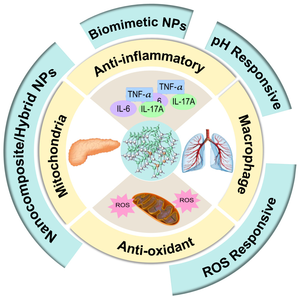
Obeng E, Ergashev A, Yao Q, Wu W, Chen G
International Journal of Nanomedicine 2025, 20:9517-9558
Published Date: 31 July 2025
Deep Tumor Penetration Using Nanoparticle Delivery Systems: Programmed Design Strategies and Emerging Evaluation Platforms
Javan M, Ajabi Zareian D, Mojarad-Jabali S
International Journal of Nanomedicine 2026, 21:563233
Published Date: 14 April 2026