Back to Journals » Nanotechnology, Science and Applications » Volume 13
Nanoparticle Drug Delivery Systems for α-Mangostin
Authors Wathoni N ![]() , Rusdin A, Motoyama K, Joni IM, Lesmana R, Muchtaridi M
, Rusdin A, Motoyama K, Joni IM, Lesmana R, Muchtaridi M ![]()
Received 19 December 2019
Accepted for publication 19 February 2020
Published 1 April 2020 Volume 2020:13 Pages 23—36
DOI https://doi.org/10.2147/NSA.S243017
Checked for plagiarism Yes
Review by Single anonymous peer review
Peer reviewer comments 2
Editor who approved publication: Professor Israel Rubinstein
Nasrul Wathoni,1 Agus Rusdin,1,2 Keiichi Motoyama,3 I Made Joni,4 Ronny Lesmana,5 Muchtaridi Muchtaridi6
1Department of Pharmaceutics and Pharmaceutical Technology, Faculty of Pharmacy, Universitas Padjadjaran, Sumedang 45363, Indonesia; 2Department of Pharmacy, Faculty of Sports and Health, Universitas Negeri Gorontalo, Gorontalo 96128, Indonesia; 3Graduate School of Pharmaceutical Sciences, Kumamoto University, Kumamoto 862-0973, Japan; 4Department of Physics, Faculty of Mathematics and Natural Sciences, Universitas Padjadjaran, Sumedang 45363, Indonesia; 5Department of Anatomy, Physiology and Biology Cell, Faculty of Medicine, Universitas Padjadjaran, Sumedang 45363, Indonesia; 6Department of Pharmaceutical Analysis and Medicinal Chemistry, Faculty of Pharmacy, Universitas Padjadjaran, Sumedang 45363, Indonesia
Correspondence: Muchtaridi Muchtaridi
Bandung-Sumedang KM 21, Sumedang, West Java 45363, Indonesia
Tel +622 842 888888 3510
Fax +622 842 888888
Email [email protected]
Abstract: α-Mangostin, a xanthone derivative from the pericarp of Garcinia mangostana L., has numerous bioactivities and pharmacological properties. However, α-mangostin has low aqueous solubility and poor target selectivity in the human body. Recently, nanoparticle drug delivery systems have become an excellent technique to improve the physicochemical properties and effectiveness of drugs. Therefore, many efforts have been made to overcome the limitations of α-mangostin through nanoparticle formulations. Our review aimed to summarise and discuss the nanoparticle drug delivery systems for α-mangostin from published papers recorded in Scopus, PubMed and Google Scholar. We examined various types of nanoparticles for α-mangostin to enhance water solubility, provide controlled release and create targeted delivery systems. These forms include polymeric nanoparticles, nanomicelles, liposomes, solid lipid nanoparticles, nanofibers and nanoemulsions. Notably, nanomicelle modification increased α-mangostin solubility increased more than 10,000 fold. Additionally, polymeric nanoparticles provided targeted delivery and significantly enhanced the biodistribution of α-mangostin into specific organs. In conclusion, the nanoparticle drug delivery system could be a promising technique to increase the solubility, selectivity and efficacy of α-mangostin as a new drug candidate in clinical therapy.
Keywords: Garcinia mangostana, solubility, controlled release, targeted delivery, nanoparticle formulations, physicochemical properties
Introduction
α-Mangostin, a xanthone derivative compound isolated from Garcinia mangostana L. peel extract, has myriad pharmacological effects: antibacterial, antifungal, anti–inflammatory, antiallergic, antioxidant and anticancer activities.1–5 The anticancer activity indicates that α-mangostin might serve as a potent anticancer agent in lung, stomach, colon, cervical, pancreatic, prostate, mammary gland, chondrosarcoma, renal, skin, tongue mucoepidermoid and breast cancers.6–18 However, α-mangostin has low solubility in water (2.03 x 10⁻⁴ mg/L at 25ºC), and many efforts have been made to improve it: structure modification, co-solvation, solid dispersion, emulsion, complexation and nanoparticle drug delivery systems.19–21 Additionally, α-mangostin and other cytotoxic drugs generally have limitations that influence their effectiveness, including a first fast metabolism reaction, an efflux reaction induced by transporter intercellular, fast drug release and a non-specific target site.22–24
Drug bioavailability is an important parameter to determine how successful the drug molecules pass through in pharmacological phases such as biopharmaceutics, pharmacokinetics, and pharmacodynamics.25 To achieve the maximum bioavailability, drug solubility is one of the primary factors that can increase the drug bioavailability.26 Currently, nanoparticle drug delivery systems are the most commonly used technique for nanomedicine-mediated treatment of diseases. Their nanosize can enhance solubility by providing a large surface area, which increases the penetration rate into a cell membrane and provides a controlled release system with passive or active targeting. This effect can serve as a cancer drug delivery system.27–29 This system becomes a promising method to overcome the limitations of α-mangostin. Several types of nanoparticles have been formulated for the α-mangostin compound, including nanolipids, nanopolymerics, nanomicelles, nanoliposomes, nanofibers and metal nanoparticles.19,20,30-34 The results are substantial, with significantly improved solubility for α-mangostin. Therefore, controlled and targeted drug delivery systems can be created by modified nanoparticle technology.
There are numerous published α-mangostin studies; however, they are usually limited and only discuss its pharmacological properties and bioactivities. Dermawan et al tried to predict the increase in α-mangostin solubility using a cyclodextrin inclusion complex. The inclusion complex formation energy values for all α-mangostin/cyclodextrins were obtained using the semi-empirical PM7 method. No researchers have performed experiments to prove the results of this in silico study.35 Taken together, we believe that our review concerning nanoparticle drug delivery systems for α-mangostin, which relates to its solubility and selectivity properties, will broaden the spectrum of α-mangostin utilisation and allow for improved efficacy.
Methodology
This review is based on the literature obtained from Scopus, PubMed and Google Scholar using the keyword “nanoparticle formulation of α-mangostin”, “nanoparticle drug delivery of α-mangostin”, and “α-mangostin nanoparticle.” We excluded opinions, reviews and unrelated topics such as pharmacological properties and bioactivities. The databases are limited to obtain the specific topic in pharmaceutical formulation. The flowchart of the methodology is shown in Figure 1.
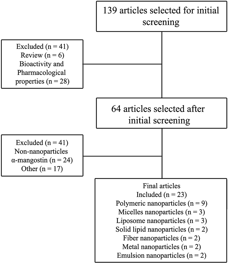 |
Figure 1 Flowchart of the methodology used in this review. |
α-Mangostin
Mangosteen (G. mangostana), the queen of tropical fruits, grows in tropical rainforests of Malaysia, Thailand and Indonesia. α-Mangostin (Figure 2) is the major compound of mangosteen peel extract; it is a xanthone derivative with the chemical name 1,3,6-trihydroxy-7-methoxy-2,8-bis(3-methyl-2-butenyl)-9H-Xanten-9-0n (Table 1). Its pharmacological activities are diverse: antibacterial, anti-allergic, anti-fungal, anti–inflammatory activity, antioxidant and anticancer.1–5
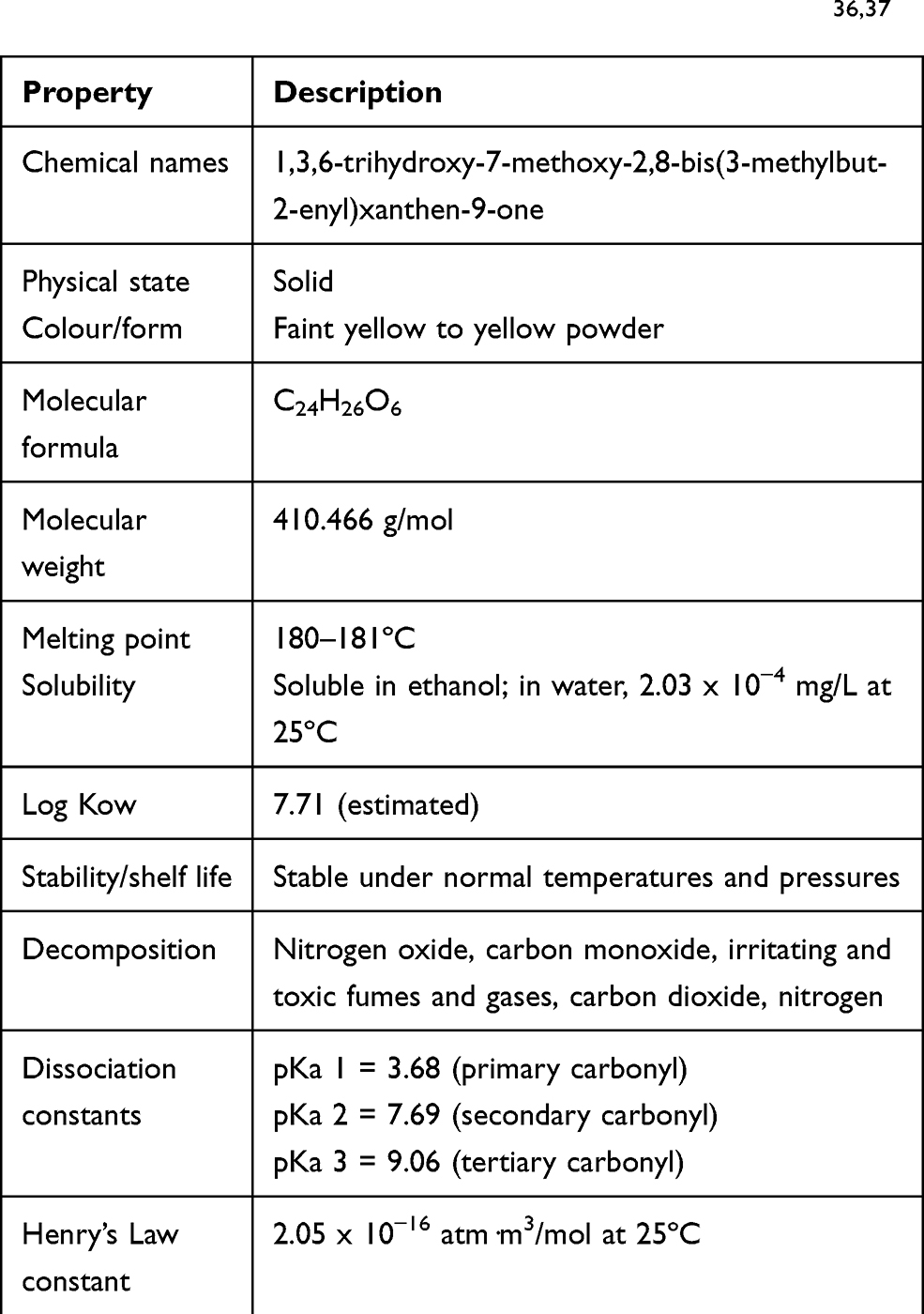 |
Table 1 Chemical and Physical Properties of α-Mangostin36,37 |
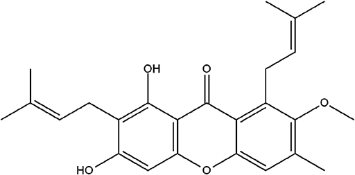 |
Figure 2 Chemical structure of α-Mangostin. |
Previous studies demonstrated that α-mangostin can act against cancer cells via multiple pathways,38–41 including inhibiting fatty acid synthase, signalling human epidermal growth factor receptor 2 (HER2)/phosphatidylinositide 3-kinase (PI3K)/Akt and mitogen-activated protein kinase (MAPK).6,42 Notwithstanding its excellent bioactivity, α-mangostin has limited solubility in water (2.03 x 10⁻⁴ mg/L at 25ºC). These problems become a basic consideration for developing α-mangostin with better efficacy.
Nanoparticle Drug Delivery Systems for α-Mangostin
Recent Nanoparticle Formulations for Improved Water Solubility, Modified Release and Targeted Drug Delivery
Nanomedicine, a nanotechnology application, has an important role in clinical therapy. Due to its nanosize (10−9 m), the large surface area of the nanocompounds enhances the surface contact with its solvent and improves the solubility or dissolution rate of slightly water-soluble compounds.43 Nanomedicine therapeutic interventions can be highly specific at the intermolecular scale to allow for curing diseases or repairing damaged tissues, such as nerves, muscles or bones. Liposomes, dendrimers, solid lipid nanoparticles, polymeric nanoparticles, silicon or carbon materials, metal and magnetic nanoparticles are examples of nanocarriers that have been formulated as drug delivery systems.44 A nanoparticle drug delivery system is a promising modification technique due to the combination of physics and chemical sciences. It is a proven, favourable technique to overcome the limitation of drugs with the poor solubility in water and provide the targeted drug delivery system.45
Table 2 and Figure 3 describe the various nanoparticle formulations that have made this technique a promising multifunctional drug delivery system. Nanoparticles are formulated as dendrimers, solid lipid nanoparticles, metal nanoparticles and liposomes, among others. They are commonly used to deliver drugs to specific targets, including cells, receptors and genes. The important aspects of the formulation depend on the selection of the right excipient, which play a crucial role in delivering active drug substances to the intended target. Folic acid (FA), mesenchymal stem cells (MSCs), mannose, hyaluronic acid, poly(lactic-co-glycolic acid) (PLGA) and chitosan conjugated with copolymers are the main excipients used to deliver active, high-affinity pharmaceutical ingredients. Nanoparticle formulations can also enhance absorption and the penetration rate as a function of small particle size, membrane transport, cellular uptake and bioadhesive interactions with the cell membrane.71,72 Finally, a drug’s bioavailability can be improved by nanoparticle formulations, a phenomenon consistent with the enhanced solubility, dissolution and absorption rate.73
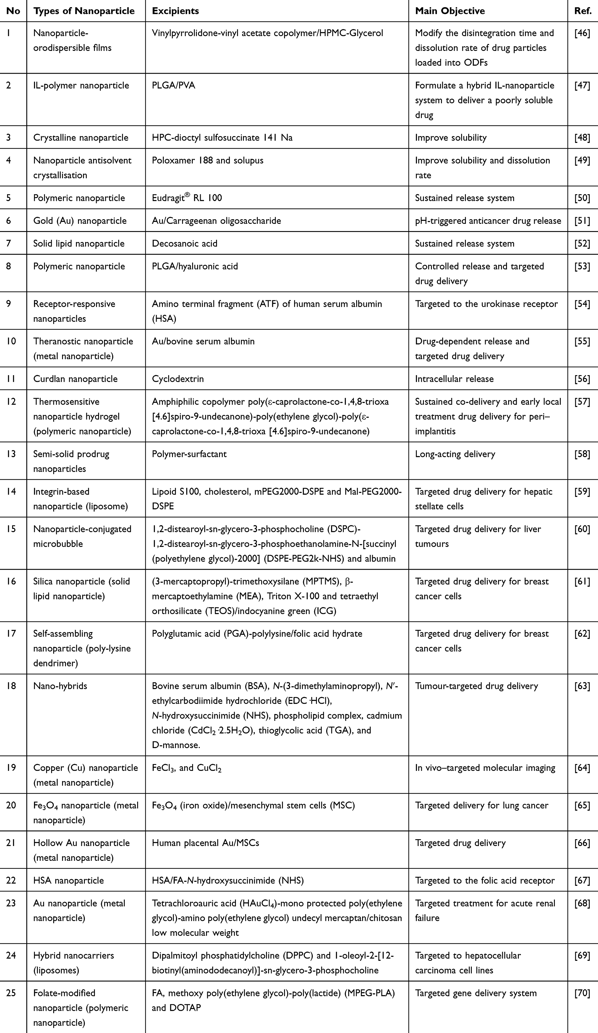 |
Table 2 Recent Nanoparticle Formulations for Improved Water Solubility, Modified Release and Targeted Drug Delivery |
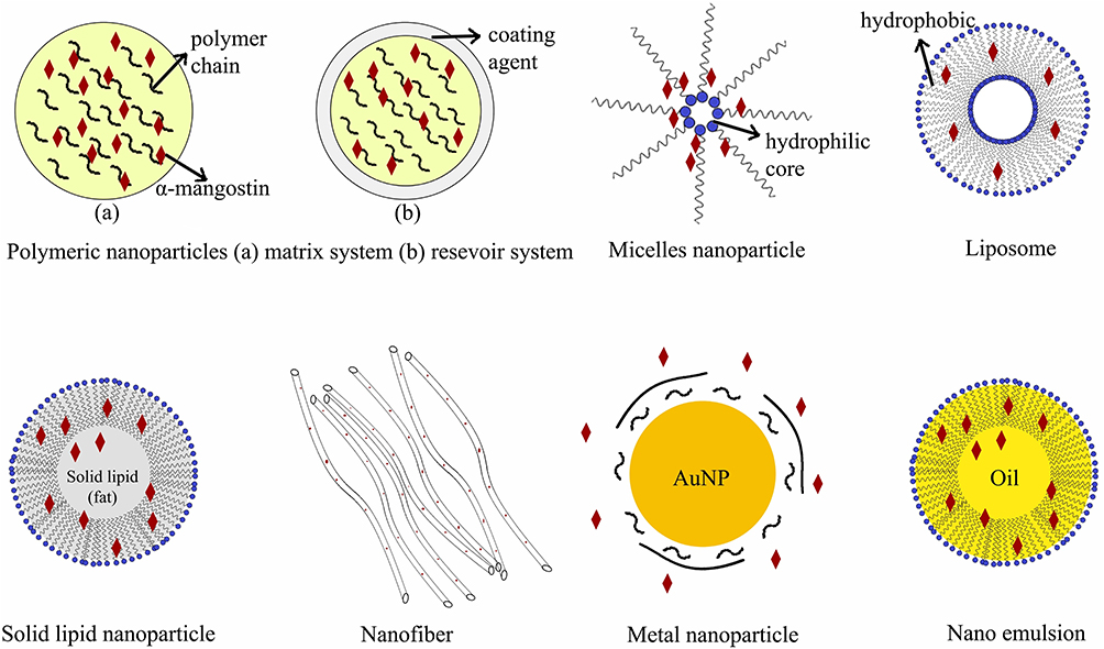 |
Figure 3 Nanoparticle drug delivery systems for α-mangostin. |
Polymeric Nanoparticles of α-Mangostin
Polymeric nanoparticles are generally used to solve the limitation of poorly soluble drugs, provide controlled release and targeted drug delivery. Several studies have been reported related to polymeric nanoparticle formulations for α-mangostin (Table 3). The first polymeric nanoparticle formulation of α-mangostin was reported in 2011; it included biodegradable PLGA copolymers. In that study, α-mangostin was encapsulated in PLGA using colloidal extraction solvent evaporation. The PLGA α-mangostin nanoparticle was less cytotoxic to the A549 lung cancer cell line compared to free α-mangostin. These results suggest that PLGA nanoparticles can be used as a micro-carrier system for the delivery of α-mangostin as a passive tumour-targeting agent.74 Another study from the same year reported an α-mangostin polymeric nanoparticle using PLGA with chitosan biopolymer. Interestingly, the formulation without chitosan was more toxic to A549 cells. The authors speculated that the mechanism of action is mediated by the high-affinity property of chitosan biopolymer can target the nanoparticle drug delivery system targeted in lung cancer tissues.75
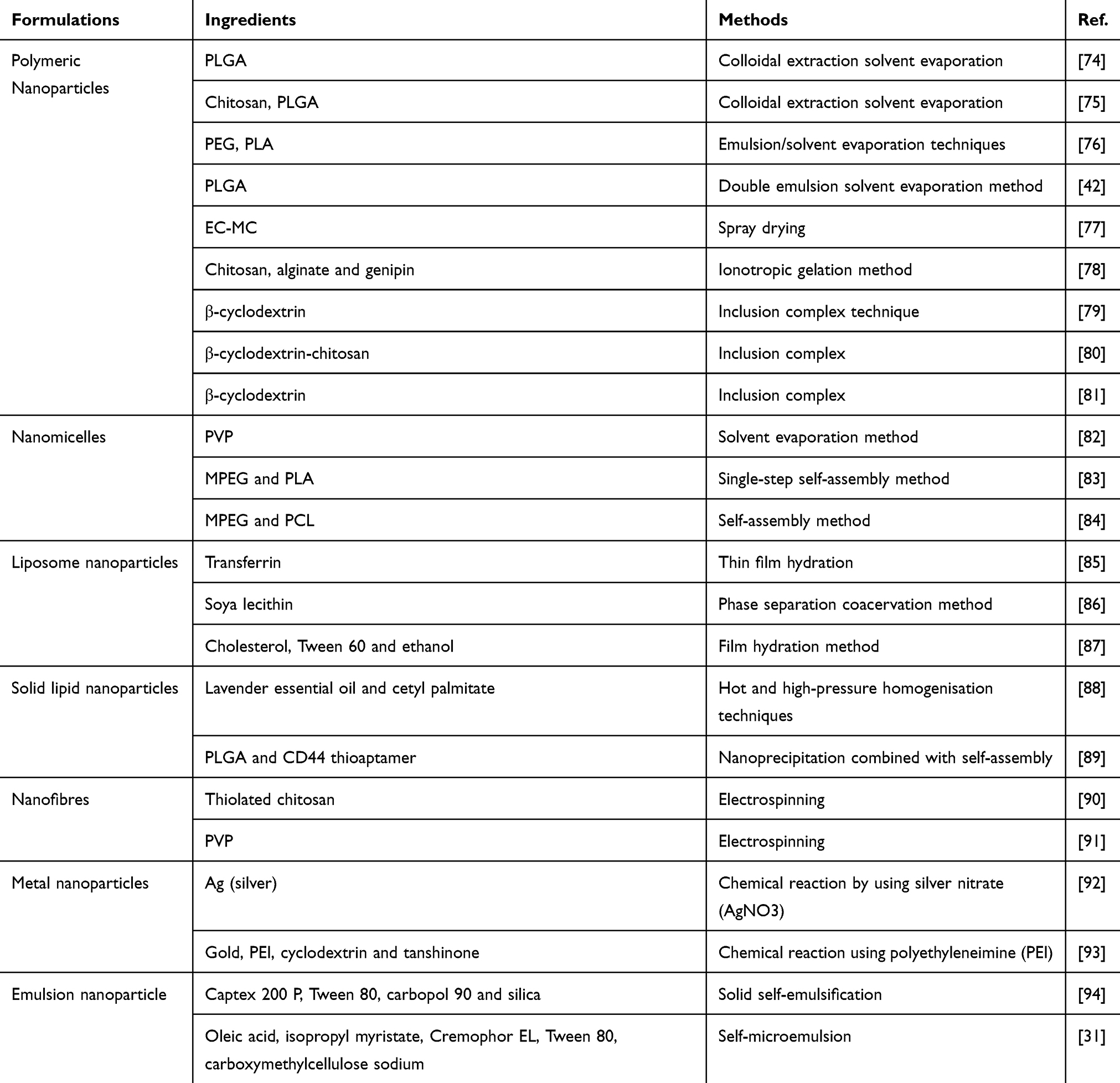 |
Table 3 Nanoparticle Formulations of α-Mangostin |
Verma et al also examined PLGA with α-mangostin as a drug payload. The authors aimed to improve the bioactivity of α-mangostin against pancreatic cancer. They prepared the nanoparticle using a double emulsion solvent evaporation method. Impressively, the nanoparticle system inhibited the proliferation of pancreatic cancer stem cells (CSCs) and pancreatic cancer cell lines and had no effect on normal human pancreatic ductal epithelial (HPNE) cells. Moreover, the nanoparticle inhibited colony formation, motility, migration and the invasion-induced apoptotic mechanism in vitro and in vivo.42
Another polymeric nanoparticle formulation used poly(ethylene glycol)-poly(L-lactide) (PEG-PLA) as a matrix for α-mangostin. The authors aimed to use this formulation for Alzheimer’s disease. The nanoparticle was prepared by emulsion/solvent evaporation techniques. The particle size, zeta-potential and entrapment efficiency of the nanoparticle were 94.26 ± 4.54 nm, −32 ± 0.43 mV and 50.47 ± 1.96%, respectively. In vitro, the drug was rapidly released in the first 24 h (approximately 50%), followed by a slow, continuous release until 72 h and 100% release by 96 h. These results demonstrated a significant improvement of the pharmacokinetic and biodistribution profiles of nanoparticle compared to free α-mangostin.76
Ethyl cellulose-methyl cellulose (EC-MC), a natural polymer from cellulose groups, was designed for α-mangostin polymeric nanoparticle formulation as an anti-acne therapy in a cosmeceutical form. The system was designed as a nanoreservoir system to achieve an extended release profile using a spray drying technique. In this study, the particle size, polydispersity and loading capacity were 300–500 nm, 0.111 ± 0.024 and 41.90 ± 0.79%, respectively. The nanoparticle formation exhibited lower skin irritation compared to controls. Approximately 80–100% of α-mangostin was released over more than 7 days. Impressively, the anti-acne activity of the nanoparticle system significantly decreased the acne severity index (ASI) value and inflammatory lesions (P < 0.05) compared to control.77
Another study developed a nanoparticle system based on chitosan/alginate and genipin (GP) as a crosslinker prepared using the ionotropic gelation method. The system aimed to achieve a controlled release system and increase the antitumor activity of α-mangostin. Cytotoxicity and antitumor activity studies confirmed that an increase in GP concentration significantly reduced cell viability and induced apoptosis in colorectal adenocarcinoma cells.78
Nguyen et al demonstrated that nanoparticles with β-cyclodextrin (β-CD) improved the solubility and enhanced the cytotoxic activity of α-mangostin, with a minimal inhibitory concentration (IC50) of 8.86 and 9.86 µg/mL for LU-1 (human lung adenocarcinoma) and HL-60 (human promyelocytic leukaemia), respectively.79 Another α-mangostin complex with β-CD was fabricated with grafted-chitosan. The system was prepared using high shear mixing techniques. The system exhibited a high of entrapment efficiency (>75%) and anti–inflammatory activity. The inclusion complex of α-mangostin and quaternised cyclodextrin grafted chitosan (QCD-g-CS) influenced cytokine secretion and inhibited inflammation during the first hour (60% inhibition). After 3 h, there was almost total inhibition (95%).80 Additionally, nanoparticles of a water-soluble β-CD and α-mangostin presented cytotoxic activities against A549 lung cancer cells, with an IC50 of 2.34 µg/mL.81
Nanomicelles
A micelle is an amphipathic molecule in water and suitable as a drug delivery carrier for drugs with high lipophilicity. The first nanomicelle for α-mangostin was generated by Aisha et al α-Mangostin in solid dispersion nanomicelles, combined with polyvinylpyrrolidone (PVP) as a main polymer, was produced by the solvent evaporation method. The solubility of α-mangostin markedly increased 10,000 fold, from 0.2 ± 0.2 pg/mL to 2743 ± 11 pg/mL. Self-assembly of anionic nanomicelles around α-mangostin was observed by transmission electron microscopy and dynamic light scattering; the diameter size was 99–127 nm. The nanomicelle uptake was mediated by endocytosis, a finding that indicated intracellular delivery of α-mangostin that could be associated with potential cytotoxicity (IC50 of 8.9 ± 0.2 μg/mL).82
In another study, α-mangostin nanomicelles with methoxypoly(ethylene glycol)-poly(lactide) (MPEG-PLA) were developed by a single-step self-assembly method for malignant glioma. In vitro and in vivo assays showed that the α-mangostin/MPEG-PLA nanoparticles inhibited cell growth and induced apoptosis—with cleaved caspase expression, DNA fragmentation, downregulation of anti-apoptotic molecules and up-regulation of apoptotic molecules. This study also successfully investigated the process of programmed cell death in malignant glioma cells after treatment with α-mangostin/MPEG-PLA.83
Yang et al recently generated α-mangostin with methoxypoly(ethylene glycol)-poly(ε-caprolactone) (MPEG-PCL) as an anti-melanoma agent. The system had a sustained release profile, high solubility, strong toxicity to tumour cells and low toxicity to non-tumour cells. Additionally, MPEG-PCL inhibited melanoma cell proliferation, induced apoptosis via intrinsic and extrinsic pathways, suppressed growth cells and restrained angiogenesis. These data suggest that α-mangostin/MPEG-PCL nanomicelles are promising potential chemotherapy agents for the treatment of melanoma.84
Liposome Nanoparticles
Liposome nanoparticle (nanoliposome) is a liposome with particle size around 80–300 nm. Liposome nanoparticles can improve the physicochemical properties and performance of drugs due to their capability to deliver a drug. Chen developed a liposome, with α-mangostin as a drug payload using transferrin, with the thin-film hydration method. On the intercellular distribution assay, liposomes presented a time-dependent property; approximately 210 Ω/cm2 α-mangostin crossed the blood–brain barrier (BBB), with horseradish peroxidase (HRP) permeability less than 5%.85
Chin et al developed an α-mangostin niosome to improve the skin permeation rate of α-mangostin. Proniosome was prepared with soya lecithin using a phase separation coacervation method. The system enhanced skin permeation of α-mangostin 1.8–8.0 fold compared to control. It also improved viable epidermis/dermis (VED) of the α-mangostin compound, where α-mangostin deposition in the VED layer was increased 2.5–2.9 fold compared to control. Moreover, the addition of spans and soya lecithin improved the solubility of α-mangostin in water.86
Another niosome formulation from Limpapayom et al utilised cholesterol, Tween 60 and ethanol as the main carrier system. The system was prepared by film hydration; the particle size was 213 ± 26.47 nm, polydispersity index (PDI) was 0.23 ± 0.19 and zeta potential was −12.67±0.90 mV. Subsequently, the noisome/α-mangostin was prepared in cream and serum forms with 2.5–5% α-mangostin. The particle size, PDI and zeta potential were 600–700 nm, 1.11 ± 0.01 and 0.58 ± 0.04 mV, respectively. A skin permeation study confirmed that about 10–40% of α-mangostin released over more than 24 h.87
Solid Lipid Nanoparticles
Solid lipid nanoparticles (SLN) are spherical carrier composed of single or double lipid layer on the surface, and solid layer in the core of the system. The complex system of SLN can provide an excellent drug control released. However, only two journals publication reported regarding solid lipid nanoparticle formulation of α-mangostin. Yostawonkul et al designed a nanostructure lipid carrier for α-mangostin (AM-NLC) by hot and high-pressure homogenisation techniques for non-surgical castration of male animals. Lavender essential oil and cetyl palmitate were the carrier system in this study. AM-NLC increased the activity of caspase-3 and caspase-7 and induced germ cell degeneration within the seminiferous tubules. Shrunken tubules were greatly depleted of germ cells. Additionally, the use of AM-NLC reduced the levels of pro-inflammatory mediators (nitric oxide and tumour necrosis factor α).88
Bonafe et al developed a lipid nanoparticle formulation to increase the activity of α-mangostin in disaggregation of MCF-7 cells. PLGA and CD44 thioaptamer used as the main carrier. A nanoparticle that contained 0.5 µg/mL α-mangostin induced disaggregation of multicellular tumour spheroid (MCTS). There was a similar dissociation effect when MCTS were cultured in matrix gel under the same conditions for 48–72 hrs. Moreover, the system with the lower α-mangostin concentration triggered damage, denoted as a substantial reduction in the MCTS size and density. The reduced spheroid expansion implied that a significant number of cells died or were in cell cycle arrest.89
Nanofibers
Nanofiber is widely used for site-specific drug released to achieve the desired therapeutic effects. Nanofiber has a diameter range around 150 nm and length 50–200 µm. Nanofibers could potentially overcome the limitation of α-mangostin. A nanofibre combined with chitosan thiolated with the electrospinning method had excellent mucoadhesive properties. Additionally, the α-mangostin nanofibre improved the bactericidal rate.90 Another nanofibre formulation was prepared using polyvinylpyrrolidone (PVP) as a carrier matrix for the active compound. The PVP nanofibre (387–586 nm) was prepared using an electrospinning apparatus. The preparation exhibited antioxidant activity, and the use of high voltage in the electrospinning technique did not apparently damage the molecular structure of α-mangostin. In vitro, α-mangostin release increased from 35% to over 90% in 60 min.91
Metal Nanoparticles
Metal nanoparticle is a metal with particle size around 1–100 nm. Several studies reported that metal nanoparticles have a bioactivity as anticancer agent and a high affinity with the cancer cells. Silver α-mangostin nanoparticles were formulated in a perfect spherical shape. These nanoparticles significantly inhibited the growth of the bacteria Escherichia coli and Bacillus subtilis and the fungus Aspergillus niger. Additionally, the presence of α-mangostin substantially reduced the silver ions in the silver nanoparticle system.92
Gold α-mangostin nanoparticles were also formulated; they comprised polyethylenimine (PEI) and cyclodextrin. Tanshinone was used as competitor drug payload in this study. The α-mangostin gold nanoparticles improved the loading efficiency approximately 15–50%, with an IC50 of 17.5 μM and 6.0 μM for PC-3 and DU145 cell lines, respectively. Comparatively, the tanshinone gold nanoparticles were very active against these cells, with a 40% improvement in the IC50 value for both PC-3 and DU145 cells.93
Emulsion Nanoparticles
Emulsion nanoparticle (nanoemulsion), a colloidal particulate system, consists of oil, water, and surfactant with high kinetic stability, low viscosity, and optically transparent. In the last decade, nanoemulsion has become a promising lipophilic drugs delivery system. Solid self-emulsification is one common modification technique to enhance the solubility and dissolution rate of α-mangostin. Droplet particles obtained from this system (using liquid-self-emulsifying drug delivery system [liquid-SEDDS]) were 106.9 ± 24.3 nm. The droplet was further converted to the solid state (solid-SEDDS) using Aeroperl 300 and Sylysia 350 silica. Solid-SEDDS with Aeroperl 300 had better flowability compared to solid-SEDDS with Sylysia 350. Based on the characterisation of X-ray diffraction (XRD) and differential scanning calorimetry (DSC) analysis, the solid-SEDDS exhibited an amorphous form. The dissolution test indicated that approximately 18.82% and 7.71% of α-mangostin was released from solid-SEDDS with Aeroperl 300 and Sylysia 350, respectively, within 60 min. However, only 0.26% of the intact α-mangostin dissolved.94
The mechanism for the improved α-mangostin solubility in emulsion was due to self-microemulsion; the particle diameter size was 24.6 nm and the encapsulation efficiency was 87.26%. These factors increased the area under the curve of α-mangostin by 4.75 fold compared to the free form. The preparation also increased α-mangostin distribution in lymphatic organs. Overall, self-microemulsion as a nano delivery system can promote the digestive tract absorption of α-mangostin and provide a specific distribution. The targeted system and high oral bioavailability of α-mangostin with self-microemulsion provides excellent performance for clinical drug efficacy.31
Perspective
In drug development, nanoparticle technology represents physical modifications intended to ameliorate solubility problems. Currently, nanotechnology can be applied for drug delivery systems, such drug controlled release,95 delayed release and sustained release.96 These nanoparticle formulations are the most commonly used in drug delivery systems.97 Our objective review highlighted that the nanoparticle technology in nanomedicine applications is divided into three general classifications: increased water solubility, controlled release and targeted drug delivery. As mentioned before, nanotechnology can be used to recover the solubility problem of drugs through multiple pathways and mechanisms. Firstly, particle size reduction in nanotechnology improves the drug solubility by expanding the surface area of particles.98,99 Secondly, the use of high water-soluble excipients as the main base of nanoparticles increase the solubility of drugs mediated by hydrogen bonding interaction between excipients and water molecules.100,101 On the other hand, the use of surface-active agent (surfactant) in nanotechnology also enhances the solubility of high lipophilicity drugs through interfacial tension reduction.102,103
Considering the effects of therapy with a dose and frequency of administration that is efficient, nanoparticle technology can be utilised to provide controlled and targeted drug delivery systems, especially for cancer therapy, to increase selectivity, mitigate potentially harmful side effects and even cause death in normal cells. The physicochemical properties of α-mangostin, especially its poor water solubility profile and its low selectivity on the target cells, limits its therapeutic applicability. Therefore, nanoparticle formulations are one option to resolve these limitations.
Numerous nanoparticle formulations have been described, including polymeric nanoparticles, solid lipid nanoparticles, nanofibers, nanomicelles and metal nanoparticles. In general, these formulations aim to increase the solubility of compounds that are poorly soluble in water through particle size modification to obtain a larger surface area. On the other hand, the type of nanoparticle and ingredients also influences the solubility of a compound. Polymeric nanoparticles are formulated with the polymer as a base for the formulations and are often made for further examinations.
Nanoparticle formulations have been developed using various nanocarriers with different techniques. Each nanocarriers are formulated by considering the aims of the studies such as to provide solubility improvement with hydrophilic polymer as a carrier,104 to provide control released system with pH-sensitive polymers or thermal-sensitive polymers,105,106 and to prevent protein degradation with liposome protection.107 In some cases, the nanocarrier is combined with targeting mediators to gain the nanoparticle targeted drug delivery system into specific target.108
Nanoparticles that mediate passive or active targeted delivery are generally prepared with ingredients that have a high affinity to the target and low affinity towards normal cells, for example, PLGA. PLGA is a copolymer formed from the combination of polymer polylactic acid (PLA) and polyglycolic acid (PGA). Research showed that PLGA has a high affinity to cancer cells, including hepatic cancer,109 prostate cancer110 and lung cancer cells,111 and many cell lines, including human umbilical vein endothelial cells,112 H1299,113 COS-7 and Cf2th.114 This high affinity allows PLGA to provide drug delivery systems or genes into the target-specific tissues or organs. Ultimately, consideration of nanoparticle shape and the materials used in the formula requires careful and thoughtful attention, especially with regards to the desired use and destination.
Conclusion
Many techniques have been considered to improve α-mangostin’s water solubility, of which nanoparticle formulations have become the most widely performed. This formulation provides many advantages. Overall, nanoparticle formulations improve α-mangostin’s water solubility and affect its biopharmaceutical, pharmacokinetic and pharmacodynamic aspects. Additionally, nanoparticle technology for α-mangostin can be a promising for controlled release and passive and active targeting. This system should help to maximise the efficacy of α-mangostin in a drug delivery system.
Acknowledgment
The authors thank the Minister of Research and Higher Education, Republic of Indonesia for the fundamental research grant (grant number 1123v/UN6.O/LT/2019), and Universitas Padjadjaran for academic leadership grant 2020.
Disclosure
The authors report no conflicts of interest in this work.
References
1. Chen L, Yang L, Wang C. Anti-inflammatory activity of mangostins from Garcinia mangostana. Food Chem Toxicol. 2008;46:688–693. doi:10.1016/j.fct.2007.09.096
2. Jung H-A, Su B-N, Keller WJ, Mehta RG, Kinghorn AD. Antioxidant xanthones from the pericarp of Garcinia mangostana (Mangosteen). J Agric Food Chem. 2006;54(6):2077–2082. doi:10.1021/jf052649z
3. Ma Y, Yu W, Shrivastava A, Srivastava RK, Shankar S. Inhibition of pancreatic cancer stem cell characteristics by α-Mangostin: molecular mechanisms involving Sonic hedgehog and Nanog. J Cell Mol Med. 2019. doi:10.1111/jcmm.14178
4. Sivaranjani M, Leskinen K, Aravindraja C, et al. Deciphering the antibacterial mode of action of alpha-mangostin on Staphylococcus epidermidis RP62A through an integrated transcriptomic and proteomic approach. Front Microbiol. 2019;10:150. doi:10.3389/fmicb.2019.00150
5. Limwikrant W, Aung T, Chooluck K, Puttipipatkhachorn S, Yamamoto K. Size reduction efficiency of Alpha-Mangostin suspension using high-pressure homogenization. Chem Pharm Bull. 2019;67(4):c18–00589.
6. Kritsanawong S, Innajak S, Imoto M, Watanapokasin R. Antiproliferative and apoptosis induction of α-mangostin in T47D breast cancer cells. Int J Oncol. 2016;48(5):2155–2165. doi:10.3892/ijo.2016.3399
7. Krajarng A, Nakamura Y, Suksamrarn S, Watanapokasin R. α-Mangostin induces apoptosis in human chondrosarcoma cells through downregulation of ERK/JNK and Akt signaling pathway. J Agric Food Chem. 2011;59(10):5746–5754. doi:10.1021/jf200620n
8. Chen CM, Hsieh SC, Lin CL, Lin YS, Tsai JP, Hsieh YH. Alpha-Mangostin suppresses the metastasis of human renal carcinoma cells by targeting MEK/ERK expression and MMP-9 transcription activity. Cell Physiol Biochem. 2017;44(4):1460–1470. doi:10.1159/000485582
9. Lee C, Ying T, Chiou H, Hsieh S. Alpha-mangostin induces apoptosis through activation of reactive oxygen species and ASK1/p38 signaling pathway in cervical cancer cells. Oncotarget. 2017;8(29):47425–47439. doi:10.18632/oncotarget.17659
10. Lee HN, Jang HY, Kim HJ, et al. Antitumor and apoptosis-inducing effects of α-mangostin extracted from the pericarp of the mangosteen fruit (Garcinia mangostana L.) in YD-15 tongue mucoepidermoid carcinoma cells. Int J Mol Med. 2016;37(4):939–948. doi:10.3892/ijmm.2016.2517
11. Novilla A, Djamhuri DS, Fauziah N, Maesaroh M, Balqis B, Widowati W. Cytotoxic activity of Mangosteen (Garcinia mangostana L.) peel extract and α-mangostin toward leukemia cell lines (HL-60 and K-562). J Nat Remedies. 2016;16(2):52. doi:10.18311/jnr/2016/842
12. Muchtaridi M, Wijaya CA. Anticancer potential of Α-Mangostin. Asian J Pharm Clin Res. 2017;10(12):440. doi:10.22159/ajpcr.2017.v10i12.20812
13. Scolamiero G, Pazzini C, Bonafè F, Guarnieri C, Muscari C. Effects of α-mangostin on viability, growth and cohesion of multicellular spheroids derived from human breast cancer cell lines. Int J Med Sci. 2018;15(1):23–30. doi:10.7150/ijms.22002
14. Moongkarndi, P., Jaisupa, N., Kosem, N., Konlata, J., Samer, J., Pattanapanyasat, K. and Rodpai, E., 2015. Effect of purified α-mangostin from mangosteen pericarp on cytotoxicity, cell cycle arrest and apoptotic gene expression in human cancer cells. World J Pharm Sci, 3(8), pp.1473–84.
15. Kwak HH, Kim IR, Kim HJ, Park BS, Yu SB. α -Mangostin induces apoptosis and cell cycle arrest in oral squamous cell carcinoma cell. Evid Based Complement Alternat Med. 2016;2016. doi:10.1155/2016/5352412
16. Phan TKT, Shahbazzadeh F, Pham TTH, Kihara T. Alpha-mangostin inhibits the migration and invasion of A549 lung cancer cells. PeerJ. 2018;6:e5027. doi:10.7717/peerj.5027
17. Wang JJ, Sanderson BJS, Zhang W. Significant anti-invasive activities of α-mangostin from the mangosteen pericarp on two human skin cancer cell lines. Anticancer Res. 2012;32(9):3805–3816.
18. Zhang C, Yu G, Shen Y. The naturally occurring xanthone α-mangostin induces ROS-mediated cytotoxicity in non-small scale lung cancer cells. Saudi J Biol Sci. 2018;25(6):1090–1095. doi:10.1016/j.sjbs.2017.03.005
19. Rungnim C, Phunpee S, Kunaseth M, et al. Co-solvation effect on the binding mode of the α-mangostin/β-cyclodextrin inclusion complex. Beilstein J Org Chem. 2015;11(1):2306–2317. doi:10.3762/bjoc.11.251
20. Zarena AS, Sankar KU. Synthesis of α− mangostin-D-glucoside in supercritical carbon dioxide media. J Food Sci Technol. 2015;52(10):6547–6555. doi:10.1007/s13197-014-1705-z
21. Elsaid Ali AA, Taher M, Mohamed F. Microencapsulation of alpha-mangostin into PLGA microspheres and optimization using response surface methodology intended for pulmonary delivery. J Microencapsul. 2013;30(8):728–740. doi:10.3109/02652048.2013.788081
22. Li L, Brunner I, Han A, Hamburger M, Kinghorn AD. Pharmacokinetics of a -mangostin in rats after intravenous and oral application. Mol Nutr Food Res. 2011;55(S1):67–74. doi:10.1002/mnfr.201000511
23. Pelivan K, Frensemeier L, Karst U, et al. Understanding the metabolism of the anticancer drug triapine: electrochemical oxidation, microsomal incubation and in vivo analysis using LC-HRMS. Analyst. 2017;142(17):3165–3176. doi:10.1039/C7AN00902J
24. Khodadadei F, Safarian S, Ghanbari N. Methotrexate-loaded nitrogen-doped graphene quantum dots nanocarriers as an efficient anticancer drug delivery system. Mater Sci Eng C. 2017;79:280–285. doi:10.1016/j.msec.2017.05.049
25. Sahoo CK, Reddy GS, Vojjala A, Reddy BV. Bioavailability enhancement for poorly soluble drugs: a review. Innoriginal Int J Sci. 2018:1–6.
26. Abuzar SM, Hyun S-M, Kim J-H, et al. Enhancing the solubility and bioavailability of poorly water-soluble drugs using supercritical antisolvent (SAS) process. Int J Pharm. 2018;538(1–2):1–13. doi:10.1016/j.ijpharm.2017.12.041
27. Wang AZ, Langer R, Farokhzad OC. Nanoparticle delivery of cancer drugs. Annu Rev Med. 2012;63:185–198. doi:10.1146/annurev-med-040210-162544
28. Singh R, Lillard JJW. Nanoparticle-based targeted drug delivery. Exp Mol Pathol. 2009;86(3):215–223. doi:10.1016/j.yexmp.2008.12.004
29. Rizvi SAA, Saleh AM. Applications of nanoparticle systems in drug delivery technology. Saudi Pharm J. 2018;26(1):64–70. doi:10.1016/j.jsps.2017.10.012
30. Chi X, Zi C, Li H, et al. RSC advances. RSC Adv. 2018;8(January):41377–41388. doi:10.1039/C8RA08409B
31. Xu W, Jiang H, Yang K, Wang Y, Zhang Q. ScienceDirect development and in vivo evaluation of self-microemulsion as delivery system for a –mangostin. Kaohsiung J Med Sci. 2017;116–123. doi:10.1016/j.kjms.2016.12.003
32. Sodalee K, Sapsuphan P, Wongsirikul R. Preparation and evaluation of alpha-mangostin solid self-emulsifying drug delivery system. Asian J Pharm Sci. 2016;11(1):225–226. doi:10.1016/j.ajps.2015.11.024
33. Jittamaro P, Ruktanonchai UR, Phunpee S. Effect of solvent on the complex between α -Mangostin and β -Cyclodextrin α. Int Conf Chem Civ Mater Eng. 2015:5–9.
34. Phunpee S, Suktham K, Surassmo S, et al. Controllable encapsulation of ␣-mangostin with quaternized -cyclodextrin grafted chitosan using high shear mixing, International Journal. Int J Pharm. 2017. doi:10.1016/j.ijpharm.2017.12.016
35. Dermawan D, Wathoni N, Muchtaridi M. Host-guest interactions of α− Mangostin with (α, β, γ)− Cyclodextrins: semi-empirical quantum mechanical methods of PM6 and PM7. J Young Pharm. 2019;11(1):31. doi:10.5530/jyp.2019.11.7
36. Wishart DS, Feunang YD, Marcu A, et al. HMDB 4.0: the human metabolome database for 2018. Nucleic Acids Res. 2017;46(D1):D608–D617. doi:10.1093/nar/gkx1089
37. Kim S, Chen J, Cheng T, et al. PubChem 2019 update: improved access to chemical data. Nucleic Acids Res. 2018;47(D1):D1102–D1109. doi:10.1093/nar/gky1033
38. Li P, Tian W, Ma X. Alpha-mangostin inhibits intracellular fatty acid synthase and induces apoptosis in breast cancer cells. Mol Cancer. 2014;13(1):138. doi:10.1186/1476-4598-13-138
39. Nakagawa Y, Iinuma M, Naoe T, Nozawa Y, Akao Y. Characterized mechanism of α-mangostin-induced cell death: caspase-independent apoptosis with release of endonuclease-G from mitochondria and increased miR-143 expression in human colorectal cancer DLD-1 cells. Bioorg Med Chem. 2007;15(16):5620–5628. doi:10.1016/j.bmc.2007.04.071
40. Shih Y-W, Chien S-T, Chen P-S, Lee J-H, Wu S-H, Yin L-T. α-Mangostin suppresses phorbol 12-myristate 13-acetate-induced MMP-2/MMP-9 expressions via αvβ3 integrin/FAK/ERK and NF-κB signaling pathway in human lung adenocarcinoma A549 cells. Cell Biochem Biophys. 2010;58(1):31–44. doi:10.1007/s12013-010-9091-2
41. Sato A, Fujiwara H, Oku H, Ishiguro K, Ohizumi Y. α-Mangostin induces Ca2+-ATPase-dependent apoptosis via mitochondrial pathway in PC12 cells. J Pharmacol Sci. 2004;95(1):33–40. doi:10.1254/jphs.95.33
42. Verma RK, Yu W, Shrivastava A, Shankar S, Srivastava RK. α-Mangostin-encapsulated PLGA nanoparticles inhibit pancreatic carcinogenesis by targeting cancer stem cells in human, and transgenic (KrasG12D, and KrasG12D/tp53R270H) mice. Sci Rep. 2016;6(May):1–13. doi:10.1038/srep32743
43. Thassu D, Deleers M, Pathak YV. Nanoparticulate Drug Delivery Systems. Vol. 166. CRC Press; 2007.
44. Wilczewska AZ, Niemirowicz K, Markiewicz KH, Car H. Nanoparticles as drug delivery systems. Pharmacol Rep. 2012;64(5):1020–1037. doi:10.1016/S1734-1140(12)70901-5
45. Langer R. Drug delivery and targeting. Nature. 1998;5–10.
46. Steiner D, Finke JH, Kwade A. Model-based description of disintegration time and dissolution rate of nanoparticle-loaded orodispersible films. Eur J Pharm Sci. 2019;132:18–26. doi:10.1016/j.ejps.2019.02.029
47. Júlio A, Lima SAC, Reis S, de Almeida TS, Fonte P. Development of ionic liquid-polymer nanoparticle hybrid systems for delivery of poorly soluble drugs. J Drug Deliv Sci Technol. 2019;100915. doi:10.1016/j.jddst.2019.01.030
48. Braig V, Konnerth C, Peukert W, Lee G. Enhanced dissolution of naproxen from pure-drug, crystalline nanoparticles: a case study formulated into spray-dried granules and compressed tablets. Int J Pharm. 2019;554:54–60. doi:10.1016/j.ijpharm.2018.09.069
49. Homayouni A, Amini M, Sohrabi M, Varshosaz J, Nokhodchi A. Curcumin nanoparticles containing poloxamer or soluplus tailored by high pressure homogenization using antisolvent crystallization. Int J Pharm. 2019;562:124–134. doi:10.1016/j.ijpharm.2019.03.038
50. Öztürk AA, Yenilmez E, Yazan Y. Dexketoprofen trometamol-loaded Eudragit® RL 100 nanoparticle formulation, characterization and release kinetics. ACTA Pharm Sci. 2019;57(1).
51. Chen X, Han W, Zhao X, Tang W, Wang F. Epirubicin-loaded marine carrageenan oligosaccharide capped gold nanoparticle system for pH-triggered anticancer drug release. Sci Rep. 2019;9(1):6754. doi:10.1038/s41598-019-43106-9
52. Tao Y, Yang F, Meng K, et al. Exploitation of enrofloxacin-loaded docosanoic acid solid lipid nanoparticle suspension as oral and intramuscular sustained release formulations for pig. Drug Deliv. 2019;26(1):273–280. doi:10.1080/10717544.2019.1580798
53. Lee P-C, Zan B-S, Chen L-T, Chung T-W. Multifunctional Plga-based nanoparticles as a controlled release drug delivery system for antioxidant and anticoagulant therapy. Int J Nanomedicine. 2019;14:1533. doi:10.2147/IJN.S174962
54. Li S, Yuan C, Chen J, et al. Nanoparticle binding to urokinase receptor on cancer cell surface triggers nanoparticle disintegration and cargo release. Theranostics. 2019;9(3):884. doi:10.7150/thno.29445
55. Saurabh CK, Gupta S, Variyar PS, Sharma A. Effect of addition of nanoclay, beeswax, tween-80 and glycerol on physicochemical properties of guar gum films. Ind Crops Prod. 2016;89:109–118. doi:10.1016/j.indcrop.2016.05.003
56. Basha RY, Sampath Kumar TS, Doble M. Dual delivery of tuberculosis drugs via cyclodextrin conjugated curdlan nanoparticles to infected macrophages. Carbohydr Polym. 2019;218:53–62. doi:10.1016/j.carbpol.2019.04.056
57. Chen W, Zhi M, Feng Z, et al. Sustained co-delivery of ibuprofen and basic fibroblast growth factor by thermosensitive nanoparticle hydrogel as early local treatment of peri-implantitis. Int J Nanomedicine. 2019;14:1347. doi:10.2147/IJN.S190781
58. Hobson JJ, Al-khouja A, Curley P, et al. Semi-solid prodrug nanoparticles for long-acting delivery of water-soluble antiretroviral drugs within combination HIV therapies. Nat Commun. 2019;10(1):1413. doi:10.1038/s41467-019-09354-z
59. Li Y, Pu S, Liu Q, et al. An integrin-based nanoparticle that targets activated hepatic stellate cells and alleviates liver fibrosis. J Control Release. 2019;303:77–90. doi:10.1016/j.jconrel.2019.04.022
60. Lee JH, Moon H, Han H, et al. Antitumor effects of intra-arterial delivery of albumin-doxorubicin nanoparticle conjugated microbubbles combined with ultrasound-targeted microbubble activation on VX2 rabbit liver tumors. Cancers (Basel). 2019;11(4):581. doi:10.3390/cancers11040581
61. Zhang X, Li Y, Wei M, Liu C, Yu T, Yang J. Cetuximab-modified silica nanoparticle loaded with ICG for tumor-targeted combinational therapy of breast cancer. Drug Deliv. 2019;26(1):129–136. doi:10.1080/10717544.2018.1564403
62. Lei M, Sha S, Wang X, et al. Co-delivery of paclitaxel and gemcitabine via a self-assembling nanoparticle for targeted treatment of breast cancer. RSC Adv. 2019;9(10):5512–5520. doi:10.1039/C9RA00276F
63. Zayed DG, Ebrahim SM, Helmy MW, et al. Combining hydrophilic chemotherapy and hydrophobic phytotherapy via tumor-targeted albumin–QDs nano-hybrids: covalent coupling and phospholipid complexation approaches. J Nanobiotechnology. 2019;17(1):7. doi:10.1186/s12951-019-0445-7
64. Fernández-Barahona I, Gutiérrez L, Veintemillas-Verdaguer S, et al. Cu-doped extremely small iron oxide nanoparticles with large longitudinal relaxivity: one-pot synthesis and in vivo targeted molecular imaging. ACS Omega. 2019;4(2):2719–2727. doi:10.1021/acsomega.8b03004
65. Wang X, Chen H, Zeng X, et al. Efficient lung cancer-targeted drug delivery via a nanoparticle/MSC system. Acta Pharm Sin B. 2019;9(1):167–176. doi:10.1016/j.apsb.2018.08.006
66. Sancho-Albero M, Navascués N, Mendoza G, et al. Exosome origin determines cell targeting and the transfer of therapeutic nanoparticles towards target cells. J Nanobiotechnology. 2019;17(1):16. doi:10.1186/s12951-018-0437-z
67. Sun Y, Zhao Y, Teng S, et al. Folic acid receptor-targeted human serum albumin nanoparticle formulation of cabazitaxel for tumor therapy. Int J Nanomedicine. 2019;14:135. doi:10.2147/IJN.S181296
68. Zayed GM, El-feky GS. Growth factor loaded functionalized gold nanoparticles as potential targeted treatment for acute renal failure. Int J Appl Pharm. 2019;11(1):66–70.
69. AlQahtani SA, Harisa GI, Badran MM, et al. Nano-erythrocyte membrane-chaperoned 5-fluorouracil liposomes as biomimetic delivery platforms to target hepatocellular carcinoma cell lines. Artif Cells Nanomed Biotechnol. 2019;47(1):989–996. doi:10.1080/21691401.2019.1577887
70. Liu X, Wang B, Li Y, et al. Powerful anticolon tumor effect of targeted gene immunotherapy using folate-modified nanoparticle delivery of CCL19 to activate the immune system. ACS Cent Sci. 2019;5:277–289. doi:10.1021/acscentsci.8b00688
71. Zeng L, Qin C, Wang W, Chi W, Li W. Absorption and distribution of chitosan in mice after oral administration. Carbohydr Polym. 2008;71(3):435–440. doi:10.1016/j.carbpol.2007.06.016
72. Yan C, Gu J, Lv Y, Shi W, Huang Z, Liao Y. 5β-cholanic acid/glycol chitosan self-assembled nanoparticles (5β-CHA/GC-NPs) for enhancing the absorption of FDs and insulin by rat intestinal membranes. AAPS PharmSciTech. 2019;20(1):30. doi:10.1208/s12249-018-1242-6
73. Pi J, Wang S, Li W, et al. A nano-cocrystal strategy to improve the dissolution rate and oral bioavailability of baicalein. Asian J Pharm Sci. 2019;14(2):154–164. doi:10.1016/j.ajps.2018.04.009
74. Elsaid Ali AA. Development of Chitosan-α-mangostin loaded nanoparticles as an anticancer agent; 2011.
75. Elsaid Ali AA. Development of alpha Mangostin-PLGA nanoparticles as an anticancer agent; 2011.
76. Yao L, Gu X, Song Q, et al. Nanoformulated alpha-mangostin ameliorates Alzheimer’s disease neuropathology by elevating LDLR expression and accelerating amyloid-beta clearance. J Control Release. 2016;226:1–14. doi:10.1016/j.jconrel.2016.01.055
77. Pan-In P, Wongsomboon A, Kokpol C, Chaichanawongsaroj N, Wanichwecharungruang S. Depositing α-mangostin nanoparticles to sebaceous gland area for acne treatment. J Pharmacol Sci. 2015;129(4):226–232. doi:10.1016/j.jphs.2015.11.005
78. Samprasit W, Akkaramongkolporn P, Jaewjira S, Opanasopit P. Design of alpha mangostin-loaded chitosan/alginate controlled-release nanoparticles using genipin as crosslinker. J Drug Deliv Sci Technol. 2018;46:312–321. doi:10.1016/j.jddst.2018.05.029
79. Nguyen PTM, Tran LD, Dang NK, Nguyen DT. Synthesis of polymeric nanoparticles of α - mangostin and its cytotoxicity to human cancer cell lines. Biomed Res Ther. 2017;4:15419. doi:10.15419/bmrat.v4iS.307
80. Phunpee S, Suktham K, Surassmo S, et al. Controllable encapsulation of α-mangostin with quaternized β-cyclodextrin grafted chitosan using high shear mixing. Int J Pharm. 2018;538(1–2):21–29. doi:10.1016/j.ijpharm.2017.12.016
81. Phương TNM, Phuong NT, Dai Lam T, Mai TT, Hop NT. Cytotoxicity of α- mangostin encapsulated polymeric nanoparticles against lung cancer cells. Tap Chi Sinh Hoc. 2018;40(1):108–114. doi:10.15625/0866-7160/v40n1.10504
82. Aisha AFA, Ismail Z, Abu-salah KM, Malik A, Abdul S. Solid dispersions of α -Mangostin improve its aqueous solubility through self-assembly of nanomicelles. J Pharm sci. 2012;101(2):815–825. doi:10.1002/jps
83. Zheng S, Liu J, Faried A, Richard SA, Gao X. Novel chemically synthesized, alpha-Mangostin-loaded nano-particles, enhanced cell death through multiple pathways against malignant glioma. J Biomed Nanotechnol. 2018;14(11):1866–1882. doi:10.1166/jbn.2018.2627
84. Yang S, Gao X, He Y, Hu Y, Xu B, Cheng Z. Applying an innovative biodegradable self-assembly nanomicelles to deliver α -mangostin for improving anti-melanoma activity. Cell Death Dis. 2019. doi:10.1038/s41419-019-1323-9
85. Chen Z, Huang M, Wang X, et al. Transferrin-modified liposome promotes α-Mangostin to penetrate the blood-brain barrier. Nanomedicine. 2015. doi:10.1016/j.nano.2015.10.021
86. Chin GS, Todo H, Kadhum R, Hamid A, Sugibayashi K. In vitro permeation and skin retention of α -mangostin proniosome. Chem Pharm Bull. 2016;64(12):1666–1673. doi:10.1248/cpb.c16-00425
87. Limphapayom W, Loylerd K, Leabwan N, Sukhasem S. Encapsulation of alpha-mangostin in cosmetic production by using nanotechnology. Int Symp Durian Other Humid Trop Fruits. 2017;189–192. doi:10.17660/ActaHortic.2017.1186.29
88. Yostawonkul J, Surassmo S, Namdee K, Khongkow M. Nanocarrier-mediated delivery of α -mangostin for non-surgical castration of male animals. Sci Rep. 2017;1–10. doi:10.1038/s41598-017-16563-3
89. Bonafè F, Pazzini C, Marchionni S, Guarnieri C, Muscari C. Complete disaggregation of MCF-7-derived breast tumour spheroids with very low concentrations of α -Mangostin loaded in CD44 thioaptamer-tagged nanoparticles. Int J Med Sci. 2019;16(1):33. doi:10.7150/ijms.28135
90. Samprasit W, Rojanarata T, Akkaramongkolporn P, Ngawhirunpat T, Kaomongkolgit R, Opanasopit P. Fabrication and in vitro/in vivo performance of mucoadhesive electrospun nanofiber mats containing α -Mangostin. AAPS PharmSciTech. 2015;16(5):1140–1152. doi:10.1208/s12249-015-0300-6
91. Miftahul M. Mangosteen pericarp extract embedded in electrospun PVP nanofiber mats: physicochemical properties and release mechanism of α –mangostin. Int J Nanomedicine. 2018;13:4927–4941. doi:10.2147/IJN.S167670
92. Karthiga P, Soranam R, Annadurai G. Alpha-mangostin, the major compound from Garcinia mangostana Linn. Responsible for synthesis of Ag nanoparticles: its characterization and evaluation studies. Res J Nanosci Nanotechnol. 2012;2(2):46–57. doi:10.3923/rjnn.2012.46.57
93. Iia T, Cyclodextrin PEI, Qiu S, et al. Delivery of tanshinone IIA and α-mangostin from Gold/PEI/cyclodextrin nanoparticle platform designed for prostate cancer chemotherapy. Bioorg Med Chem Lett. 2016. doi:10.1016/j.bmcl.2016.03.097
94. Sodalee K, Sapsuphan P, Wongsirikul R, Puttipipatkhachorn S. Preparation and evaluation of alpha-mangostin solid self-emulsifying drug delivery system. Asian J Pharm Sci. 2016;11(1):225–226. doi:10.1016/j.ajps.2015.11.024
95. Liu T, Qiao Z, Wang J, et al. Molecular imprinted S-nitrosothiols nanoparticles for nitric oxide control release as cancer target chemotherapy. Colloids Surf B Biointerfaces. 2019;173:356–365. doi:10.1016/j.colsurfb.2018.09.078
96. Kashyap S, Singh A, Mishra A, Singh V. Enhanced sustained release of furosemide in long circulating chitosan-conjugated PLGA nanoparticles. Res Pharm Sci. 2019;14(2):93–106. doi:10.4103/1735-5362.253356
97. Sharma G, Parchur AK, Jagtap JM, Hansen CP, Joshi A. Hybrid nanostructures in targeted drug delivery. In: Hybrid Nanostructures for Cancer Theranostics. Elsevier; 2019: 139–158.
98. Sun J, Wang F, Sui Y, et al. Effect of particle size on solubility, dissolution rate, and oral bioavailability: evaluation using coenzyme Q10 as naked nanocrystals. Int J Nanomedicine. 2012;7:5733.
99. Khadka P, Ro J, Kim H, et al. Pharmaceutical particle technologies: an approach to improve drug solubility, dissolution and bioavailability. Asian J Pharm Sci. 2014;9(6):304–316. doi:10.1016/j.ajps.2014.05.005
100. Agarwal R, Singh V, Jurney P, Shi L, Sreenivasan SV, Roy K. Scalable imprinting of shape-specific polymeric nanocarriers using a release layer of switchable water solubility. ACS Nano. 2012;6(3):2524–2531. doi:10.1021/nn2049152
101. Patel T, Zhou J, Piepmeier JM, Saltzman WM. Polymeric nanoparticles for drug delivery to the central nervous system. Adv Drug Deliv Rev. 2012;64(7):701–705. doi:10.1016/j.addr.2011.12.006
102. Attwood D. Surfactant Systems: Their Chemistry, Pharmacy and Biology. Springer Science & Business Media; 2012.
103. Nagarajan R. Self-Assembly: From Surfactants to Nanoparticles. Wiley; 2019.
104. Kumari A, Yadav SK, Yadav SC. Biodegradable polymeric nanoparticles based drug delivery systems. Colloids Surf B Biointerfaces. 2010;75(1):1–18. doi:10.1016/j.colsurfb.2009.09.001
105. Sahoo B, Devi KSP, Banerjee R, Maiti TK, Pramanik P, Dhara D. Thermal and pH responsive polymer-tethered multifunctional magnetic nanoparticles for targeted delivery of anticancer drug. ACS Appl Mater Interfaces. 2013;5(9):3884–3893. doi:10.1021/am400572b
106. Yoshida T, Lai TC, Kwon GS, Sako K. pH-and ion-sensitive polymers for drug delivery. Expert Opin Drug Deliv. 2013;10(11):1497–1513. doi:10.1517/17425247.2013.821978
107. Sercombe L, Veerati T, Moheimani F, Wu SY, Sood AK, Hua S. Advances and challenges of liposome assisted drug delivery. Front Pharmacol. 2015;6:286. doi:10.3389/fphar.2015.00286
108. Rusdin A, Wathoni N, Motoyama K, Joni IM, Lesmana R. Nanoparticles targeted drug delivery system via epidermal growth factor receptor. J Pharm. 2019;1(3):77–91.
109. Kou G, Gao J, Wang H, et al. Preparation and characterization of paclitaxel-loaded PLGA nanoparticles coated with cationic SM5-1 single-chain antibody. BMB Rep. 2007;40(5):731–739. doi:10.5483/BMBRep.2007.40.5.731
110. Dhar S, Gu FX, Langer R, Farokhzad OC, Lippard SJ. Targeted delivery of cisplatin to prostate cancer cells by aptamer functionalized Pt (IV) prodrug-PLGA–PEG nanoparticles. Proc Natl Acad Sci. 2008;105(45):17356–17361. doi:10.1073/pnas.0809154105
111. Nafee N, Taetz S, Schneider M, Schaefer UF, Lehr C-M. Chitosan-coated PLGA nanoparticles for DNA/RNA delivery: effect of the formulation parameters on complexation and transfection of antisense oligonucleotides. Nanomedicine. 2007;3(3):173–183. doi:10.1016/j.nano.2007.03.006
112. Danhier F, Vroman B, Lecouturier N, et al. Targeting of tumor endothelium by RGD-grafted PLGA-nanoparticles loaded with paclitaxel. J Control Release. 2009;140(2):166–173. doi:10.1016/j.jconrel.2009.08.011
113. Nguyen J, Steele TWJ, Merkel O, Reul R, Kissel T. Fast degrading polyesters as siRNA nano-carriers for pulmonary gene therapy. J Control Release. 2008;132(3):243–251. doi:10.1016/j.jconrel.2008.06.010
114. Koby G, Ofra B, Dganit D, Marcelle M. Poly (d, l-lactide-co-glycolide acid) nanoparticles for DNA delivery: waiving preparation complexity and increasing efficiency. Biopolym Orig Res Biomol. 2007;85(5–6):379–391.
 © 2020 The Author(s). This work is published and licensed by Dove Medical Press Limited. The
full terms of this license are available at https://www.dovepress.com/terms
and incorporate the Creative Commons Attribution
- Non Commercial (unported, 3.0) License.
By accessing the work you hereby accept the Terms. Non-commercial uses of the work are permitted
without any further permission from Dove Medical Press Limited, provided the work is properly
attributed. For permission for commercial use of this work, please see paragraphs 4.2 and 5 of our Terms.
© 2020 The Author(s). This work is published and licensed by Dove Medical Press Limited. The
full terms of this license are available at https://www.dovepress.com/terms
and incorporate the Creative Commons Attribution
- Non Commercial (unported, 3.0) License.
By accessing the work you hereby accept the Terms. Non-commercial uses of the work are permitted
without any further permission from Dove Medical Press Limited, provided the work is properly
attributed. For permission for commercial use of this work, please see paragraphs 4.2 and 5 of our Terms.