Back to Journals » International Journal of Nanomedicine » Volume 20
Nanomaterials Application for STING Pathway-Based Tumor Immunotherapy
Authors Zhao W, Tang X, Qin Y, Wang X, Zhong K, Gong N, Li T
Received 19 April 2025
Accepted for publication 22 August 2025
Published 3 September 2025 Volume 2025:20 Pages 10771—10793
DOI https://doi.org/10.2147/IJN.S535460
Checked for plagiarism Yes
Review by Single anonymous peer review
Peer reviewer comments 5
Editor who approved publication: Prof. Dr. RDK Misra
Wenrui Zhao,1,2 Xiaolin Tang,2 Yucui Qin,2 Xiaochang Wang,2 Keqing Zhong,2 Ningqiang Gong,3 Tao Li2,4
1Department of Infectious Diseases, Shandong Provincial Hospital, Shandong University, Jinan, People’s Republic of China; 2Department of Infectious Diseases, Shandong Provincial Hospital Affiliated to Shandong First Medical University, Jinan, People’s Republic of China; 3Department of School of Basic Medical Sciences, Division of Life Sciences and Medicine, Hefei National Research Center for Physical Science at the Microscale, University of Science and Technology of China, Hefei, People’s Republic of China; 4National Medical Center for Infectious Diseases, Huashan Hospital, Shanghai, People’s Republic of China
Correspondence: Tao Li, Department of Infectious Diseases, Shandong Provincial Hospital Affiliated to Shandong First Medical University, 324#, Jing 5 Road, Jinan, 250021, People’s Republic of China, Tel +86 531 85188250, Email [email protected] Ningqiang Gong, Department of School of Basic Medical Sciences, Division of Life Sciences and Medicine, Hefei National Research Center for Physical Science at the Microscale, University of Science and Technology of China, 96#, Jinzhai Road, Hefei, 230026, People’s Republic of China, Email [email protected]
Abstract: The STING pathway has emerged as a therapeutic target in tumor immunotherapy due to its ability to induce interferon responses, enhance antigen presentation and activate T cells. Despite its therapeutic potential, STING pathway-based tumor immunotherapy has been limited by challenges in poor cellular delivery, rapid degradation of STING agonists, and potential systemic toxicity. Recently, advancements in nanotechnology have tried to overcome these limitations by providing platforms for more accurate and efficient targeted delivery of agonists, more moderate sustained STING pathway activation, and more efficient immune presentation and anti-tumor immune response. This review systematically examines the application of nanomaterials in STING pathway-based tumor immunotherapy, focusing on three principal strategies: enhancing tumor vaccine efficacy, modulating the tumor microenvironment, and improving T cell mediated tumor immunotherapy. The challenges to clinical translation, including clinical trial research updates, regulatory hurdles, and biosafety considerations, are also discussed. Overall, STING pathway-based nanomaterials offer promising potential for clinical translation in tumor immunotherapy.
Keywords: STING pathway, STING agonist, nanomaterial, tumor immunotherapy
Introduction
Tumor immunotherapy has emerged as a promising therapeutic strategy, offering considerable potential when combined with typical treatments across various tumor types. However, a substantial proportion of patients respond poorly to tumor immunotherapy. Developing effective strategies to enhance immune responses within the tumor microenvironment has become a promising solution to improve immunotherapeutic effects.1–4
The cyclic GMP-AMP synthase–stimulator of interferon genes (cGAS–STING) pathway, abbreviated as the STING pathway, plays a critical role in initiating innate and adaptive immune responses. Activation of the STING pathway can induce the production of type I interferons, enhance antigen presentation, and amplify anti-tumor immunity, positioning it as an important focus in tumor immunotherapy.5 To date, many types of STING agonists have entered clinical trials, with agents such as ADU-S100, DMXAA, and RBS2418 currently in Phase II.6 Nevertheless, current strategies directly targeting STING pathway activation encounter several significant challenges, including STING agonists’ poor cellular delivery, rapid degradation and potential systemic toxicity, all of which hinder the broader clinical application of STING pathway-based tumor immunotherapy.7,8
Advancements in nanotechnology have significantly addressed the aforementioned challenges by enhancing drug stability, improving transmembrane efficiency, enabling targeted tumor delivery, and reducing systemic toxicity.9–13 Nanomaterial-based delivery systems promote dendritic cell (DCs) maturation and stimulate CD8⁺ T-cell activation, recruitment, and proliferation. These actions enhance antigen presentation and anti-tumor immunity by achieving more efficient, sustained STING pathway activation and remodeling the tumor immune microenvironment.14,15 However, clinical translation of nanomaterials remains hindered by challenges such as clinical trial research updates, regulatory hurdles and biosafety considerations, despite promising preclinical outcomes.
This article provides a comprehensive review of the nanomaterial application in STING pathway-based tumor immunotherapy, focusing on their application in enhancing the efficacy of tumor vaccines, modulating the tumor immune microenvironment, and improving T cell-based tumor immunotherapy. Utilization of nanomaterials provides new directions and broad prospects for clinical applications.
Overview of STING Pathway and Its Clinical Translation in Tumor Immunotherapy
STING Pathway and Tumor Immunotherapy
The stimulator of interferon genes (STING) protein, a 379 amino acid transmembrane protein, is located in the mammalian endoplasmic reticulum (ER). STING protein comprises an N-terminal transmembrane domain (AA 1–154), a central spherical domain (AA 155–341) and an acidic C-terminal tail (AA 342–379).16 Human STING and murine STING protein share 68% amino acid sequence homology.17,18 As shown in Figure 1, STING is localized to the ER through interaction with the calcium sensor stromal interaction molecule 1 (STIM1).19 When the cyclic GMP-AMP synthase (cGAS) detects intracellular double-stranded DNA (dsDNA) released from necrotic self-cells or foreign pathogens, it can induce the conversion of ATP and GTP into cyclic GMP-AMP (cGAMP), a second messenger for STING activation.20 After forming the cGAMP–STING complex, it migrates to the perinuclear region, and undergoes post-translational modification in the Golgi apparatus.21 Modified STING recruits IκB kinase (IKK) and TANK-binding kinase 1 (TBK1) can lead to NF-κB and IRF3 activation and translocation to the nucleus. Activated NF-κB and IRF3 induce the expression of type I interferon (IFN) and inflammatory cytokines.22 The STING pathway recognizes and eliminates both self and foreign dsDNA, preventing viral and bacterial infections and maintaining immune balance.23
 |
Figure 1 Overview of STING pathway activation. STING is activated by cGAMP synthesized by cGAS upon detection of dsDNA. Activated STING recruits IKK and TBK1, leading to NF-κB and IRF3 phosphorylation and nuclear translocation to induce type I interferons and inflammatory cytokines expression. |
The STING pathway has been demonstrated to play a critical role in anti-tumor immune responses, attracting significant attention in tumor immunotherapy. As shown in Figure 2, during anti-tumor treatments, DNA fragments, which are released from dying tumor cells, are taken up by dendritic cells (DCs) and macrophages. These DNA fragments, a vital form of intracellular dsDNA, can activate the STING pathway in these two cell types, leading to the expression of type I IFNs and inflammatory cytokines (TNF-α, IL-6, etc.).20,21 Subsequently, type I IFNs activate antigen-presenting cells (APCs, including DCs and macrophages) by binding to IFN receptors, upregulating MHC I and costimulatory molecules to enhance antigen presentation and CD8+ T cell activation. Meanwhile, inflammatory cytokines can create a pro-inflammatory environment and support APC maturation and T cell proliferation. Chemokines CXCL recruit CD8+ T cells to the tumor site and enhance its anti-tumor cytotoxicity.24,25 Additionally, through IFN receptors of APCs, type I IFNs upregulate cGAS and STING RNA expression, triggering a feedback loop that amplifies IFN production and strengthens anti-tumor immune response.26,27
 |
Figure 2 Overview of STING pathway activation that induces the activation of DCs, macrophages and T cells, further augmenting anti-tumor immune responses. DNA fragments released from dying tumor cells activate the STING pathway in APCs (including DCs and macrophages), inducing the expression of type I IFNs and inflammatory cytokines. These cytokines further promote APC maturation and CD8+ T cell activation, recruitment, and proliferation. Subsequent engagement of IFN receptors further up-regulates interferon-stimulated genes, including cGAS and STING, establishing a positive feedback loop that amplifies STING signaling and ultimately enhances anti-tumor immune responses. |
Clinical Translation of STING Pathway-Based Tumor Immunotherapy
STING pathway-based tumor immunotherapy is attracting significant attention for its potential to enhance immune responses and therapeutic outcomes. This section explores its clinical application in tumor vaccines, immune checkpoint inhibitors (ICIs), and chimeric antigen receptor (CAR)-T cell therapy.
Application in Adjuvant of Tumor Vaccine
Adjuvants are crucial for improving the efficacy of tumor vaccines, which often have low immunogenicity despite targeting specific antigens. As a critical mediator for modulating immune responses, the STING pathway is currently under investigation as a potential vaccine adjuvant.28 Kinkead et al developed the targeted antigen vaccine PancVAX and combined it with ADU-V16, one STING agonist used as an adjuvant, to induce specific CD8+ T cell responses.29 Hanson et al also confirmed that liposome c-di-GMP (CDG) significantly enhances antigen accumulation in lymph nodes and CD8+ T cell responses when combined with ovalbumin (OVA) and gp100.30
Formulated into lipid nanoparticles, a cationic gemini amphiphile simultaneously activated the STING pathway and enabled robust transfection of plasmid DNA or mRNA into DCs, ultimately significant enhancing tumor vaccine efficacy.31 When nitro-oleic acid was encapsulated into lipid nanoparticles, it alleviated STING-induced inflammation and minimized systemic toxicity.32 Strategies that facilitate the efficient and sustain activation of the STING pathway are desirable for improving vaccine efficacy.
Application of STING Pathway-Based Strategy in ICIs Therapy
Another promising clinical application is combining STING pathway-based strategies with ICI therapy. ICIs, targeting the PD-1/PD-L1, can reverse exhausted T-cells to effective T-cells, facilitating anti-tumor responses. When combined with ICIs, STING-based strategies can further sustain the cytotoxic activity of CD8+ T cells by STING pathway activation, ultimately improving its effects.33–35 Fu et al formulated c-di-AMP (CDA), a common STING agonist, in irradiated GM-CSF-producing cellular tumor vaccines (STINGVAX). When combined with ICIs, STINGVAX enhances tumor eradication by significantly increasing CD8+ T cell proportions and decreasing PD-L1 expression in tumor cells.36 Additionally, c-di-GMP, another STING agonist, is loaded into lipid nanoparticles to form STING-LNPs, which activate NK cells and induce PD-L1 expression in tumor cells. When used in conjunction with ICIs, this strategy reduces PD-L1 expression, thereby augmenting the overall anti-tumor efficacy.37 What’s more, Hao et al also developed a tumor-penetrating cytopharmaceutical by conjugating STING agonist DMXAA to neutrophils. It promotes DC maturation and enhances T cell infiltration. The combination of this approach with ICIs significantly suppresses tumor growth and extends mouse survival.38 On another note, by using the ability of IL-12 to restore exhausted T cells’ functions, Wang et al utilized lipid nanoparticle DMT7 to encapsulate IL-12 mRNA and developed DMT7-IL12 LNP. DMT7-IL12 LNP markedly increased the secretion of TNF-α and IFN-γ and decreased PD-1 expression on CD8⁺ T cells, effectively reversing T cell exhaustion. With the assistance of STING agonist MSA-2, these nanoparticles more facilitated CD8⁺ T cells intratumoral infiltration and improved tumor immunotherapy efficacy.39 The excessive immune responses induced by the combination of STING agonists and ICIs warrant significant attention, as they may potentially lead to life-threatening adverse effects. It is crucial to address these potential risks through the optimization of the combination strategies.
Application of STING Pathway-Based Strategies in CAR-T Therapy
Moreover, STING pathway-based strategies in CAR-T therapy also hold significant potential for clinical application. CAR-T cell therapy involves genetically modifying T lymphocytes to target and activate against tumor-specific antigens.40 Accumulating evidences indicated that the STING pathway activation can significantly promote the anti-tumor efficacy of CAR-T therapy. Li et al demonstrated that STING pathway activation promotes the differentiation of cell-like CD8+ T cells through TCF1 regulation, and enhances the anti-tumor efficacy of CAR-T therapy.41 Xu et al confirmed that DMXAA-induced STING activation enhances CAR-T cell trafficking, persistence, and recruitment in immunosuppressive environments, enhancing anti-tumor efficacy.42 Conde’s studies also showed that combining cGAMP with CAR-T cells induced tumor cell death and inhibited tumor growth.43 The efficacy of CAR-T therapy can be enhanced by combining STING agonists to overcome immune suppression, improve CAR-T cell expansion, persistence, and tumor infiltration, ultimately boosting therapeutic effectiveness.
Challenges for STING Pathway-Based Tumor Immunotherapy
STING Agonists
Cyclic dinucleotides (CDN), characterized by their cyclic dinucleotide structure, are an intrinsic class of STING agonists, including 2′,3′-cGAMP, 3′,3′-cGAMP, c-di-GMP, and c-di-AMP (CDA). Among these, 2′,3′-cGAMP is produced by cGAS in mammalian cells, while the others are synthesized by bacterial cGAS.44 Notably, large-scale production of CDA can be achieved by modulating the expression of diadenylate cyclase in E. coli 1917, supporting its broad applicability as a STING agonist.45 These agonists are characterized by large size, high polarity, poor intracellular delivery, and susceptibility to degradation by ecto-nucleotide phosphodiesterase 1 (ENPP1), presenting considerable challenges to their clinical application.46,47 Thus, novel, stable STING agonists, including CDN/non-CDN molecules, metal ions, and ENPP1 inhibitors, have been developed and are detailed in Table 1 and Supplementary Figure 1.
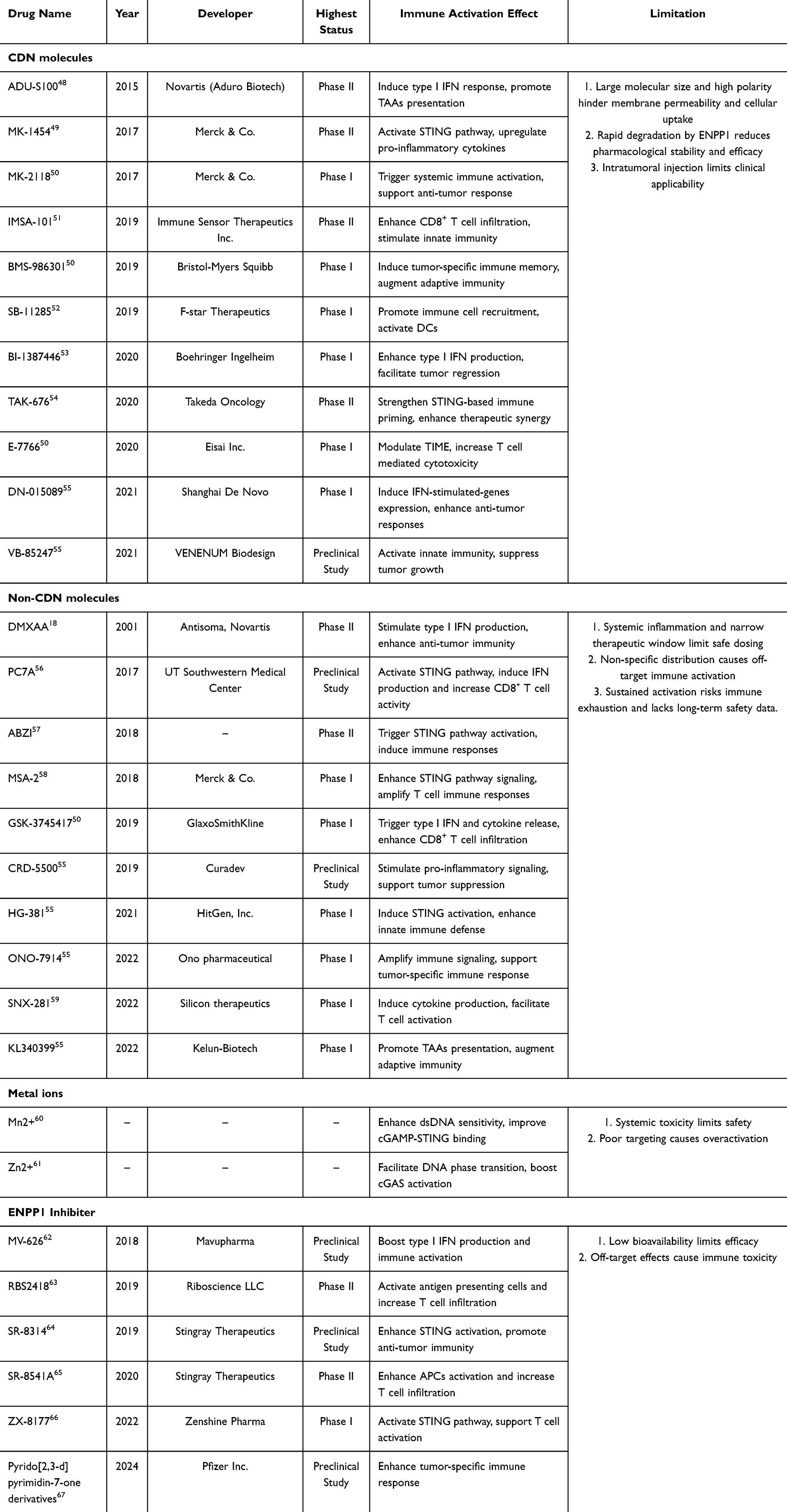 |
Table 1 List of STING Agonists Under Development |
Small Molecules
CDN Molecules
Researchers have synthesized a series of CDN class STING agonists based on the intrinsic CDN structure to enhance their anti-hydrolysis of ENPP1 and optimize their degradation resistance. These modified CDN molecules have also shown notable anti-tumor efficacy through expeditious STING pathway activation. A series of CDN class STING agonists (including ADU-S100, MK-1454, MK-2118, etc.48–55,68–70) and their clinical status are listed in Table 1. Among these, ADU-S100, stabilized by thiophosphate substitution in the intrinsic CDN structure, was the first CDN class STING agonist to advance to clinical trials,71 although its safety and efficacy still require further improvement. Another novel CDN class STING agonist, disulfide phosphate analog 2′3′-cCsAsMP, was designed for anti-hydrolysis of ENPP1 and degradation resistance. Compared to intrinsic cGAMP, 2′3′-cCsAsMP can achieve 10 times more effective in stimulating IFN secretion in human THP1 cells.72 Similarly, MK-1454 exhibited a high affinity for STING and a significant type I IFN induction. When MK-1454 was injected intratumorally into tumor-bearing mice, it was demonstrated to induce complete tumor regression and enhance the efficacy of ICI therapy and entered Phase II clinical trials.73 Unfortunately, CDN class STING agonists still require intratumoral injection to achieve therapeutic effects, limiting their broader clinical translation.
Non-CDN Molecules
Novel STING agonists, suitable for intravenous injection, oral, and subcutaneous administration, were developed to address the limitations of existing intratumoral injection delivery methods for CDN class STING agonists. DMXAA, the first intravenous injection non-CDN class STING agonist, showed significant anti-tumor effects in mouse models. However, its poor binding to human STING led to suboptimal results in a Phase II clinical trial of non-small cell lung cancer.17,74,75 Thus, as a way to address the need for a high binding affinity of human STING, aminobenzoimidazole (ABZI), another novel non-CDN class STING agonist, exhibited superior efficacy in binding both mouse and human STING.57 Compared to cGAMP, ABZI achieved a 400-fold greater STING pathway activation in human PBMCs. Intravenous injection of ABZI exhibited powerful tumor growth inhibition and extend survival in mouse models.76
Apart from intravenous injection of ABZI, MSA-2, a novel non-CDN STING agonist for oral and subcutaneous administration, induces elevated IFN expression in plasma and tumors, resulting in durable anti-tumor immune responses.58 By conjugating the triggering receptor expressed on myeloid cells 2 (TREM2) inhibitor artesunate to MSA-2, Deng et al developed the novel STING agonist prodrug named GB2. Intravenous injection of GB2 promoted M1 polarization of tumor-associated macrophages (TAMs), enhanced immune response and induced tumor regression.77 Several novel non-CDN class STING agonists (including SNX-281, GSK-3745417, ONO-7914, etc.50,55,56,59) and their clinical status are listed in Table 1. More multi-center clinical trials are encouraged to evaluate its clinical biosafety and efficacy.
Metal Ions
Metal ions, due to their superior chemical stability, effectively address the challenge of insufficient stability often observed in STING agonists. Manganese ions (Mn²⁺), released from cellular organelles like the Golgi apparatus and mitochondria, can accumulate cytoplasmically and enhance sensitivity to dsDNA, improving cGAMP-STING binding and pathway activation.60 Similarly, zinc ions (Zn²⁺), primarily stored in mitochondria and the ER, promote cGAS activation by facilitating intracellular DNA phase transition.61 However, the systemic toxicity of metal ions has gained increasing attention. It has been reported that excessive exposure to Mn²⁺ led to manganism, displaying serious central nervous system damage and an elevated risk of Parkinson’s disease.78 In addition, excessive exposure to Zn²⁺ resulted in multi-system damage, including neurological suppression, immune system dysfunction, digestive systems injury, etc.79 Nanomaterials are expected to minimize their systemic toxicity by means of metal ions control release and targeted delivery.
ENPP1 Inhibitors
ENPP1 inhibitors can reduce the hydrolysis and degradation of extracellular cGAMP, thereby elevating its concentration and facilitating STING pathway activation.80 For instance, through inhibition of extracellular cGAMP degradation and subsequent STING pathway activation, ZX-8177 can increase the infiltration of CD8+ T cells and NK cells into the tumor microenvironment (TME) and improve tumor immunotherapy efficacy.66 MV-626, another highly potent and selective ENPP1 inhibitor, showed completely oral bioavailability in mouse models. When combined with radiation or ICIs, MV-626 can effectively block cGAMP hydrolysis, delay tumor growth, and significantly prolong overall survival.62 Several ENPP1 inhibitors, such as SR-8541A65 and RBS2418,63 have significantly improved CD8+ T cells intratumoral infiltration in mouse models and progressed to Phase II clinical research. While SR-8314 and Pyrido[2,3-d]pyrimidine-7-one derivatives64,67 remain in preclinical study stages (Table 1). More efficient inhibition of cGAMP degradation and better oral bioavailability of ENPP1 inhibitors needed to be explored. Moreover, the combined application of ENPP1 inhibitors with radiotherapy and ICIs is expected to further enhance the anti-tumor effect.
Delivery Challenges and Solutions
There are several critical delivery challenges of STING agonists, which primarily manifest in low cell transmembrane transport efficiency, rapid degradation, and systemic toxicity. As illustrated in Figure 3, recent advances in nanotechnology, especially in the development of novel delivery systems, have partially addressed these challenges.81,82
 |
Figure 3 Application of nanotechnology in addressing challenges of STING pathway-based tumor immunotherapy. Application of nanotechnology, as exemplified by c-di-AMP (CDA) formulated as ZnCDA with a zinc phosphate core and PEG-lipid shell, can reduce STING agonist degradation, promote transmembrane transport, enhance APC uptake, and increase IFN production. It also increases accumulation at tumor sites and eventually reduces systemic toxicity. Reprinted with permission from Yang K, Han W, Jiang X, et al. Zinc cyclic di-AMP nanoparticles target and suppress tumours via endothelial STING activation and tumour-associated macrophage reinvigoration. Nat Nanotechnol. 2022;17(12):1322–1331. Copyright © 2022, Springer Nature.83 |
Cell Transmembrane Transport and Rapid Degradation
Efficient transmembrane transport is crucial for the STING pathway activation due to STING’s localization in ER. Nevertheless, large molecular size, high polarity, and susceptibility to hydrolysis of STING agonists significantly impede their delivery, reducing bioavailability and therapeutic efficacy.7,8 These problems also potentially increase the requirement for higher doses or more frequent injections.84 Wilson et al employed cationic poly(beta-amino ester) nanoparticles to deliver CDNs to the cytoplasm, achieving significant STING and IRF3 activation. Notably, the dosage required was 100-fold lower than that needed without the use of nanoparticles.85 Similarly, Webb et al utilized polymeric vesicles to synthesize STING-NPs, which can significantly enhance cGAMP uptake and cytoplasmic delivery. After intravenous injection, this approach resulted in a 40-fold increase in the half-life of cGAMP and significantly elevated serum levels of IFN, TNF-α, IL-12, etc.86 Additionally, Ariosa et al synthesized one pre-formed loadable lipid nanoparticle aimed at reducing ENPP1 activity and achieved the result of inhibition of 2′3′-cGAMP rapid degradation.87,88
Systemic Toxicity
Overactivated STING pathway and heightened immune responses pose a risk of excessive inflammatory cytokine secretion and cytokine storms, which can lead to systemic toxicity.58
Dou et al developed and synthesized high-mesoporous Mn-based nanomaterials with metformin nanoparticles named Mn-MSN@Met-M NPs. Through retaining tumor cell membrane proteins and adhesion molecules, these nanocarriers can facilitate homologous binding to tumor cells, and reduce accumulation in other organs and systemic toxicity.89 In addition, Yang et al encapsulated bacterial-derived CDA in nanoscale coordination polymers with a zinc phosphate core and PEG-conjugated phospholipid bilayer. This nanoparticle enhances CDA tumor accumulation and penetration by disrupting endothelial cell vasculature, as well as reducing systemic toxicity.83 Furthermore, Cui et al combined the photosensitizer BODTPE and the targeting ligand dibenzocyclooctyne to design nanoparticles. This nanoparticle specifically target tumor cells via a click chemistry reaction with azide receptors of the cell membrane, improving drug accumulation and stability while maintaining minimal systemic toxicity.90
STING Pathway-Based Nanomaterial for Tumor Vaccine
Tumor vaccine was designed to trigger anti-tumor immune responses by targeting tumor-specific antigens, activating and amplifying corresponding immune cell responses.91 Recent nanomaterial applications, such as metal ion-loaded, pH-sensitive, and biologically or chemically modified nanoparticles, significantly enhance STING pathway activation, promote MHC I antigen presentation and upregulate costimulatory molecule expression. These approaches also preserve vaccine integrity, extend duration, and regulate release, further enhancing their therapeutic potential and tumor vaccine efficacy.92,93
Metal-Based Nano-Tumor Vaccine
Metal nanoparticles, including Fe2O₃, α-Al2O₃, and MnO2, possess properties of enhanced membrane penetration, streamlined synthesis and robust stability. These properties render them particularly suitable as carriers for the precise delivery of STING agonists.94,95 Notably, apart from serving as carriers, MnO2 nanoparticles also act as potent STING agonists.
Using metal iron nanoparticles as carriers, a PEIM@Mem iron nano-tumor vaccine, engineered by loading the STING agonist MSA-2 and an acidic copolymer, significantly enhanced antigen uptake and presentation by APCs, leading to a marked CD8+ T cell response and effective tumor regression.96 In the same way, an α-Al2O₃-UPs-4T1/EPB nano-tumor vaccine was developed using α-Al2O₃ nanoparticles as carriers via the covalent conjugation of ubiquitin-binding proteins. This innovative vaccine improved CD8+ T cell responses and tumor-infiltrating lymphocyte receptor diversity. Notably, the incorporation of the STING agonist DMXAA, this vaccine further enhanced DCs migration to lymph nodes and lymphocyte infiltration into tumors, resulting in a significant anti-tumor effect in mouse model.97 Unlike merely serving as carriers, the dual function of MnO2 nanoparticles plays a unique efficacy when developing a MnP-PEG nano-tumor vaccine. This vaccine efficiently delivers Mn²⁺ via endocytosis, and activates STING pathway, further inducing IFN and inflammatory cytokine expression, enhancing CD8⁺ T cell-mediated anti-tumor activity.98
To further stimulate DCs maturation and upregulate co-stimulatory molecules, OVA, a model antigen to enhance immunogenicity, was integrated into nanoparticles.99 Zhao et al incorporated MnO2 nanoparticles and cationic polymers to construct the MPO nano-tumor vaccine. Upon draining to the lymph nodes, Mn²⁺ activated the STING pathway and induced the generation of tumor-associated antigens (TAAs). The MPO nano-vaccine promoted DCs maturation, ultimately enhancing anti-tumor immune responses by utilizing the synergy of OVA and TAAs.100 Qiao et al further improved vaccine efficacy by incorporating Mn²⁺ and Al³⁺ into their MnO2-Al-OVA nano-vaccine system. This delivery platform effectively directs the vaccine to lymph nodes, facilitates efficient endocytic uptake by DCs and significantly amplifies the antigen-specific anti-tumor immune response.101 To target the overexpressed mannose receptors on TAMs and DCs, Gu et al synthesized mannose-modified, OVA-coated MnO2 nanoparticles. This nano-tumor vaccine released Mn²⁺, activated the STING pathway, induced M2 to M1 phenotype switch and upregulated CD80/CD86 on DCs, and enhanced CD8⁺ T cell activation.102 Nevertheless, the cytotoxicity of metal nanoparticles (including their potential to disrupt cellular membranes, affect cytoskeletal components, and damage DNA, etc.) remains a concern.103 Recently, several strategies have been employed to address this issue. PEGylation was used in MnP-PEG nanoparticles to improve endocytic drug delivery, confining STING pathway activation to the tumor microenvironment rather than acting systemically.98 The MnO2-Al-OVA platform utilizes TME-responsive release and dual-metal coordination, enabling dose reduction, minimizing systemic metal ion exposure and potential neurotoxicity.101 Another novel mannose-modified MnO2 nanoparticles achieve selective targeting of immune cells via receptor-mediated uptake, enhancing delivery specificity and reducing off-target effects.102 Although these strategies have been explored, additional approaches are still encouraged to improve the biosafety of metal-based nano-tumor vaccine.
PH-Sensitive Nano-Tumor Vaccine
PH-sensitive nanoparticles, which exhibit the ability to undergo structural changes in response to specific pH environments, have been engineered to facilitate the precision and efficacy of STING agonist delivery.
Using reversible addition-fragmentation chain transfer (RAFT) polymerization, Zhou et al synthesized a PEG-b-PDPA copolymer that can be activated at pH5.9–6.2. The PEG-b-PDPA copolymer was conjugated with DMXAA and tumor-specific antigens to develop a nano-tumor vaccine. This vaccine enhanced DC's endocytosis, facilitated rapid antigen release into the cytoplasm, and achieved a better CD8+ T cell response in the mouse model of subcutaneous administration.104 Unfortunately, poor binding to human STING of DMXAA hinders its clinical application. In a related study, Su et al developed another pH-sensitive nano-tumor vaccine incorporating cGAMP and tumor-specific antigens with multivesicular nanoparticles. This vaccine can self-assemble at physiological pH and disassemble in acidic conditions, which precisely delivered cGAMP to DCs and induced a robust CD8+ T cell response upon subcutaneous administration.105
Meanwhile, pH-sensitive nanoparticles with the capacity to activate the STING pathway have also been explored. A series of ultra-pH-sensitive (UPS) nanoparticles were developed from copolymers with linear or cyclic tertiary amines, tunable across physiological pH (4–7.4), and had greater ability to activate the STING pathway simultaneously. Among these, PC7A was selected for its ability to induce the highest CTL responses through STING pathway activation, resulting in significant anti-tumor effects in mouse models. Gradual degradation of PC7A in acidic environments can also prolong STING pathway activation, enabling a more sustained and controlled immune response compared to cGAMP, highlighting its promising clinical potential.56,106 Intratumoral injection of the PC7A nano-vaccine effectively promoted T cell proliferation and infiltration within tumors, facilitated the IFN-γ-expressing CD8+ T cell recruitment, and enhanced the efficacy of anti-tumor immune response.107 Although significant progression of targeting precision and delivery methods has been made in PH-sensitive nano-tumor vaccine, their clinical effectiveness and safety are needed more improvement and validation. For instance, instead of intratumoral injection, oral or subcutaneous administration of the PC7A may indicate a promise prospect for clinical application.
Biologically/Chemically Modified Nano-Tumor Vaccine
Biologically/chemically modified nanoparticles are emerging as novel tools for delivering STING agonists and activating STING pathway. Guo et al utilized bio-inspired tumor cell membrane technology to modify poly(lactic-co-glycolic acid) nanoparticles with the CBP-12 peptide. These biologically modified nano-tumor vaccine can precisely deliver cGAMP and tumor-specific antigens to Clec9a+ DCs, induced STING pathway activation, markedly improved antigen presentation and the key immunoregulatory factor production.108,109 Additionally, using self-degradable poly(beta-amino esters), Liu et al engineered a chemically modified nano-tumor vaccine to deliver cGAMP and the protein antigen OVA to APCs. This vaccine effectively enhanced MHC I-mediated antigen presentation of APCs, increased IFN production and improved tumor vaccine efficacy.110 Moreover, Chen et al designed cGAMP-loaded lipidoid nanoparticles (LNPs/cGAMP). LNPs/cGAMP were injected to the apoptotic site of tumor induced by a low dose of DOX. These nanoparticles can capture TAAs and develop as LNP/cGAMP/TAAs. This lipidoid-based nanosystem tumor vaccine promoted the presentation of tumor antigens, activated the STING pathway, promoted T cell activation, and significantly enhanced the anti-tumor immune response.111 Different from the direct STING pathway activation, Zhao et al developed chitosan-derived composite nanoparticles to promote intracellular DNA release and subsequently activate the STING pathway. By means of these nanoparticles as adjuvants, this tumor vaccine facilitated DC maturation, stimulated macrophage activation, and enhanced overall immune responses.112,113 However, it should be concerned that complex synthetic technology of these biologically/chemically modified nano-tumor vaccine may impede their mass production and clinical translation.
While recent nanomaterials have significantly enhanced tumor vaccine efficacy via improved STING activation and targeted antigen delivery, each strategy presents unique advantages and critical limitations. Specifically, metal-based nano-tumor vaccine exhibits robust delivery and potent immune activation, yet raise biosafety concerns due to inherent cytotoxicity and systemic toxicity risks. Conversely, pH-sensitive nano-tumor vaccine enables precise antigen release responsive to tumor acidity but faces ongoing challenges in clinical validation and systemic administration feasibility. Biologically or chemically modified nano-tumor vaccine, despite their superior targeting specificity and immunogenicity, encounter substantial hurdles related to complex synthesis, regulatory standards, and scalable manufacturing.
STING Pathway-Based Nanomaterial for Modulating Tumor Immune Microenvironment
The tumor immune microenvironment (TIME), characterized by polarization of TAM toward the M2 phenotype, accumulation of regulatory T (Treg) cells, and secretion of cytokines inhibiting T cell activation, plays a pivotal role in tumor cell immune evasion and metastasis.114–116 In addition to single STING pathway activation, the application of nanomaterial can more effectively modulate the immunosuppressive TIME by integrating multiple synergistic effects (such as oxidative stress, radiotherapy, and TLR pathway activation) with STING pathway activation.117
Single STING Pathway Activation
Various STING agonists-loaded nanoparticle can independently modulate the TIME. By encapsulating c-di-GMP in lipid nanoparticles, Nakamura et al developed targeted lipid nanoparticle formulations (STING-LNP). STING-LNP was verified to activate immune cells infiltrating the TIME and induce anti-tumor immune responses in various types of malignant tumors.118 Luo et al constructed Ln-GAMP-NPs through the self-assembly of lanthanides with AMP/GMP (1:1 M ratio) in aqueous solutions. These nanoparticles effectively activated the STING pathway, upregulated CD8+ T cell proportion, and remodeled the TIME.119 Additionally, Xu et al developed a supramolecular CDN nanoparticle composed of oleic acid, deoxycytidine-derived CDG, and the hydrophobic ligand 3′,5′-diOA-dC. This nanoparticle markedly improved intracellular CDG transport and facilitated STING pathway activation. It also modified the ratio of Treg cells and M2 macrophages, leading to the remodeling of the immunosuppressive TIME.120 Furthermore, by incorporating STING agonist cGAMP into polymeric vesicles, Wang et al synthesized STING-activated nanoparticles (STANs) using the RAFT method. The result indicated that intravenous injection of STANs promoted T cell recruitment and activation and normalized the TIME in mouse models.121 Unlike direct agonist for STING pathway, Liu et al encapsulated a STING mutant with strong IFN-inducing capabilities (STINGR284S) in lipid nanoparticles. This approach can deliver STINGR284S to STING-deficient tumor cells, stimulate anti-tumor cytokines production, and effectively activate T cells while remodeling the TIME.122 Compared with single STING agonist delivery systems, novel nanoparticles, which can induce both STING pathway activation and other synergistic biological effects (including oxidative stress and TLR pathway activation), are expected to modulate the immunosuppressive TIME more effectively.
Synergy of Oxidative Stress Inducing and STING Pathway Activation
Oxidative stress resulting from cellular damage can effectively enhance the expression of TAAs, activate immune cells, and markedly modulate immunosuppressive TIME. Utilizing ultrasound and the polycarbonate membrane extrusion method, Bao et al developed membrane fusion lipid-encapsulated Fe-STING nanoparticles containing Fe²⁺ ions and cGAMP. Fe²⁺ ions can generate reactive hydroxyl radicals and induce oxidative stress, while cGAMP activates the STING pathway. The synergy between oxidative stress and STING pathway activation transformed the TIME from “cold” to “hot”, characterized by intratumoral infiltration of CD8+ T cells and reprogramming of macrophages from the M2 to M1 phenotype.123 Additionally, using the combination of the STING agonist SR-717 and the photosensitizer TCPP, Zhou et al developed a novel nanoparticle named SR@PMOF. Following intravenous injection and subsequent irradiation at the tumor site, light activation of TCPP within SR@PMOF induced reactive oxygen species (ROS) generation, and promoted TAAs and tumor DNA fragments release. In synergy with SR-717-mediated STING pathway activation, SR@PMOF can significantly promote DCs and CD8⁺ T cell maturation, reversing the immunosuppressive TIME.124 Similarly, Li et al constructed a near-infrared-responsive nanoenzyme by doping Mn²⁺ into OVA-templated Prussian blue nanoparticles. These nanoparticles induced oxidative stress through photothermal conversion and simultaneously released Mn²⁺ to activate the STING pathway and promote DCs maturation and antigen presentation, resulting in significant T cell activation and the TIME modulation.125 Moreover, Zhou et al synthesized Mn₃O₄@Au-dsDNA/DOX nanoparticles, which integrated chemotherapy with STING pathway activation functions. The synergy between DOX-mediated oxidative stress and STING pathway activation by dsDNA and Mn²⁺ enhanced CD8⁺ T cell activity and intratumoral infiltration. Notably, through the enhanced permeability and retention effect, Mn₃O₄@Au-dsDNA/DOX nanoparticles can accumulate at tumor sites and significantly improve immunosuppressive TIME.126 Excessive oxidative stress was known to induce cellular damage and DNA mutations.127 Although synergy of oxidative stress inducing and STING pathway activation can better modulate the immunosuppressive TIME, nanotechnology application in the future is expected to balance efficiency and safety of oxidative stress response.
Synergy of TLRs and STING Pathway Activation
Toll-like receptors (TLRs), one of the key innate immune receptors, detect pathogens and damage and activate anti-tumor immune responses.128 The combined activation of TLRs and the STING pathway can produce synergistic effects for more effective immune system activation and immunosuppressive TIME reversal.129 Zhang et al developed multicomponent nano-vaccines (McNVs) by conjugating TLR7/8 agonist 522 and STING agonist CDGSF. TLR7/8 pathway activation can promote a series of inflammatory and immune cytokine production. When synergized with STING pathway activation, these McNVs nanoparticles effectively enhanced APCs presentation and increased lymphocyte intratumoral infiltration, reversing the immunosuppressive TIME.130 Similarly, Liu et al developed TME-targeting nanoparticles (PMM NPs) by conjugating TLR4 agonists and Mn₃O₄. In synergy with TLR4-mediated NF-κB activation, the PMM NPs can amplify STING-mediated immune response and increase IFN and pro-inflammatory cytokine secretion. PMM NPs can also reprogram macrophages from the M2 to M1 phenotype, alleviating immunosuppressive TIME.131 However, co-activation of these pathways may trigger excessive cytokine release or cytokine storms.132 Therefore, optimizing therapeutic parameters (including dosage, timing, administration sequence, and route of administration) represents an effective strategy to balance efficacy and safety. Additionally, novel TME-responsive nanomaterials capable of precise spatiotemporal modulation of immune activation are expected to facilitate controlled regulation of immune responses.133
Synergy of Radiotherapy and STING Pathway Activation
Radiotherapy, a classical anti-tumor therapy, induces tumor cell death through DNA damage and immunogenic cell death (ICD). Radiotherapy can release damage-associated molecular patterns and TAAs and subsequently activate immune cells. When combined with STING pathway activation, it can further promote APC maturation and T cell activation, thereby modifying the immunosuppressive TIME.134 By conjugating cGAMP to nanoscale metal-organic layers (MOLs), Luo et al developed cGAMP/MOL nanoparticles. These MOLs effectively penetrated and were retained within the TIME, exhibited strong radiosensitizing effects, and promoted tumor ICD. The incorporation of STING pathway activation by cGAMP. This dual effect resulted in increased radiosensitivity, facilitated the intratumoral infiltration of APCs, and enhanced antigen presentation, ultimately promoting anti-tumor responses.135 In the same way, Deng et al synthesized MnO2 nanoparticles using biomimetic mineralization techniques. Mn²⁺ ions released from nanoparticles not only activated the STING pathway but also overcame hypoxia-induced radioresistance in tumor cells and enhanced ICD, ultimately reversing the immunosuppressive TIME.136 As illustrated in Figure 4, these STING‑centric nanomaterials synergistically enhance tumor‑vaccine efficacy and modulate the immunosuppressive microenvironment.
 |
Figure 4 STING pathway-based nanomaterial in tumor vaccines and immune microenvironment modulation. The application of nanomaterials to enhance tumor vaccine efficacy and modulate the immune microenvironment, focusing on strategies to activate the STING pathway and integrate multiple synergistic effects (such as oxidative stress, radiotherapy, and TLR pathway activation), results in enhanced CD8⁺ T cell proliferation and an M2-to-M1 macrophage phenotype switch. Reproduced from Gao M, Xie YQ, Lei K, et al. A Manganese Phosphate Nanocluster Activates the cGAS‐STING Pathway for Enhanced Cancer Immunotherapy. Advanced Therapeutics 2021;4(8). © 2021 The Authors. Advanced Therapeutics published by Wiley‐VCH GmbH. Creative Commons CC-BY-NC license.98 Su T, Cheng F, Qi J, et al. Responsive Multivesicular Polymeric Nanovaccines that Codeliver STING Agonists and Neoantigens for Combination Tumor Immunotherapy. Adv Sci (Weinh) 2022;9(23):e2201895. © 2022 The Authors. Advanced Science published by Wiley‐VCH GmbH. Creative Commons CC BY license.105 Chen J, Qiu M, Ye Z, et al. In situ cancer vaccination using lipidoid nanoparticles. Sci Adv 2021;7(19). Copyright © 2021 The Authors, some rights reserved; exclusive licensee American Association for the Advancement of Science. No claim to original US Government Works. Distributed under a Creative Commons Attribution NonCommercial License 4.0 (CC BY-NC).111 Luo Z, Liang X, He T, et al. Lanthanide-Nucleotide Coordination Nanoparticles for STING Activation. J Am Chem Soc 2022;144(36):16366–16377. Copyright © 2022 American Chemical Society.119 Zhou M, Wang X, Lin S, et al. Multifunctional STING-Activating Mn(3) O(4) @Au-dsDNA/DOX Nanoparticle for Antitumor Immunotherapy. Adv Healthc Mater 2020;9(13):e2000064. © 2020 WILEY‐VCH Verlag GmbH & Co. KGaA, Weinheim.126 Zhang B-D, Wu J-J, Li W-H, et al. STING and TLR7/8 agonists-based nanovaccines for synergistic antitumor immune activation. Nano Research 2022;15(7):6328–6339. © Tsinghua University Press 2022. This article is made available via the PMC Open Access Subset for unrestricted research re-use and secondary analysis in any form or by any means with acknowledgement of the original source. These permissions are granted for the duration of the World Health Organization (WHO) declaration of COVID-19 as a global pandemic.130 Deng Z, Xi M, Zhang C, et al. Biomineralized MnO(2) Nanoplatforms Mediated Delivery of Immune Checkpoint Inhibitors with STING Pathway Activation to Potentiate Cancer Radio-Immunotherapy. ACS Nano 2023;17(5):4495–4506. Copyright © 2023 American Chemical Society.136 |
Despite significant progress in nanomaterials for TIME modulation, distinct strategies exhibit varied therapeutic efficacy and safety profiles. Single STING pathway activation provides controlled immune stimulation yet is insufficient against complex immunosuppressive environments. Combining oxidative stress markedly enhances antigen release and immune cell infiltration but poses potential cytotoxic risks. Similarly, combined activation of TLR and STING pathways strongly reverses immunosuppression but requires careful management of cytokine release. Additionally, integrating radiotherapy amplifies immune activation and antigen presentation, though further clinical validation of safety and optimal dosage is required.
STING Pathway-Based Nanomaterial for T Cell Tumor Immunotherapy
T cell tumor immunotherapy aims to enhance T cell responses for effective tumor recognition and elimination by targeting tumor-specific antigens.137 Developments in nanomaterial have demonstrated the ability to enhance DC antigen presentation and promote T cell activity, holding substantial potential in improving T cell tumor immunotherapy effects. This potential is especially significant in CAR-T cell therapy, where STING pathway activation markedly amplifies CAR-T cell proliferation and cytotoxicity.138,139
Enhancing DC Antigen-Presentation Ability and T Cell Tumor Immunotherapy
By means of chemically modified and mesoporous nanoparticles, nanotechnology optimizes STING agonist delivery to DCs, promoting their antigen-presentation ability and T cell activation.
Chemically Modified Nanoparticles
It is reported that chemically modified nanoparticles with mannan can enhance their recognition by DCs, induce mannan-associated antigens phagocytosis, and promote DCs maturation and antigen-presentation ability.140,141 Stearic acid (SA) was grafted onto chitosan and subsequently modified with mannose to form M-CS-SA micelles. These micelles can effectively target DCs through interaction with the mannose receptor and adsorb TAAs released from Oxaliplatin-induced ICD, creating an autologous tumor vaccine. By activating the STING pathway and promoting DCs maturation and antigen-presentation ability, these micelles increased CD8⁺ T cells intratumoral infiltration, effectively inhibiting tumor growth.142 Similarly, bovine serum albumin (BSA) was grafted onto mannose to construct BSA-Man@Mn²⁺. This nanoparticle also effectively facilitates DC-targeting through interaction with the mannose receptors on DCs. Concurrently, ferritin-encapsulated β-lapachone (Lap) released from BSA-Man@Mn²⁺ induced tumor cells ICD and released abundant dsDNA for STING pathway activation. In combination with STING agonist Mn²⁺, BSA-Man@Mn²⁺ ultimately promoted CD8⁺ T cell activation, significantly enhanced T cell tumor immunotherapy effects.143 Additionally, Li et al developed nanoparticles, ONc-Mn-A-malF127, with antigen-capturing capabilities through chemically grafting the protein capture groups malF127. By prolonging the retention of antigens released through the photosensitizer ONc-induced ICD at tumor sites, the malF127 component enhanced their phagocytic uptake by DCs. Concurrently, the incorporation of STING agonist Mn²⁺ and ABZI collectively promoted DC maturation, ultimately inducing CD8⁺ T cells to mediate anti-tumor immune responses.144
Mesoporous Nanoparticles
Mesoporous nanoparticles, novel nanoparticles featuring a large surface area and easy functionalization for loading drugs such as cisplatin, are a promising drug delivery platform for tumor treatment.145,146 By utilizing mesoporous silica nanoparticles functionalized with PEG and loaded with c-di-GMP, Chen et al developed novel nanoparticles named cdG@RMSN-PEG-TA. Through prolonging antigen retention time and modulating immune cell interactions in tumor sites, these nanoparticles significantly enhanced DC antigen presentation and co-stimulatory molecule expression, resulting in the recruitment of CD8+ T cells, amplifying T cell tumor immunotherapy effects.147 Another mesoporous nanoparticles are composed of polymerized alginate porous scaffolds, exhibiting high porosity and controlled degradability.148 Smith et al utilized porous scaffolds loaded with c-di-GMP and CAR-T cells. When these implants were subsequently placed at solid tumor sites, the CAR-T cells effectively eradicated tumor cells and released TAAs. Concurrently, c-di-GMP activated DCs and enhanced antigen presentation, providing immunological support for CAR-T cells and improving the efficacy of CAR-T cell-mediated tumor immunotherapy.149 By means of simple structure and high loading capacity, mesoporous nanoparticles are expected to address the complex synthetic technology of chemically modified nanoparticles.
Enhancing Drug Tumor Accumulation and T Cell Tumor Immunotherapy
Nanomaterials can be engineered to prolong the circulation time of STING agonists, enable their sustained release within the TME, enhance agonist accumulation in tumors, promote CD8⁺ T cell infiltration, and ultimately augment the T cell immunotherapy effects.150
Prolonged Drug Circulation Time
Given the irregular and complex structure of tumor vasculature, a prolonged drug circulation time can significantly enhance the concentration of drugs within tumor sites.151 A novel highly hydrophobic nanoparticle composed of ROS-sensitive polymer (P1) and mPEG2k-DSPE can significantly enhance the stability of the loaded drugs, prolong their circulation time, and increase their accumulation within tumor sites. These nanoparticles, loaded with cisplatin and camptothecin, induced DNA damage and activated the STING pathway, increasing CD8⁺ T cells infiltration and enhancing T cell tumor immunotherapy effects.152 Additionally, red blood cell (RBC) membranes were used to develop novel nanoparticles to extend the circulation time and elevate their concentration in tumor sites. Building on this approach, Li et al synthesized nanoparticles named m-PUNCs by integrating ultrasmall iron oxide nanoparticles with the STING agonist PHMA diblock copolymer. These nanoparticles were significantly uptaken by TAMs, activated the STING pathway, increased CD8⁺ T cell populations and enhanced T cell tumor immunotherapy.153 Prolonged drug circulation time has a trend to increase systemic toxicity, suggesting the critical need for precisely targeted delivery to minimize the systemic toxicity.
Precisely Targeted Drug Delivery at Tumor Sites
Precisely targeted drug delivery strategies can enhance drug accumulation at tumor sites, reduce systemic toxicity and improve T cell tumor immunotherapy. Intratumoral injection is the simplest way to distribute drug in tumors.154 Cheng et al encapsulated cGAMP in a silk fibroin (SF) hydrogel and injected it into the tumor site. Hydrogel, characterized with the thermoresponsive properties, can transition from a liquid to a solid state at physiological temperatures (37 °C). The hydrogel’s stable properties ensured their retention at tumor site and facilitated the sustained delivery of the STING agonist cGAMP to DCs, promoting CD8⁺ T cell proliferation within both the tumor and associated lymph nodes, ultimately inducing a robust anti-tumor immune response.155
Under common subcutaneous or intravenous administration, achieving precisely targeted drug delivery at tumor sites remains to be addressed. Novel magnetic iron oxide nanoparticles can achieve highly precise, drug delivery to tumor site through directional guidance under an externally magnetic field.156 Huang et al utilized magnetic iron oxide nanoparticles loaded with Mn²⁺ and Ca²⁺ to synthesize Fe₃O₄@Ca/MnCO₃. Under an external magnetic field, the nanoparticles upon subcutaneous administration were directed to the tumor sites and interacted with DCs, facilitating TAAs internalization. Mn²⁺ significantly activated immune responses via the STING pathway activation, while Ca²⁺ induced autophagy in DCs, enhanced antigen presentation, ultimately promoted CD8⁺ T cell activation and improved T cell tumor immunotherapy effects.157 It is expected to conduct further clinical applications to evaluate its efficacy and safety. As shown in Figure 5, these targeted delivery strategies enhance T cell tumor immunotherapy by improving antigen presentation, prolonging in‑vivo exposure of STING agonists, and achieving precise tumor‑site accumulation.
 |
Figure 5 STING pathway-based nanomaterial for enhancing T cell tumor immunotherapy. Application of nanomaterial in enhancing T cell tumor immunotherapy, focusing on improving tumor-associated antigen presentation, prolonging drug (STING agonist etc) circulation, and achieving targeted drug delivery at tumor sites. |
Nanomaterial-based strategies have markedly improved DC antigen presentation and STING agonist accumulation at tumor sites, though notable limitations persist. Chemically modified nanoparticles effectively enhance DC targeting, but their complex synthetic process impedes large-scale production. Mesoporous nanoparticles offer simple structures and high loading capacity yet raise biodegradability concerns. Approaches prolonging drug circulation time significantly increase tumor accumulation but concurrently heighten systemic toxicity. In contrast, precise targeted drug delivery effectively reduces off-target effects, yet clinical effectiveness and feasibility remain to be confirmed.
Challenges in Clinical Translation
Despite extensive research on improving the therapeutic outcomes with STING pathway-based nanomaterials, clinical translation remains limited. Some drawbacks and challenges should be considered.
Clinical Trial Research Updates
Most nanomaterial-based delivery systems are still assessed in murine tumor models, especially melanoma,56,96,119 that inadequately reflect human biological heterogeneity and immune diversity,158–160 leaving their efficacy in other tumors uncertain and impeding clinical translation. Therefore, exploring animal models that can better recapitulate human immune microenvironments is expected to generate reliable efficacy data and accelerate clinical translation. In this regard, patient-derived xenografts or humanized mouse models, which successfully recreate patient-specific tumor-immune interactions,161–163 can bring new insights for this challenge.
Regulatory Hurdles
The complex synthesis process of nanomaterials, especially those incorporating biological or chemical modifications, increasingly presents regulatory challenges that impede clinical translation.164,165 Researchers are encouraged to identify robust formulations through systematic evaluation and the establishment of Good Manufacturing Practice standards for nanomaterial synthesis,166,167 aiming to ensure batch consistency and scalable production for clinical translation.168,169
Biosafety Considerations
Clinical biosafety considerations primarily arise from immune hyperactivation induced by STING pathway activation, as well as nanomaterial-induced systemic toxicity, exemplified by Mn²⁺- and Zn²⁺-related neurotoxicity.58,103 Recently, surface modifications like PEGylation, ligand decoration and TME-responsive activation enhance targeting of nano-delivery and reduce dosing, effectively alleviating immune hyperactivation and systemic toxicity. PEGylated MnP-PEG nanoparticles significantly enhanced intratumoral STING activation by improving endocytic drug delivery into APCs.98 Mannose-modified MnO2 nanoparticles improved delivery specificity to immune cells via receptor-mediated uptake.102 A series of TME-responsive nanomaterials can be activated under tumor-specific acidic pH, enabling controlled drug release and enhanced targeting of nano-delivery.104,105,107 Nevertheless, the long-term biosafety of these approaches requires further investigation, including comprehensive evaluation of pharmacokinetics and biodistribution.86,170
Outlook
The integration of nanomaterial with STING pathway modulation opens a new prospect for tumor immunotherapy, characterized by improved efficacy, precision, and adaptability.
It has been reported that basal activation of the STING pathway varies across tumor types, with patient genetic backgrounds and tumor immune statuses further affecting tumor immunotherapeutic effects.21,171–173 In particular, the latter, characterized by differences in immune cell infiltration and activation within the tumor immune microenvironment, represents a key challenge for effective STING pathway-based immunotherapy. Hence, the assessment of tumor mutational burden, neoantigen levels, and cytokine expression using genomic and proteomic data can support the design of STING pathway-based nanomaterial and personalized treatment. Additionally, combining ICI therapy to restore exhausted T cells helps enhance immune cell infiltration and activation in the tumor microenvironment, offering a promising approach to modulate immune status.
In summary, the future of STING pathway-based nanomaterial in tumor immunotherapy lies in refining nanocarrier systems for optimized delivery of STING agonist. Innovations in biocompatible, multifunctional nanoparticles and biodegradable, TME-responsive materials will enhance the targeting of nano-delivery and minimize systemic toxicity.
Abbreviations
STING, stimulator of interferon genes; ER, endoplasmic reticulum; STIM1, stromal interaction molecule 1; cGAS, cyclic GMP-AMP synthase; dsDNA, double-stranded DNA; cGAMP, cyclic GMP-AMP; IKK, IκB kinase; TBK1, TANK-binding kinase 1; IFN, interferon; DCs, dendritic cells; APCs, antigen-presenting cells; ICIs, immune checkpoint inhibitors; CAR, chimeric antigen receptor; CDG, c-di-GMP; OVA, ovalbumin; CDA, c-di-AMP; CDN, cyclic dinucleotide; ENPP1, ecto-nucleotide phosphodiesterase 1; ABZI, aminobenzoimidazole; TREM2, triggering receptor expressed on myeloid cells 2; TAMs, tumor-associated macrophages; TME, tumor microenvironment; TAAs, tumor-associated antigens; RAFT, reversible addition-fragmentation chain transfer; UPS, ultra-pH-sensitive; TIME, tumor immune microenvironment; Treg, regulatory T; ROS, reactive oxygen species; TLRs, Toll-like receptors; ICD, immunogenic cell death; MOLs, metal-organic layers; SA, stearic acid; BSA, bovine serum albumin; RBC, red blood cell; SF, silk fibroin.
Acknowledgments
Figures in the manuscript were made by Figdraw.
Author Contributions
All authors made a significant contribution to the work reported. ZWR took part in drafting and revising the article. TXL, QYC, WXC, and ZKQ contributed to the critical review and editing of the manuscript. LT and GNQ conceived the study, supervised its execution, approved the final version for publication, and agree to be accountable for all aspects of the work. All authors have agreed on submission to the International Journal of Nanomedicine.
Funding
This work was supported by Natural Science Foundation of Shandong Province (ZR2021MH324), Research on Reducing the Incidence of Liver Cancer in Hepatitis B Patients in China (Lvzhou) Project Research Project (LZGC2022-05), Youth Foundation of Shandong Natural Science Foundation (No. ZR2022QH32), National Key Research and Development Program of China (No. 2024YFA1208301), National Natural Science Foundation of China (No. 22477118).
Disclosure
The authors report no conflicts of interest in this work.
References
1. Chen Y, Yu D, Qian H, Shi Y, Tao Z. CD8(+) T cell-based cancer immunotherapy. J Transl Med. 2024;22(1):394. doi:10.1186/s12967-024-05134-6
2. Rui R, Zhou L, He S. Cancer immunotherapies: advances and bottlenecks. Front Immunol. 2023;14:1212476. doi:10.3389/fimmu.2023.1212476
3. Bai R, Chen N, Li L, et al. Mechanisms of Cancer Resistance to Immunotherapy. Front Oncol. 2020;10:1290. doi:10.3389/fonc.2020.01290
4. Lei X, Lei Y, Li JK, et al. Immune cells within the tumor microenvironment: biological functions and roles in cancer immunotherapy. Cancer Lett. 2020;470:126–133. doi:10.1016/j.canlet.2019.11.009
5. Li A, Yi M, Qin S, et al. Activating cGAS-STING pathway for the optimal effect of cancer immunotherapy. J Hematol Oncol. 2019;12(1):35. doi:10.1186/s13045-019-0721-x
6. Zheng J, Mo J, Zhu T, et al. Comprehensive elaboration of the cGAS-STING signaling axis in cancer development and immunotherapy. Mol Cancer. 2020;19(1):133. doi:10.1186/s12943-020-01250-1
7. Huang C, Shao N, Huang Y, et al. Overcoming challenges in the delivery of STING agonists for cancer immunotherapy: a comprehensive review of strategies and future perspectives. Mater Today Bio. 2023;23:100839. doi:10.1016/j.mtbio.2023.100839
8. Tabar MMM, Fathi M, Kazemi F, Bazregari G, Ghasemian A. STING pathway as a cancer immunotherapy: progress and challenges in activating anti-tumor immunity. Mol Biol Rep. 2024;51(1):487. doi:10.1007/s11033-024-09418-4
9. Zheng YF, Wu JJ. Overcoming STING agonists barriers: peptide, protein, and biomembrane-based biocompatible delivery strategies. Chem Asian J. 2022;17(6):e202101400. doi:10.1002/asia.202101400
10. Wu YT, Fang Y, Wei Q, et al. Tumor-targeted delivery of a STING agonist improvescancer immunotherapy. Proc Natl Acad Sci U S A. 2022;119(49):e2214278119. doi:10.1073/pnas.2214278119
11. Wang J, Meng F, Yeo Y. Delivery of STING agonists for cancer immunotherapy. Curr Opin Biotechnol. 2024;87:103105. doi:10.1016/j.copbio.2024.103105
12. Garland KM, Sheehy TL, Wilson JT. Chemical and biomolecular strategies for STING pathway activation in cancer immunotherapy. Chem Rev. 2022;122(6):5977–6039. doi:10.1021/acs.chemrev.1c00750
13. Wells K, Liu T, Zhu L, Yang L. Immunomodulatory nanoparticles activate cytotoxic T cells for enhancement of the effect of cancer immunotherapy. Nanoscale. 2024;16(38):17699–17722. doi:10.1039/D4NR01780C
14. Gupta J, Safdari HA, Hoque M. Nanoparticle mediated cancer immunotherapy. Semin Cancer Biol. 2021;69:307–324. doi:10.1016/j.semcancer.2020.03.015
15. Chen X, Xu Z, Li T, et al. Nanomaterial-encapsulated STING agonists for immune modulation in cancer therapy. Biomark Res. 2024;12(1):2. doi:10.1186/s40364-023-00551-z
16. Bao T, Liu J, Leng J, Cai L. The cGAS-STING pathway: more than fighting against viruses and cancer. Cell Biosci. 2021;11(1):209. doi:10.1186/s13578-021-00724-z
17. Gao P, Ascano M, Zillinger T, et al. Structure-function analysis of STING activation by c[G(2′,5′)pA(3′,5′)p] and targeting by antiviral DMXAA. Cell. 2013;154(4):748–762. doi:10.1016/j.cell.2013.07.023
18. Ishikawa H, Barber GN. STING is an endoplasmic reticulum adaptor that facilitates innate immune signalling. Nature. 2008;455(7213):674–678. doi:10.1038/nature07317
19. Barber GN. STING: infection, inflammation and cancer. Nat Rev Immunol. 2015;15(12):760–770. doi:10.1038/nri3921
20. Kwon J, Bakhoum SF. The cytosolic DNA-sensing cGAS-STING pathway in cancer. Cancer Discov. 2020;10(1):26–39. doi:10.1158/2159-8290.CD-19-0761
21. Samson N, Ablasser A. The cGAS-STING pathway and cancer. Nat Cancer. 2022;3(12):1452–1463. doi:10.1038/s43018-022-00468-w
22. Flood BA, Higgs EF, Li S, Luke JJ, Gajewski TF. STING pathway agonism as a cancer therapeutic. Immunol Rev. 2019;290(1):24–38. doi:10.1111/imr.12765
23. Maekawa H, Fain ME, Wasano K. Pathophysiological Roles of the cGAS-STING Inflammatory Pathway. Physiology. 2023;38(4):167–177. doi:10.1152/physiol.00031.2022
24. Jiang M, Chen P, Wang L, et al. cGAS-STING, an important pathway in cancer immunotherapy. J Hematol Oncol. 2020;13(1):81. doi:10.1186/s13045-020-00916-z
25. Wang H, Hu S, Chen X, et al. cGAS is essential for the antitumor effect of immune checkpoint blockade. Proc Natl Acad Sci U S A. 2017;114(7):1637–1642. doi:10.1073/pnas.1621363114
26. Hopfner KP, Hornung V. Molecular mechanisms and cellular functions of cGAS-STING signalling. Nat Rev Mol Cell Biol. 2020;21(9):501–521. doi:10.1038/s41580-020-0244-x
27. Bashash D, Zandi Z, Kashani B, et al. Resistance to immunotherapy in human malignancies: mechanisms, research progresses, challenges, and opportunities. J Cell Physiol. 2022;237(1):346–372. doi:10.1002/jcp.30575
28. Van Herck S, Feng B, Tang L. Delivery of STING agonists for adjuvanting subunit vaccines. Adv Drug Deliv Rev. 2021;179:114020. doi:10.1016/j.addr.2021.114020
29. Kinkead HL, Hopkins A, Lutz E, et al. Combining STING-based neoantigen-targeted vaccine with checkpoint modulators enhances antitumor immunity in murine pancreatic cancer. JCI Insight. 2018;3(20). doi:10.1172/jci.insight.122857
30. Hanson MC, Crespo MP, Abraham W, et al. Nanoparticulate STING agonists are potent lymph node-targeted vaccine adjuvants. J Clin Invest. 2015;125(6):2532–2546. doi:10.1172/JCI79915
31. Le Z, Qian J, Chen H, et al. A versatile gemini amphiphile-based platform with STING-activating properties for efficient gene delivery into dendritic cells. Chem Eng J. 2024;497:14. doi:10.1016/j.cej.2024.154513
32. Patel MN, Tiwari S, Wang Y, et al. Safer non-viral DNA delivery using lipid nanoparticles loaded with endogenous anti-inflammatory lipids. Nat Biotechnol. 2025. doi:10.1038/s41587-025-02556-5
33. Dougan M, Dranoff G, Dougan SK. Cancer immunotherapy: beyond checkpoint blockade. Annu Rev Cancer Biol. 2019;3:55–75. doi:10.1146/annurev-cancerbio-030518-055552
34. Nagasaki J, Ishino T, Togashi Y. Mechanisms of resistance to immune checkpoint inhibitors. Cancer Sci. 2022;113(10):3303–3312. doi:10.1111/cas.15497
35. Vornholz L, Isay SE, Kurgyis Z, et al. Synthetic enforcement of STING signaling in cancer cells appropriates the immune microenvironment for checkpoint inhibitor therapy. Sci Adv. 2023;9(11):eadd8564. doi:10.1126/sciadv.add8564
36. Fu J, Kanne DB, Leong M, et al. STING agonist formulated cancer vaccines can cure established tumors resistant to PD-1 blockade. Sci Transl Med. 2015;7(283):283ra252. doi:10.1126/scitranslmed.aaa4306
37. Nakamura T, Sato T, Endo R, et al. STING agonist loaded lipid nanoparticles overcome anti-PD-1 resistance in melanoma lung metastasis via NK cell activation. J Immunother Cancer. 2021;9(7):e002852. doi:10.1136/jitc-2021-002852
38. Hao M, Zhu L, Hou S, et al. Sensitizing tumors to immune checkpoint blockage via STING agonists delivered by tumor-penetrating neutrophil cytopharmaceuticals. ACS Nano. 2023;17:1663–1680. doi:10.1021/acsnano.2c11764
39. Wang B, Tang M, Chen Q, et al. Delivery of mRNA encoding Interleukin-12 and a stimulator of interferon genes agonist potentiates antitumor efficacy through reversing T cell exhaustion. ACS Nano. 2024;18(24):15499–15516. doi:10.1021/acsnano.4c00063
40. Feins S, Kong W, Williams EF, Milone MC, Fraietta JA. An introduction to chimeric antigen receptor (CAR) T-cell immunotherapy for human cancer. Am J Hematol. 2019;94(S1):S3–S9. doi:10.1002/ajh.25418
41. Li W, Lu L, Lu J, et al. cGAS-STING-mediated DNA sensing maintains CD8(+) T cell stemness and promotes antitumor T cell therapy. Sci Transl Med. 2020;12(549). doi:10.1126/scitranslmed.aay9013
42. Xu N, Palmer DC, Robeson AC, et al. STING agonist promotes CAR T cell trafficking and persistence in breast cancer. J Exp Med. 2021;218(2). doi:10.1084/jem.20200844
43. Conde E, Vercher E, Soria-Castellano M, et al. Epitope spreading driven by the joint action of CART cells and pharmacological STING stimulation counteracts tumor escape via antigen-loss variants. J Immunother Cancer. 2021;9(11):e003351. doi:10.1136/jitc-2021-003351
44. Lu Z, Fu Y, Zhou X, Du H, Chen Q. Cyclic dinucleotides mediate bacterial immunity by dinucleotide cyclase in Vibrio. Front Microbiol. 2022;13:1065945. doi:10.3389/fmicb.2022.1065945
45. Jiang Y, Li X, Qian F, et al. Fine-tuning bacterial Cyclic di-AMP production for durable antitumor effects through the activation of the STING pathway. Research. 2023;6:0102. doi:10.34133/research.0102
46. Rueckert C, Rand U, Roy U, et al. Cyclic dinucleotides modulate induced type I IFN responses in innate immune cells by degradation of STING. FASEB J. 2017;31(7):3107–3115. doi:10.1096/fj.201601093R
47. Ahn J, Xia T, Konno H, et al. Inflammation-driven carcinogenesis is mediated through STING. Nat Commun. 2014;5:5166. doi:10.1038/ncomms6166
48. Yang H, Lee WS, Kong SJ, et al. STING activation reprograms tumor vasculatures and synergizes with VEGFR2 blockade. J Clin Invest. 2019;129(10):4350–4364. doi:10.1172/JCI125413
49. Harrington KJ, Brody J, Ingham M, et al. Preliminary results of the first-in-human (FIH) study of MK-1454, an agonist of stimulator of interferon genes (STING), as monotherapy or in combination with pembrolizumab (pembro) in patients with advanced solid tumors or lymphomas. Ann Oncol. 2018;29:viii712. doi:10.1093/annonc/mdy424.015
50. Ding C, Song Z, Shen A, Chen T, Zhang A. Small molecules targeting the innate immune cGAS‒STING‒TBK1 signaling pathway. Acta Pharm Sin B. 2020;10(12):2272–2298. doi:10.1016/j.apsb.2020.03.001
51. Lee P, Malhotra J, Salsamendi J, et al. Two phase 2A clinical trials to evaluate the safety and efficacy of IMSA101 in combination with radiotherapy and checkpoint inhibitors in oligometastatic and oligoprogressive solid tumor malignancies. J Clin Oncol. 2024;42(16_suppl):TPS2685. doi:10.1200/JCO.2024.42.16_suppl.TPS2685
52. Abbas A, Strauss J, Janku F, et al. P01.01 A Phase 1a/1b dose-escalation study of intravenously administered SB 11285 alone and in combination with nivolumab in patients with advanced solid tumors. J Immunother Cancer. 2020;8:A7–A8.
53. Harrington K, Parkes E, Weiss J, et al. 408 Phase I, first-in-human trial evaluating BI 1387446 (stimulator of interferon genes [STING] agonist) alone and combined with BI 754091 (anti-programmed cell death [PD]-1) in solid tumors. J Immunother Cancer. 2020;8:A248.241–A248. doi:10.1136/jitc-2020-SITC2020.0408
54. Carideo Cunniff E, Sato Y, Mai D, et al. TAK-676: a novel stimulator of interferon genes (STING) agonist promoting durable IFN-dependent antitumor immunity in preclinical studies. Cancer Res Commun. 2022;2(6):489–502. doi:10.1158/2767-9764.CRC-21-0161
55. Huang R, Ning Q, Zhao J, et al. Targeting STING for cancer immunotherapy: from mechanisms to translation. Int Immunopharmacol. 2022;113(Pt A):109304. doi:10.1016/j.intimp.2022.109304
56. Luo M, Wang H, Wang Z, et al. A STING-activating nanovaccine for cancer immunotherapy. Nat Nanotechnol. 2017;12(7):648–654. doi:10.1038/nnano.2017.52
57. Xi Q, Wang M, Jia W, et al. Design, synthesis, and biological evaluation of amidobenzimidazole derivatives as stimulator of interferon genes (STING) receptor agonists. J Med Chem. 2020;63(1):260–282. doi:10.1021/acs.jmedchem.9b01567
58. Pan BS, Perera SA, Piesvaux JA, et al. An orally available non-nucleotide STING agonist with antitumor activity. Science. 2020;369(6506). doi:10.1126/science.aba6098
59. Wang J, Falchook G, Nabhan S, et al. 495 Trial of SNX281, a systemically delivered small molecule STING agonist, in solid tumors and lymphomas. J Immunother Cancer. 2021;9(Suppl 2):A527.
60. Zhang K, Qi C, Cai K. Manganese-Based Tumor Immunotherapy. Adv Mater. 2023;35(19):e2205409. doi:10.1002/adma.202205409
61. Du M, Chen ZJ. DNA-induced liquid phase condensation of cGAS activates innate immune signaling. Science. 2018;361(6403):704–709. doi:10.1126/science.aat1022
62. Baird J, Dietsch G, Florio V, et al. MV-626, a potent and selective inhibitor of ENPP1 enhances STING activation and augments T-cell mediated anti-tumor activity in vivo.
63. Marron TU, Misleh JG, Chen C, et al. 1025MO Preliminary safety, pharmacokinetics and immunomodulatory activity of RBS2418, an oral ENPP1 inhibitor, alone and in combination with pembrolizumab in patients with solid tumors. Ann Oncol. 2023;34:S623–S624. doi:10.1016/j.annonc.2023.09.2164
64. Weston A, Thode T, Munoz R, et al. Abstract 3077: preclinical studies of SR-8314, a highly selective ENPP1 inhibitor and an activator of STING pathway. Cancer Res. 2019;79(13_Supplement):3077. doi:10.1158/1538-7445.AM2019-3077
65. Weston A, Thode T, Ng S, et al. Abstract LB323: inhibition of ENPP1 using small molecule, SR-8541A, enhances the effect of checkpoint inhibition in cancer models. Cancer Res. 2023;83(8–Sup):3. doi:10.1158/1538-7445.AM2023-LB323
66. Li YY, Peng Z, Sun S, et al. Abstract 5486: ENPP1 inhibitor ZX-8177 enhances anti-tumor activity of conventional therapies by modulating tumor microenvironment. Cancer Res. 2022;82(12_Supplement):5486. doi:10.1158/1538-7445.AM2022-5486
67. Sun Y, Chen M, Han Y, et al. Discovery of Pyrido[2,3-d]pyrimidin-7-one derivatives as highly potent and efficacious ectonucleotide pyrophosphatase/phosphodiesterase 1 (ENPP1) inhibitors for cancer treatment. J Med Chem. 2024;67(5):3986–4006. doi:10.1021/acs.jmedchem.3c02288
68. Amouzegar A, Chelvanambi M, Filderman JN, Storkus WJ, Luke JJ. STING Agonists as Cancer Therapeutics. Cancers. 2021;13(11):2695. doi:10.3390/cancers13112695
69. McIntosh JA, Liu Z, Andresen BM, et al. A kinase-cGAS cascade to synthesize a therapeutic STING activator. Nature. 2022;603(7901):439–444. doi:10.1038/s41586-022-04422-9
70. Kim DS, Endo A, Fang FG, et al. E7766, a macrocycle-bridged stimulator of interferon genes (STING) agonist with potent pan-genotypic activity. ChemMedChem. 2021;16(11):1740–1743. doi:10.1002/cmdc.202100068
71. Novotna B, Vanekova L, Zavrel M, et al. Enzymatic preparation of 2′-5′,3′-5′-cyclic dinucleotides, their binding properties to stimulator of interferon genes adaptor protein, and structure/activity correlations. J Med Chem. 2019;62(23):10676–10690. doi:10.1021/acs.jmedchem.9b01062
72. Li L, Yin Q, Kuss P, et al. Hydrolysis of 2′3′-cGAMP by ENPP1 and design of nonhydrolyzable analogs. Nat Chem Biol. 2014;10(12):1043–1048. doi:10.1038/nchembio.1661
73. Chang W, Altman MD, Lesburg CA, et al. Discovery of MK-1454: a potent cyclic dinucleotide stimulator of interferon genes agonist for the treatment of cancer. J Med Chem. 2022;65(7):5675–5689. doi:10.1021/acs.jmedchem.1c02197
74. Lara PN, Douillard JY, Nakagawa K, et al. Randomized Phase III placebo-controlled trial of carboplatin and paclitaxel with or without the vascular disrupting agent vadimezan (ASA404) in advanced non-small-cell lung cancer. J Clin Oncol. 2011;29(22):2965–2971. doi:10.1200/JCO.2011.35.0660
75. McKeage MJ, Jameson MB; AS1404-201 Study Group Investigators. Comparative outcomes of squamous and non-squamous non-small cell lung cancer (NSCLC) patients in phase II studies of ASA404 (DMXAA) - retrospective analysis of pooled data. J Thorac Dis. 2010;2(4):199–204. doi:10.3978/j.issn.2072-1439.2010.02.04.1
76. Ramanjulu JM, Pesiridis GS, Yang J, et al. Design of amidobenzimidazole STING receptor agonists with systemic activity. Nature. 2018;564(7736):439–443. doi:10.1038/s41586-018-0705-y
77. Deng A, Fan R, Hai Y, et al. A STING agonist prodrug reprograms tumor-associated macrophage to boost colorectal cancer immunotherapy. Theranostics. 2025;15(1):277–299. doi:10.7150/thno.101001
78. Mezzaroba L, Alfieri DF, Colado Simao AN, Vissoci Reiche EM. The role of zinc, copper, manganese and iron in neurodegenerative diseases. Neurotoxicology. 2019;74:230–241. doi:10.1016/j.neuro.2019.07.007
79. Schoofs H, Schmit J, Rink L. Zinc Toxicity: understanding the Limits. Molecules. 2024;29(13):3130. doi:10.3390/molecules29133130
80. Ruiz-fernández de Córdoba B, Martínez-Monge R, Lecanda F. ENPP1 immunobiology as a therapeutic target. Clin Cancer Res. 2023;29(12):2184–2193. doi:10.1158/1078-0432.CCR-22-1681
81. Guo J, Huang L. Nanodelivery of cGAS-STING activators for tumor immunotherapy. Trends Pharmacol Sci. 2022;43(11):957–972. doi:10.1016/j.tips.2022.08.006
82. Zhou Q, Zhou Y, Li T, Ge Z. Nanoparticle-mediated STING agonist delivery for enhanced cancer immunotherapy. Macromol Biosci. 2021;21(8):e2100133. doi:10.1002/mabi.202100133
83. Yang K, Han W, Jiang X, et al. Zinc cyclic di-AMP nanoparticles target and suppress tumours via endothelial STING activation and tumour-associated macrophage reinvigoration. Nat Nanotechnol. 2022;17(12):1322–1331. doi:10.1038/s41565-022-01225-x
84. Petrovic M, Borchard G, Jordan O. Considerations for the delivery of STING ligands in cancer immunotherapy. J Control Release. 2021;339:235–247. doi:10.1016/j.jconrel.2021.09.033
85. Wilson DR, Sen R, Sunshine JC, et al. Biodegradable STING agonist nanoparticles for enhanced cancer immunotherapy. Nanomedicine. 2018;14(2):237–246. doi:10.1016/j.nano.2017.10.013
86. Wehbe M, Wang-Bishop L, Becker KW, et al. Nanoparticle delivery improves the pharmacokinetic properties of cyclic dinucleotide STING agonists to open a therapeutic window for intravenous administration. J Control Release. 2021;330:1118–1129. doi:10.1016/j.jconrel.2020.11.017
87. Fang J, Islam W, Maeda H. Exploiting the dynamics of the EPR effect and strategies to improve the therapeutic effects of nanomedicines by using EPR effect enhancers. Adv Drug Deliv Rev. 2020;157:142–160. doi:10.1016/j.addr.2020.06.005
88. Ariosa A, Taylor D, Forsyth V, Rzeczycki P, Clements L. Abstract 2349 Modulation of ENPP1 activity and 2′3′-cGAMP degradation in ovarian cancer cell lines via loadable pre-formed lipid nanoparticles. J Biol Chem. 2024;300(3):106788. doi:10.1016/j.jbc.2024.106788
89. Dou Y, Zheng J, Kang J, et al. Mesoporous manganese nanocarrier target delivery metformin for the co-activation STING pathway to overcome immunotherapy resistance. Iscience. 2024;27(7):110150. doi:10.1016/j.isci.2024.110150
90. Cui M, Tang D, Wang B, et al. Bioorthogonal guided activation of cGAS‐STING by AIE photosensitizer nanoparticles for targeted tumor therapy and imaging. Adv Mater. 2023;35(52). doi:10.1002/adma.202305668
91. Liang J, Yao L, Liu Z, et al. Nanoparticles in subunit vaccines: immunological foundations, categories, and applications. Small. 2024;21:e2407649. doi:10.1002/smll.202407649
92. Feng C, Li Y, Ferdows BE, et al. Emerging vaccine nanotechnology: from defense against infection to sniping cancer. Acta Pharm Sin B. 2022;12(5):2206–2223. doi:10.1016/j.apsb.2021.12.021
93. Schunke J, Mailander V, Landfester K, Fichter M. Delivery of immunostimulatory cargos in nanocarriers enhances anti-tumoral nanovaccine efficacy. Int J Mol Sci. 2023;24(15):12174. doi:10.3390/ijms241512174
94. Kwon HJ, Shin K, Soh M, et al. Large-scale synthesis and medical applications of uniform-sized metal oxide nanoparticles. Adv Mater. 2018;30(42):e1704290. doi:10.1002/adma.201704290
95. Liu Q, Zhang A, Wang R, Zhang Q, Cui D. A review on metal- and metal oxide-based nanozymes: properties, mechanisms, and applications. Nanomicro Lett. 2021;13(1):154. doi:10.1007/s40820-021-00674-8
96. Chen F, Li T, Zhang H, et al. Acid-ionizable iron nanoadjuvant augments STING activation for personalized vaccination immunotherapy of cancer. Adv Mater. 2023;35(10):e2209910. doi:10.1002/adma.202209910
97. Huang F, Pan N, Wei Y, et al. Effects of combinatorial ubiquitinated protein-based nanovaccine and STING agonist in mice with drug-resistant and metastatic breast cancer. Front Immunol. 2021;12:707298. doi:10.3389/fimmu.2021.707298
98. Gao M, Xie YQ, Lei K, et al. A manganese phosphate nanocluster activates the cGAS‐STING pathway for enhanced cancer immunotherapy. Adv Ther. 2021;4(8). doi:10.1002/adtp.202100065
99. Watanabe A, Yamashita K, Fujita M, et al. Vaccine based on dendritic cells electroporated with an exogenous ovalbumin protein and pulsed with invariant natural killer T cell ligands effectively induces antigen-specific antitumor immunity. Cancers. 2021;14(1):171. doi:10.3390/cancers14010171
100. Zhao X, Zhang J, Chen B, et al. Rough nanovaccines boost antitumor immunity through the enhancement of vaccination cascade and immunogenic cell death induction. Small Methods. 2023;7(5):e2201595. doi:10.1002/smtd.202201595
101. Qiao N, Wang H, Xu Y, et al. A MnAl double adjuvant nanovaccine to induce strong humoral and cellular immune responses. J Control Release. 2023;358:190–203. doi:10.1016/j.jconrel.2023.04.036
102. Gu Y, Lin S, Wu Y, et al. Targeting STING activation by antigen-inspired MnO(2) nanovaccines optimizes tumor radiotherapy. Adv Healthc Mater. 2023;12(12):e2300028. doi:10.1002/adhm.202300028
103. Attarilar S, Yang J, Ebrahimi M, et al. The toxicity phenomenon and the related occurrence in metal and metal oxide nanoparticles: a brief review from the biomedical perspective. Front Bioeng Biotechnol. 2020;8:822. doi:10.3389/fbioe.2020.00822
104. Zhou L, Hou B, Wang D, et al. Engineering polymeric prodrug nanoplatform for vaccination immunotherapy of cancer. Nano Lett. 2020;20(6):4393–4402. doi:10.1021/acs.nanolett.0c01140
105. Su T, Cheng F, Qi J, et al. Responsive multivesicular polymeric nanovaccines that codeliver STING agonists and neoantigens for combination tumor immunotherapy. Adv Sci. 2022;9(23):e2201895. doi:10.1002/advs.202201895
106. Li S, Luo M, Wang Z, et al. Prolonged activation of innate immune pathways by a polyvalent STING agonist. Nat Biomed Eng. 2021;5(5):455–466. doi:10.1038/s41551-020-00675-9
107. Jiang X, Wang J, Zheng X, et al. Intratumoral administration of STING-activating nanovaccine enhances T cell immunotherapy. J Immunother Cancer. 2022;10(5):e003960. doi:10.1136/jitc-2021-003960
108. Gou S, Liu W, Wang S, et al. Engineered nanovaccine targeting Clec9a(+) dendritic cells remarkably enhances the cancer immunotherapy effects of STING agonist. Nano Lett. 2021;21(23):9939–9950. doi:10.1021/acs.nanolett.1c03243
109. Yaman S, Ramachandramoorthy H, Oter G, et al. Melanoma peptide MHC specific TCR expressing T-cell membrane camouflaged PLGA nanoparticles for treatment of melanoma skin cancer. Front Bioeng Biotechnol. 2020;8:943. doi:10.3389/fbioe.2020.00943
110. Liu H, Hu Z, Chen H, et al. Self-degradable poly(beta-amino ester)s promote endosomal escape of antigen and agonist. J Control Release. 2022;345:91–100. doi:10.1016/j.jconrel.2022.03.006
111. Chen J, Qiu M, Ye Z, et al. In situ cancer vaccination using lipidoid nanoparticles. Sci Adv. 2021;7(19):eabf1244.
112. Zhao Z, Peng Y, Shi X, Zhao K. Chitosan derivative composite nanoparticles as adjuvants enhance the cellular immune response via activation of the cGAS-STING pathway. Int J Pharm. 2023;636:122847. doi:10.1016/j.ijpharm.2023.122847
113. Jin Z, Hu G, Zhao K. Mannose-anchored quaternized chitosan/thiolated carboxymethyl chitosan composite NPs as mucoadhesive carrier for drug delivery. Carbohydr Polym. 2022;283:119174. doi:10.1016/j.carbpol.2022.119174
114. Binnewies M, Roberts EW, Kersten K, et al. Understanding the tumor immune microenvironment (TIME) for effective therapy. Nat Med. 2018;24(5):541–550. doi:10.1038/s41591-018-0014-x
115. Quail DF, Joyce JA. Microenvironmental regulation of tumor progression and metastasis. Nat Med. 2013;19(11):1423–1437. doi:10.1038/nm.3394
116. Peng Y, Liang S, Liu D, et al. Multi-metallic nanosheets reshaping immunosuppressive tumor microenvironment through augmenting cGAS-STING innate activation and adaptive immune responses for cancer immunotherapy. Adv Sci. 2024;11:e2403347. doi:10.1002/advs.202403347
117. Martin JD, Cabral H, Stylianopoulos T, Jain RK. Improving cancer immunotherapy using nanomedicines: progress, opportunities and challenges. Nat Rev Clin Oncol. 2020;17(4):251–266. doi:10.1038/s41571-019-0308-z
118. Nakamura T, Kawakami K, Nomura M, et al. Combined nano cancer immunotherapy based on immune status in a tumor microenvironment. J Control Release. 2022;345:200–213. doi:10.1016/j.jconrel.2022.03.026
119. Luo Z, Liang X, He T, et al. Lanthanide-nucleotide coordination nanoparticles for STING activation. J Am Chem Soc. 2022;144(36):16366–16377. doi:10.1021/jacs.2c03266
120. Xu L, Deng H, Wu L, et al. Supramolecular cyclic dinucleotide nanoparticles for STING-mediated cancer immunotherapy. ACS Nano. 2023;17(11):10090–10103. doi:10.1021/acsnano.2c12685
121. Wang-Bishop L, Kimmel BR, Ngwa VM, et al. STING-activating nanoparticles normalize the vascular-immune interface to potentiate cancer immunotherapy. Sci Immunol. 2023;8(83):eadd1153. doi:10.1126/sciimmunol.add1153
122. Liu W, Alameh MG, Yang JF, et al. Lipid nanoparticles delivering constitutively active STING mRNA to stimulate antitumor immunity. Int J Mol Sci. 2022;23(23):14504.
123. Bao P, Zheng ZT, Ye JJ, Zhang XZ. Apoptotic body-mediated intracellular delivery strategy for enhanced STING activation and improved tumor immunogenicity. Nano Lett. 2022;22(6):2217–2227. doi:10.1021/acs.nanolett.1c03996
124. Zhou Q, Dutta D, Cao Y, Ge Z. Oxidation-responsive PolyMOF nanoparticles for combination photodynamic-immunotherapy with enhanced STING activation. ACS Nano. 2023;17(10):9374–9387. doi:10.1021/acsnano.3c01333
125. Li Q, Yang M, Sun X, et al. NIR responsive nanoenzymes via photothermal ablation and hypoxia reversal to potentiate the STING-dependent innate antitumor immunity. Mater Today Bio. 2023;19:100566. doi:10.1016/j.mtbio.2023.100566
126. Zhou M, Wang X, Lin S, et al. Multifunctional STING-activating Mn(3) O(4) @Au-dsDNA/DOX nanoparticle for antitumor immunotherapy. Adv Healthc Mater. 2020;9(13):e2000064. doi:10.1002/adhm.202000064
127. Jomova K, Raptova R, Alomar SY, et al. Reactive oxygen species, toxicity, oxidative stress, and antioxidants: chronic diseases and aging. Arch Toxicol. 2023;97(10):2499–2574. doi:10.1007/s00204-023-03562-9
128. Chen X, Zhang Y, Fu Y. The critical role of Toll-like receptor-mediated signaling in cancer immunotherapy. Med Drug Discov. 2022;14:100122. doi:10.1016/j.medidd.2022.100122
129. Danielson M, Nicolai CJ, Vo TT, Wolf N, Burke TP. Cytosolic bacterial pathogens activate TLR pathways in tumors that synergistically enhance STING agonist cancer therapies. bioRxiv. 2024.
130. Zhang B-D, Wu -J-J, Li W-H, et al. STING and TLR7/8 agonists-based nanovaccines for synergistic antitumor immune activation. Nano Res. 2022;15(7):6328–6339. doi:10.1007/s12274-022-4282-x
131. Liu D, Liang S, Ma K, et al. Tumor microenvironment-responsive nanoparticles amplifying STING signaling pathway for cancer immunotherapy. Adv Mater. 2024;36(6):e2304845. doi:10.1002/adma.202304845
132. Tisoncik JR, Korth MJ, Simmons CP, et al. Into the eye of the cytokine storm. Microbiol Mol Biol Rev. 2012;76(1):16–32. doi:10.1128/MMBR.05015-11
133. Li M, Jiang H, Hu P, Shi J. Nanocatalytic anti-tumor immune regulation. Angew Chem Int Ed Engl. 2024;63(13):e202316606. doi:10.1002/anie.202316606
134. Deng L, Liang H, Fu S, Weichselbaum RR, Fu YX. From DNA damage to nucleic acid sensing: a strategy to enhance radiation therapy. Clin Cancer Res. 2016;22(1):20–25. doi:10.1158/1078-0432.CCR-14-3110
135. Luo T, Nash GT, Jiang X, et al. A 2D nanoradiosensitizer enhances radiotherapy and delivers STING agonists to potentiate cancer immunotherapy. Adv Mater. 2022;34(39):e2110588. doi:10.1002/adma.202110588
136. Deng Z, Xi M, Zhang C, et al. Biomineralized MnO(2) nanoplatforms mediated delivery of immune checkpoint inhibitors with STING pathway activation to potentiate cancer radio-immunotherapy. ACS Nano. 2023;17(5):4495–4506. doi:10.1021/acsnano.2c10352
137. Leko V, Rosenberg SA. Identifying and targeting human tumor antigens for T cell-based immunotherapy of solid tumors. Cancer Cell. 2020;38(4):454–472. doi:10.1016/j.ccell.2020.07.013
138. Nguyen NT, Le XT, Lee WT, et al. STING-activating dendritic cell-targeted nanovaccines that evoke potent antigen cross-presentation for cancer immunotherapy. Bioact Mater. 2024;42:345–365. doi:10.1016/j.bioactmat.2024.09.002
139. Richter F, Paget C, Apetoh L. STING-driven activation of T cells: relevance for the adoptive cell therapy of cancer. Cell Stress. 2023;7(11):95–104. doi:10.15698/cst2023.11.291
140. Sheng KC, Pouniotis DS, Wright MD, et al. Mannan derivatives induce phenotypic and functional maturation of mouse dendritic cells. Immunology. 2006;118(3):372–383. doi:10.1111/j.1365-2567.2006.02384.x
141. Tang CK, Lodding J, Minigo G, et al. Mannan-mediated gene delivery for cancer immunotherapy. Immunology. 2007;120(3):325–335. doi:10.1111/j.1365-2567.2006.02506.x
142. Zhang S, Zeng Y, Wang K, et al. Chitosan-based nano-micelles for potential anti-tumor immunotherapy: synergistic effect of cGAS-STING signaling pathway activation and tumor antigen absorption. Carbohydr Polym. 2023;321:121346. doi:10.1016/j.carbpol.2023.121346
143. Wang X, Liu Y, Xue C, et al. A protein-based cGAS-STING nanoagonist enhances T cell-mediated anti-tumor immune responses. Nat Commun. 2022;13(1):5685. doi:10.1038/s41467-022-33301-0
144. Li J, Ren H, Qiu Q, et al. Manganese coordination micelles that activate stimulator of interferon genes and capture in situ tumor antigens for cancer metalloimmunotherapy. ACS Nano. 2022;16(10):16909–16923. doi:10.1021/acsnano.2c06926
145. Choi JY, Gupta B, Ramasamy T, et al. PEGylated polyaminoacid-capped mesoporous silica nanoparticles for mitochondria-targeted delivery of celastrol in solid tumors. Colloids Surf B Biointerfaces. 2018;165:56–66. doi:10.1016/j.colsurfb.2018.02.015
146. Guo Y, Li B, Xie H, et al. The therapeutic efficacy of different configuration nano-polydopamine drug carrier systems with photothermal synergy against head and neck squamous cell carcinoma. Regen Biomater. 2024;11:rbae073. doi:10.1093/rb/rbae073
147. Chen YP, Xu L, Tang TW, et al. STING activator c-di-GMP-loaded mesoporous silica nanoparticles enhance immunotherapy against breast cancer. ACS Appl Mater Interfaces. 2020;12(51):56741–56752. doi:10.1021/acsami.0c16728
148. Baldwin AD, Kiick KL. Polysaccharide-modified synthetic polymeric biomaterials. Biopolymers. 2010;94(1):128–140. doi:10.1002/bip.21334
149. Smith TT, Moffett HF, Stephan SB, et al. Biopolymers codelivering engineered T cells and STING agonists can eliminate heterogeneous tumors. J Clin Invest. 2017;127(6):2176–2191. doi:10.1172/JCI87624
150. Hu T, Gong H, Xu J, et al. Nanomedicines for overcoming cancer drug resistance. Pharmaceutics. 2022;14(8):1606. doi:10.3390/pharmaceutics14081606
151. Zheng Y, Li Z, Chen H, Gao Y. Nanoparticle-based drug delivery systems for controllable photodynamic cancer therapy. Eur J Pharm Sci. 2020;144:105213. doi:10.1016/j.ejps.2020.105213
152. Cao L, Tian H, Fang M, et al. Activating cGAS-STING pathway with ROS-responsive nanoparticles delivering a hybrid prodrug for enhanced chemo-immunotherapy. Biomaterials. 2022;290:121856. doi:10.1016/j.biomaterials.2022.121856
153. Li T, Song R, Sun F, et al. Bioinspired magnetic nanocomplexes amplifying STING activation of tumor-associated macrophages to potentiate cancer immunotherapy. Nano Today. 2022;43:101400. doi:10.1016/j.nantod.2022.101400
154. Su D, Ma R, Zhu L. Numerical study of nanofluid infusion in deformable tissues for hyperthermia cancer treatments. Med Biol Eng Comput. 2011;49(11):1233–1240. doi:10.1007/s11517-011-0819-y
155. Cheng SL, Lee HM, Li CP, et al. Robust and sustained STING pathway activation via hydrogel-based in situ vaccination for cancer immunotherapy. ACS Nano. 2024;18(43):29439–29456. doi:10.1021/acsnano.3c12337
156. Elahi N, Rizwan M. Progress and prospects of magnetic iron oxide nanoparticles in biomedical applications: a review. Artif Organs. 2021;45(11):1272–1299. doi:10.1111/aor.14027
157. Huang L, Liu Z, Wu C, Lin J, Liu N. Magnetic nanoparticles enhance the cellular immune response of dendritic cell tumor vaccines by realizing the cytoplasmic delivery of tumor antigens. Bioeng Transl Med. 2023;8(2):e10400. doi:10.1002/btm2.10400
158. Chulpanova DS, Kitaeva KV, Rutland CS, Rizvanov AA, Solovyeva VV. Mouse tumor models for advanced cancer immunotherapy. Int J Mol Sci. 2020;21(11):4118. doi:10.3390/ijms21114118
159. Mosely SI, Prime JE, Sainson RC, et al. Rational selection of syngeneic preclinical tumor models for immunotherapeutic drug discovery. Cancer Immunol Res. 2017;5(1):29–41. doi:10.1158/2326-6066.CIR-16-0114
160. Ireson CR, Alavijeh MS, Palmer AM, Fowler ER, Jones HJ. The role of mouse tumour models in the discovery and development of anticancer drugs. Br J Cancer. 2019;121(2):101–108. doi:10.1038/s41416-019-0495-5
161. Xu L, Li B, Pi C, et al. Targeting CD89 on tumor-associated macrophages overcomes resistance to immune checkpoint blockade. J Immunother Cancer. 2022;10(12):e005447. doi:10.1136/jitc-2022-005447
162. Byrne AT, Alférez DG, Amant F, et al. Interrogating open issues in cancer precision medicine with patient-derived xenografts. Nat Rev Cancer. 2017;17(4):254–268. doi:10.1038/nrc.2016.140
163. Gao H, Korn JM, Ferretti S, et al. High-throughput screening using patient-derived tumor xenografts to predict clinical trial drug response. Nat Med. 2015;21(11):1318–1325. doi:10.1038/nm.3954
164. Venturini J, Chakraborty A, Baysal MA, Tsimberidou AM. Developments in nanotechnology approaches for the treatment of solid tumors. Exp Hematol Oncol. 2025;14(1):76. doi:10.1186/s40164-025-00656-1
165. Allan J, Belz S, Hoeveler A, et al. Regulatory landscape of nanotechnology and nanoplastics from a global perspective. Regul Toxicol Pharmacol. 2021;122:104885. doi:10.1016/j.yrtph.2021.104885
166. Ajmal CS, Yerram S, Abishek V, et al. Innovative approaches in regulatory affairs: leveraging artificial intelligence and machine learning for efficient compliance and decision-making. AAPS J. 2025;27(1):22. doi:10.1208/s12248-024-01006-5
167. Beach MA, Nayanathara U, Gao Y, et al. Polymeric nanoparticles for drug delivery. Chem Rev. 2024;124(9):5505–5616. doi:10.1021/acs.chemrev.3c00705
168. de Souza Cardoso Delfino C, de Paula Pereira MC, Dos Santos Oliveira M, et al. Scaling nanopharmaceutical production for personalized medicine: challenges and strategies. J Nanopart Res. 2025;27(4):108. doi:10.1007/s11051-025-06293-3
169. Fury B, Bauer G. GMP manufacturing of cell and gene therapy products: challenges, opportunities, and pathways forward. Mol Ther. 2025;33(5):1886–1888. doi:10.1016/j.ymthe.2025.02.022
170. Van Herck S, De Geest BG. Nanomedicine-mediated alteration of the pharmacokinetic profile of small molecule cancer immunotherapeutics. Acta Pharmacol Sin. 2020;41(7):881–894. doi:10.1038/s41401-020-0425-3
171. Corrales L, McWhirter SM, Dubensky TW, Gajewski TF. The host STING pathway at the interface of cancer and immunity. J Clin Invest. 2016;126(7):2404–2411. doi:10.1172/JCI86892
172. Hayman TJ, Baro M, MacNeil T, et al. STING enhances cell death through regulation of reactive oxygen species and DNA damage. Nat Commun. 2021;12(1):2327. doi:10.1038/s41467-021-22572-8
173. Wang J, Xiao Y, Loupakis F, et al. Genetic variants involved in the cGAS-STING pathway predict outcome in patients with metastatic colorectal cancer: data from FIRE-3 and TRIBE trials. Eur J Cancer. 2022;172:22–30. doi:10.1016/j.ejca.2022.05.016
 © 2025 The Author(s). This work is published and licensed by Dove Medical Press Limited. The
full terms of this license are available at https://www.dovepress.com/terms
and incorporate the Creative Commons Attribution
- Non Commercial (unported, 4.0) License.
By accessing the work you hereby accept the Terms. Non-commercial uses of the work are permitted
without any further permission from Dove Medical Press Limited, provided the work is properly
attributed. For permission for commercial use of this work, please see paragraphs 4.2 and 5 of our Terms.
© 2025 The Author(s). This work is published and licensed by Dove Medical Press Limited. The
full terms of this license are available at https://www.dovepress.com/terms
and incorporate the Creative Commons Attribution
- Non Commercial (unported, 4.0) License.
By accessing the work you hereby accept the Terms. Non-commercial uses of the work are permitted
without any further permission from Dove Medical Press Limited, provided the work is properly
attributed. For permission for commercial use of this work, please see paragraphs 4.2 and 5 of our Terms.