
Back to Journals » International Journal of Nanomedicine » Volume 20
Nanomaterial-Based Anti-Angiogenic Gene Therapy for Retinal Neovascular Diseases: Mechanistic Insights and Preclinical Advances
Authors Zeng L, Wei Y, Qiu Y, Bi R, Peng H, Hu B, Li Y 
Received 11 February 2025
Accepted for publication 8 July 2025
Published 18 September 2025 Volume 2025:20 Pages 11361—11388
DOI https://doi.org/10.2147/IJN.S521960
Checked for plagiarism Yes
Review by Single anonymous peer review
Peer reviewer comments 2
Editor who approved publication: Professor Eng San Thian
Longhai Zeng,* Yanhao Wei,* Yanmei Qiu,* Rentang Bi, Haokun Peng, Bo Hu, Ya’nan Li
Department of Neurology, Union Hospital, Tongji Medical College, Huazhong University of Science and Technology, Wuhan, People’s Republic of China
*These authors contributed equally to this work
Correspondence: Ya’nan Li, Email [email protected] Bo Hu, Email [email protected]
Abstract: Retinal neovascular diseases (RNVs) are the leading cause of preventable vision loss worldwide, including diabetic retinopathy, age related macular degeneration, retinopathy of prematurity, and retinal vein occlusion. Anti-VEGF therapy remains central to current clinical management, while emerging molecular targets, including ANG-2, PDGF, Sema4D, integrins, and inflammatory mediators, are gaining therapeutic relevance. The current standard anti-VEGF intravitreal injection (administered every 4– 8 weeks) regimen significantly increases the risk of complications such as endophthalmitis and elevated intraocular pressure, which has driven interest in one-time gene therapy approaches. However, traditional viral delivery systems for gene therapy are limited by limited drug loading and poor biocompatibility. This review systematically investigated nanomaterial mediated gene therapy options for anti-angiogenesis in RNVs, focusing on six distinct nanomaterial categories: metal nanoparticles, carbon/silicon nanostructures, lipid nanoparticles, polymers, dendrimers, and nanocomposites. The advantages and limitations of various nanomaterials in terms of gene-loading capacity, controlled release profiles, biocompatibility, and transfection efficiency in the preclinical application of anti-angiogenic gene therapy for RNV diseases were compared. It also provides unique insights into the future multi-target therapy of nanomaterials and hybrid nanomaterial delivery.
Keywords: nanomaterials, gene therapy, retinal neovascularization, VEGF
Introduction
Retinal neovascular diseases (RNVs) are considered the leading cause of preventable blindness worldwide.1,2 With the aging global population, the prevalence of age-related macular degeneration (AMD) and diabetic retinopathy (DR) is projected to surge in future. By 2040, an estimated 288 million individuals will have AMD, while the global diabetic population is projected to reach 585 million, with one-third of these individuals likely to develop DR lesions.3,4 In addition to DR And AMD, RNVs also include retinopathy of prematurity(ROP) and retinal vein occlusion(RVO), which are characterized by pathological neovascularization caused by retinal ischemia or hypoxia. Notably, although AMD is classified pathologically as choroidal neovascularization (CNV), retinal neovascularization is an important feature in the late stage of wet AMD (nAMD) and is therefore often discussed together.5–7 Intravitreal injections(IV)of anti-vascular endothelial growth factor (VEGF) drugs are considered the clinical gold standard for treating retinal vascular complications.
However, there are still some challenges with intravitreal injection. Firstly, intravitreal injection requires repeated administration to maintain effective drug concentrations.8,9 A multicenter clinical trial found that regardless of the drug used, reducing injection frequency led to a loss of efficacy, and intermittent treatment was less safe.10 Second, intravitreal anti-VEGF injections require a high frequency of 12 to 14 procedures per year, resulting in more than a quarter of patients being lost to follow-up within 2 years. Treatment costs can range from $1500 to $3000 per treatment, and complications such as endophthalmitis and cataract progression are significantly related to the number of injections, leading to serious vision loss risk.11,12 Furthermore, intravitreal therapy is a technically challenging invasive procedure that is not easily performed in non-tertiary medical centers, which also increases the difficulty of treatment to a certain extent.
Following the success of mRNA vaccines in combating COVID-19, gene therapy has gained renewed attention from researchers. Gene therapy achieves sustained therapeutic effects through a single injection, eliminating the need for frequent intravitreal injections and reducing the complications associated with frequent intraocular injections. Clinical trials of gene therapy in AMD and DR have also entered Phase III Table 1. High transfection efficiency is a key factor for the success of gene therapy.13,14 Although viral delivery systems have been widely used for targeted delivery of gene drugs, the immunogenicity and limited loading capacity of viral vectors still limit the transfection efficiency of gene drugs in the posterior segment of the eye.15–17 In recent years, the emergence of nanomaterials has garnered significant attention as an alternative to viral vector systems. Nanodrug delivery systems not only exhibit similar transfection rates to viral vectors but also have higher drug loading capacity, lower biotoxicity and better controlled release.18–20 In addition, the good plasticity of nanomaterials not only be applied to the same type of materials, but also combine the advantages of different types of nanomaterials to provide more possibilities for anti-vascular tissue engineering.21 These features endow them with significant potential as efficient gene therapy delivery vectors, particularly in addressing the multi-target requirements of future anti-angiogenic gene therapies for RNVs. However, no study has systematically investigated nanomaterial-based gene therapy in anti-angiogenesis.
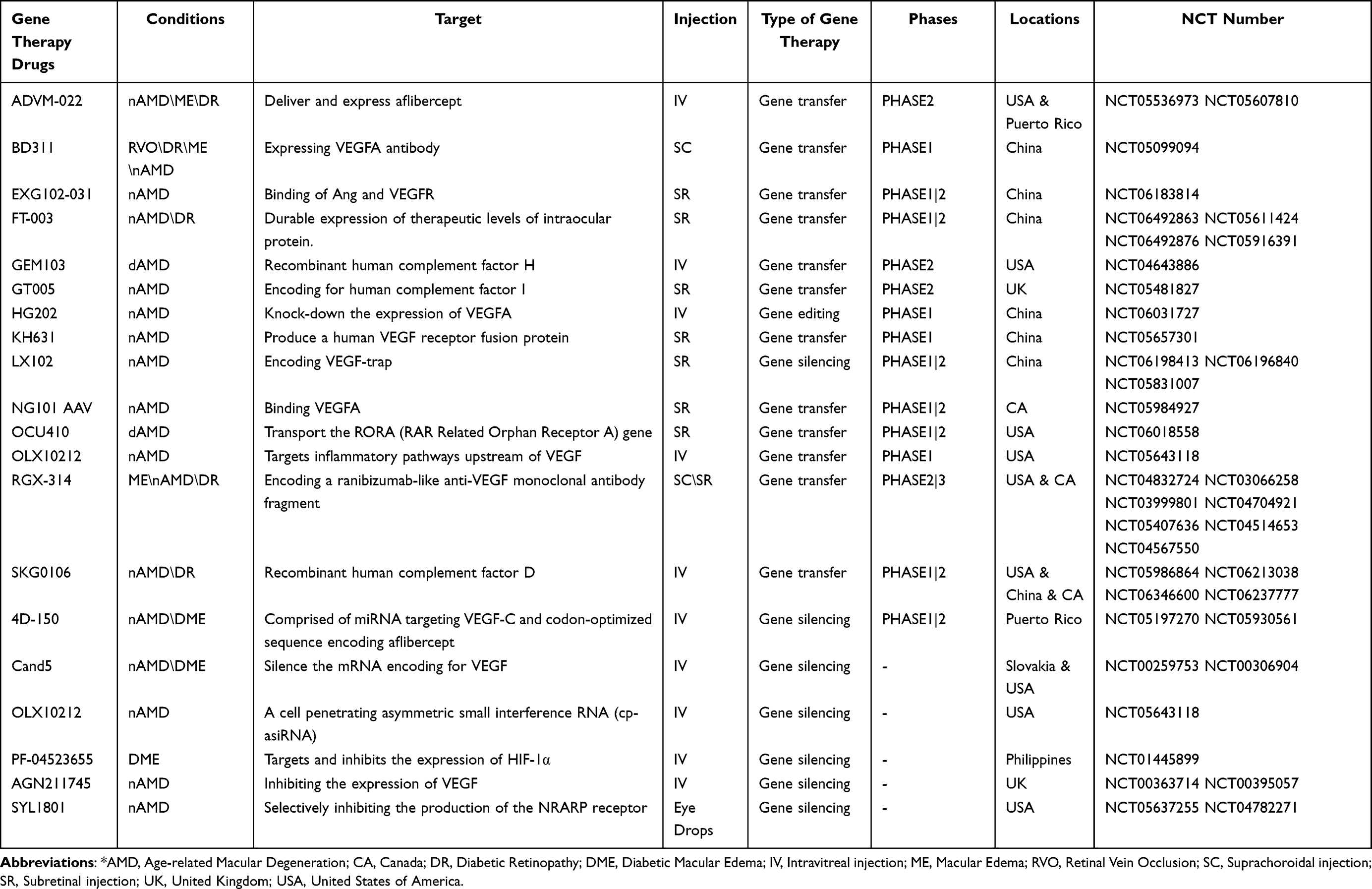 |
Table 1 Ongoing or Completed RNVs Gene Therapy Clinical Trials |
Therefore, we review the advancements in nanomaterial-based anti-angiogenic gene therapy for RNVs, summarizing current therapeutic targets and commonly used nanomaterials to support the future development of potential single-administration gene therapies for RNVs.
The Mechanism and Targets of RNVs Treatment
Regulation of VEGF and Anti-Angiogenic Therapy
Pathological neovascularization is the core pathological feature of RNV, primarily mediated by abnormal VEGF and its receptors.22 Elevated levels of VEGF have been found people with RNV, such as individuals diagnosed with AMD, PDR, or RVO.23–25 Overexpression of VEGF increases the phosphorylation of tight junction (TJ) proteins between endothelial cells, leading to the release and degradation of the key protein from the cell membrane, which compromises the integrity of the BRB, significantly increasing the permeability of the endothelial cell layer, allowing plasma components and inflammatory mediators to enter the retinal tissue more easily, causing macular edema and more severe vision loss.26,27 On the other hand, VEGF binding to VEGFR activates signaling pathways within target cells, leading to migration and proliferation of retinal endothelial cells into the vitreous, with the formation of fibrous vascular membranes further damaging the hypoxic retina28(Figure 1). These changes exacerbate the hypoxic condition in AMD, leading to severe complications such as blindness, intraocular inflammation, or glaucoma. Additionally, in addition, fragile neovascularization can be easily destroyed, especially in diabetic patients under hyperglycemic conditions. Elevated glucose levels not only mediate the overexpression of VEGF through IGF-1, but also causes oxidative stress and increased inflammatory factors in retinal cells. Under the dual pressure of ischemia and hypoxia, neovascularization occurs pathological leakage or hemorrhage, which leads to retinal detachment and severe visual loss.29
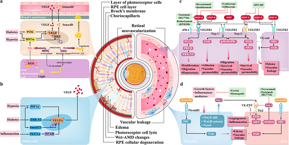 |
Figure 1 The pathogenesis and target of retinal neovascularization. (a) Light is an exogenous stressor, raising ROS in retinal cells. Visible light mimics RPE cell phagocytosis, inducing superoxide. This stabilizes HIF-1, promoting VEGF transcription. Imbalance of light and photosensitive molecules can cause RPE cell degeneration. (b) Hypoxic-ischemic HIF-1 isoforms up-regulate VEGF. In insulin - treated diabetics, IGF-1 induces VEGF via ERK1/2 in Muller and RPE cells. (c) In retinal vascular endothelial cells, VEGF family activates PLCγ - PKC pathway, regulating cell functions. PI3K and p38 MAPK also play roles. VEGF stimulation leads to permeability changes and abnormal vascular growth. (d) Ang-2, up-regulated in pathologies, inhibits Tie2 activation by Ang-1, disrupting vascular stability. Sema4D/PlexinB1 signaling causes endothelial and pericyte dysfunction in DR. |
Anti-angiogenic therapy is the core strategy for treating RNV, primarily targeting VEGF and its receptors in the retina. The primary sources of VEGF are the retinal pigment epithelium (RPE) and Müller cells, with endothelial cells serving as both target cells and contributors to VEGF secretion.30,31 Anti-VEGF therapies include bevacizumab, ranibizumab, aflibercept, among others, which specifically bind to VEGF-A and other members, preventing their interaction with receptors and thereby inhibiting angiogenesis.32 The multi-targeted tyrosine kinase inhibitor sorafenib further blocks VEGF signaling by inhibiting VEGFR activity.33
Other Potential Targets for Anti-Angiogenesis
In recent years, more and more anti-angiogenic targets have been developed for RNV diseases, which can be divided into “direct inhibition of angiogenesis” and “regulation of vascular microenvironment” according to the mechanism of action. The former includes the Angiopoietin/Tie2 pathway, PDGF-BB pathway, integrin, and Semaphorin 4D (Sema4D), which are thought to be directly involved in key steps of neovascularization. Ang-2 disrupts vascular stability by antagonizing Tie2 receptor activation and collaborates with VEGF to aggravate the leakage of DR And AMD.34 Faricimab, an intravitreal VEGF-A/Ang-2 dual antibody being developed by Roche/Genentech, was tested in phase III trials (NCT03622580 and NCT03622593) has been shown to prolong the treatment interval for patients with diabetic macular edema (DME) and wet AMD.35 In addition, Tie2 activator AKB-9778 significantly enhanced vascular stability and eliminated macular edema caused by RNV disease in early clinical trials.36 PDGF-BB pathway promotes the maturation of neovascularization through the recruitment of perivascular cells, which is related to anti-VEGF resistance. Although the phase IIb trial of anti-PDGF-BB drug Fovista combined with VEGF inhibitors is not as expected, its mechanism provides the basis for a multi-target strategy.37,38 The integrin antagonist Risuteganib improved visual acuity and inhibited pathological neovascularization in patients with dry AMD and DME in Phase II trials, but more data are needed before the benefit of using risuteganib in the clinic can be truly assessed.39 Our previous studies have shown that the nanonet developed based on Sema4D/PlexinB1 pathway can effectively trap sSema4D to reduce angiogenesis in DR, which is being developed for other RNV diseases.40
On the other hand, more targets have been developed to regulate the vascular microenvironment to inhibit angiogenesis in RNV disease, including hypoxia-inducible factor (HIF), complement system, inflammatory mediators, and fibrosis factors. Their application has received much attention in the gene regulation of VEGF.
Gene Regulation of VEGFA
The expression of the VEGFA gene is regulated at both the transcriptional and translational levels by multiple factors, with hypoxia being one of the most well-known regulators.41 The hypoxia-dependent regulation of VEGF is primarily mediated by the hypoxia-inducible factor (HIF) transcription factor family, particularly HIF-1.42,43 HIF-1 consists of HIF-1α and HIF-1β, and under hypoxic conditions, HIF-1α is not degraded but binds to the hypoxia response element (HRE), activating the transcription of target genes like VEGF.44 Additionally, different subtypes of HIF can regulate VEGF expression, making them potential therapeutic targets for patients with RNVs.45,46
In addition to hypoxia, the complement system also plays a critical role in the pathogenesis of AMD. Mutations in complement factor C3 and complement factor H (CFH) genes increase the risk of AMD [56], while complement terminal component C5a in the vitreous promotes the development of CNV by upregulating VEGF in the RPE.47,48
Moreover, NF-κB, as a key transcription factor, also participates in the transcriptional regulation of VEGFA, although clear κB binding sites have not been identified in the VEGF promoter.49 Recently, NF-κB-based anti-angiogenic strategies have been developed for gene therapy targeting DR.50 Additionally, inflammatory cytokines, such as IL-1α, IL-1β, IL-6, and IL-8, have been found to induce VEGFA expression in various cell types51,52 (Figure 1). Anti-inflammatory treatments, such as triamcinolone, can slow the progression of RNV, and melatonin, as an anti-inflammatory and antioxidant treatment, has also shown potential in inhibiting VEGF expression.53–55 In addition, connective tissue growth factor (CTGF) has been paid attention to the treatment of retinal fibrosis and other complications after anti-VEGF therapy in recent years.56 The dual-target protocol of CTGF and VEGF-A developed by Hu et al in the previous study can better improve the damage of retinal microvascular ultrastructure.56 Recently, Farzad et al are trying to construct umbelliprenin-containing niosome nanoparticles to inhibit the gene expression of VEGF-A and CTGF in retinal pigment epithelial cells.57 These anti-VEGF gene therapy strategies still need to be validated through clinical trials for their efficacy and safety, with optimization of drug delivery methods being crucial.
The Progress of Gene Therapy in Anti-Angiogenesis
Intravitreal injection is the standard method for delivering anti-angiogenic drugs, but repeated long-term injections cause complications while also imposing a significant financial burden.11 With the development of gene therapy technology in recent years, these issues seem to have found a “standard answer”.
Gene therapy is described as the ability to improve genetic alterations by inserting, removing, or correcting any mutated genes. Since the FDA’s first approval of a gene therapy drug for the treatment of hereditary retinal diseases related to biallelic RPE65 mutations, the application of gene therapy in ocular diseases has regained attention, especially in pathological neovascularization.58 Firstly, the eye is considered an ideal target for gene therapy for several reasons: (1) the presence of the blood-retinal barrier provides the eye with a certain level of immune privilege;59 (2) the highly compartmentalized eye tissues offer an advantage for targeted pathways;60 (3) the transparent ocular medium allows easy observation of pathological changes post-treatment;61 (4) visual function and retinal structure can be monitored non-invasively at any time;62 (5) In the same subject, one eye was designated as the experimental target, while the other eye served as the control.63
Secondly, unlike most hereditary retinal diseases, although retinal neovascularization involves a complex network of various pro-angiogenic and anti-angiogenic factors, these diseases are often associated with VEGF-A related gene mutations, making RNVs particularly responsive to anti-angiogensic gene therapy.64 Gene therapy includes three methods: gene transfer to increase target cell function by introducing exogenous genes (gene replacement), gene silencing to inhibit target gene expression by suppressing mRNA in target cells, and gene editing to directly modify target genes (Figure 2).
 |
Figure 2 The barrier for retinal gene therapy. (a) For gene therapy in the posterior segment of the eye, the four routes of administration need to overcome intraocular barriers. Eye drops need to traverse most intraocular structures. Intravitreal injection, being closest to the retina, is widely used. Suprachoroidal injection and subretinal injection are at a relatively greater distance from the retina but can exert a sustained - release effect. (b) After the surface groups of the gene vector recognize the receptors on the surface of the target cells and localize to the target cells, the gene vector needs to overcome three major barriers: the cell membrane, the cytoplasm, and the cell nucleus to complete gene delivery. |
Gene Transfer
Gene replacement or transfer involves the insertion of a corrected copy of the gene into a target cell to induce synthesis of the desired protein. This is the most common form of gene therapy (often used interchangeably with gene therapy). One of the key factors for this process’s success is identifying appropriate targets. Anti-angiogenic gene targets are also abundant based on the multipath network of VEGF-mediated angiogenic signaling pathways. Established targets of anti-vascular gene therapy include PEDF, sFLT-1, and anti-VEGF antibodies. Injection of AAV5 vector carrying PEDF significantly inhibited neovascularization in the nAMD mouse model. Although mild inflammation occurs in a quarter of patients, this gene therapy product is safe and well tolerated compared with long-term injections.58 sFLT-1, a VEGF inhibitor, has been shown to effectively inhibit pathological angiogenesis in RNVs and is one of the earliest anti-VEGF gene therapy strategies. Two clinical trials involving sFLT-1 gene therapy (rAAV.sFLT-1) have completed phase II controlled trials, and the results show that the treatment group effectively prolongs the duration of drug efficacy and reduces the number of injections compared with the control group.65–67 In addition, significant progress has been made in the genetic engineering of proven anti-VEGF agents, such as Ranibizumab, Bevacizumab, and Aflibercept (Table 1). Other drug targets for pathological retinal neovascularization have also progressed toward gene therapy, such as angiostatin and endostatin, platelet-derived growth factor, and Tie-2 tyrosine kinase receptor.68 These gene targets expand drug diversity, addressing the selective nature of gene therapy across different populations.
Gene Silencing
Gene silencing refers to the inhibition of gene expression-related functional proteins by silencing mRNA in the cytoplasm, typically using non-coding RNAs (ncRNAs) that bind to mRNA to interfere with protein synthesis, also known as RNA interference (RNAi) technology. In the retina, ncRNAs have been found to not only influence angiogenesis by silencing mRNA but also to regulate DNA methylation or histone methylation and other epigenetic modifications, thereby influencing gene transcription and the progression of diabetic vascular complications.69,70 NcRNAs primarily include microRNAs (miRNAs), small interfering RNAs (siRNAs), long non-coding RNAs (lncRNAs), short hairpin RNAs (shRNAs), and antisense oligonucleotides (ASOs). Gene therapy by siRNA in the eye has been described in detail by Kavita et al and mainly involves the delivery of anti-VEGF siRNA into target cells by adenoviruses, liposomes, and polymers. And quite a few have entered the early stage of clinical trials (Table 1).71,72 lncRNA is another kind of non-coding RNA with great therapeutic potential. It can achieve post-transcriptional gene regulation by competitive binding with miRNA in the treatment of eye diseases, also known as “miRNA sponge”. Studies have shown that the expression of MALAT1 (a long non-coding RNA) is increased in DR Patients, which may activate retinal damage caused by oxidative stress.73 In addition, injection of miR-124-3p and shMALAT1 reduced Egr1 expression and inhibited retinal neovascularization, highlighting the MALAT1/miR-124-3p/Egr1 axis as a potential target for ROP gene therapy. The shRNA was artificial stem loop RNA, and the siRNA was generated by Dicer and then accurately degraded the target mRNA. Its complete complementary pairing is more specific than the partial matching of miRNA, and its stability is better than that of the siRNA that needs repeated transfection.74 The construction of shRNA against mTOR and HIF-1α has received good anti-vascular effect in vivo and in vitro experiments, but further clinical experiments are needed to verify.
Gene Editing
Gene editing refers to the technology of artificially intervening to alter the DNA sequence in an organism’s genome to modify, insert, or delete target genes. Since Jennifer Doudna and Emmanuelle Charpentier used CRISPR-Cas9 as an editing tool in 2012, it has become the most used technology in gene editing due to its strong advantages.75 Compared to ZFNs and TALENs, CRISPR-Cas9 has higher editing efficiency and lower off-target effects, and it is also easier to operate.76
Currently, CRISPR-Cas9 gene editing strategies for ocular angiogenesis primarily target VEGF-A, VEGF-R, and HIF-1α. Early CRISPR genome editing used AAV9 to package smaller CRISPR nucleases, such as Cas9 and their respective gRNAs, targeting HIF-1α or VEGF-A, finding similar neovascularization inhibition effects compared to aflibercept, but gene editing likely offers a permanent cure.77,78 Additionally, rAAV1-packaged spCas9 targeting VEGFR2 in endothelial cells successfully inhibited neovascularization in a mouse OIR model.64,79,80 However, viral delivery vectors such as AAV face the problems of immunogenicity and low loading capacity. Through the strategy of delivering Cas9 ribonucleoprotein (RNP) complex directly instead of viral vector DNA, gene editing technology can not only circumvent the host immune recognition of the viral capsid, but also avoid long-term immune stimulation due to its transient expression characteristics. At the same time, the non-viral delivery system breaks through the physical limitations of the viral capsid and significantly improves the payload capacity of gene vectors.81,82. Therefore, Sahel et al recently developed a cRGD-modified lipid polymer nanocomposite loaded with Cas9 RNP and found to have lower toxicity and faster transfection efficiency.83 Nanoformulations are expected to offer more benefits than current approaches. Many nanometry-based products have been approved by the FDA or are under investigation (Table 2).
 |
Table 2 Ongoing or Completed Clinical Trials of RNVs Nanomaterials |
Retinal Barrier and Route of Delivery for Gene Therapy
The key to the success of gene therapy lies in how gene therapy drugs are delivered to target cells and how to improve delivery efficiency. Due to the eye’s unique position, local administration methods for retinal diseases have more options, including non-invasive (eye drops) and invasive methods (intravitreal injection, subretinal injection, suprachoroidal injection)(Figure 2). Although eye drops are the most common modality in eye therapy, no gene therapy trials appear to employ this route (Table 1). Notably, in recent years, researchers have been actively developing nanocomposite materials to overcome the limitations of eye drops in delivering drugs to the posterior segment of the eye.84 Compared with the glass body cavity injection, even if the drug concentration in eye drops is twice that of intravitreal injections, the therapeutic effect is still lower and may increase the patient’s burden.85
Administration Routes for Retinal Gene Therapy
The preferred method for targeting the posterior segment of the eye in gene therapy remains invasive administration, particularly intravitreal injection. Through this route, therapeutic agents rapidly diffuse throughout the vitreous body, reaching the retina quickly. After overcoming barriers such as the inner limiting membrane, nerve fiber layer, and blood-retinal barrier, these agents act on endothelial cells and RPE cells86(Figure 2). However, this method often requires frequent injections and carries risks of endophthalmitis, increased intraocular pressure, retinal detachment, and cataracts.8–10 Subretinal injection is an effective method for directly delivering drugs to photoreceptor membranes and RPE cells. Despite the complexity of the surgical procedure, which requires specialized equipment and techniques, the significant potential benefits of gene therapy make subretinal injection a rational choice for treating retinal degenerative diseases.87 However, clinicians must carefully weigh the pros and cons due to potential complications, such as temporary focal retinal detachment, and the limited range of therapeutic agent action due to the confined space.88 Suprachoroidal injection (SC) is an emerging ocular drug delivery method that involves injecting drugs into the anatomical space between the choroid and sclera using specialized microneedles. Recent studies show that SC can achieve extensive expression of AAV transgene drugs in the outer retinal cells, with higher vector coverage, though mainly limited to the peripheral retina.89 However, SC also relies on advanced technology and equipment, like microneedle injection systems, for safe and effective suprachoroidal drug delivery. And its safety and efficacy still lack validation from large-scale clinical trials.90
Extracellular Barrier for Retinal Gene Therapy
Before reaching the target cells, gene therapy drugs for the posterior segment of the eye must not only overcome natural anatomical structures but also pass through the immune system’s examination. Although it is difficult for living cells of immune substances in the blood to enter the eye due to the blood-eye barrier, unique mechanisms of immune regulation still exist in various anatomical structures. In the ocular surface, tear derived secretory IgA complexes combine with TGF-β to mediate the inhibition of dendritic cell maturation to enhance immune quiescence. In the anterior chamber, aqueous humor has immunomodulatory function. The specific elevated soluble Fas ligand (sFasL) induces the expression of Foxp3 in activated regulatory T cells, which helps to clear pathogens. The vascular-rich iris/ciliary contact-dependent PD-L1/CTLA-2α signaling inhibits T cell activation. The vitreous body, as the clearest gelatinous substance filled in the eye, is usually not rich in immune cells, but it secrets CCL5 to recruit CXCR3-T cells during disease inflammation. The retina and choroid are rich in microvessels and contain small numbers of Muller cells and dendritic cells. These immune mechanisms create multiple layers of defense against the delivery vector, presenting both challenges and opportunities for gene therapy delivery.91 When target cells are localized in endothelial cells, the extracellular barrier also needs to overcome BRB, which limits nanoparticle delivery through size exclusion and electrostatic repulsion by TJ proteins. Alternatively, pathological BRB leakage enhanced passive penetration but impaired targeting specificity. Heterogeneous receptor-mediated dynamic barrier integrity across diseases, such as AMD versus DR, further challenges standardized nanocophore design, requiring customized ligand functionalization or stimulus-response systems.92,93
Intracellular Barrier for Retinal Gene Therapy
After gene therapy drugs enter the target cells, they still face challenges from intracellular clearance mechanisms. These intracellular barriers include cellular uptake and endocytosis, lysosomal degradation, nuclear membrane barriers, and cellular immune responses. More important for gene delivery is how to ensure that nucleic acids remain intact during lysosomal degradation. In acidic environments such as endosomes or lysosomes, even for lipid nanoparticles widely used in clinical practice, the cell escape efficiency of siRNA was reported to be less than 5%.94 How to cleverly design the carrier to improve the efficiency of endosomal escape is one of the keys to solve this problem. Ph-responsive nanomaterials can be designed to be protonated and positively charged. They can promote endosomal escape, mainly through the proton sponge effect.95 Our previous studies are trying to use it in the treatment of angiogenesis96,97 (Figure 2). Understanding intracellular barriers effectively improve delivery efficiency, and often, leveraging these characteristics to construct delivery materials serve gene therapy. Therefore, nanomaterials are being widely used for targeted delivery of gene therapy due to their good plasticity and stability.98
The Advances of Nanomaterials in Gene Therapy
Gene therapy advancements have brought immense hope for the treatment of ocular diseases. Effective retinal gene therapy has always relied on the development of suitable delivery systems. Viral vectors are the most commonly used gene vectors for ocular disease treatment.99 However, using viral vectors for in vivo gene expression still faces limitations such as low payload, complex customization, and risks associated with viral antigens (such as immune responses).15–17 Given these limitations, advancements in non-viral delivery methods, such as nanoparticles, are essential.
Nanomaterials are materials with at least one dimension (such as length, width, or thickness) within the range of 1 to 100 nanometers (nm). This small size endows nanomaterials with unique properties, including a high surface area to volume ratio, quantum effects, and tunable chemical properties. For gene delivery to the posterior segment of the eye, these characteristics of nanomaterials offer an almost ideal solution, overcoming the limitations of viral delivery systems constrained by the eye’s natural barriers and limited drug load. Typically, surface modification of nanomaterials is used to enhance targeting specificity, such as introducing near-infrared (NIR) light-active fluorophores (indocyanine green) and conjugating drugs like Avastin to nano-capsules, paired with micro-laser systems for effective in vitro targeted anti-VEGF drug delivery.100 Additionally, many nanomaterials inherently possess anti-angiogenic properties, making them ideal vectors for treating retinal neovascular diseases, including Implantable anti-angiogenic stents, which enhance the slow release of peptides from nanofibers, enabling faster anti-angiogenic or anti-inflammatory effects to synergistically combat retinal neovascularization and edema.21 According to the composition of nanomaterials, they can be divided into inorganic and organic nanomaterials101(Figure 3).
 |
Figure 3 Structure and characteristics of nano-delivery vectors. |
Inorganic Nanoparticles Drug Delivery for Gene Therapy
Inorganic nanoparticles, typically derived from metals, metal oxides, and non-metallic materials like carbon and silica, offer numerous advantages as drug carriers, including increased quantum yield and drug-loading capacity.102 Compared to organic nanoparticles, inorganic nanoparticles possess unique properties such as photosensitivity, electrical conductivity, magnetic attraction, and thermal efficiency, making them valuable not only as drug carriers but also as therapeutic agents.103,104
Metal Nano Delivery Materials
Gold nanoparticles(AuNPs), with stability, malleability and metal properties, are widely used in nanobiotechnology.105 Especially in ocular diseases, AuNPs photothermal therapy based on NIR has been shown to be effective in treating intraocular inflammation and tumor angiogenesis.101 Studies focus on both the intrinsic anti-angiogenic properties of gold nanoparticles and their advantages as carriers for delivering anti-angiogenic drugs.106 Due to their chemical inertness and biocompatibility, gold nanoparticles effectively evade clearance by the intraocular immune system, allowing them to reach retinal target cells.107 Previous studies indicate that AuNPs enhanced their cellular uptake success rate by altering their nanoparticle size, shape, and surface charge. Among gold nanoparticles ranging from 10 to 100 nm, 50 nm AuNPs were found to be more efficiently taken up by cells.108,109 Additionally, the shape of gold nanoparticles determines different endocytosis pathways, with triangular AuNPs showing the highest cellular uptake rate. Gold nanostructures exhibit anti-angiogenic effects and superior biodegradability, electrical properties, and molecular recognition characteristics.110 Hollow gold nanoshells inhibit VEGF-165 gene expression to suppress Human Aortic Endothelial Cells (HAEC) proliferation, thus inhibiting angiogenesis. Gold nanoparticles coated with different functional peptides activate or inhibit angiogenesis in vitro, but they cannot prevent HAEC tube formation.111
Silver nanoparticles(AgNPs) have also gained attention in ocular anti-angiogenesis applications in recent years. As the concentration of AgNPs gradually increased, the concentration gradient effect of AgNPs on the inhibition of VEGF gradually increased, mainly by inhibiting the gene expression of HIF-1, the upstream regulator of VEGF. And the mRNA expression levels of VEGFR-2, protein kinase Cγ(PKCγ), Rac encoding genes, inhibit HUVEC tube formation, and also affect optic nerve development.112,113 Although metallic nanomaterials have been widely used in the diagnosis and treatment of RNV diseases, there are still considerable challenges. Most metals catalyze the production of ROS in the eye through Fenton-like reactions, leading to retinal lipid peroxidation. Coating the surface with a silica shell reduced oxidative damage by 70%, but the shell defects still triggered local hot-spot effects.104,114,115
Oxide Nanomaterials
Metal oxide nanomaterials have shown great prospects in cancer treatment. In recent years, it has received extensive attention in the treatment of retinal neovascular diseases. Common ones include titanium, platinum, zinc, and cerium oxides. Although in previous studies, metal titanium and its oxide have good antibacterial and pro-angiogenic effects and are widely used in orthopedics.116 However, studies have found that local injection of nano-titanium dioxide is non-toxic to oxygen-induced retinopathy mice in vitro and inhibits angiogenesis. Although the mechanism is unclear, its inhibition of VEGFR-2 phosphorylation suggests that its effect may be similar to that of gold nanoparticles, leading to VEGFR-2 inactivation.117 In addition, copper oxide nanoparticles reduced angiogenesis in transgenic zebrafish by downregulating the expression of VEGF and inducing apoptosis.118 However, compared with organic and metallic nano-delivery materials, oxide nanomaterials are more likely to act as therapeutic agents against oxidative stress.119,120 In the light-induced retinal degeneration model, Pt nanoparticles showed strong antioxidant capacity, with 42% reduction of ROS compared with the control group, which effectively protected the retina from oxidative stress damage and was an ideal nanoenzyme for the treatment of oxidative stress-related retinal diseases.121 In addition, CeO2 has been shown to effectively scavenge iron-induced ROS, improve retinal cell survival, and ameliorate retinal damage observed in AMD or other degenerative diseases.122 In addition to the significant anti-oxidative stress ability, nano-zno nanomaterials were found to effectively reduce blood glucose levels in DR rat models and have excellent anti-inflammatory effects123 them. Although iron oxide nanoparticles (Fe3O4) have the advantage of magnetic targeting, when the release rate of iron ion in the vitreous is too fast, it exceeds the ferritin storage capacity of RPE cells and causes ferroptosis. Therefore, although there are many options for metal oxide nanomaterials, to become a good delivery material or anti-vascular material, researchers need to find the right balance.124
Carbon-Silicon Nanomaterials
Unlike metal and other inorganic nanomaterials, the application of nanoparticles formed by nonmetallic oxides in anti-angiogenesis gene therapy has been practically proven. SiNPs significantly reduce the mRNA expression of macrophage activity-related genes MIF and angiogenesis-related genes VEGFR2, mainly by downregulating the MAPK signaling pathway.125 Additionally, since most anti-angiogenesis drugs under development are hydrophobic, using inorganic nanomaterials as carriers significantly enhances drug biocompatibility and hydrophilicity, greatly improving bioavailability and drug duration. For instance, a recent study designed and prepared a graphene oxide (GO)-based carrier for delivering VEGF-siRNA. Due to its good water dispersibility and surface area, it rapidly reaches target tissues and effectively inhibits angiogenesis. Other studies show that surface modification of GO also significantly increases the success rate of cellular transfection.126 Similarly, silicate nanocarriers, relying on their hydrophilicity, enhance the bioavailability of anti-angiogenic drugs and exhibit very low toxicity to cells.127 More interestingly, Mesoporous carbon/silica Nanoparticle has been widely studied for gene delivery in recent years. Because of its controlled nonporous structure, it may be used as a drug delivery carrier. This can be doped with various therapeutic ions (strontium, copper, magnesium, zinc, lithium, silver, etc.) and loaded with specific biomolecules (for example, therapeutic drugs, antibiotics, growth factors) to achieve controlled loading and release kinetics.128,129
Organic Nanoparticles Drug Delivery for Gene Therapy
Polymers
Polymers are important macromolecular compounds consisting of multiple repeating units (monomers) linked by covalent bonds. They are classified into naturally occurring and synthetically prepared types. Natural polymers, such as cyclodextrin, chitosan, hyaluronic acid, and fibrin, are widely present in biological organisms and have good biocompatibility and biodegradability.130 In contrast, synthetic polymers, such as polyethyleneimine (PEI) and polypropyleneimine (PPI), are obtained through artificial synthesis and offer a broader range of tunable physicochemical properties.131 The good controllability of polymers allows for customization based on target cells, providing a foundation for gene therapy targeting various cells in RNVs. The composition of polymers determines their targeting specificity. In RNVs, endothelial and RPE cells are the primary target cells. Shmueli et al synthesized a new polymer PBAEs for gene delivery to endothelial cells and found its transfection rate in HREC reached 85%.132 These polymer nanoparticles can be used as experimental tools in vitro or for targeting and treating vascular-specific diseases in vivo. Another study compared the diffusion abilities of seven different polymer self-assemblies in the retina, finding that nanoparticles could overcome biological barriers in the vitreous and deep retinal structures, possibly related to interactions with specific cell types like Müller cells.133
These excellent targeting properties provide a basis for developing gene delivery systems for treating RNVs. Liu et al used polymers to load siRNA targeting NF-kB for gene therapy of DR, showing that in 25% fetal bovine serum, DMAPA-Glyp/siRNA and DMAPA-Amp/siRNA nanoparticles could protect siRNA from nuclease degradation.50
The size of polymers significantly affects their impact on angiogenesis. Previous studies have shown that short-fiber polymers sNAG (40–60 nm) promote angiogenesis and wound healing in diabetic mice.134 Another study used modified ionic gelation to prepare chitosan-hyaluronic acid nanocomposites (186±4.3 nm) loaded with siRNA for retinal delivery, showing that intravitreal injection of VEGFR-2 siRNA nanocomposites significantly reduced laser-induced choroidal neovascularization size compared to the control group.135
The surface charge of polymers is closely related to their biotoxicity. Recently, Zhao et al developed a cationic polymer SSL-AG, poly(disulfide-l-lysine)-g-agmatine, and evaluated its ability to transfer plasmid (pZNF580) into HUVECs, showing excellent buffering capacity and rapid release ability, with an average cellular uptake 3.6 times higher than the control group, indicating good biocompatibility.136 Therefore, the polymers provide significant potential for retinal gene therapy, deserving further exploration in treating RNVs. Notably, the results of a recent study exploring the long-term effects of polystyrene nanoparticles on the retina showed that oxidative stress and inflammation mediated adverse effects of polystyrene nanoparticles on retinal function. Future attention should be paid to the long-term effects of other polymers on the retina to increase safety.137
Dendrimers
Dendrimers are highly branched, three-dimensional spherical molecules composed of a central core and multiple repeating layers of branched units (generations). Compared to linear polymers, the main advantage of dendrimers lies in their spherical structure, which affects their size and lipophilicity, making them ideal drug delivery systems due to their ability to penetrate cell walls easily.138 Like polymers, the branched structure of dendrimers allows for the presence of numerous surface groups, which be considered active sites, providing templates for drug immobilization and grafting. You, S. al. developed seven types of surface-functionalized water-soluble dendritic nanocarriers to compare their physicochemical effects on bioavailability, distribution, and targeting efficiency in the retina. DNSG accumulated in the photoreceptor layer and RPE, while DPEA showed higher membrane permeability and faster retinal penetration, indicating its potential for effective intravitreal use.139 Wathsala et al developed PAMAM-dendrimer-siRNA conjugates that can effectively cross the blood-brain barrier and seem to have potential as retinal gene delivery.140
Dendritic molecules have good biocompatibility and low cytotoxicity. Studies show that second-generation polypropylenimine (PPI) dendrimers effectively bind to DNA, exhibit low cytotoxicity, and optimize in vitro gene transfer activity.141 Dendrimers have good targeting properties. A drug based on hydroxyl-terminated poly(amidoamine) (PAMAM) dendrimers, this nanocarrier achieves ligand-free cell targeting through systemic administration and is undergoing clinical studies for the treatment of wet age-related macular degeneration (wet AMD).142
Lipid Nanoparticle
Lipid nanoparticle (LNP)-based deli very systems have become one of the most advanced non-viral delivery technologies in clinical settings. Actually, COVID-19 vaccines are LNP-based mRNA delivered drugs. LNP inherit almost all the biocompatibility properties of lipids, which gives it significant advantages as gene therapy vectors, including minimal toxicity and immunogenicity, easy degradation and metabolism, large loading capacity, good cellular permeability, and controllability.143 LNPs mainly include solid lipid nanoparticles (SLNPs) and lipid complexes (liposome-polycation-DNA complexes).
SLNPs are composed of solid lipids with bilayer structures. The lipid bilayer firmly binds most hydrophobic drugs, and the solid-state characteristics help extend the drug’s sustained release effect, prolonging its duration of action. Therefore, SLNPs are often used to deliver hydrophobic drugs.144 Compared to SLNPs, cationic liposomes utilize the properties of electrostatic interactions and are widely used to introduce nucleic acids into cells both in vitro and in vivo, as nucleic acids carry a negative charge at normal physiological pH. The formation of cationic lipid-nucleic acid complexes promotes binding to the cell membrane and allows the complex to enter cells via endocytosis.145,146 Additionally, cationic liposomes interacting with a high proportion of dendritic cells enhanced intracellular localization and improved targeting efficiency.147 Therefore, cationic lipid nanoparticles have unique advantages for delivering gene therapy substances, including siRNA, mRNA, and DNA plasmids. Currently, gene therapy based on cationic lipid nanoparticles has been widely used in treating RNV.148 Recent studies have revealed a breakthrough application of LNPs in the treatment of eye diseases. For mRNA delivery, intravitreal injection of LNP can target mouse and human retinal Muller glia and RPE without causing significant inflammation.149,150 The novel prefabricated vesicle method (PFV-LNP) improved retinal transfection efficiency by 50% through the surface mRNA localization strategy, and its safety was verified in a non-human primate model. In the siRNA delivery domain, bioionizing LNP (2N12H), which enables efficient delivery of siRNA through a degradable lipid design, inhibits retinal neovascularization as effectively as ranibizumab.151 These explorations bring limitless possibilities for gene therapy of RNV.
However, LNPs in gene therapy still face limitations, including poor stability, potentially due to changes in serum protein charges under pathological conditions, leading to reduced delivery efficiency and potential biotoxicity.152 Despite widespread clinical trials of LNP-based therapies in the central nervous system, their validation in RNVs appears lacking153 (Table 3). Recent studies have further explored the potential of LNP surface hybridization with other materials, such as natural polymer groups, to solve the current dilemma.154
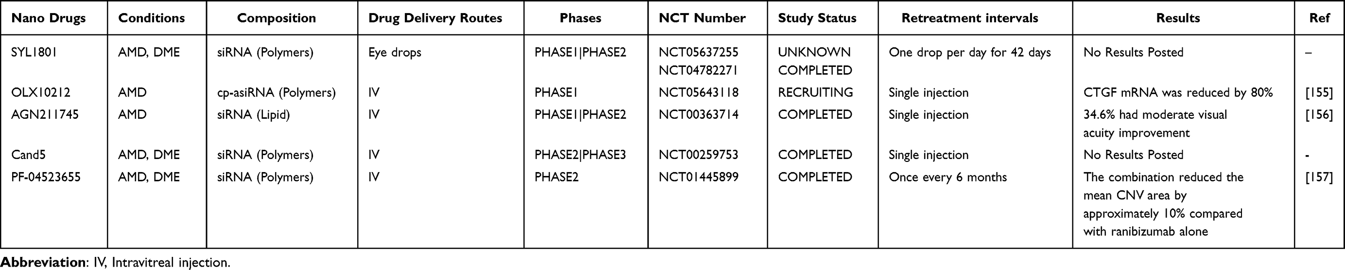 |
Table 3 Nanomaterial-Based Gene Therapy for RNVs |
Nanocomposites Drug Delivery for Gene Therapy
In recent years, in order to overcome the limitations of single nanomaterials, researchers have begun to explore the role of composite nanomaterials in gene drug delivery. Nanocomposites (nanohybrids) refer to material systems composed of two or more different materials (usually including nanoscale components).84
Nanocomposites can effectively combine the advantages of a variety of single materials. For example, inorganic nanomaterials are often poorly targeted, but the delivery efficiency can be improved by recombination with polymers with good targeting properties. Song et al constructed a pegylated gold nanorod (PEGF-GNRs) that specifically blocks the TGF-β pathway in endothelial cells and inhibits actin assembly at contractile rings, thereby preventing angiogenesis in the OIR model.158 In addition, the adsorption of serum proteins on the surface of liposomes was prevented by the surface modification of lipid nanoparticles with polyethylene glycol, thereby triggering the rapid uptake of LNPs by the macrophages-phagocytes system.159 Gautam et al used fluorescently labeled PEG-LNPs carrying mRNA to transfect mouse retinal cells and found the highest expression in RPE cells when the PEG content in nanoparticles was 0.5%.160 Sahel et al found that cRGD-modified lipopolymer nanocomposites were less toxic and more efficient in delivering the CRISPR/Cas9 RNP payload to retinal tissue.83
Nanocomposites further improve the current situation of gene therapy delivery in the posterior segment of the eye. Although gene therapy can effectively avoid repeated injection problems of invasive drug administration such as intravitreal injection, invasive drug administration is still limited by technology and equipment. Therefore, the development of local drug administration for gene therapy is still of great significance for the popularization of RNVs.161 Lu et al constructed cyclodextrin-liposome nanocomposite delivery materials to overcome the ocular barrier encountered by topical eye drops and found that more than 20% of the nanocomposite could reach the choroid-retina after 60 min in vivo.162 Additionally, Wang used the organic-inorganic hybrid nanocomposite drug delivery system to successfully deliver loaded drugs to the retina and maintain a high concentration for more than 3 hours.163
Safety and Biocompatibility of Nanomaterials in Retinal Gene Therapy
Although nanomaterials offer transformative potential for retinal gene therapy, their clinical translation depends on rigorous evaluation of safety and biocompatibility, which requires critical considerations of immune responses, retinal toxicity, and systemic clearance mechanisms. As mentioned above, reasonable design of nanomaterials can achieve anti-inflammatory effects, such as NF-κB and CTGF, to counteract the immune response in the retina. However, a balance is needed to achieve safety, as excessive immunosuppression may increase the risk of intraocular infection and reduce targeting.91,164 Similarly, there is a delicate balance between retinal toxicity and long-term efficacy of nanomaterials. AgNPs triggered endothelial damage through the Fenton reaction as previously mentioned, while silica or polymer coatings inhibited ROS generation.112,114 Notably, although PPI/PEI has reduced acute toxicity, retinal biocompatibility under chronic exposure still needs to be validated.141 In addition, mesoporous silica can induce chronic inflammation and photoreceptor dysfunction through long-term accumulation in retinal cells even at subtoxic doses.137 Effective clearance can minimize the off-target effects of nanomaterials, but this also requires full consideration of long-term effectiveness.165,166 The clearance rate comparison of different sizes of nanomaterials in animal models is shown (Table 4).
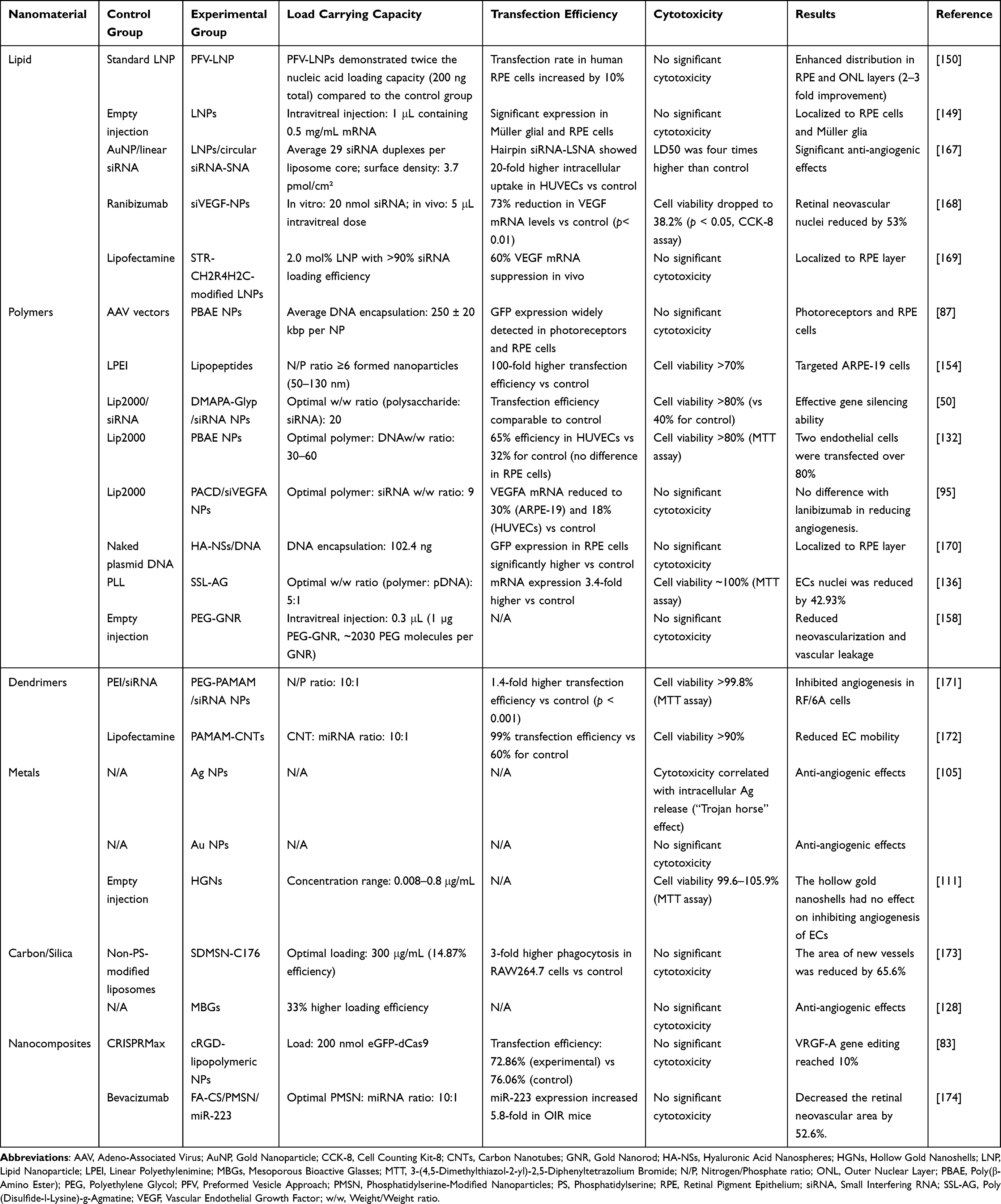 |
Table 4 Comparative Analysis of Nanomaterial-Based Delivery Systems for Retinal Therapeutics |
The Application of Gene Therapy Based on Nanomaterials in RNV
Age-Related Macular Degeneration (AMD)
AMD is a common chronic progressive macular retinal degenerative disease that affects older adults, leading to central vision impairment and the leading cause of vision loss in the elderly. Early AMD usually does not cause significant vision loss, only small hard exudates and pigmented macules in the macular area, and no significant vascular abnormalities or lesions. When AMD progresses from the early stage to the late stage, it can be further divided into two main types: non-neovascular (dry; atrophic) AMD and neovascular (wet) AMD.175 In wet or exudative AMD, the destruction of retinal structures is mainly caused by abnormal blood vessels invading the retina from the choroid. There is currently no definitive treatment for dry AMD, and clinical trials of gene therapy for dry AMD are still being actively explored (Table 1). The difference is that in neovascular AMD, glass anti-VEGF drugs new blood vessels in the body has been widely recognized.176–178
Although the latest advancements in intravitreal anti-VEGF drug injections have allowed for intervals of up to 24 weeks between injections,179 potential ocular adverse events remain after intravitreal injections of anti-VEGF drugs, such as endophthalmitis, retinal detachment, and traumatic scleral injury. To avoid the side effects of intravitreal injections, researchers have made efforts in gene therapy in recent years to enable one-time treatment for AMD. Following a single intravitreal injection of anti-VEGF gene-targeting drugs, effectiveness persisted at 15 months, despite evidence of reduced peak responses.180 The sustained effect significantly reduces the frequency of injections and improves patient compliance. Although AAV vectors are effective, their carrying capacity is limited, necessitating improvements for broader therapeutic applications.181 Several novel drug delivery technologies are being widely researched for posterior segment drug delivery, especially the nano delivery systems.
As early as 2009, Singh et al found that surface-functionalized nanoparticles carrying anti-VEGF receptor plasmids, through RGD/transferrin/both functionalization, significantly reduced the area of neovascularization. Compared to the naked plasmid group, the CNV area decreased by 77.3%, 62.9%, and 54.5% in the RGD peptide, transferrin, and dual-functional nanoparticle groups, respectively. This study first used nanoparticles to deliver anti-VEGF plasmids intravenously, indicating that this method enhances safety and patient compliance compared to local intravitreal injections and provides a preventive effect on AMD progression in the non-affected eye.182 In 2013, Luo et al delivered a single dose of biodegradable nanoparticles, specifically designed to target retinal neovascular lesions and carrying recombinant Flt23k receptor plasmids, via intravenous injection. This treatment led to a notable improvement in vision within a mouse model of CNV. Remarkably, the therapy restored vision by approximately 40% and was free of any observable ocular or systemic toxicity.183 These studies provide a nanoparticle-based platform for targeted, systemic delivery of non-viral gene therapy. Recently, Dasari et al used reducible PEG-POD nanoparticles to deliver human FLT1 genes to the retina, successfully reducing choroidal neovascularization by 50% in an AMD mouse model.184 Naash et al later attempted to encapsulate an enhanced GFP expression cassette controlled by an RPE-specific promoter into compact nanoparticles and injected them into the eyes of adult BALB/c mice twice, showing good targeting of compact NPs to RPE and sustained VEGF inhibition gene expression enhancement.185 Simultaneously, combined therapy as a supplement to standalone anti-VEGF treatment urgently needs pathways with long-term efficacy and efficient delivery to targeted sites, achievable through carefully designed nanocarriers. Zhu et al designed a biocompatible shell with good antioxidant and anti-angiogenic activity for CNV disease intervention, showing that the nanomaterial effectively delivers VEGF-siRNA and causes EGCG release, repairing oxidative stress in new blood vessel endothelial cells, thereby achieving combined treatment for AMD.186
Diabetic Retinopathy (DR)
DR is a microvascular complication occurring that occurs in people with diabetes. Approximately one-third of diabetic patients exhibit signs of DR, including microangiopathy, hemorrhage, exudates, and hard degeneration. Among them, one-third may have vision-threatening retinopathy, defined as severe retinopathy, including proliferative diabetic retinopathy (PDR) or diabetic macular edema (DME).187 Among the many factors contributing to DR, VEGF undoubtedly plays a central role. Therefore, the primary clinical goal is to prevent vision impairment or more severe blindness by overcoming DR-related macular edema. Most clinical trials related to diabetic eye diseases involve macular edema and retinopathy focusing on anti-angiogenic drug therapy and corticosteroid therapy (Table 1).56,188–190 Additionally, other potential targets are being studied, including inflammatory modulators TNF, the kallikrein-kinin system (KKS) non-steroidal anti-inflammatory drugs, and vascular regulators such as the RAS and angiopoietin.188
Despite identifying many drug targets, improving drug bioavailability and duration remains a technical challenge. Moreover, repeated injections are required over time to maintain visual benefits for many patients.10 Gene therapy is expected to achieve lifelong benefits through a single injection, but there are still many limitations in gene drug delivery in the posterior segment of the eye.62,191 To overcome the challenges of gene therapy, researchers have introduced nanomaterials in recent years, achieving significant results.192 Liang et al encapsulated the CRISPR-Cas9 vector into an aptamer-functionalized PEG-PEI-cholesterol nanocarrier, targeting and reducing VEGF gene expression in vivo and in vitro.193 With a deeper understanding of the DR pathogenesis, other potential gene therapy targets besides VEGF are being developed. Numerous negative regulators of VEGF exist in vivo, directly or indirectly reducing VEGF overexpression to maintain normal vascular development.194,195 For instance, Liu et al used two polymers to load siRNA targeting NF-κB for gene therapy of DR. Their study demonstrated that in 25% fetal bovine serum, DMAPA-Glyp/siRNA and DMAPA-Amp/siRNA nanoparticles effectively protected siRNA from nuclease degradation and significantly increased the inhibition of NF-κB p65 mRNA and protein expression in transfected hRPE cells.50 Sardoiwala et al developed a PDG nanomaterial to promote melatonin absorption, showing a 20% improvement in protective efficiency without differences in tissue distribution in vivo, indicating good tissue compatibility of the nanomaterial.196 Park et al used nanoparticles to deliver the plasminogen 5 Kringle domain expression vector, and compared with the control group, DR Mice injected with K5-NP showed decreased VEGF overexpression, leukocytosis, and vascular leakage for at least 4 weeks without toxic effects on retinal structure and function.197
Oxidative stress also plays an important role in DR progression. Compared to more potent and selective VEGF antagonists, targeting upstream factors of VEGF, such as HIF. Active HIF-2α promotes VEGF overexpression by activating plasminogen activator inhibitor-1 (PAI-1). Qin et al used HIF-2α inhibitors (PT-2385) or nanoparticle-mediated targeting of PAI-1 RNA interference, demonstrating that the HIF-2α/PAI-1 axis is essential for retinal neovascularization in mice. Targeting the HIF-2α/PAI-1 pathway could serve as an effective adjunct therapy for patients with PDR.46
Monotherapy with anti-VEGF agents may lead to an elevated expression of CTGF in the retina, potentially heightening the risk of fibrosis and tractional retinal detachment. To mitigate this issue, Hu et al introduced an innovative dual-target strategy that combined ranibizumab with CTGF shRNA. Their research demonstrated that the transcriptional levels of both VEGF and CTGF were significantly reduced in the dual-target group compared to those receiving only a single-target treatment. These results indicate that dual-target therapy could be more effective than single-target approaches in treating diabetic retinopathy, presenting a promising alternative for managing the condition.56,57 In addition, researchers found that SLNPs loaded vildagliptin not only effectively controlled blood glucose levels in diabetes, but also improved retinal microvascular blood flow and delayed the progression of DR, which also provides a basis for future multi-target therapy.198
Combining gene therapy and nanotechnology for treating diabetic retinopathy (DR) shows potential. By promoting the expression of VEGF-negative regulators using siRNA and nanoparticles, the targeted and sustained effects are demonstrated.199 Future trends include further optimization of personalized treatments and safety assessments.
Retinopathy of Prematurity (ROP)
ROP is an ischemic neovascular disease that primarily affects premature infants and is a leading cause of vision impairment and blindness in premature infants. The current belief is that the etiology of ROP may be due to the fluctuation of retinal oxygen concentration from the intrauterine environment to atmospheric levels, causing a halt in retinal vascular development (hyperoxia), and subsequently, due to avascularity-induced hypoxia, leading to abnormal angiogenesis.200 Premature infants transition from a high-oxygen state to hypoxia, where low-oxygen-inducing factors are the primary stimuli for VEGF production.201
The current gold standard for the treatment of severe central vision loss caused by ROP is retinal laser therapy, which involves continuous burns to the periphery of the vascular retina of premature infants under local or general anesthesia to reduce VEGF production caused by hypoxia of the avascular retina.202,203 Simultaneously, gene therapy has shown results in drug duration and reducing dosage frequency in anti-angiogenesis treatment of AMD and DR. Thus, exploring gene therapy for anti-angiogenesis in ROP is also a potential treatment direction. The role of miRNA in ROP treatment has been reported. The role of miRNAs in ROP treatment has been reported previously. In the OIR mouse model, miR16, 17, 18, and 20 have been shown to down-regulate the expression of HIF-1α and VEGF in the retina to exert anti-angiogenic effects.204–206 However, miRNA-based therapies face challenges due to low stability and non-specific cell targeting by delivery systems. To overcome these issues, Li et al developed a nanomaterial composed of folic acid-chitosan (FA-CS) modified porous silicon nanoparticles (PMSNs), which protect miRNA from nuclease degradation. They utilized these nanoparticles to deliver miR-223, which modulates retinal microglia polarization. miR-223 can shift retinal microglia from an M1 to an M2 phenotype, where the M1 phenotype promotes vascular permeability, proliferation, and neovascularization by releasing high levels of inflammatory cytokines, while the M2 phenotype contributes to the natural resolution of OIR and has anti-inflammatory effects.174,207 This treatment strategy is considered a promising new approach for treating neovascular eye diseases like ROP. Additionally, siRNA-mediated gene therapy has also gained recognition in ROP applications. Researchers constructed PLGA carriers to load siRNA against annexin A2, downregulating annexin A2 and aiding in ROP adjunctive therapy.208,209 In summary, the application of nanomaterial-based gene therapy in ROP primarily focuses on OIP models, targeting oxidative induction mechanisms and VEGF-mediated angiogenesis, offering significant insights for future ROP treatments.
Retinal Vein Occlusion (RVO)
RVO is a retinal vascular disease caused by thrombosis within the retinal vein, leading to obstructed blood flow and the fifth leading cause of blindness. Macular edema is the most common complication of RVO and the primary cause of vision loss. The occurrence of macular edema is closely associated with pathological neovascularization, making VEGF-A play a crucial role in the development and progression of RVO.210,211
Currently, clinical interventions for RVO primarily address complications like macular edema to improve vision or prevent blindness. Intravitreal injection of anti-VEGF drugs is typically used to improve clinical outcomes in RVO patients.212 Monthly injections of ranibizumab or aflibercept for six months significantly improve the mean visual acuity in eyes with BRVO or CRVO (with mean BCVA improvement >15 letters).213 Clinical trials on gene therapy for RVO are ongoing (NCT05099094), with the main treatment outcome still focused on controlling macular edema. Recently, researchers developed a novel nanocarrier constructed from thiolates chitosan, capable of gradually releasing anti-VEGF over at least a week in cell culture, sustaining anti-angiogenic effects, reducing dosing frequency, and extending drug action time. Additionally, Gounari et al first attempted in vivo to use a novel thiolates chitosan anti-VEGF nanocarrier to treat an RVO model, showing that it could effectively increase local drug duration and adhesion in the administration environment with lower cytotoxicity.214 This provides a foundation for applying nanomaterials in gene therapy for RVO.
Conclusion and Prospect
Challenges and Blind Spots
The application of nanomaterials in anti-angiogenic gene therapy has brought new hope for the treatment of retinal neovascular diseases. Nanomaterials, with their high drug-loading capacity, low toxicity, and controlled release properties, have become ideal carriers for the effective delivery of gene therapy drugs. However, several significant challenges remain to be addressed. One such challenge is optimizing the delivery method to target cells. Currently, the use of nano-eye drops for delivering gene therapy drugs has not achieved significant results, which limits the popularization of gene therapy in patients with RNV disease. Although researchers have begun to pay attention to the anti-angiogenic effect of nano-material eye drops, there is still a long way to go before the appropriate carrier delivers gene drugs. How to overcome the physiological barrier of the eyeball to reach the target cells is one of the key factors. Additionally, there are issues related to the stability and efficiency of nanomaterial-based delivery systems, particularly in minimizing potential immunogenicity. Although natural polymer delivery carriers, such as beta-cyclodextrin, chitosan and hyaluronic acid, have been developed to address these challenges, no material has yet demonstrated absolute superiority. These challenges are compounded by the need for extended safety evaluations to assess potential off-target effects and chronic toxicity, which are essential for regulatory approval.
Future Directions
Future research should focus on overcoming these barriers to fully unlock the potential of gene therapy for retinal diseases. In fact, researchers are currently making efforts and attempts, including dual-target gene therapy and the application of hybrid nanomaterials. Dual-target and even multi-target gene therapy methods are mainly developed around the core target VEGF, such as targeted VEGF combined with Ang-2, which has been commercialized (such as Faricimab), but further development of gene therapy programs is needed. Hybrid nanomaterials can combine the advantages of multiple nanomaterials to improve anti-angiogenic ability, among which functionalized group modification and Mesoporous Silica/Carbon Nanoparticle are several completely new attempts to overcome the current challenges, which should receive more attention. Furthermore, alternative gene-editing platforms including CRISPR-based systems and aptamer-mediated gene silencing warrant comparative investigation, as these technologies may offer complementary advantages in specificity or delivery efficiency.
The field is at a critical juncture, where technological innovation must be balanced with rigorous translational science. Continued research should focus not only on improving efficacy but also on establishing robust safety through longitudinal studies. As we progress, interdisciplinary collaboration will be essential to bridge the gap between laboratory breakthroughs and clinically viable therapies. With careful development of technological and translational challenges, nanomaterials may eventually enable personalized, long-lasting therapies that transform the treatment of retinal vascular diseases.
Abbreviations
AAV, Adenovirus Associated Vector; AMD, Age-Related Macular Degeneration; ASO, Antisense Oligonucleotides; BRB, Blood-Retinal Barrier; CNV, Choroidal Neovascularization; CRISPR, Clustered Regularly Interspaced Short Palindromic Repeats; CFH, Complement Factor H; Cas, Crispr-Associated Proteins; DR, Diabetic Retinopathy; FGF, Fibroblast Growth Factor; AuNPs, Gold Nps; GO, Graphene Oxide; HRECs, Human Retinal Endothelial Cells; HRE, Hypoxia Response Elements; HIF, Hypoxia-Inducible Factor; IL, Interleukin; LCA, Leber Congenital Amaurosis; LV, Lentivirus Vector; LNP, Lipid Nanoparticle; LncRNAs, Long Non-Coding RNAs; miRNAs, Micro RNAs; MAPK, Mitogen Activated Protein Kinases; NPs, Nanoparticle; NIR, Near-Infrared-Ray; ncRNAs, Non-Coding Rnas; NF-κB, Nuclear Factor-Kappab; ZFNs, Nucleases Like Zinc Finger Nucleases; PEDF, Pigment Epithelium-Derived Factor; PAI, Plasminogen Activator Inhibitor; PAMAM, Polyamide-Amine Dendrimer; PEI, polyethyleneimine; PPI, Polypropylenimine; PKCγ, Protein Kinase Cγ; RTK, Receptor Tyrosine Kinase; RNVs, Retinal Neovascular Diseases; RPE, Retinal Pigment Epithelium; RVO, Retinal Vein Obstruction; ROP, Retinopathy Of Prematurity; shRNAs, Short Hairpin Rnas; siRNAs, Small Interfering Rnas; TJ, Tight Junction; TALENs, Transcription Activator-Like Effector Nucleases; TGF, Transforming Growth Factor; VEGF, Vascular Endothelial Growth Factor.
Data Sharing Statement
No datasets were generated or analysed during the current study.
Author Contributions
All authors made a significant contribution to the work reported, whether that is in the conception, study design, execution, acquisition of data, analysis and interpretation, or in all these areas; took part in drafting, revising or critically reviewing the article; gave final approval of the version to be published; have agreed on the journal to which the article has been submitted; and agree to be accountable for all aspects of the work.
Funding
This work was supported by the National Natural Science Foundation of China (No. 82571526 to Yanan Li, No. 82330040, No. 82090044 and No. 81820108010 to Bo Hu), Hubei Province Key R&D Program (No. 2020CF763 to Yanan Li, No. 2022BCA008 to B.H, No. 2022CFB296 to Pengcheng Li), General Project of Natural Science Foundation of Hubei Province (2024AFB623 to Chengye Yao), and the National Key Research and Development Program of China (grant 2024YFC3044800 to B.H).
Disclosure
The authors declare no competing interests.
References
1. Campochiaro PA. Molecular pathogenesis of retinal and choroidal vascular diseases. Prog Retin Eye Res. 2015;49:67–81. doi:10.1016/j.preteyeres.2015.06.002
2. Mohamed Q, Gillies MC, Wong TY. Management of diabetic retinopathy: a systematic review. JAMA. 2007;298(8):902–916. doi:10.1001/jama.298.8.902
3. Saeedi P, Petersohn I, Salpea P, et al. Global and regional diabetes prevalence estimates for 2019 and projections for 2030 and 2045: results from the international diabetes federation diabetes atlas, 9(th) edition. Diabetes Res Clin Pract. 2019;157:107843. doi:10.1016/j.diabres.2019.107843
4. Wong WL, Su X, Li X, et al. Global prevalence of age-related macular degeneration and disease burden projection for 2020 and 2040: a systematic review and meta-analysis. Lancet Glob Health. 2014;2(2):e106–116. doi:10.1016/S2214-109X(13)70145-1
5. Cheung CMG, Arnold JJ, Holz FG, et al. Myopic choroidal neovascularization: review, guidance, and consensus statement on management. Ophthalmology. 2017;124(11):1690–1711. doi:10.1016/j.ophtha.2017.04.028
6. Bird AC. Therapeutic targets in age-related macular disease. J Clin Invest. 2010;120(9):3033–3041. doi:10.1172/JCI42437
7. Xu Y, Huang S, Zhou S, et al. Iron chelator deferiprone restores iron homeostasis and inhibits retinal neovascularization in experimental neovascular age-related macular degeneration. Invest Ophthalmol Vis Sci. 2024;65(10):5. doi:10.1167/iovs.65.10.5
8. Barequet D, Shor R, Segal O, et al. Treatment patterns and visual acuity change of AMD patients, before, during and after the COVID-19 pandemic lockdowns: a large cohort. Acta Ophthalmol. 2024;102(3):e322–e327. doi:10.1111/aos.15756
9. Volkmann I, Knoll K, Wiezorrek M, Greb O, Framme C. Individualized treat-and-extend regime for optimization of real-world vision outcome and improved patients’ persistence. BMC Ophthalmol. 2020;20(1):122. doi:10.1186/s12886-020-01397-x
10. Chakravarthy U, Harding SP, Rogers CA, et al. Alternative treatments to inhibit VEGF in age-related choroidal neovascularisation: 2-year findings of the IVAN randomised controlled trial. Lancet. 2013;382(9900):1258–1267. doi:10.1016/S0140-6736(13)61501-9
11. Falavarjani KG, Nguyen QD. Adverse events and complications associated with intravitreal injection of anti-VEGF agents: a review of literature. Eye. 2013;27(7):787–794. doi:10.1038/eye.2013.107
12. Khurana RN, Li C, Lum F. Loss to follow-up in patients with neovascular age-related macular degeneration treated with anti-VEGF therapy in the United States in the IRIS® registry. Ophthalmology. 2023;130(7):672–683. doi:10.1016/j.ophtha.2023.02.021
13. Bainbridge JW, Mehat MS, Sundaram V, et al. Long-term effect of gene therapy on Leber’s congenital amaurosis. N Engl J Med. 2015;372(20):1887–1897. doi:10.1056/NEJMoa1414221
14. Campochiaro PA, Lauer AK, Sohn EH, et al. Lentiviral vector gene transfer of endostatin/angiostatin for macular degeneration (GEM) study. Hum Gene Ther. 2017;28(1):99–111. doi:10.1089/hum.2016.117
15. Kotin RM. Large-scale recombinant adeno-associated virus production. Hum Mol Genet. 2011;20(R1):R2–6. doi:10.1093/hmg/ddr141
16. Mingozzi F, High KA. Immune responses to AAV vectors: overcoming barriers to successful gene therapy. Blood. 2013;122(1):23–36. doi:10.1182/blood-2013-01-306647
17. Wang D, Tai PWL, Gao G. Adeno-associated virus vector as a platform for gene therapy delivery. Nat Rev Drug Discov. 2019;18(5):358–378. doi:10.1038/s41573-019-0012-9
18. Dias AP, da Silva Santos S, da Silva JV, et al. Dendrimers in the context of nanomedicine. Int J Pharm. 2020;573:118814. doi:10.1016/j.ijpharm.2019.118814
19. Uhrich KE, Cannizzaro SM, Langer RS, Shakesheff KM. Polymeric systems for controlled drug release. Chem Rev. 1999;99(11):3181–3198. doi:10.1021/cr940351u
20. Wang Y, Rajala A, Cao B, et al. Cell-specific promoters enable lipid-based nanoparticles to deliver genes to specific cells of the retina in vivo. Theranostics. 2016;6(10):1514–1527. doi:10.7150/thno.15230
21. Sarkar B, Siddiqui Z, Kim KK, et al. Implantable anti-angiogenic scaffolds for treatment of neovascular ocular pathologies. Drug Deliv Transl Res. 2020;10(5):1191–1202. doi:10.1007/s13346-020-00753-0
22. Pérez-Gutiérrez L, Ferrara N. Biology and therapeutic targeting of vascular endothelial growth factor A. Nat Rev Mol Cell Biol. 2023;24(11):816–834. doi:10.1038/s41580-023-00631-w
23. Aiello LP, Avery RL, Arrigg PG, et al. Vascular endothelial growth factor in ocular fluid of patients with diabetic retinopathy and other retinal disorders. N Engl J Med. 1994;331(22):1480–1487. doi:10.1056/NEJM199412013312203
24. Young TL, Anthony DC, Pierce E, Foley E, Smith LE. Histopathology and vascular endothelial growth factor in untreated and diode laser-treated retinopathy of prematurity. J Aapos. 1997;1(2):105–110. doi:10.1016/S1091-8531(97)90008-2
25. Kvanta A, Algvere PV, Berglin L, Seregard S. Subfoveal fibrovascular membranes in age-related macular degeneration express vascular endothelial growth factor. Invest Ophthalmol Vis Sci. 1996;37(9):1929–1934.
26. Murakami T, Felinski EA, Antonetti DA. Occludin phosphorylation and ubiquitination regulate tight junction trafficking and vascular endothelial growth factor-induced permeability. J Biol Chem. 2009;284(31):21036–21046. doi:10.1074/jbc.M109.016766
27. Argaw AT, Gurfein BT, Zhang Y, Zameer A, John GR. VEGF-mediated disruption of endothelial CLN-5 promotes blood-brain barrier breakdown. Proc Natl Acad Sci U S A. 2009;106(6):1977–1982. doi:10.1073/pnas.0808698106
28. Kim LA, Wong LL, Amarnani DS, et al. Characterization of cells from patient-derived fibrovascular membranes in proliferative diabetic retinopathy. Mol Vis. 2015;21:673–687.
29. Arrigo A, Aragona E, Bandello F. VEGF-targeting drugs for the treatment of retinal neovascularization in diabetic retinopathy. Ann Med. 2022;54(1):1089–1111. doi:10.1080/07853890.2022.2064541
30. Ferrara N, Gerber HP, LeCouter J. The biology of VEGF and its receptors. Nat Med. 2003;9(6):669–676. doi:10.1038/nm0603-669
31. Bai Y, Ma JX, Guo J, et al. Müller cell-derived VEGF is a significant contributor to retinal neovascularization. J Pathol. 2009;219(4):446–454. doi:10.1002/path.2611
32. Uludag G, Hassan M, Matsumiya W, et al. Efficacy and safety of intravitreal anti-VEGF therapy in diabetic retinopathy: what we have learned and what should we learn further? Expert Opin Biol Ther. 2022;22(10):1275–1291. doi:10.1080/14712598.2022.2100694
33. Madhusudhan S, Gupta NV, Rahamathulla M, et al. Subconjunctival delivery of sorafenib-tosylate-loaded cubosomes for facilitated diabetic retinopathy treatment: formulation development, evaluation, Pharmacokinetic and pharmacodynamic (PKPD) studies. Pharmaceutics. 2023;15(10):2419. doi:10.3390/pharmaceutics15102419
34. Joussen AM, Ricci F, Paris LP, Korn C, Quezada-Ruiz C, Zarbin M. Angiopoietin/Tie2 signalling and its role in retinal and choroidal vascular diseases: a review of preclinical data. Eye. 2021;35(5):1305–1316. doi:10.1038/s41433-020-01377-x
35. Wong TY, Haskova Z, Asik K, et al. Faricimab treat-and-extend for diabetic macular edema: two-year results from the randomized phase 3 YOSEMITE and RHINE trials. Ophthalmology. 2024;131(6):708–723. doi:10.1016/j.ophtha.2023.12.026
36. Campochiaro PA, Khanani A, Singer M, et al. Enhanced benefit in diabetic macular edema from AKB-9778 Tie2 Activation combined with vascular endothelial growth factor suppression. Ophthalmology. 2016;123(8):1722–1730. doi:10.1016/j.ophtha.2016.04.025
37. Vahidi A, Khosravi T, Dastaviz F, Sheikh Arabi M, Khosravi A, Oladnabi M. Niosome-encapsulated auraptene reduced the mRNA expression of VEGF-A and PDGFs genes in human retina-derived RPE cell line. Int J Ophthalmol. 2024;17(6):1028–1035. doi:10.18240/ijo.2024.06.06
38. Jaffe GJ, Ciulla TA, Ciardella AP, et al. Dual antagonism of PDGF and VEGF in neovascular age-related macular degeneration: a phase IIb, multicenter, randomized controlled trial. Ophthalmology. 2017;124(2):224–234. doi:10.1016/j.ophtha.2016.10.010
39. Shaw LT, Mackin A, Shah R, et al. Risuteganib-a novel integrin inhibitor for the treatment of non-exudative (dry) age-related macular degeneration and diabetic macular edema. Expert Opin Investig Drugs. 2020;29(6):547–554. doi:10.1080/13543784.2020.1763953
40. Li YN, Liang HW, Zhang CL, et al. Ophthalmic solution of smart supramolecular peptides to capture semaphorin 4D against diabetic retinopathy. Adv Sci. 2023;10(3):e2203351. doi:10.1002/advs.202203351
41. Shweiki D, Itin A, Soffer D, Keshet E. Vascular endothelial growth factor induced by hypoxia may mediate hypoxia-initiated angiogenesis. Nature. 1992;359(6398):843–845. doi:10.1038/359843a0
42. Plastino F, Pesce NA, André H. MicroRNAs and the HIF/VEGF axis in ocular neovascular diseases. Acta Ophthalmol. 2021;99(8):e1255–e1262. doi:10.1111/aos.14845
43. Ke Q, Costa M. Hypoxia-inducible factor-1 (HIF-1). Mol Pharmacol. 2006;70(5):1469–1480. doi:10.1124/mol.106.027029
44. Kuroki M, Voest EE, Amano S, et al. Reactive oxygen intermediates increase vascular endothelial growth factor expression in vitro and in vivo. J Clin Invest. 1996;98(7):1667–1675. doi:10.1172/JCI118962
45. Atalor RE, Dieckmann BW, Penn JS, Uddin MI. Method to regulate monocyte function by silencing HIF-1α mRNA in a model of retinal neovascularization. ACS Appl Nano Mater. 2023;6(24):22939–22946. doi:10.1021/acsanm.3c04300
46. Qin Y, Zhang J, Babapoor-Farrokhran S, et al. PAI-1 is a vascular cell-specific HIF-2-dependent angiogenic factor that promotes retinal neovascularization in diabetic patients. Sci Adv. 2022;8(9):eabm1896. doi:10.1126/sciadv.abm1896
47. Ricci F, Bandello F, Navarra P, Staurenghi G, Stumpp M, Zarbin M. Neovascular age-related macular degeneration: therapeutic management and new-upcoming approaches. Int J Mol Sci. 2020;21(21):8242. doi:10.3390/ijms21218242
48. Yang S, Li T, Jia H, et al. Targeting C3b/C4b and VEGF with a bispecific fusion protein optimized for neovascular age-related macular degeneration therapy. Sci Transl Med. 2022;14(647):eabj2177. doi:10.1126/scitranslmed.abj2177
49. Tischer E, Mitchell R, Hartman T, et al. The human gene for vascular endothelial growth factor. Multiple protein forms are encoded through alternative exon splicing. J Biol Chem. 1991;266(18):11947–11954. doi:10.1016/S0021-9258(18)99049-6
50. Liu Z, Gong H, Zeng R, et al. Efficient delivery of NF-κB siRNA to human retinal pigment epithelial cells with hyperbranched cationic polysaccharide derivative-based nanoparticles. Int J Nanomedicine. 2015;10:2735–2749. doi:10.2147/IJN.S75188
51. Shchors K, Shchors E, Rostker F, Lawlor ER, Brown-Swigart L, Evan GI. The Myc-dependent angiogenic switch in tumors is mediated by interleukin 1beta. Genes Dev. 2006;20(18):2527–2538. doi:10.1101/gad.1455706
52. Mizukami Y, Jo WS, Duerr EM, et al. Induction of interleukin-8 preserves the angiogenic response in HIF-1alpha-deficient colon cancer cells. Nat Med. 2005;11(9):992–997. doi:10.1038/nm1294
53. Palacka K, Hermankova B, Cervena T, et al. The immunomodulatory effect of silver nanoparticles in a retinal inflammatory environment. Inflammation. 2024;48(3):1378–1390. doi:10.1007/s10753-024-02128-w
54. Wu Y, Li X, Fu X, et al. Innovative nanotechnology in drug delivery systems for advanced treatment of posterior segment ocular diseases. Adv Sci. 2024;11(32):e2403399. doi:10.1002/advs.202403399
55. Lu Y, Zhu F, Zhou X, et al. A supramolecular deferoxamine-crisaborole nanoparticle targets ferroptosis, inflammation, and oxidative stress in the treatment of retinal ischemia/reperfusion injury. Nano Lett. 2025;25(3):1058–1066. doi:10.1021/acs.nanolett.4c05012
56. Hu B, Zhang Y, Zeng Q, et al. Intravitreal injection of ranibizumab and CTGF shRNA improves retinal gene expression and microvessel ultrastructure in a rodent model of diabetes. Int J Mol Sci. 2014;15(1):1606–1624. doi:10.3390/ijms15011606
57. Dastaviz F, Vahidi A, Khosravi T, et al. Impact of umbelliprenin-containing niosome nanoparticles on VEGF-A and CTGF genes expression in retinal pigment epithelium cells. Int J Ophthalmol. 2024;17(1):7–15. doi:10.18240/ijo.2024.01.02
58. Wei-Zhang S, Cui B, Xing M, et al. Chimpanzee adenovirus-mediated multiple gene therapy for age-related macular degeneration. iScience. 2023;26(10):107939. doi:10.1016/j.isci.2023.107939
59. Benhar I, London A, Schwartz M. The privileged immunity of immune privileged organs: the case of the eye. Front Immunol. 2012;3:296. doi:10.3389/fimmu.2012.00296
60. Boye SE, Boye SL, Lewin AS, Hauswirth WW. A comprehensive review of retinal gene therapy. Mol Ther. 2013;21(3):509–519. doi:10.1038/mt.2012.280
61. Boon CJ, Jeroen Klevering B, Keunen JE, Hoyng CB, Theelen T. Fundus autofluorescence imaging of retinal dystrophies. Vision Res. 2008;48(26):2569–2577. doi:10.1016/j.visres.2008.01.010
62. Guimaraes TAC, Georgiou M, Bainbridge JWB, Michaelides M. Gene therapy for neovascular age-related macular degeneration: rationale, clinical trials and future directions. Br J Ophthalmol. 2021;105(2):151–157. doi:10.1136/bjophthalmol-2020-316195
63. Tawfik M, Chen F, Goldberg JL, Sabel BA. Nanomedicine and drug delivery to the retina: current status and implications for gene therapy. Naunyn Schmiedebergs Arch Pharmacol. 2022;395(12):1477–1507. doi:10.1007/s00210-022-02287-3
64. Chung SH, Frick SL, Yiu G. Targeting vascular endothelial growth factor using retinal gene therapy. Ann Transl Med. 2021;9(15):1277. doi:10.21037/atm-20-4417
65. Lai CM, Shen WY, Brankov M, et al. Long-term evaluation of AAV-mediated sFlt-1 gene therapy for ocular neovascularization in mice and monkeys. Mol Ther. 2005;12(4):659–668. doi:10.1016/j.ymthe.2005.04.022
66. Lai CM, Estcourt MJ, Himbeck RP, et al. Preclinical safety evaluation of subretinal AAV2.sFlt-1 in non-human primates. Gene Ther. 2012;19(10):999–1009. doi:10.1038/gt.2011.169
67. Lai CM, Estcourt MJ, Wikstrom M, et al. rAAV.sFlt-1 gene therapy achieves lasting reversal of retinal neovascularization in the absence of a strong immune response to the viral vector. Invest Ophthalmol Vis Sci. 2009;50(9):4279–4287. doi:10.1167/iovs.08-3253
68. Hussain RM, Ciulla TA. Emerging vascular endothelial growth factor antagonists to treat neovascular age-related macular degeneration. Expert Opin Emerg Drugs. 2017;22(3):235–246. doi:10.1080/14728214.2017.1362390
69. Lu J, Huang Y, Zhang X, Xu Y, Nie S. Noncoding RNAs involved in DNA methylation and histone methylation, and acetylation in diabetic vascular complications. Pharmacol Res. 2021;170:105520. doi:10.1016/j.phrs.2021.105520
70. Bannister AJ, Kouzarides T. Regulation of chromatin by histone modifications. Cell Res. 2011;21(3):381–395. doi:10.1038/cr.2011.22
71. Akhtar MS, Mohsin N, Zahak A, Ahmed K, Alhazmi Y, Taleuzzaman M. siRNA treatments for diabetic neuropathy: obstacles and delivery techniques. Curr Mol Med. 2024. doi:10.2174/0115665240307413240531111140
72. Supe S, Upadhya A, Singh K. Role of small interfering RNA (siRNA) in targeting ocular neovascularization: a review. Exp Eye Res. 2021;202:108329. doi:10.1016/j.exer.2020.108329
73. Lee SHS, Chang H, Kim JH, et al. Inhibition of mTOR via an AAV-delivered shRNA tested in a rat OIR model as a potential antiangiogenic gene therapy. Invest Ophthalmol Vis Sci. 2020;61(2):45. doi:10.1167/iovs.61.2.45
74. Nasrolahi A, Khojasteh Pour F, Mousavi Salehi A, et al. Potential roles of lncRNA MALAT1-miRNA interactions in ocular diseases. J Cell Commun Signal. 2023;17(4):1203–1217. doi:10.1007/s12079-023-00787-2
75. Cong L, Ran FA, Cox D, et al. Multiplex genome engineering using CRISPR/Cas systems. Science. 2013;339(6121):819–823. doi:10.1126/science.1231143
76. Mali P, Yang L, Esvelt KM, et al. RNA-guided human genome engineering via Cas9. Science. 2013;339(6121):823–826. doi:10.1126/science.1232033
77. Koo T, Park SW, Jo DH, et al. CRISPR-LbCpf1 prevents choroidal neovascularization in a mouse model of age-related macular degeneration. Nat Commun. 2018;9(1):1855. doi:10.1038/s41467-018-04175-y
78. Kim E, Koo T, Park SW, et al. In vivo genome editing with a small Cas9 orthologue derived from Campylobacter jejuni. Nat Commun. 2017;8:14500. doi:10.1038/ncomms14500
79. Chung SH, Sin TN, Ngo T, Yiu G. CRISPR technology for ocular angiogenesis. Front Genome Ed. 2020;2:594984. doi:10.3389/fgeed.2020.594984
80. Huang X, Zhou G, Wu W, et al. Genome editing abrogates angiogenesis in vivo. Nat Commun. 2017;8(1):112. doi:10.1038/s41467-017-00140-3
81. Salman A, Kantor A, McClements ME, Marfany G, Trigueros S, MacLaren RE. Non-viral delivery of CRISPR/Cas Cargo to the retina using nanoparticles: current possibilities, challenges, and limitations. Pharmaceutics. 2022;14(9):1842. doi:10.3390/pharmaceutics14091842
82. Patra JK, Das G, Fraceto LF, et al. Nano based drug delivery systems: recent developments and future prospects. J Nanobiotechnology. 2018;16(1):71. doi:10.1186/s12951-018-0392-8
83. Sahel DK, Goswami SG, Jatyan R, et al. cRGD-modified hybrid lipopolymeric nanoplexes for gene editing in the posterior segment of the eye. Int J Biol Macromol. 2024;271(Pt 2):132426. doi:10.1016/j.ijbiomac.2024.132426
84. Wang Y, Huang L. Composite nanoparticles for gene delivery. Adv Genet. 2014;88:111–137.
85. Ozaki T, Nakazawa M, Yamashita T, et al. Intravitreal injection or topical eye-drop application of a μ-calpain C2L domain peptide protects against photoreceptor cell death in royal college of surgeons’ rats, a model of retinitis pigmentosa. Biochim Biophys Acta. 2012;1822(11):1783–1795. doi:10.1016/j.bbadis.2012.07.018
86. Rowe-Rendleman CL, Durazo SA, Kompella UB, et al. Drug and gene delivery to the back of the eye: from bench to bedside. Invest Ophthalmol Vis Sci. 2014;55(4):2714–2730. doi:10.1167/iovs.13-13707
87. Shen J, Lima ESR, Zhang M, et al. Suprachoroidal gene transfer with nonviral nanoparticles in large animal eyes. Sci Adv. 2024;10(10):eadl3576. doi:10.1126/sciadv.adl3576
88. Peng Y, Tang L, Zhou Y. Subretinal injection: a review on the novel route of therapeutic delivery for vitreoretinal diseases. Ophthalmic Res. 2017;58(4):217–226. doi:10.1159/000479157
89. Yiu G, Chung SH, Mollhoff IN, et al. Suprachoroidal and subretinal injections of AAV using transscleral microneedles for retinal gene delivery in nonhuman primates. Mol Ther Methods Clin Dev. 2020;16:179–191. doi:10.1016/j.omtm.2020.01.002
90. Hartman RR, Kompella UB. Intravitreal, subretinal, and suprachoroidal injections: evolution of microneedles for drug delivery. J Ocul Pharmacol Ther. 2018;34(1–2):141–153. doi:10.1089/jop.2017.0121
91. Mochizuki M, Sugita S, Kamoi K. Immunological homeostasis of the eye. Prog Retin Eye Res. 2013;33:10–27. doi:10.1016/j.preteyeres.2012.10.002
92. Martins B, Pires M, Ambrósio AF, Girão H, Fernandes R. Contribution of extracellular vesicles for the pathogenesis of retinal diseases: shedding light on blood-retinal barrier dysfunction. J Biomed Sci. 2024;31(1):48. doi:10.1186/s12929-024-01036-3
93. Wang R, Liu Y, Xiao W, et al. Framework nucleic acids as blood-retinal-barrier-penetrable nanocarrier for periocular administration. ACS Appl Mater Interfaces. 2023;15(1):541–551. doi:10.1021/acsami.2c18042
94. Gilleron J, Querbes W, Zeigerer A, et al. Image-based analysis of lipid nanoparticle-mediated siRNA delivery, intracellular trafficking and endosomal escape. Nat Biotechnol. 2013;31(7):638–646. doi:10.1038/nbt.2612
95. Guo S, Li C, Wang C, et al. pH-responsive polymer boosts cytosolic siRNA release for retinal neovascularization therapy. Acta Pharm Sin B. 2024;14(2):781–794. doi:10.1016/j.apsb.2023.09.001
96. Yang H, Luo Y, Hu H, et al. pH-Sensitive, Cerebral Vasculature-Targeting Hydroxyethyl Starch Functionalized Nanoparticles for Improved Angiogenesis and Neurological Function Recovery in Ischemic Stroke. Adv Healthc Mater. 2021;10(12):e2100028. doi:10.1002/adhm.202100028
97. Douar AM, Poulard K, Stockholm D, Danos O. Intracellular trafficking of adeno-associated virus vectors: routing to the late endosomal compartment and proteasome degradation. J Virol. 2001;75(4):1824–1833. doi:10.1128/JVI.75.4.1824-1833.2001
98. Bisht R, Jaiswal JK, Rupenthal ID. Nanoparticle-loaded biodegradable light-responsive in situ forming injectable implants for effective peptide delivery to the posterior segment of the eye. Med Hypotheses. 2017;103:5–9. doi:10.1016/j.mehy.2017.03.033
99. Bordet T, Behar-Cohen F. Ocular gene therapies in clinical practice: viral vectors and nonviral alternatives. Drug Discov Today. 2019;24(8):1685–1693. doi:10.1016/j.drudis.2019.05.038
100. Stoia D, Nistor M, Suciu M, et al. NIR photothermal-activable drug-conjugated microcapsules for in vitro targeted delivery and release: an alternative treatment of diabetic retinopathy. Int J Pharm. 2023;635:122700. doi:10.1016/j.ijpharm.2023.122700
101. Postovalova AS, Tishchenko YA, Istomina MS, et al. Comparison of passive targeted delivery of inorganic and organic nanocarriers among different types of tumors. Nanomedicine. 2024;59:102753. doi:10.1016/j.nano.2024.102753
102. Nag S, Mitra O, Tripathi G, et al. Nanomaterials-assisted photothermal therapy for breast cancer: state-of-the-art advances and future perspectives. Photodiagnosis Photodyn Ther. 2024;45:103959. doi:10.1016/j.pdpdt.2023.103959
103. Amaldoss MJN, Yang JL, Koshy P, Unnikrishnan A, Sorrell CC. Inorganic nanoparticle-based advanced cancer therapies: promising combination strategies. Drug Discov Today. 2022;27(12):103386. doi:10.1016/j.drudis.2022.103386
104. Wong KY, Nie Z, Wong MS, Wang Y, Liu J. Metal-drug coordination nanoparticles and hydrogels for enhanced delivery. Adv Mater. 2024;36(26):e2404053. doi:10.1002/adma.202404053
105. Liu J, Zhang F, Shi X. The role of metal nanocarriers, liposomes and chitosan-based nanoparticles in diabetic retinopathy treatment: a review study. Int J Biol Macromol. 2025;291:139017. doi:10.1016/j.ijbiomac.2024.139017
106. Scheive M, Yazdani S, Hajrasouliha AR. The utility and risks of therapeutic nanotechnology in the retina. Ther Adv Ophthalmol. 2021;13:25158414211003381. doi:10.1177/25158414211003381
107. Kanu GA, Parambath JBM, Abu Odeh RO, Mohamed AA. Gold nanoparticle-mediated gene therapy. Cancers. 2022;14(21):5366. doi:10.3390/cancers14215366
108. Chithrani BD, Ghazani AA, Chan WC. Determining the size and shape dependence of gold nanoparticle uptake into mammalian cells. Nano Lett. 2006;6(4):662–668. doi:10.1021/nl052396o
109. Fernández TD, Pearson JR, Leal MP, et al. Intracellular accumulation and immunological properties of fluorescent gold nanoclusters in human dendritic cells. Biomaterials. 2015;43:1–12. doi:10.1016/j.biomaterials.2014.11.045
110. Xie X, Liao J, Shao X, Li Q, Lin Y. The effect of shape on cellular uptake of gold nanoparticles in the forms of stars, rods, and triangles. Sci Rep. 2017;7(1):3827. doi:10.1038/s41598-017-04229-z
111. Gu C, Wu H, Ge G, et al. In vitro effects of hollow gold nanoshells on human aortic endothelial cells. Nanoscale Res Lett. 2016;11(1):397. doi:10.1186/s11671-016-1620-5
112. Yang T, Yao Q, Cao F, Liu Q, Liu B, Wang XH. Silver nanoparticles inhibit the function of hypoxia-inducible factor-1 and target genes: insight into the cytotoxicity and antiangiogenesis. Int J Nanomedicine. 2016;11:6679–6692. doi:10.2147/IJN.S109695
113. Lu C, Liu Y, Liu Y, et al. Silver nanoparticles cause neural and vascular disruption by affecting key neuroactive ligand-receptor interaction and VEGF signaling pathways. Int J Nanomedicine. 2023;18:2693–2706. doi:10.2147/IJN.S406184
114. Geng T, Zhang J, Wang Z, Shi Y, Shi Y, Zeng L. Ultrasmall gold decorated bimetallic metal-organic framework based nanoprobes for enhanced chemodynamic therapy with triple amplification. J Mater Chem B. 2023;11(10):2249–2257. doi:10.1039/D2TB02548E
115. Niżnik Ł, Noga M, Kobylarz D, et al. Gold nanoparticles (AuNPs)-toxicity, safety and green synthesis: a critical review. Int J Mol Sci. 2024;25(7):4057. doi:10.3390/ijms25074057
116. Awad NK, Edwards SL, Morsi YS. A review of TiO(2) NTs on Ti metal: electrochemical synthesis, functionalization and potential use as bone implants. Mater Sci Eng C Mater Biol Appl. 2017;76:1401–1412. doi:10.1016/j.msec.2017.02.150
117. Jo DH, Kim JH, Son JG, et al. Anti-angiogenic effect of bare titanium dioxide nanoparticles on pathologic neovascularization without unbearable toxicity. Nanomedicine. 2014;10(5):1109–1117. doi:10.1016/j.nano.2014.02.007
118. Chang J, Ichihara G, Shimada Y, et al. Copper oxide nanoparticles reduce vasculogenesis in transgenic zebrafish through down-regulation of vascular endothelial growth factor expression and induction of apoptosis. J Nanosci Nanotechnol. 2015;15(3):2140–2147. doi:10.1166/jnn.2015.9762
119. Wang K, Zheng M, Lester KL, Han Z. Light-induced Nrf2(-/-) mice as atrophic age-related macular degeneration model and treatment with nanoceria laden injectable hydrogel. Sci Rep. 2019;9(1):14573. doi:10.1038/s41598-019-51151-7
120. Zhou X, Wong LL, Karakoti AS, Seal S, McGinnis JF. Nanoceria inhibit the development and promote the regression of pathologic retinal neovascularization in the Vldlr knockout mouse. PLoS One. 2011;6(2):e16733. doi:10.1371/journal.pone.0016733
121. Su L, Gong X, Fan R, et al. Mechanism of action of platinum nanoparticles implying from antioxidant to metabolic programming in light-induced retinal degeneration model. Redox Biol. 2023;65:102836. doi:10.1016/j.redox.2023.102836
122. Kieffer J, Singh S, Dhillon BS, et al. Ceria nanoparticles mitigate the iron oxidative toxicity of human retinal pigment epithelium. Cureus. 2020;12(8):e9675. doi:10.7759/cureus.9675
123. Zhang L, Chu W, Zheng L, et al. Zinc oxide nanoparticles from Cyperus rotundus attenuates diabetic retinopathy by inhibiting NLRP3 inflammasome activation in STZ-induced diabetic rats. J Biochem Mol Toxicol. 2020;34(12):e22583. doi:10.1002/jbt.22583
124. Bae S, Jeoung JW, Jeun M, et al. Magnetically softened iron oxide (MSIO) nanofluid and its application to thermally-induced heat shock proteins for ocular neuroprotection. Biomaterials. 2016;101:165–175. doi:10.1016/j.biomaterials.2016.05.049
125. Duan J, Hu H, Feng L, Yang X, Sun Z. Silica nanoparticles inhibit macrophage activity and angiogenesis via VEGFR2-mediated MAPK signaling pathway in zebrafish embryos. Chemosphere. 2017;183:483–490. doi:10.1016/j.chemosphere.2017.05.138
126. Li J, Ge X, Cui C, et al. Preparation and characterization of functionalized graphene oxide carrier for siRNA delivery. Int J Mol Sci. 2018;19(10).
127. Ren H, Zhu C, Li Z, Yang W, Song E. Emodin-loaded magnesium silicate hollow nanocarriers for anti-angiogenesis treatment through inhibiting VEGF. Int J Mol Sci. 2014;15(9):16936–16948. doi:10.3390/ijms150916936
128. Zhu H, Zheng K, Boccaccini AR. Multi-functional silica-based mesoporous materials for simultaneous delivery of biologically active ions and therapeutic biomolecules. Acta Biomater. 2021;129:1–17. doi:10.1016/j.actbio.2021.05.007
129. Breznan D, Das DD, MacKinnon-Roy C, et al. Physicochemical properties can be key determinants of mesoporous silica nanoparticle potency in vitro. ACS Nano. 2018;12(12):12062–12079. doi:10.1021/acsnano.8b04910
130. Teng W, Wang Q, Chen Y, Huang H. Controllably local gene delivery mediated by polyelectrolyte multilayer films assembled from gene-loaded nanopolymersomes and hyaluronic acid. Int J Nanomedicine. 2014;9:5013–5024. doi:10.2147/IJN.S70952
131. Chen A, Wang H, Su Y, et al. Exosomes: biomarkers and therapeutic targets of diabetic vascular complications. Front Endocrinol. 2021;12:720466. doi:10.3389/fendo.2021.720466
132. Shmueli RB, Sunshine JC, Xu Z, Duh EJ, Green JJ. Gene delivery nanoparticles specific for human microvasculature and macrovasculature. Nanomedicine. 2012;8(7):1200–1207. doi:10.1016/j.nano.2012.01.006
133. Koo H, Moon H, Han H, et al. The movement of self-assembled amphiphilic polymeric nanoparticles in the vitreous and retina after intravitreal injection. Biomaterials. 2012;33(12):3485–3493. doi:10.1016/j.biomaterials.2012.01.030
134. Scherer SS, Pietramaggiori G, Matthews JC, et al. Poly-N-acetyl glucosamine fibers induce angiogenesis in ADP inhibitor-treated diabetic mice. J Trauma. 2011;71(2 Suppl 1):S183–186. doi:10.1097/TA.0b013e318225585b
135. Chaharband F, Daftarian N, Kanavi MR, et al. Trimethyl chitosan-hyaluronic acid nano-polyplexes for intravitreal VEGFR-2 siRNA delivery: formulation and in vivo efficacy evaluation. Nanomedicine. 2020;26:102181. doi:10.1016/j.nano.2020.102181
136. Zhao J, Ullah I, Gao B, et al. Agmatine-grafted bioreducible poly(l-lysine) for gene delivery with low cytotoxicity and high efficiency. J Mater Chem B. 2020;8(12):2418–2430. doi:10.1039/C9TB02641J
137. He J, Xiong S, Zhou W, et al. Long-term polystyrene nanoparticles exposure reduces electroretinal responses and exacerbates retinal degeneration induced by light exposure. J Hazard Mater. 2024;473:134586. doi:10.1016/j.jhazmat.2024.134586
138. Kambhampati SP, Kannan RM. Dendrimer nanoparticles for ocular drug delivery. J Ocul Pharmacol Ther. 2013;29(2):151–165. doi:10.1089/jop.2012.0232
139. You S, Kim H, Jung HY, et al. Tuning surface functionalities of sub-10 nm-sized nanocarriers to target outer retina in designing drug delivery agents for intravitreal administration. Biomaterials. 2020;255:120188. doi:10.1016/j.biomaterials.2020.120188
140. Liyanage W, Wu T, Kannan S, Kannan RM. Dendrimer–siRNA conjugates for targeted intracellular delivery in glioblastoma animal models. ACS Appl Mater Interfaces. 2022;14(41):46290–46303. doi:10.1021/acsami.2c13129
141. Heo JY, Kang SH, Kim YH, et al. Toward redesigning the PEG surface of nanocarriers for tumor targeting: impact of inner functionalities on size, charge, multivalent binding, and biodistribution. Chem Sci. 2017;8(7):5186–5195. doi:10.1039/C6SC05640G
142. Kannan RM, Pitha I, Parikh KS. A new era in posterior segment ocular drug delivery: translation of systemic, cell-targeted, dendrimer-based therapies. Adv Drug Deliv Rev. 2023;200:115005. doi:10.1016/j.addr.2023.115005
143. van Rooijen N, van Nieuwmegen R. Liposomes in immunology: multilamellar phosphatidylcholine liposomes as a simple, biodegradable and harmless adjuvant without any immunogenic activity of its own. Immunol Commun. 1980;9(3):243–256. doi:10.3109/08820138009065997
144. Müller RH, Radtke M, Wissing SA. Solid lipid nanoparticles (SLN) and nanostructured lipid carriers (NLC) in cosmetic and dermatological preparations. Adv Drug Deliv Rev. 2002;54(Suppl 1):S131–155. doi:10.1016/S0169-409X(02)00118-7
145. Wang Y, Rajala A, RV R. Lipid nanoparticles for ocular gene delivery. J Funct Biomater. 2015;6(2):379–394. doi:10.3390/jfb6020379
146. Pozzi D, Marchini C, Cardarelli F, et al. Mechanistic evaluation of the transfection barriers involved in lipid-mediated gene delivery: interplay between nanostructure and composition. Biochim Biophys Acta. 2014;1838(3):957–967. doi:10.1016/j.bbamem.2013.11.014
147. Foged C, Arigita C, Sundblad A, Jiskoot W, Storm G, Frokjaer S. Interaction of dendritic cells with antigen-containing liposomes: effect of bilayer composition. Vaccine. 2004;22(15–16):1903–1913. doi:10.1016/j.vaccine.2003.11.008
148. Huang X, Chau Y. Enhanced delivery of siRNA to retinal ganglion cells by intravitreal lipid nanoparticles of positive charge. Mol Pharm. 2021;18(1):377–385. doi:10.1021/acs.molpharmaceut.0c00992
149. Chambers CZ, Soo GL, Engel AL, et al. Lipid nanoparticle-mediated delivery of mRNA Into the mouse and human retina and other ocular tissues. Transl Vis Sci Technol. 2024;13(7):7. doi:10.1167/tvst.13.7.7
150. Eygeris Y, Henderson MI, Curtis AG, et al. Preformed vesicle approach to LNP manufacturing enhances retinal mRNA delivery. Small. 2024;20(37):e2400815. doi:10.1002/smll.202400815
151. Cao X, Su L, Chen H. A potent bioreducible ionizable lipid nanoparticle enables siRNA delivery for retinal neovascularization inhibition. Eur J Pharm Biopharm. 2024;199:114296. doi:10.1016/j.ejpb.2024.114296
152. Peer D. Immunotoxicity derived from manipulating leukocytes with lipid-based nanoparticles. Adv Drug Deliv Rev. 2012;64(15):1738–1748. doi:10.1016/j.addr.2012.06.013
153. Susa F, Arpicco S, Pirri CF, Limongi T. An overview on the physiopathology of the blood-brain barrier and the lipid-based nanocarriers for central nervous system delivery. Pharmaceutics. 2024;16(7). doi:10.3390/pharmaceutics16070849
154. Albuquerque LJC, de Oliveira FA, Christoffolete MA, et al. Nucleic acid delivery to retinal cells using lipopeptides as a potential tool towards ocular gene therapies. J Colloid Interface Sci. 2024;655:346–356. doi:10.1016/j.jcis.2023.11.003
155. Hwang J, Chang C, Kim JH, et al. Development of cell-penetrating asymmetric interfering RNA targeting connective tissue growth factor. J Invest Dermatol. 2016;136(11):2305–2313. doi:10.1016/j.jid.2016.06.626
156. Kaiser PK, Symons RC, Shah SM, et al. RNAi-based treatment for neovascular age-related macular degeneration by Sirna-027. Am J Ophthalmol. 2010;150(1):33–39.e32. doi:10.1016/j.ajo.2010.02.006
157. Nguyen QD, Schachar RA, Nduaka CI, et al. Evaluation of the siRNA PF-04523655 versus ranibizumab for the treatment of neovascular age-related macular degeneration (MONET Study). Ophthalmology. 2012;119(9):1867–1873. doi:10.1016/j.ophtha.2012.03.043
158. Song H, Guo T, Zhao Z, et al. Biocompatible PEGylated gold nanorods function as cytokinesis inhibitors to suppress angiogenesis. Biomaterials. 2018;178:23–35. doi:10.1016/j.biomaterials.2018.06.006
159. Hu CM, Zhang L, Aryal S, Cheung C, Fang RH, Zhang L. Erythrocyte membrane-camouflaged polymeric nanoparticles as a biomimetic delivery platform. Proc Natl Acad Sci U S A. 2011;108(27):10980–10985. doi:10.1073/pnas.1106634108
160. Gautam M, Jozic A, Su GL, et al. Lipid nanoparticles with PEG-variant surface modifications mediate genome editing in the mouse retina. Nat Commun. 2023;14(1):6468. doi:10.1038/s41467-023-42189-3
161. Simunovic MP, Xue K, Jolly JK, MacLaren RE. Structural and functional recovery following limited iatrogenic macular detachment for retinal gene therapy. JAMA Ophthalmol. 2017;135(3):234–241. doi:10.1001/jamaophthalmol.2016.5630
162. Lu J, Zhu X, Zhang M, et al. In vitro and in vivo assessment of structural integrity for HPCD complex@Liposome nanocomposites from ocular surface to the posterior segment of the eye. Carbohydr Polym. 2023;315:120960. doi:10.1016/j.carbpol.2023.120960
163. Wang Y, Zhou L, Fang L, Cao F. Multifunctional carboxymethyl chitosan derivatives-layered double hydroxide hybrid nanocomposites for efficient drug delivery to the posterior segment of the eye. Acta Biomater. 2020;104:104–114. doi:10.1016/j.actbio.2020.01.008
164. Gaballa SA, Kompella UB, Elgarhy O, et al. Corticosteroids in ophthalmology: drug delivery innovations, pharmacology, clinical applications, and future perspectives. Drug Deliv Transl Res. 2021;11(3):866–893. doi:10.1007/s13346-020-00843-z
165. Xu M, Qi Y, Liu G, Song Y, Jiang X, Du B. Size-dependent in vivo transport of nanoparticles: implications for delivery, targeting, and clearance. ACS Nano. 2023;17(21):20825–20849. doi:10.1021/acsnano.3c05853
166. Kaur IP, Kakkar S. Nanotherapy for posterior eye diseases. J Control Release. 2014;193:100–112. doi:10.1016/j.jconrel.2014.05.031
167. Vasher MK, Evangelopoulos M, Mirkin CA. Transforming hairpin-like siRNA-based spherical nucleic acids into biocompatible constructs. ACS Appl Bio Mater. 2023;6(9):3912–3918. doi:10.1021/acsabm.3c00574
168. Wang Z, Liu A, Zhang H, et al. Inhibition of retinal neovascularization by VEGF siRNA delivered via bioreducible lipid-like nanoparticles. Graefes Arch Clin Exp Ophthalmol. 2020;258(11):2407–2418. doi:10.1007/s00417-020-04797-3
169. Nishida S, Takashima Y, Udagawa R, Ibaraki H, Seta Y, Ishihara H. A multifunctional hybrid nanocarrier for non-invasive siRNA delivery to the retina. Pharmaceutics. 2023;15(2):611. doi:10.3390/pharmaceutics15020611
170. Crane R, Makia MS, Zeibak S, et al. Effective intravitreal gene delivery to retinal pigment epithelium with hyaluronic acid nanospheres. Mol Ther Nucleic Acids. 2024;35(2):102222. doi:10.1016/j.omtn.2024.102222
171. Xu L, Shen W, Wang B, et al. Efficient siRNA delivery using PEG-conjugated PAMAM dendrimers targeting vascular endothelial growth factor in a CoCl2-induced neovascularization model in retinal endothelial cells. Curr Drug Deliv. 2016;13(4):590–599. doi:10.2174/1567201812666150817123049
172. Masotti A, Miller MR, Celluzzi A, et al. Regulation of angiogenesis through the efficient delivery of microRNAs into endothelial cells using polyamine-coated carbon nanotubes. Nanomedicine. 2016;12(6):1511–1522. doi:10.1016/j.nano.2016.02.017
173. Shao A, Jin L, Ge Y, et al. C176-loaded and phosphatidylserine-modified nanoparticles treat retinal neovascularization by promoting M2 macrophage polarization. Bioact Mater. 2024;39:392–405. doi:10.1016/j.bioactmat.2024.05.038
174. Huang K, Lin Z, Ge Y, et al. Immunomodulation of MiRNA-223-based nanoplatform for targeted therapy in retinopathy of prematurity. J Control Release. 2022;350:789–802. doi:10.1016/j.jconrel.2022.08.006
175. Ferris FL 3rd, Wilkinson CP, Bird A, et al. Clinical classification of age-related macular degeneration. Ophthalmology. 2013;120(4):844–851. doi:10.1016/j.ophtha.2012.10.036
176. Ye L, Jiaqi Z, Jianchao W, Zhaohui F, Liang Y, Xiaohui Z. Comparative efficacy and safety of anti-vascular endothelial growth factor regimens for neovascular age-related macular degeneration: systematic review and Bayesian network meta-analysis. Ther Adv Chronic Dis. 2020;11:2040622320953349. doi:10.1177/2040622320953349
177. Funk M, Karl D, Georgopoulos M, et al. Neovascular age-related macular degeneration: intraocular cytokines and growth factors and the influence of therapy with ranibizumab. Ophthalmology. 2009;116(12):2393–2399. doi:10.1016/j.ophtha.2009.05.039
178. Brown DM, Regillo CD. Anti-VEGF agents in the treatment of neovascular age-related macular degeneration: applying clinical trial results to the treatment of everyday patients. Am J Ophthalmol. 2007;144(4):627–637. doi:10.1016/j.ajo.2007.06.039
179. Heier JS, Khanani AM, Quezada Ruiz C, et al. Efficacy, durability, and safety of intravitreal faricimab up to every 16 weeks for neovascular age-related macular degeneration (TENAYA and LUCERNE): two randomised, double-masked, phase 3, non-inferiority trials. Lancet. 2022;399(10326):729–740. doi:10.1016/S0140-6736(22)00010-1
180. Cideciyan AV, Jacobson SG, Ho AC, et al. Durable vision improvement after a single treatment with antisense oligonucleotide sepofarsen: a case report. Nat Med. 2021;27(5):785–789. doi:10.1038/s41591-021-01297-7
181. Pugazhendhi A, Hubbell M, Jairam P, Ambati B. Neovascular macular degeneration: a review of etiology, risk factors, and recent advances in research and therapy. Int J Mol Sci. 2021;22(3):1170. doi:10.3390/ijms22031170
182. Singh SR, Grossniklaus HE, Kang SJ, Edelhauser HF, Ambati BK, Kompella UB. Intravenous transferrin, RGD peptide and dual-targeted nanoparticles enhance anti-VEGF intraceptor gene delivery to laser-induced CNV. Gene Ther. 2009;16(5):645–659. doi:10.1038/gt.2008.185
183. Luo L, Zhang X, Hirano Y, et al. Targeted intraceptor nanoparticle therapy reduces angiogenesis and fibrosis in primate and murine macular degeneration. ACS Nano. 2013;7(4):3264–3275. doi:10.1021/nn305958y
184. Dasari BC, Cashman SM, Kumar-Singh R. Reducible PEG-POD/DNA nanoparticles for gene transfer in vitro and in vivo: application in a mouse model of age-related macular degeneration. Mol Ther Nucleic Acids. 2017;8:77–89. doi:10.1016/j.omtn.2017.06.004
185. Han Z, Koirala A, Makkia R, Cooper MJ, Naash MI. Direct gene transfer with compacted DNA nanoparticles in retinal pigment epithelial cells: expression, repeat delivery and lack of toxicity. Nanomedicine. 2012;7(4):521–539. doi:10.2217/nnm.11.158
186. Zhu H, Ye J, Wu Y, et al. A synergistic therapy with antioxidant and anti-VEGF: toward its safe and effective elimination for corneal neovascularization. Adv Healthc Mater. 2024;13(5):e2302192. doi:10.1002/adhm.202302192
187. Cheung N, Mitchell P, Wong TY. Diabetic retinopathy. Lancet. 2010;376(9735):124–136. doi:10.1016/S0140-6736(09)62124-3
188. Bolinger MT, Antonetti DA. Moving past anti-VEGF: novel therapies for treating diabetic retinopathy. Int J Mol Sci. 2016;17(9):1498. doi:10.3390/ijms17091498
189. Francois J. Cortisone and eye strain. Ann Ocul. 1954;187(9):805–816.
190. Boyer DS, Yoon YH, Belfort R Jr, et al. Three-year, randomized, sham-controlled trial of dexamethasone intravitreal implant in patients with diabetic macular edema. Ophthalmology. 2014;121(10):1904–1914. doi:10.1016/j.ophtha.2014.04.024
191. Campochiaro PA, Avery R, Brown DM, et al. Gene therapy for neovascular age-related macular degeneration by subretinal delivery of RGX-314: a Phase 1/2a dose-escalation study. Lancet. 2024;403(10436):1563–1573. doi:10.1016/S0140-6736(24)00310-6
192. Chien Y, Hsiao YJ, Chou SJ, et al. Nanoparticles-mediated CRISPR-Cas9 gene therapy in inherited retinal diseases: applications, challenges, and emerging opportunities. J Nanobiotechnology. 2022;20(1):511. doi:10.1186/s12951-022-01717-x
193. Liang C, Li F, Wang L, et al. Tumor cell-targeted delivery of CRISPR/Cas9 by aptamer-functionalized lipopolymer for therapeutic genome editing of VEGFA in osteosarcoma. Biomaterials. 2017;147:68–85. doi:10.1016/j.biomaterials.2017.09.015
194. Yoshida A, Yoshida S, Khalil AK, Ishibashi T, Inomata H. Role of NF-kappaB-mediated interleukin-8 expression in intraocular neovascularization. Invest Ophthalmol Vis Sci. 1998;39(7):1097–1106.
195. Kroon ME, Koolwijk P, van der Vecht B, van Hinsbergh VW. Hypoxia in combination with FGF-2 induces tube formation by human microvascular endothelial cells in a fibrin matrix: involvement of at least two signal transduction pathways. J Cell Sci. 2001;114(Pt 4):825–833. doi:10.1242/jcs.114.4.825
196. Sardoiwala MN, Nagpal S, Bhatt B, Roy Choudhury S, Karmakar S. Improved melatonin delivery by a size-controlled polydopamine nanoformulation attenuates preclinical diabetic retinopathy. Mol Pharm. 2023;20(6):2899–2910. doi:10.1021/acs.molpharmaceut.2c01039
197. Park K, Chen Y, Hu Y, et al. Nanoparticle-mediated expression of an angiogenic inhibitor ameliorates ischemia-induced retinal neovascularization and diabetes-induced retinal vascular leakage. Diabetes. 2009;58(8):1902–1913. doi:10.2337/db08-1327
198. Ramadan AEH, Elsayed MMA, Elsayed A, et al. Development and optimization of vildagliptin solid lipid nanoparticles loaded ocuserts for controlled ocular delivery: a promising approach towards treating diabetic retinopathy. Int J Pharm X. 2024;7:100232. doi:10.1016/j.ijpx.2024.100232
199. Amadio M, Pascale A, Cupri S, et al. Nanosystems based on siRNA silencing HuR expression counteract diabetic retinopathy in rat. Pharmacol Res. 2016;111:713–720. doi:10.1016/j.phrs.2016.07.042
200. Wang Y, Wu S, Yang Y, et al. Differentially expressed miRNAs in oxygen‑induced retinopathy newborn mouse models. Mol Med Rep. 2017;15(1):146–152. doi:10.3892/mmr.2016.5993
201. Liu J, Jiang F, Jiang Y, et al. Roles of exosomes in ocular diseases. Int J Nanomedicine. 2020;15:10519–10538. doi:10.2147/IJN.S277190
202. Kinnunen K, Ylä-Herttuala S. Vascular endothelial growth factors in retinal and choroidal neovascular diseases. Ann Med. 2012;44(1):1–17. doi:10.3109/07853890.2010.532150
203. Prow T, Grebe R, Merges C, et al. Nanoparticle tethered antioxidant response element as a biosensor for oxygen induced toxicity in retinal endothelial cells. Mol Vis. 2006;12:616–625.
204. Shao B, Fu X, Yu Y, Yang D. Regulatory effects of miRNA‑181a on FasL expression in bone marrow mesenchymal stem cells and its effect on CD4+T lymphocyte apoptosis in estrogen deficiency‑induced osteoporosis. Mol Med Rep. 2018;18(1):920–930. doi:10.3892/mmr.2018.9026
205. Desjarlais M, Rivera JC, Lahaie I, et al. MicroRNA expression profile in retina and choroid in oxygen-induced retinopathy model. PLoS One. 2019;14(6):e0218282. doi:10.1371/journal.pone.0218282
206. Nunes DN, Dias-Neto E, Cardó-Vila M, et al. Synchronous down-modulation of miR-17 family members is an early causative event in the retinal angiogenic switch. Proc Natl Acad Sci U S A. 2015;112(12):3770–3775. doi:10.1073/pnas.1500008112
207. Li J, Yu S, Lu X, et al. The phase changes of M1/M2 phenotype of microglia/macrophage following oxygen-induced retinopathy in mice. Inflamm Res. 2021;70(2):183–192. doi:10.1007/s00011-020-01427-w
208. Mukerjee A, Shankardas J, Ranjan AP, Vishwanatha JK. Efficient nanoparticle mediated sustained RNA interference in human primary endothelial cells. Nanotechnology. 2011;22(44):445101. doi:10.1088/0957-4484/22/44/445101
209. Sharma MC, Sharma M. The role of annexin II in angiogenesis and tumor progression: a potential therapeutic target. Curr Pharm Des. 2007;13(35):3568–3575. doi:10.2174/138161207782794167
210. Rogers S, McIntosh RL, Cheung N, et al. The prevalence of retinal vein occlusion: pooled data from population studies from the United States, Europe, Asia, and Australia. Ophthalmology. 2010;117(2):313–319.e311. doi:10.1016/j.ophtha.2009.07.017
211. Scott IU, Campochiaro PA, Newman NJ, Biousse V. Retinal vascular occlusions. Lancet. 2020;396(10266):1927–1940. doi:10.1016/S0140-6736(20)31559-2
212. Campa C, Alivernini G, Bolletta E, Parodi MB, Perri P. Anti-VEGF therapy for retinal vein occlusions. Curr Drug Targets. 2016;17(3):328–336. doi:10.2174/1573399811666150615151324
213. Holash J, Davis S, Papadopoulos N, et al. VEGF-Trap: a VEGF blocker with potent antitumor effects. Proc Natl Acad Sci U S A. 2002;99(17):11393–11398. doi:10.1073/pnas.172398299
214. Gounari E, Komnenou A, Kofidou E, et al. Intravitreal administration effect of adipose-derived mesenchymal stromal cells combined with anti-VEGF nanocarriers, in a pharmaceutically induced animal model of retinal vein occlusion. Stem Cells Int. 2022;2022:2760147. doi:10.1155/2022/2760147
 © 2025 The Author(s). This work is published and licensed by Dove Medical Press Limited. The
full terms of this license are available at https://www.dovepress.com/terms
and incorporate the Creative Commons Attribution
- Non Commercial (unported, 4.0) License.
By accessing the work you hereby accept the Terms. Non-commercial uses of the work are permitted
without any further permission from Dove Medical Press Limited, provided the work is properly
attributed. For permission for commercial use of this work, please see paragraphs 4.2 and 5 of our Terms.
© 2025 The Author(s). This work is published and licensed by Dove Medical Press Limited. The
full terms of this license are available at https://www.dovepress.com/terms
and incorporate the Creative Commons Attribution
- Non Commercial (unported, 4.0) License.
By accessing the work you hereby accept the Terms. Non-commercial uses of the work are permitted
without any further permission from Dove Medical Press Limited, provided the work is properly
attributed. For permission for commercial use of this work, please see paragraphs 4.2 and 5 of our Terms.