
Back to Journals » International Journal of Nanomedicine » Volume 20
Nanodelivery Strategies for STING Agonists: Toward Efficient Cancer Immunotherapy
Authors Zhang M, Ji Y  , Liu M, Liu Y, Tong S, Cai Y, Liu M, Qu N
Received 10 June 2025
Accepted for publication 15 October 2025
Published 22 October 2025 Volume 2025:20 Pages 12805—12829
DOI https://doi.org/10.2147/IJN.S543991
Checked for plagiarism Yes
Review by Single anonymous peer review
Peer reviewer comments 2
Editor who approved publication: Dr Yan Shen
Meng Zhang,1 Yating Ji,1 Mingxia Liu,1 Yang Liu,1 Shiyu Tong,1 Yuqing Cai,1 Mengjiao Liu,2 Na Qu1
1School of Pharmaceutical Science, Liaoning University, Shenyang, 110036, People’s Republic of China; 2Institute for Experimental Molecular Imaging, RWTH Aachen University Clinic, Aachen, North Rhine-Westphalia, 52074, Germany
Correspondence: Na Qu, Email [email protected]
Abstract: cGAS-STING (cyclic GMP-AMP synthetase-interferon gene stimulator) signaling pathway has great potential in tumor treatment, and its superior performance in tumor treatment makes it a new method for tumor treatment. However, the delivery of STING agonists alone has some limitations, such as easy degradation and low bioavailability. Moreover, STING agonists are usually injected intratumorally to avoid systemic side effects caused by strong immune responses, which is not suitable for metastatic lesions. Using nanocarriers to deliver STING agonists can overcome these limitations to some extent. This review explores the collaborative foundation between nanodelivery systems and the STING signaling pathway. It systematically summarizes the existing types of STING agonists and the types of nanoscale delivery systems and analyzes the delivery strategies of STING agonist delivery systems from multiple aspects, including the tumor microenvironment (TME), tumor cell targeting, multifunctional integrated drug delivery systems, and novel biomaterial. The review elaborates on the practical applications of these strategies as well as the challenges and problems they face. Finally, it analyzes and discusses the potential future directions of STING nanodelivery systems, aiming to provide references for research in related fields and to promote the efficient application of STING agonists in cancer therapy.
Keywords: cGAS-STING, Anti-tumor therapy, Drug delivery system, Synergistic basis, Delivery strategy
Introduction
Currently, the treatment methods for tumors are diverse, including traditional surgery, radiotherapy, chemotherapy, as well as the rapidly developing immunotherapy and targeted therapy in recent years. These treatment methods have achieved significant progress in tumor therapy, but still have certain limitations. For example, compared with traditional treatment methods, immunotherapy has milder side effects, but it can cause the immune system to become overly active, causing damage to normal tissues.1 Immunotherapy works by removing the “constraints” of the host immune system on cancer cells. But this process may stimulate the immune system to misidentify and attack normal cells and tissues, thereby causing adverse reactions.2 The infiltration of immune cells in solid tumors is limited by the immunosuppressive tumor microenvironment (TME).3 Targeted drug delivery offers several benefits in tumor treatment, including improved permeability, the Enhanced Permeability and Retention (EPR) effect, minimized side effects, and accurate targeting of tumor cells.4 Improved permeability refers to Nanoparticles boost drug passage through biological membranes, aiding in reaching target sites. The EPR effect refers to the phenomenon in solid tumors where large molecules or nanoparticles enter tumor tissue via leaky blood vessels and stay there due to poor lymphatic drainage. However, during targeted therapy, cancer cells may evade the suppression of drugs through genomic alterations, changes in signaling pathways, and other means, leading to drug resistance.5 Hence, it remains highly challenging to design and develop more advanced methods for tumor treatment.
The superior performance of STING pathway in tumor treatment has made it a novel method for tumor therapy. Activating STING pathway can boost the development of dendritic cells (DC) and enhance antigen presentation, which in turn initiates T cell-mediated immune reactions.6 However, the use of STING agonists in tumor therapy are rarely stemming from their poor stability and rapid clearance. Nanoparticle delivery systems can encapsulate STING agonists, significantly improving their stability, bioavailability, and cellular uptake efficiency. Moreover, nanoparticle delivery systems can precisely deliver STING agonists to tumor sites, achieving targeted drug delivery and reducing side effects on normal tissues.
In this review, we summarize the existing types of STING agonists, the types of nanoscale delivery systems and analyze the strategies and challenges of STING agonist delivery systems in terms of TME, tumor cell targeting, multifunctional integrated drug delivery systems, and novel biomaterials. We also analyze and predict the future development directions based on the current research status and challenges.
STING Signaling Pathway: A Powerful Target for Tumor Immunotherapy
STING is a crucial immunoregulatory protein that acts as a pivotal component in innate immune responses. STING is composed of 379 amino acids,7 STING consists of five parts: four transmembrane helices in the N-terminal transmembrane domain (NTD), namely TM1 (residues 21–41), TM2 (47–67), TM3 (87–106), and TM4 (116–136), as well as a globular C-terminal domain (CTD, residues 157–379).8 The N-terminal transmembrane region anchors STING to the endoplasmic reticulum membrane or the membranes of other organelles.9 CTD, which is exposed in the cytoplasm, contains a ligand-binding domain (LBD) and a C-terminal tail (CTT).10 LBD interacts with cGAS (cyclic GMP-AMP synthase), which is responsible for converting intracellular double-stranded DNA into the second messenger 2´,3´-cyclic GMP-AMP (cGAMP), thereby activating STING. In the reSTING state of the cell, the dimerized conformation of STING predominantly resides within the endoplasmic reticulum (ER) membrane system.11 Upon stimulation, the STING protein initiates intracellular translocation, beginning its journey from the endoplasmic reticulum membrane system and subsequently progressing toward the Golgi, facilitated by the coat protein complex II.9,12
The structure of STING enables it to sense DNA that abnormally exists within cells. Pharmaceutical agents designed to target genomic material induce structural DNA lesions in the cytoplasmic compartment of malignant cells. This biochemical alteration subsequently triggers the generation of chromatin fragments, which undergo specific recognition by the cyclic GMP-AMP synthase (cGAS) detection pathway.13 This recognition triggers the production of cGAMP.14 STING becomes activated upon binding to cGAMP and subsequently translocated from the endoplasmic reticulum to the Golgi apparatus.15 As STING relocates, it recruits TANK-binding kinase 1 (TBK1) and phosphorylates it.16 Subsequently, the CTT domain of STING facilitates the recruitment of IRF3, which subsequently undergoes phosphorylation.17 Phosphorylated IRF3 promotes the production of type I interferons (IFN-I), and IRF3 also stimulates the production of pro-inflammatory cytokines together with activated NF-κB.18,19 The cGAS-STING pathway generates interferons and pro-inflammatory cytokines, creating an inflammatory microenvironment that makes tumor cells more susceptible to recognition and attack by the immune system.20 Dendritic cells (DCs) take up DNA derived from tumor cells through phagocytosis, activating the cGAS-STING pathway in DCs to produce type I IFN.21 This promotes the maturation of DCs. The mature DCs then facilitate the clonal expansion of effector T cells, which go on to kill tumor cells.22
Upon activation of the STING signaling pathway, tumor cells and immune cells produce large amounts of IFN-I, which possess multiple antitumor functions. Interferon can recruit immune cells and activate dendritic cells. It can also increase the number and activity of cytotoxic T cells (CD8+T) and natural killer cells (NK), thus enhancing the immune system’s attack on tumors.23,24 Figure 1 shows the mechanism of activing the cGAS-STING signaling pathway for anti-tumor therapy.
 |
Figure 1 The mechanism of activing the cGAS-STING signaling pathway for anti-tumor therapy. |
Classification of STING Agonist Nanodelivery Systems
Polymer Nanoparticles
Polymeric nanoparticles are typically composed of natural or synthetic polymer materials that serve as carriers, with drugs loaded internally or on the surface to deliver drugs and enhance their bioavailability.25 The carrier materials of polymeric nanoparticles generally exhibit good biocompatibility and can be safely metabolized and degraded in the body, thereby reducing toxicity to the organism.26 Moreover, polymeric nanoparticles can improve the solubility and stability of drugs, thereby enhancing their bioavailability in the body.27 However, during the preparation process of polymeric nanoparticles, some impurities or residues may be introduced, which can affect biological safety.28 Additionally, the preparation process of nanoparticles is complex and it is difficult to achieve large-scale production and quality control.
Liposomes
Liposomes are nanovesicles composed of a phospholipid bilayer.29 The composition of liposomes is similar to that of biological membranes, which gives them good biocompatibility.30 They can encapsulate drugs to prevent degradation and promote drug penetration into cells. However, the phospholipid components of liposomes are prone to oxidation, which can affect the stability and drug encapsulation efficiency of liposomes during long-term storage.31 Moreover, the encapsulation rate of liposomes is relatively low, leading to insufficient drug loading capacity.32
Inorganic Nanoparticles
Inorganic nanoparticles are nanoscale particles made from inorganic materials. They commonly include silica, metal oxides, and metallic nanoparticles.33 Compared with organic nanoparticles such as liposomes and polymeric nanoparticles, inorganic nanoparticles have several distinct features, including good controllability of size and shape, and a large specific surface area.34 However, some heavy metal components in inorganic nanoparticles have potential toxicity, and their accumulation in the body may lead to organ damage.35 Additionally, inorganic nanoparticles generally have low solubility.35 Table 1 lists the advantages and disadvantages of different nanodelivery systems.
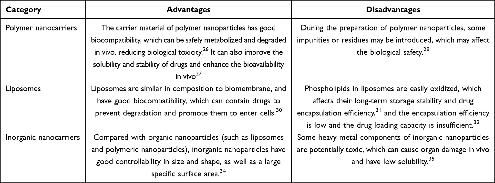 |
Table 1 Advantages and Disadvantages of Different Nanodelivery Systems |
Synergistic Basis of Nanodelivery System and STING Signal Pathway
Characteristics and Delivery Requirements of STING Agonists
Cyclic dinucleotide (CDN) agonists are natural ligands or analogs of STING that can directly bind to the STING receptor, activate the STING protein and trigger downstream signaling pathways, and promote the production of IFN-I, initiating a critical immune response cascade for antitumor activity.36 ADU-S100, being the initial STING agonist, can induce the activation of the STING pathway through intratumoral injection, which induces the generation of CD8+ T cells that are tumor-specific. Cardin et al37 synthesized a carbon cyclopyrimidine compound CDN (15a), which can activate the STING pathway via intravenous injection, thereby promoting cytokine secretion and the activation of CD8+ cytotoxic T cells, producing adaptive immune response.
Unlike traditional CDN agonists, non-CDN agonists do not rely on imitating the natural cyclic dinucleotide structure. Compared with CDN agonists, Non-CDN agonists have higher metabolic stability in vivo and are not easily degraded, so they are more suitable for systemic administration.38 Non-CDN agonists include aminobenzimidazole, xanthone, acridone, and benzothiophene classes. Unlike CDNs, non-CDN agonists activate the STING pathway through a distinct mechanism, and the binding mechanisms of different agonists vary. For example, SR-717 can act as a cGAMP mimic, directly activating the STING pathway and enhancing the activation of CD8+ T cells and DCs within associated tissues;39 MSA-2 induces conformational changes in STING, converting it to an active state and thereby activating the STING pathway.40
ENPP1 inhibitors have been developed as novel STING agonists. ENPP1 is a transmembrane phosphodiesterase that downregulates cGAS-STING pathway signaling by degrading cGAMP.41 Cells lacking ENPP1 exhibit higher capacity for activating STING with cGAMP, and inhibiting ENPP1 enhances signaling through the cGAS-STING pathway.42 Li et al43 discovered an effective ENPP1 inhibitor, ZX-8177, which prevents ENPP1 from hydrolyzing cGAMP, thereby increasing the expression level of IFN-β in vivo. This inhibitor significantly inhibits tumor growth and enhances antitumor immune responses. Table 2 lists the types and route of delivery of some STING agonists.
 |
Table 2 Clinical Stages and Types of STING Agonists |
STING agonists face many challenges in cancer treatment.44 For example, CDN-derived STING agonists have significant effects in activating STING. However, they have poor stability in vivo, are prone to degradation, have a short half-life, and have difficulty crossing the cell membrane barrier.45,46 This limits their effective duration of action in the body and affects the therapeutic effect. Non-CDN STING agonists have overcome the above limitations to a certain extent, but they have poor water solubility, and injection administration often leads to systemic toxicity and significant adverse reactions.47 Therefore, STING agonists are mainly administered by intratumoral injection, but this mode of administration is not suitable for metastatic lesions and tumors that are located in hard-to-reach deep organs or in the central nervous system.48,49 For example, CDN-based STING agonists such as ADU-S100 require intratumoral injection,50,51 which not only limits their therapeutic effect on deep-seated tumors or metastatic lesions, but also increases the complexity of clinical operations and the pain of patients. The use of STING agonists can also lead to serious toxic and side effects, such as rheumatoid arthritis, non-alcoholic fatty liver disease, systemic lupus erythematosus and other autoimmune diseases, which limit their widespread clinical application.52–54
In the in vivo environment, STING agonists are prone to hydrolysis, nanocarriers can enhance the stability of drugs and improve the water solubility of poorly soluble drugs, so as to increase their bioavailability in vivo.55,56 In addition, STING agonists need to be specifically delivered to target cells such as tumor cells or immune cells. Nanocarriers can deliver STING agonists to target diseased tissues and alleviate the side effects and reduce the impact of drugs on normal tissues.57 The application of nanocarriers can also increase the drug concentration in tumors, thus the efficacy of cGAS-STING agonist is enhanced to some extent.58
Regulatory Potential of Nanodelivery System on STING Signaling Pathway
Nanodelivery systems have the potential to boost the effectiveness of STING agonist immunotherapy. Shaji et al59 compared the antitumor effects of cGAMP encapsulated in lipid nanoparticles (LNPs) and free cGAMP using a mouse model. The study demonstrated that the administration of cGAMP encapsulated in LNPs to mice effectively suppressed tumor growth.
Nanodelivery systems can also modulate immunosuppressive cells. Nanoparticles can be modified on their surface or loaded with specific drugs to modulate immunosuppressive cells within the TME.60 For instance, nanocarriers carrying immunomodulatory agents can boost the percentage of CD8⁺ T cells within tumors and diminish the infiltration of immunosuppressive cells into tumors.61
Additionally, the properties of nanomaterials themselves (such as surface charge, chemical composition, and topological structure) may have direct or indirect effects on the STING signaling pathway. Some nanomaterials can act as agonists to directly activate the STING pathway. Zhang et al62 found that MnO2 nanomaterials, by converting to Mn²⁺, show potential as cGAS agonists. Mn²⁺ not only boosts the DNA-sensing capability of cGAS and the formation of cGAMP, but also enhances STING’s affinity for cGAMP, thus efficiently activating the cGAS-STING pathway.60 Cheng et al63 merged exosomes from M1 macrophages and tumor cells that were genetically modified to overexpress CD47, designing and synthesizing gene-engineered hybrid exosomes (gHE). To activate the STING pathway, they further encapsulated DNA-damaging chemotherapeutic agent SN38 coupled with MnO2 nanoparticles. In the acidic TME, MnO2 nanoparticles decompose into Mn²⁺, which, together with cytoplasmic DNA produced by immunogenic cell death induced by the therapeutic efficacy of SN38 in chemotherapy, synergistically activates cGAS-STING pathway.
Nanomaterials can also indirectly promote the intracellular action of STING agonists by regulating cell uptake and endosomal escape mechanisms, revealing their intrinsic synergy. For example, Shae et al64 designed STING-activating nanoparticles (STING-NPs) that can respond in the acidic endosomal environment, releasing polymer fragments that disrupt the endosomal membrane, and promote the intracellular release of cGAMP, thereby enhancing the activation of the STING pathway and the antitumor immune response.
Design Strategy of Drug Delivery System
Drug Delivery System Based on Tumor Microenvironment Response
Tumor Specific Enzyme Activation
The aberrant expression of enzymes such as matrix metalloproteinases (MMPs), cathepsins, phospholipases and redox enzymes in TME is due to the faster proliferation of tumor cells compared to normal tissues, which requires more enzymes to provide functional support.65 MMPs are enzymes that promote tumor invasion and metastasis.66 MMPs degrade the extracellular matrix (ECM), compromising its structural integrity and facilitating tumor invasion and metastasis.67 Additionally, MMPs can promote tumor angiogenesis by degrading the ECM.68 The degradation of the ECM and vascular basement membrane by MMPs creates space for the formation of new blood vessels, allowing endothelial cells to proliferate and extend toward the tumor.69 Hyaluronic acid (HA) is a high-molecular-weight polysaccharide that is widely present in the ECM and plays a significant role in tumor development and progression.70 HA engages with cell surface receptors and activates signal pathways that enhance the proliferation, migration and invasion of tumor cells.71 Hyaluronidase can degrade hyaluronic acid, thereby improving the TME, inhibiting tumor growth, and enhancing the penetration of antitumor drugs into the tumor.72 Cathepsin B (CtsB) is a lysosomal cysteine protease capable of directly degrading ECM components, including collagen and fibronectin.73 It also indirectly promotes tumor invasion and metastasis by activating MMPs. Moreover, CtsB can degrade specific ECM components such as tenascin-C, which promotes angiogenesis and thus supports tumor growth.74
Therefore, key enzymes in the TME can be designed as enzyme-responsive nanoparticles to inhibit tumor growth. By designing prodrug structures that can be cleaved by specific enzymes (such as MMPs, and hyaluronidase, HAase), the specific release of STING agonists in the TME can be achieved. Using nanoparticles as carriers to encapsulate STING agonists and incorporating enzyme-responsive linkers can enable targeted drug release. For example, Zhu et al75 designed an enzyme-responsive STING agonist nanoparticle (NEs@STING-Mal-NP) that is responsive to hyaluronidase (Figure 2A). This nanoparticle uses neutrophils as carriers to actively penetrate tumor tissues and release the STING agonist in response to hyaluronidase. This nanoparticle significantly enhances the tumor penetration of the STING agonist, activates the STING pathway, and improves the immunosuppressive TME. Li et al76 developed a multilayer liposome (HLHC) based on the physical cross-linking of HA and lipid molecules for efficient delivery of the STING agonist cGAMP (Figure 2B).
 |
Figure 2 (A) The design strategy of NEs@STING-Mal-NP. Reproduced from Hao M, Zhu L, Hou S, Chen S et al. Sensitizing tumors to immune checkpoint blockage via STING agonists delivered by tumor-penetrating neutrophil cytopharmaceuticals. ACS nano. 2023;17(2):1663–1680. Copyright © 2023 American Chemical Society.75 (B) Preparation process of multilayer liposomes based on physical crosslinking of hyaluronic acid and lipid molecules for delivering STING agonist cGAMP. Reproduced from Yu J, Li X, Li J, Sun N, Cheng P et al. Single-Dose Physically Cross-Linked Hyaluronic Acid and Lipid Hybrid Nanoparticles Containing Cyclic Guanosine Monophosphate–Adenosine Monophosphate Eliminate Established Tumors. ACS nano. 2024;18(43):29,942–29955. Copyright © 2024 American Chemical Society.76 |
Immune cells in TME can be activated by the release of STING agonists from enzyme-responsive drug delivery systems, which enhances the efficacy of immunotherapy. Although there are currently few studies on the combination of enzyme-responsive drug delivery systems and STING agonists, this strategy holds significant potential for clinical application and is worthy of further research and exploration. In the future, personalized drug delivery regimens could be tailored based on the enzyme expression profiles in a patient’s TME, further improving therapeutic outcomes.
PH Response Mechanism
Tumor cells primarily generate energy through glycolysis, a metabolic process that leads to the generation of substantial quantities of lactic acid.77 Lactic acid is an acidic metabolic byproduct, and its accumulation significantly reduces the extracellular pH, thereby establishing a microenvironment that is acidic.78 This acidic microenvironment within tumors facilitates the invasion of tumor cells while stimulating their colonization and expansion at metastatic sites.79 This acidic microenvironment can inhibit the function of immune cells by inhibiting the differentiation of monocytes into dendritic cells and the anti-tumor activity of natural killer cells and cytotoxic T cells, thus promoting the immune escape of tumor cells.80 Moreover, an acidic pH can promote the formation of premetastatic niches in distant organs, creating favorable conditions for tumor cell metastasis.79 A pH-sensitive cationic polymer modified lipid nanoparticle (LNP-B) loaded with CDNs has been developed.81 Figure 3A shows the preparation process of LNP-B. In order to efficiently encapsulate the negatively charged CDNs, a distinctive functional phospholipid known as DSPE-PCB+ was synthesized, featuring multiple positive charges at one end.81 This enhances the ability of CDNs to load drugs in the lipid nanoparticles. In an acidic environment, DSPE-PCB+ experiences hydrolysis, promoting the release of CDNs from the lipid nanoparticles, thereby enhancing immunogenic cell death (ICD) in tumor cells. Compared with free CDNs, LNP-B prolonged the circulation of CDNs and enhanced their accumulation in tumors. Wang et al82 designed a nanomicelle formulation called D-SAM, encapsulating the STING agonist cGAMP within micelles of another STING agonist, PC7A. The PC7A in D-SAM contains ultra-pH-sensitive tertiary amines that can be protonated under weakly acidic conditions, creating a proton sponge effect. This buffers the lysosomal pH to delay the degradation of STING, allowing the agonist to be released and activate STING more sustainably. Compared with the classical STING agonist ADU-S100, D-SAM not only effectively prolonged the activation and infiltration of CD8-positive T cells but also avoided T cell exhaustion and apoptosis (Figure 3B).
 |
Figure 3 (A) Preparation of pH-sensitive polycationic polymer modified lipid nanoparticles (LNP-B) system for tumor delivery of intravenous CDNs. Reproduced from He Y, Zheng K, Qin X, Wang S, Li X, Liu H, Liu M, Xu R, Peng S, Pang Z. Intravenous delivery of STING agonists using acid-sensitive polycationic polymer-modified lipid nanoparticles for enhanced tumor immunotherapy. Acta Pharm Sin B. 2025 Mar;15(3):1211–1229. © 2025 The Authors. CC BY-NC-ND license.81 (B) D-SAM extends the activation duration and infiltration period of CD8-positive T cells. (Some data in this paper are expressed by mean standard deviation (SD). The significant difference analysis method of each group data is one-way ANOVA. Reproduced from Wang J, Wang X, Xiong Q, Gao S, Wang S, Zhu S, Xiang S, Li M, Xie H, Li S. A dual-STING-activating nanosystem expands cancer immunotherapeutic temporal window. Cell Rep Med. 2024 Nov 19;5(11):101,797. © 2024 The Author(s). CC BY-NC-ND license.82 The degree of difference between each group is marked as: n.s., and there is no obvious difference; *P<0.05; **P<0.01; ***P<0.001) (C) Synthesis and Mechanism of AMSC MOFs. Reproduced from Zhou Y, Huang X, Wu D, Xie X et al. HIFU postoperative hypoxia enables metal-organic frameworks amplifying banoxantrone and STING activation for enhanced immunotherapy. Chem Eng J. 2025;159,704. © 2025 Elsevier B.V. All rights are reserved, including those for text and data mining, AI training, and similar technologies.83 |
Currently, pH-responsive drug delivery systems still face many challenges in practical applications. Many pH-responsive materials have poor stability and are prone to degradation. For example, some biopolymer carriers are easily degraded by enzymes in the body, leading to premature drug release.84 Moreover, pH-responsive carriers in the body may be interfered with by blood components, metabolic products, and other factors, which can affect their responsive performance.85
Hypoxia Response
The hypoxic condition within tumor tissues is a key feature of the TME.86 The formation of a hypoxic environment in tumors is caused by processes such as the incomplete structure and poor permeability of tumor blood vessels.87 The STING signaling pathway is inhibited by tumor hypoxia.88 Improving the hypoxic state helps to restore the normal function of the STING signaling pathway and boosts the immune response against tumors. Some nanoparticles can carry oxygen or substances that can generate oxygen, thereby improving the hypoxic environment of tumor tissues. For example, Zhou et al83 developed a metal-organic framework (MOF) nano-system, which was composed of Mn2+, CaCO3, a prodrug of barnesonone (AQ4N) and STING agonist SR-717 (AMSC MOFs). Figure 3C shows the preparation process and mechanism of AMSC MOFs. CaCO3 consumes lactic acid in TME to alleviate immunosuppression caused by hypoxia. The nanosystem effectively activates the STING pathway through SR-717 and CaCO3 consuming lactic acid. Yu et al89 developed a high-valence bismuth nanoparticle platform NaBiVO3-PEG, which can generate of reactive oxygen species (ROS). Because tumor hypoxia can inhibit the activation of STING signaling pathway, the nano-platform has the potential to trigger the STING signaling pathway, thereby enhancing the anti-tumor immune response. Table 3 lists some design strategies of drug delivery system based on TME reaction.
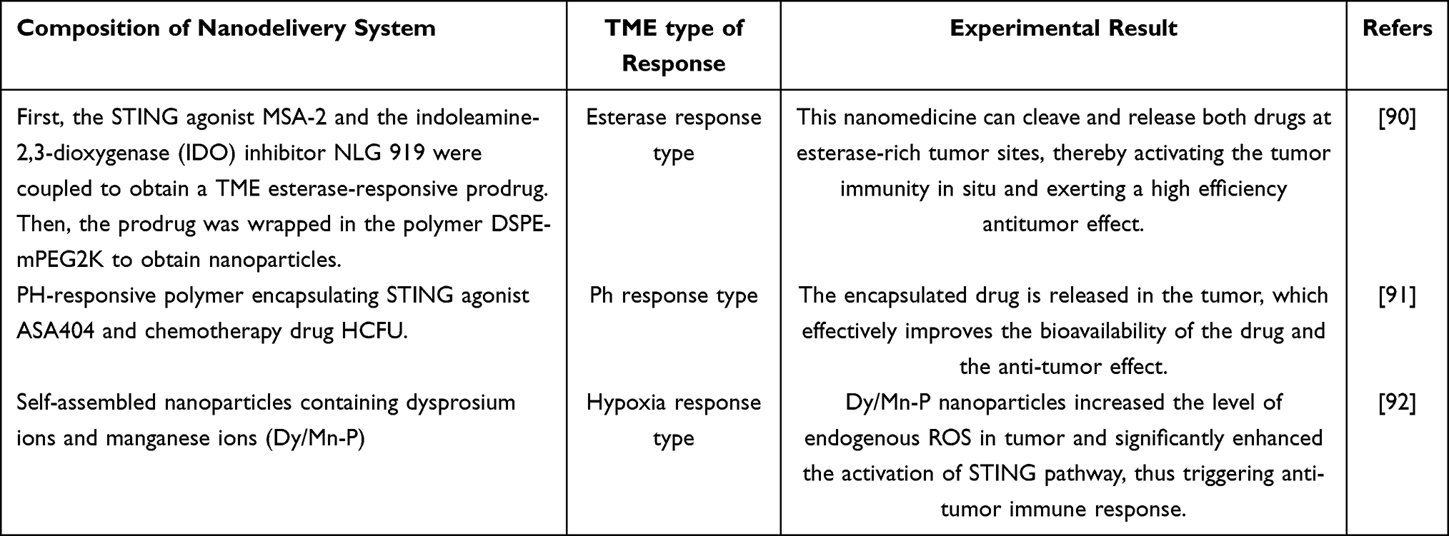 |
Table 3 Drug Delivery System Based on Tumor Microenvironment (TME) Response |
Cell Targeted Drug Delivery System
Immune Cell Targeting
The STING signaling pathway is an essential component of innate immunity, capable of activating immune responses by recognizing intracellular DNA damage and pathogen DNA. Additionally, activation of the STING pathway can enhance the TME and boost the infiltration of immune cells.60 The activation of STING promotes the maturation of DCs and enhances their antigen-presenting ability, thereby activating T cells.93 It also promotes the secretion of chemokines, guiding T cells to infiltrate the tumor site.94 STING activation can transform an immunosuppressive TME into an immunosupportive one.95 For example, activation of STING can induce the polarization of tumor-associated macrophages (TAMs) from the M2 phenotype, which is immunosuppressive, to the immunostimulatory M1 phenotype.96
DCs, as antigen-presenting cells,97 play a crucial role in both innate and adaptive immunity and are central to antitumor immunity. DCs are capable of capturing and processing tumor antigens, activating cytotoxic T cells and natural killer (NK) cells, and thus play an essential role in antitumor immune pathways.98 After recognizing tumor cells, DCs present processed tumor antigens to T cells via antigenic peptides bound to major histocompatibility complexes as the first signal for activation.99 The upregulation of costimulatory molecules such as CD80 and CD86 on the surface of DCs provides the second signal for activation.100 Naive T cells, stimulated by these two signals, become specific cytotoxic T lymphocytes (CTLs),101 thereby exerting targeted antitumor effects. DCs can release a variety of chemokines, such as CXCL9, CXCL10, and CCL5.101,102 These chemokines recruit T cells expressing CXCR3 to infiltrate the tumor and also recruit NK cells. Activated NK cells leave the lymph nodes and travel to the tumor site to kill cancer cells.103 Within the tumor microenvironment (TME), macrophages exhibit functional heterogeneity and are principally categorized into two distinct subtypes: pro-inflammatory M1 macrophages and immunomodulatory properties M2 macrophages. M1 macrophages can secrete a variety of pro-inflammatory cytokines and chemokines.104 These cytokines and chemokines can activate T cells and enhance antitumor immune responses. M2 macrophages have anti-inflammatory properties and can release immunosuppressive factors, which can promote tumor progression, angiogenesis, and immune evasion.104,105
Targeting nanoparticles to immune cells is a feasible design strategy. By selecting appropriate nanomaterials, such as mannosylated liposomes and nanoparticles conjugated with anti-CD11c antibodies, precise targeting and recognition of immune cells can be achieved. Additionally, optimizing the drug loading and release patterns to match the intracellular environment of immune cells can optimize the performance of drug delivery.
Mannose is a type of sugar ligand that can be recognized and endocytosed by mannose receptors on the exterior of immune cells like macrophages and dendritic cells, and then enter the lysosomes within the cell. Mannose-modified liposomes can achieve precise targeting of immune cells through specific binding to mannose receptors. Liao et al106 developed D-mannose-modified nanoliposomes (MLipo@OVA) for the targeted delivery of ovalbumin (OVA) to macrophages. This type of liposome significantly increased the uptake efficiency of macrophages. Studies have shown that mannose-modified liposomes can be efficiently absorbed by DCs, promote the maturation of DCs, and significantly inhibit tumor metastasis in vivo.107 Although there are currently relatively few research examples of using mannitol-modified nanoparticles to target immune cells to activate the STING pathway, this method shows great potential in terms of both theory and application prospects. CD11c is an antigen primarily expressed on the surface of dendritic cells. Nanoparticles conjugated with anti-CD11c antibodies can achieve precise targeting through specific binding to the CD11c antigen on the surface of dendritic cells. Zhang et al108 synthesized mannose-modified stearic acid grafted chitosan micelles (M-CS-SA) and cooperated with oxaliplatin. The nano-micelle can target dendritic cells, activate cGAS-STING signal pathway in cooperation with oxaliplatin, and enhance the anti-tumor effect. Figure 4A shows a schematic diagram of the synergy between oxaliplatin and M-CS-SA.
 |
Figure 4 (A) Schematic diagram of synergy between oxaliplatin and M-CS-SA. Reproduced from Nguyen NT, Le XT, Lee WT, Lim YT, Oh KT, Lee ES, Choi HG, Youn YS. STING-activating dendritic cell-targeted nanovaccines that evoke potent antigen cross-presentation for cancer immunotherapy. Bioact Mater. 2024 Sep 6;42:345–365. © 2024 The Authors. CC BY-NC-ND license.109 (B) Compared the fluorescence intensity between targeted liposomes and non-targeted liposomes, the targeted liposomes showed higher uptake. Reproduced from Wu YT, Fang Y, Wei Q, Shi H, Tan H, Deng Y, Zeng Z, Qiu J, Chen C, Sun L, Chen ZJ. Tumor-targeted delivery of a STING agonist improvescancer immunotherapy. Proc Natl Acad Sci U S A. 2022 Dec 6;119(49):e2214278119. Copyright © 2022 the Author(s). Published by PNAS. Creative Commons Attribution License 4.0 (CC BY).110 (The degree of difference between each group is marked as: n.s., and there is no obvious difference; *P<0.05; **P<0.01). |
Although immune cell targeting has shown remarkable potential for the therapy of neoplasms and infectious illnesses, it also faces many challenges. For example, immunosuppressive cells and metabolic products in TME can weaken the capacity of immune cells and promote drug resistance.111 Moreover, the heterogeneity and mutability of tumor cell surface antigens present obstacles for immune cells to exactly recognize and target tumor cells.112 To address these issues, immune cell therapies that can target multiple tumor antigens simultaneously can be developed to enhance targeting specificity. Additionally, combining multiple therapies to eliminate immunosuppressive cells can reduce drug resistance.
Tumor Cell Targeting
Tumor cells express a variety of specific receptors on their surface, which have a major impact on occurrence, development, metastasis, and treatment of tumors. The number of epidermal growth factor receptors (EGFR) is significantly increased in various tumor cells.113 The EGFR signaling pathway is crucial for regulating cell proliferation, differentiation, survival, and development. Upon activation, it promotes cell proliferation, survival, migration, and angiogenesis through downstream signaling pathways such as Ras-Raf-MEK-ERK and PI3K-Akt-mTOR.114
Human epidermal growth factor receptor 2 (HER2) is overexpressed in several cancers, including breast, gastric, and ovarian cancers.115 HER2 forms heterodimers with other HER family members to activate the PI3K-Akt-mTOR and Ras-Raf-MEK-ERK pathways, thereby promoting cell proliferation, survival, migration, and invasion.116
Prostate-specific membrane antigen (PSMA) is highly expressed in malignant prostate epithelial cells, with expression levels increasing as the malignancy of prostate cancer progresses.117,118 PSMA activates the PI3K-Akt pathway and intracellular calcium release by stimulating the glutamate receptor (mGluR), upregulating the mTOR pathway to promote cell proliferation and survival.119 PSMA inhibitors can target its binding sites to delay or even terminate the proliferation of prostate cancer cells.120,121
The abnormal expression and signaling mechanisms of these receptors on the surface of tumor cells are important factors in the occurrence, development, and malignant transformation of tumors, and have become key targets for cancer therapy. Sun et al122 developed HER2-targeted nanoliposomes for co-delivering viral peptides and the STING agonist diABZI. By targeting HER2 specifically, diABZI was delivered specifically to tumor cells. Figure 4B shows a comparison of the fluorescence intensity between targeted and non-targeted liposomes, with the targeted liposomes demonstrating superior uptake.
Targeted therapy for tumor cells is of significant importance in cancer treatment but also faces many challenges. Targeted drugs for tumor cells can specifically recognize antigens on the surface of cancer cells, but drug’efficiency entry into cancer cells is still limited.123 For example, the TME is complex, containing a variety of cells and extracellular matrix components.124 These components form physical and chemical barriers that hinder the penetration and uptake of drugs.125 Currently, innovative drug delivery systems are being developed to improve the efficiency of drug entry into tumor cells. Moreover, as treatment progresses, tumor cells may develop drug resistance through genetic mutations, reducing the effectiveness of treatment. Table 4 lists some design strategies of cell targeted drug delivery system.
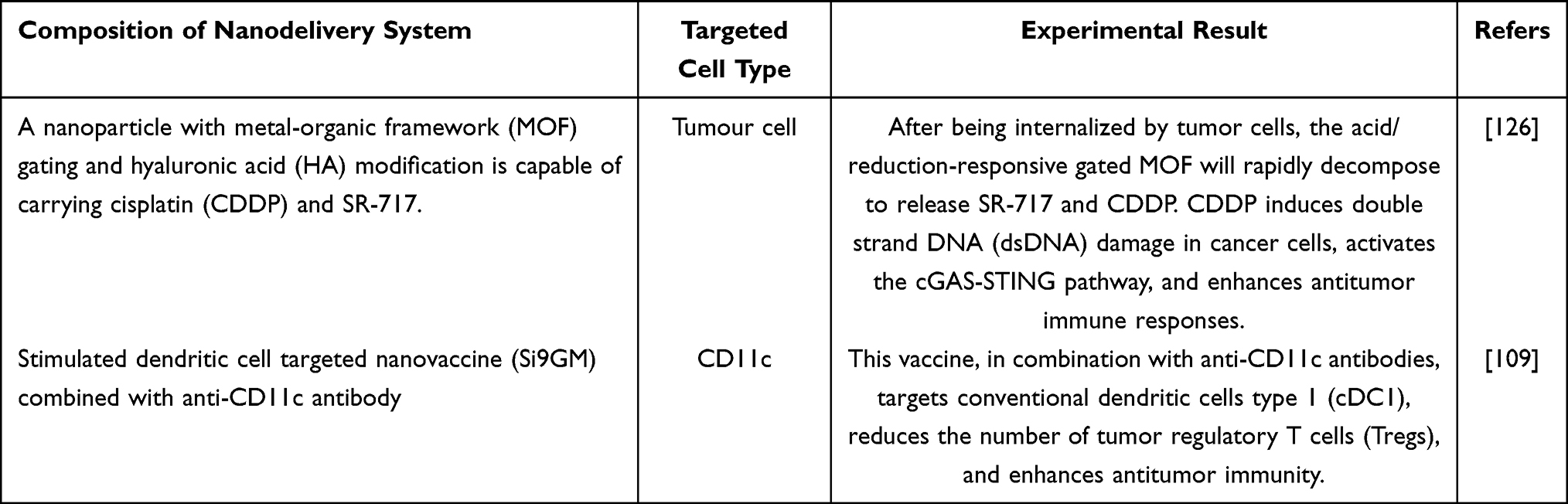 |
Table 4 Cell Targeted Drug Delivery System |
Multifunctional Integrated Drug Delivery System
Combination of Imaging and Treatment
Nanoplatforms can integrate therapeutic drugs and imaging contrast agents into one entity, enabling simultaneous diagnosis and treatment. This integrated design not only allows for precise localization of the tumor’s position, size, and boundaries but also enables real-time monitoring of treatment efficacy.127,128 Nanoplatforms can also carry targeting molecules that specifically bind to receptors or antigens outer membrane of tumor cells, thereby achieving tumor targeting and reducing distribution of drugs in normal tissues, which in turn reduces toxicity to normal tissues.129 The imaging function of nanoplatforms can monitor in real-time the distribution, release of the drug, and the tumor’s response.
Biosensors constructed via nanotechnology can monitor in real-time the changes in STING pathway and the body’s immune reaction during tumor therapy.130,131 A fluorescent molecular probe designated as BN-O was developed by Song er al.132 through structural optimization of an N-oxide framework. This engineered compound demonstrates hypoxia-responsive characteristics, enabling simultaneous generation of both near-infrared II (NIR-II) region fluorescence emissions and photoacoustic detection signals under oxygen-deficient microenvironmental conditions (Figure 5A). This mechanism additionally stimulates cGAS-STING pathway, thereby facilitating the secretion of damage-associated molecular patterns along with inflammatory cytokines from neoplastic cells. This, in turn, enhances the development of dendritic cells and the activation of T cells, ultimately accomplishing precise tumor diagnosis and effective therapy.133,134 Sun et al135 designed a NIR-II photoactivatable immunotheranostic nanoparticle (SAPTN) based on a STING agonist. SAPTN has high spatial and temporal resolution deep tissue NIR-II imaging capabilities, which can clearly diSTINGuish tumor margins from surrounding tissues, thereby guiding phototherapy.
 |
Figure 5 (A) Schematic diagram of nano-platform that can be activated in low oxygen environment, which is used to start NIR-II fluorescence imaging, photoacoustic imaging and enhance cancer immunotherapy. Reproduced from Song, J., Wang, H., Meng, X. et al. A hypoxia-activated and microenvironment-remodeling nanoplatform for multifunctional imaging and potentiated immunotherapy of cancer. Nat Commun 15, 10,395 (2024). Creative Commons Attribution 4.0 International License.132 (B) Effect of intratumoral injection of STING alone on tumor treatment by the fusion of systemic α-PD-1 and/or α-CTLA-4 therapies. Reproduced from Dorta-Estremera S, Hegde VL, Slay RB, Sun R, Yanamandra AV, Nicholas C, Nookala S, Sierra G, Curran MA, Sastry KJ. Targeting interferon signaling and CTLA-4 enhance the therapeutic efficacy of anti-PD-1 immunotherapy in preclinical model of HPV+ oral cancer. J Immunother Cancer. 2019 Sep 18;7(1):252. © The Author(s). 2019. Creative Commons Attribution 4.0 International License.136 (The degree of difference between each group is marked as: *P<0.05; ***P<0.001; ****P<0.0001). |
Nanoplatforms integrating imaging and therapeutic functions hold significant importance in tumor treatment. They can significantly improve diagnostic accuracy, enhance therapeutic efficacy, improve the TME, reduce side effects, and support personalized treatment. However, the preparation of nanoplatforms faces numerous complex challenges. For instance, the extremely small size of nanoplatforms necessitates highly precise control techniques during the preparation process. Moreover, the preparation process of nanoplatforms is often quite complex. Overcoming the limitations of nanoplatforms is an important direction for the future development of precision oncology.
Combination Therapy and Synergistic Administration
Cancer cells exhibit significant heterogeneity in their genetics, phenotype, and function, and different cells have varying sensitivities to treatment. Monotherapy may fail to cover all cancer cells comprehensively, leading to the survival of some cells and subsequent drug resistance. Moreover, monotherapy is often insufficient to address the complexity of tumors.137 Immunotherapy, such as PD-1/PD-L1 inhibitors, relies on the infiltration and activation of immune cells in the TME. However, some tumors are characterized as “immune-cold” tumors, meaning that immune cells have difficulty infiltrating the tumor tissue, resulting in poor efficacy of immunotherapy.138,139 Chemotherapy and radiotherapy, while capable of directly killing cancer cells, also have significant toxicity to normal tissues and are prone to inducing drug resistance.140,141
Monotherapy has many limitations in cancer treatment, while combination therapy can act on cancer cells through multiple mechanisms, making it difficult for cancer cells to develop resistance mutations to multiple drugs simultaneously, thereby significantly improving treatment efficacy. For example, radiotherapy can induce immunogenic cell death (ICD), releasing tumor antigens to activate the immune system.142 Immunotherapy, on the other hand, can further increase the local bioavailability of the drug, thereby enhancing the efficacy of immunotherapy.143 Sun et al144 significantly enhanced the antitumor immune response through local radiotherapy combined with dual-immune-activating nanoadjuvants (STING-TLR9). The dual-immune-activating nanoadjuvants were able to counteract the immunosuppressive effects associated with radiotherapy, and this combination therapy achieved full eradication of tumors and metastatic lesions in various syngeneic mouse tumor models. The combined administration of PD-1 and CTLA-4 immune checkpoint inhibitors with intralesional ADU-S100 delivery exhibited significant therapeutic efficacy, inducing persistent tumor volume reduction in 71% of treated neoplastic lesions.136 The efficacy of the combination therapy was superior to that of PD-1 antibody monotherapy. Figure 5B shows the effect of intratumoral injection of STING alone on tumor treatment compared with systemic α-PD-1 and/or α-CTLA-4 treatment combination. Hao et al145 also used the combination of photodynamic therapy and STING agonist to treat colorectal cancer and experimentally validated that the efficacy of the combined therapy was significant and superior to that of monotherapy. It is also possible to achieve a synergistic antitumor effect by combining metal-based nanosystems that can activate the STING pathway with other therapeutic modalities such as chemotherapy, radiotherapy, and immune checkpoint inhibitors.146
Moreover, due to the synergistic effects of combination therapy, the required dosage of each drug can be appropriately reduced, which helps to mitigate toxic side effects. For example, the combination of targeted therapy with chemotherapy or immunotherapy can inhibit tumor growth at multiple stages, thereby reducing the risk of drug resistance. The combination of the STING agonist MSA-2 with PD-1 antibodies can reduce the likelihood of resistance.147 Table 5 lists some design strategies of multifunctional integrated drug delivery system.
 |
Table 5 Multifunctional Integrated Drug Delivery System |
Novel Biomaterial-Driven Drug Delivery System
Biodegradable Material
Natural biodegradable materials are mainly derived from renewable natural resources and possess excellent biocompatibility and environmental friendliness. Common natural biodegradable materials include starch-based materials, cellulose and lignin, and protein-based materials. These natural biodegradable materials can rapidly absorb moisture from blood, promoting coagulation, and can be used as hemostatic materials. Wu et al151 developed a novel starch-based hemostatic sponge incorporating serotonin (SLS sponge) by integrating serotonin into a starch matrix through esterification and amidation reactions. The SLS sponge is able to quickly set off the coagulation cascade, promote the grouping of red blood cells and platelets, and enhance platelet activation. Natural biodegradable materials can also be used as scaffold materials to guide cell growth and tissue repair. Cordeiro R et al152 tested the porosity, mechanical properties and biocompatibility of different types of cellulose (including microcrystalline cellulose, methylcellulose, and corn core cellulose) and found that the addition of cellulose can promote cell adhesion and proliferation. Xun et al153 developed a macroporous bacterial cellulose (BC) scaffold crosslinked by calcium ions for cranial bone regeneration. Natural biodegradable materials such as starch-based nanomaterials, owing to their small size, high specific surface area, and biocompatibility, have been used to develop precision drug delivery systems that can actively recognize cancer cells and achieve targeted drug release.154 Jeong et al155 developed a urease-powered nanomotor (STING@nanomotor) using chitosan, a natural biodegradable material, as the base material. The biocompatibility and mucosal adhesion of chitosan enable it to effectively bind to and penetrate the mucus layer on the bladder wall, thereby increasing retention time of the nanomotor in the bladder and the efficiency of drug delivery. Figure 6A shows the fabrication method of a nano-motor driven by urease and loaded with a STING agonist. Wang et al156 fused different glioma targeting parts to ferritin nanoparticles (RGE-HFn NPs), and wrapped or bound STING agonist SR717 in nanoparticles (SR717@RGE-HFn NPs). Although the research and application of natural biodegradable materials such as cellulose in the delivery of STING agonists are relatively few, these materials show great potential for diverse applications in the field of STING agonist delivery because of their biodegradability and environmental friendliness.
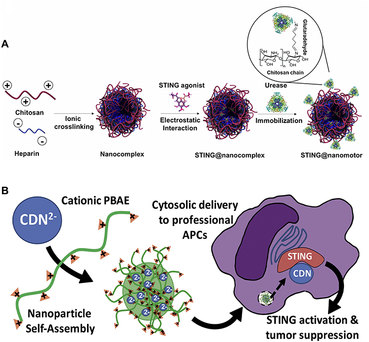 |
Figure 6 (A) The fabrication procedure of the urease-driven nano-motor incorporating a STING agonist. Reproduced from Choi H, Jeong SH, Simó C, Bakenecker A, Liop J, Lee HS, Kim TY, Kwak C, Koh GY, Sánchez S, Hahn SK. Urease-powered nanomotor containing STING agonist for bladder cancer immunotherapy. Nat Commun. 2024 Nov 15;15(1):9934. © The Author(s) 2024. Creative Commons Attribution-NonCommercial-NoDerivatives 4.0 International License.155 (B) PBAE nanoparticles deliver CDNs and act on STING signaling pathway. Reproduced from Wilson DR, Sen R, Sunshine JC, Pardoll DM, Green JJ, Kim YJ. Biodegradable STING agonist nanoparticles for enhanced cancer immunotherapy. Nanomedicine: Nanotechnology. Biology and Medicine. 2018;14(2):237–246. © 2017 Elsevier Inc. All rights reserved.157 |
In the sphere of medication delivery system development, synthetic biodegradable materials like polylacticacid (PLA), polyglycolicacid (PGA), and their copolymer PLGA are utilized. These materials can encapsulate drugs and achieve targeted drug delivery and sustained release in the form of nanoparticles, microspheres, or films. For example, PLGA nanoparticles have been used to encapsulate STING agonists, significantly improving drug delivery efficiency and immune activation. Wilson et al157 used biodegradable poly (β-amino ester) (PBAE) nanoparticles to deliver CDNs, effectively inhibiting tumor growth (Figure 6B). PBAE nanoparticles possess excellent biocompatibility and degradability and can naturally degrade in the body, avoiding the risk of immune reactions arising from prolonged retention. Gu et al158 integrated the cGAS-STING-activating vascular disrupting agent DMXAA with poly (ethylene glycol)-grafted poly (lactic-co-glycolic acid) copolymer (PLGA-PEG) to obtain PLGA-PEG/DMXAA (PPD) nanoparticles to induce tumor-specific vascular destruction, which was used for multiple synergistic treatment of cancer. Although biodegradable materials generally have good biocompatibility, the intermediate products generated during their degradation in the body may trigger immune responses in the organism, thus limiting their applications.159
Intelligent Hydrogel
Intelligent hydrogels are a type of polymeric material featuring a three-dimensional network structure, which enables them adapting to these shifts in the surrounding environment,160 such as temperature, pH, light, pressure, etc. Intelligent hydrogels can be categorized based on their response mechanisms into several types, including temperature-sensitive hydrogels, pH-sensitive hydrogels, redox-sensitive hydrogels, enzyme-sensitive hydrogels, electric field-sensitive hydrogels, light-sensitive hydrogels, and those that are multi-responsive.161,162 Wang et al163 developed a ROS-responsive hydrogel system (GEM-STING@Gel), which was used to co-deliver gemcitabine and STING agonist DMXAA. GEM-STING@Gel can form in situ at the tumor site and release drugs in response to ROS stimulation, thereby achieving targeted drug release and long-term treatment. A temperature-responsive hydrogel encapsulating STING agonist and a photothermal agent has been developed.164 This hydrogel can rapidly release drugs upon near-infrared phototherapy, which prompts rapid heating. The release of the STING agonist activates the STING pathway, promoting the release of IFN-I and further enhancing the immune response. Due to the small pore size of the hydrogel network, the transport of biomacromolecules is relatively difficult. Therefore, the response of intelligent hydrogels to biomacromolecules is slow, which restricts their widespread application.165 Table 6 lists some design strategies of the novel biomaterial-driven drug delivery system.
 |
Table 6 Novel Biomaterial-Driven Drug Delivery System |
In addition, we summarize the advantages and disadvantages of various drug delivery systems in Table 7.
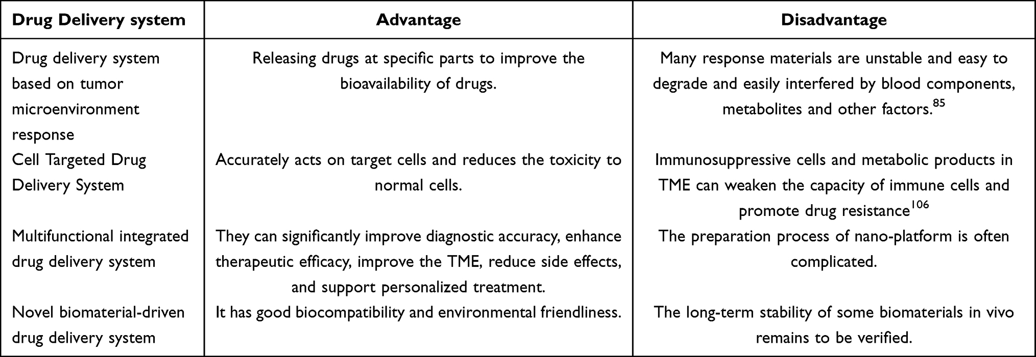 |
Table 7 The Advantages and Disadvantages of Various Drug Delivery Systems |
Prospects for STING Agonist Nanodelivery
Nanotechnology has effectively enhanced the piling up of STING agonists at neoplastic sites, while the triggering effect on the STING pathway has strengthened immune response to combat tumors.168 However, the exiSTING delivery systems all have certain limitations. By optimizing nanomaterials or developing nanomaterials with higher stability, the stability and effectiveness of drug delivery systems can be improved, thereby improving therapeutic potency of drugs. For example, biodegradable nanoparticles can be employed in combination with metal ions through coordination, which leverages the charge-shielding effect to alter the surface properties of the nanoparticles.169 This approach enhances the stability of the particles and regulates the targeting ability of the nanocarriers, thereby achieving precise treatment. Additionally, biomimetic cell membrane nanoparticles can be utilized. By mimicking the structure and function of cell membranes, these nanoparticles can evade clearance by the immune system and prolong the circulation time of the drug within the body.
The properties of STING agonists has the potential to be optimized to improve therapeutic efficacy. The physicochemical properties of STING agonists may limit their loading efficiency in nanocarriers, thereby affecting their performance in cancer immunotherapy. Chemical modification or structural remodeling of STING agonists can enhance their properties,170 such as introducing groups at specific sites of the STING agonist or by modifying the chemical bonds, making them more readily loaded by nanocarriers, not only can their cellular uptake efficiency be increased, but also their bioavailability can be enhanced, thus fully exerting their therapeutic effects in inhibiting tumor growth.
In addition to optimizing materials and drugs themselves, exploring more effective combination therapy strategies is also an important direction for enhancing therapeutic efficacy. For example, combining nanomedicine with radiotherapy. For example, nanomedicine can be used as a radiosensitizer linked to selenide bonds that are sensitive to X-rays. Through the triple dynamic phase shift effect induced by X-rays, precise treatment can be achieved.171 Moreover, big data and artificial intelligence technologies can be utilized to conduct in-depth analyses of patients’ clinical data and treatment responses, enabling the customization of the most suitable treatment plans for each individual patient and realizing personalized precision therapy. Figure 7 illustrates the methods that can improve the limitations of the combination of STING agonists and nanodelivery systems.
 |
Figure 7 The methods that can improve the limitations of the combination of STING agonists and nanodelivery systems. |
Conclusion
Currently, there are a variety of methods for treating tumors, including traditional surgery, radiotherapy, chemotherapy, as well as immunotherapy and targeted therapy, which have developed rapidly in recent years. These treatment methods have made significant progress in tumor therapy, but still have some limitations. The approach of delivering STING agonists via nanodelivery systems has brought a new direction to tumor treatment. Nanodelivery systems for STING agonists can not only overcome the problems of poor stability and rapid clearance of STING agonists, but also precisely deliver STING agonists to tumor sites, achieving targeted drug delivery and reducing side effects on normal tissues.
The synergistic approach of nanodelivery systems and the STING signaling pathway has shown significant advantages in tumor treatment. In this review, we explored and analyzed the design strategies of several types of drug delivery systems for STING agonists. Nanotechnology was used to effectively increase the accumulation of STING agonists at tumor sites, while the activation of the STING signaling pathway enhanced the antitumor immune response. However, the existing drug delivery systems all have certain limitations. For example, nanocarrier materials may have potential toxicity and side effects, the synergistic mechanisms between STING agonists and other drugs in combination therapy are not fully understood, and the stability of nanocarriers is difficult to ensure. Future research needs to explore new therapeutic strategies to overcome these limitations.
In summary, we hope that these strategies will not only bring new hope to cancer patients but also provide new ideas and orientations for the future vision of overcoming cancer challenges and offer some forward-looking guidance for subsequent research.
Data Sharing Statement
No data was used for the research described in the article.
Acknowledgments
This work was supported by the Youth Scientific Research Fund Project of Liaoning University (grant numbers LDZDJC2401).
Disclosure
The authors declare that they have no known competing financial interests or personal relationships that could have appeared to influence the work reported in this paper.
References
1. Hsu CY, Pallathadka H, Jasim SA, et al. Innovations in cancer immunotherapy: a comprehensive overview of recent breakthroughs and future directions. Crit Rev Oncol Hemat. 2024:104588. doi:10.1016/j.critrevonc.2024.104588
2. Weigmann K. Releasing the brakes to fight cancer: the recent discovery of checkpoints has boosted the field of cancer immunotherapy. EMBO Rep. 2016;17(9):1257–1260. doi:10.15252/embr.201643038
3. Haas AR, Tanyi JL, O’Hara MH, et al. Phase I study of lentiviral-transduced chimeric antigen receptor-modified T cells recognizing mesothelin in advanced solid cancers. Mol Ther. 2019;27(11):919–1929. doi:10.1016/j.ymthe.2019.07.015
4. Maeda H, Bharate GY, Daruwalla J. Polymeric drugs for efficient tumor-targeted drug delivery based on EPR-effect. Eur J Pharm Biopharm. 2009;71(3):409–419. doi:10.1016/j.ejpb.2008.11.010
5. Manstein VV, Yang CM, Richter D, Delis N, Vafaizadeh V, Groner B. Resistance of cancer cells to targeted therapies through the activation of compensating signaling loops. Curr Signal Transduct Ther. 2013;8(3):193–202. doi:10.2174/1574362409666140206221931
6. Thim-Uam A, Prabakaran T, Tansakul M, et al. STING mediates lupus via the activation of conventional dendritic cell maturation and plasmacytoid dendritic cell differentiation. Iscience. 2020;23(9):101706. doi:10.1016/j.isci.2020.101530
7. Zhang H, You QD, Xu XL. Targeting stimulator of interferon genes (STING): a medicinal chemistry perspective. J Med Chem. 2019;63(8):3785–3816. doi:10.1021/acs.jmedchem.9b01039
8. Zou Y, Zhang M, Zhou J. Recent trends in STING modulators: structures, mechanisms, and therapeutic potential. Drug Discov Today. 2023;28(9):03694. doi:10.1016/j.drudis.2023.103694
9. Xu Q, Xing J, Wang S, Peng H, Liu Y. The role of the cGAS-STING pathway in metabolic diseases. Heliyon. 2024;10(12). doi:10.1016/j.heliyon.2024.e33093
10. Shu C, Yi G, Watts T, Kao CC, Li P. Structure of STING bound to cyclic di-GMP reveals the mechanism of cyclic dinucleotide recognition by the immune system. Nat Struct Mol Biol. 2012;19(7):722–724. doi:10.1038/nsmb.2331
11. Ouyang S, Song X, Wang Y, et al. Structural and functional analysis of STING sheds new light on cyclic di-GMP mediated immune signaling mechanism. Immunity. 2012;36(6):1073–1086. doi:10.1016/j.immuni.2012.03.019
12. Gui X, Yang H, Li T, et al. Autophagy induction via STING trafficking is a primordial function of the cGAS pathway. Nature. 2019;567(7747):262–266. doi:10.1038/s41586-019-1006-9
13. Yu L, Liu P. cGAS/STING signalling pathway in senescence and oncogenesis. Semin Cancer Biol. 2024. doi:10.1016/j.semcancer.2024.08.007
14. Kato K, Omura H, Ishitani R, Nureki O. Cyclic GMP–AMP as an endogenous second messenger in innate immune signaling by cytosolic DNA. Ann Rev Biochem. 2017;86(1):541–566. doi:10.1146/annurev-biochem-061516-044813
15. Dobbs N, Burnaevskiy N, Chen D, Gonugunta VK, Alto NM, Yan N. STING activation by translocation from the ER is associated with infection and autoinflammatory disease. Cell Host Microbe. 2015;18(2):157–168. doi:10.1016/j.chom.2015.07.001
16. Zhang C, Shang G, Gui X, Zhang X, Bai X, Chen ZJ. Structural basis of STING binding with and phosphorylation by TBK1. Nature. 2019;567(7748):394–398. doi:10.1038/s41586-019-1000-2
17. de Oliveira Mann CC, Orzalli MH, King DS, Kagan JC, Lee ASY, Kranzusch PJ. Modular architecture of the STING C-terminal tail allows interferon and NF-κB signaling adaptation. Cell Rep. 2019;27(4):1165–1175.e5. doi:10.1016/j.celrep.2019.03.098
18. Li X, Yang M, Yu Z, et al. The tyrosine kinase Src promotes phosphorylation of the kinase TBK1 to facilitate type I interferon production after viral infection. Sci Signal. 2017;10(460):eaae0435. doi:10.1126/scisignal.aae0435
19. Park GB, Hur DY, Kim YS, Lee K, Yang JW, Kim D. TLR 3/TRIF signalling pathway regulates IL‐32 and IFN‐β secretion through activation of RIP‐1 and TRAF in the human cornea. J Cell Mol Med. 2015;19(5):1042–1054. doi:10.1111/jcmm.12495
20. Chen Q, Sun L, Chen ZJ. Regulation and function of the cGAS-STING pathway of cytosolic DNA sensing. Nat Immunol. 2016;17(10):1142–1149. doi:10.1038/ni.3558
21. Fyrstenberg Laursen M, Kofod-Olsen E, Agger R. Activation of dendritic cells by targeted DNA: a potential addition to the armamentarium for anti-cancer immunotherapy. Cancer Immunol Immunother. 2019;68(11):1875–1880. doi:10.1007/s00262-019-02400-1
22. Du J, Qian M, Yuan T, et al. cGAS and cancer therapy: a double-edged sword. Acta Pharmacol Sinica. 2022;43(9):2202–2211. doi:10.1038/s41401-021-00839-6
23. Duong E, Fessenden TB, Lutz E, et al. Type I interferon activates MHC class I-dressed CD11b+ conventional dendritic cells to promote protective anti-tumor CD8+ T cell immunity. Immunity. 2022;55(2):308–323.e9. doi:10.1016/j.immuni.2021.10.020
24. Yi M, Zheng X, Niu M, et al. Combination strategies with PD-1/PD-L1 blockade: current advances and future directions. Mol Cancer. 2022;21(1):28. doi:10.1186/s12943-021-01489-2
25. Chandra S. Natural and synthetic polymer. In: Polymers in Concrete. CRC Press; 2020:5–25.
26. Calzoni E, Cesaretti A, Polchi A, et al. Biocompatible polymer nanoparticles for drug delivery applications in cancer and neurodegenerative disorder therapies. J Func Biomater. 2019;10(1):4. doi:10.3390/jfb10010004
27. Begines B, Ortiz T, Pérez-Aranda M, et al. Polymeric nanoparticles for drug delivery: recent developments and future prospects. Nanomaterials. 2020;10(7):1403. doi:10.3390/nano10071403
28. Rao JP, Geckeler KE. Polymer nanoparticles: preparation techniques and size-control parameters. Prog Polym Sci. 2011;36(7):887–913. doi:10.1016/j.progpolymsci.2011.01.001
29. Yadav D, Sandeep K, Pandey D, et al. Liposomes for drug delivery. J Biotechnol Biomater. 2017;7(04):276. doi:10.4172/2155-952X.1000276
30. Wang Y. Liposome as a delivery system for the treatment of biofilm-mediated infections. J Appl Microbiol. 2021;131(6):2626–2639. doi:10.1111/jam.15053
31. Mosca M, Ceglie A, Ambrosone L. Effect of membrane composition on lipid oxidation in liposomes. Chem Phys Lipids. 2011;164(2):158–165. doi:10.1016/j.chemphyslip.2010.12.006
32. Kirby CJ, Gregoriadis G. A simple procedure for preparing liposomes capable of high encapsulation efficiency under mild conditions. In: Liposome Technology. CRC Press; 2019:19–27.
33. Pokharkar VB, Dhapte VV, Kadam SS. Metallic nanoparticulate drug delivery systems. Nanotechnol Drug Deliv. 2014;1:41.
34. Nam J, Won N, Bang J, et al. Surface engineering of inorganic nanoparticles for imaging and therapy. Adv Drug Deliv Rev. 2013;65(5):622–648. doi:10.1016/j.addr.2012.08.015
35. Mitchell MJ, Billingsley MM, Haley RM, et al. Engineering precision nanoparticles for drug delivery. Nat Rev Drug Discov. 2021;20(2):101–124. doi:10.1038/s41573-020-0090-8
36. Magand J, Roy V, Meudal H, et al. Synthesis of novel 3’, 3’‐cyclic dinucleotide analogues targeting STING protein. Asian J Org Chem. 2023;12(1):e202200597. doi:10.1002/ajoc.202200597
37. Vyskocil S, Cardin D, Ciavarri J, et al. Identification of novel carbocyclic pyrimidine cyclic dinucleotide STING agonists for antitumor immunotherapy using systemic intravenous route. J Med Chem. 2021;64(10):6902–6923. doi:10.1021/acs.jmedchem.1c00374
38. Caldwell SE, Janosko CP, Deiters A. Development of a light-activated STING agonist. Org Biomol Chem. 2024;22(2):302–308. doi:10.1039/D3OB01578E
39. Chin EN, Yu C, Vartabedian VF, et al. Antitumor activity of a systemic STING-activating non-nucleotide cGAMP mimetic. Science. 2020;369(6506):993–999. doi:10.1126/science.abb4255
40. Yang J, Luo Z, Ma J, Wang Y, Cheng N. A next-generation STING agonist MSA-2: from mechanism to application. J Control Release. 2024;371(2024):273–287. doi:10.1016/j.jconrel.2024.05.042
41. Kato K, Nishimasu H, Oikawa D, et al. Structural insights into cGAMP degradation by Ecto-nucleotide pyrophosphatase phosphodiesterase 1. Nat Commun. 2018;9(1):4424. doi:10.1038/s41467-018-06922-7
42. Li L, Yin Q, Kuss P, et al. Hydrolysis of 2′ 3′-cGAMP by ENPP1 and design of nonhydrolyzable analogs. Nat Chem Biol. 2014;10(12):1043–1048. doi:10.1038/nchembio.1661
43. Li Y, Peng Z, Sun S, et al. ENPP1 inhibitor ZX-8177 enhances anti-tumor activity of conventional therapies by modulating tumor microenvironment. Cancer Res. 2022;82(12_Supplement):5486. doi:10.1158/1538-7445.AM2022-5486
44. Wang R, Hussain A, Guo Q, Ma M. cGAS-STING at the crossroads in cancer therapy. Crit Rev Oncol Hemat. 2024;193:104194. doi:10.1016/j.critrevonc.2023.104194
45. Petrovic M, Borchard G, Jordan O. Considerations for the delivery of STING ligands in cancer immunotherapy. J Control Release. 2021;339:235–247. doi:10.1016/j.jconrel.2021.09.033
46. Zhou S, Cheng F, Zhang Y, Su T, Zhu G. Engineering and delivery of cGAS-STING immunomodulators for the immunotherapy of cancer and autoimmune diseases. Accounts Chem Res. 2023;56(21):2933–2943. doi:10.1021/acs.accounts.3c00394
47. Irvine DJ, Aung A, Silva M. Controlling timing and location in vaccines. Adv Drug Deliv Rev. 2020;158:91–115. doi:10.1016/j.addr.2020.06.019
48. Harrington K, Freeman DJ, Kelly B, Harper J, Soria JC. Optimizing oncolytic virotherapy in cancer treatment. Nat Rev Drug Discov. 2019;18(9):689–706. doi:10.1038/s41573-019-0029-0
49. Bommareddy PK, Shettigar M, Kaufman HL. Integrating oncolytic viruses in combination cancer immunotherapy. Nat Rev Immunol. 2018;18(8):498–513. doi:10.1038/s41577-018-0014-6
50. Esteves AM, Papaevangelou E, Dasgupta P, Galustian C. Combination of interleukin-15 with a STING agonist, ADU-S100 analog: a potential immunotherapy for prostate cancer. Front Oncol. 2021;11:621550. doi:10.3389/fonc.2021.621550
51. Filderman JN, Luke JJ, Storkus WJ. Platelet STING agonism and venous thrombosis: translational implications for improved disease outcomes. J Leukocyte Biol. 2023:195–198. doi:10.1093/jleuko/qiad073
52. Shen H, Jin L, Zheng Q, et al. Synergistically targeting synovium STING pathway for rheumatoid arthritis treatment. Bioact Mater. 2023;24:37–53. doi:10.1016/j.bioactmat.2022.12.001
53. Maher JJ. Macrophages steal STING from the infectious disease playbook to promote nonalcoholic fatty liver disease. Gastroenterology. 2018;155(6):1687. doi:10.1053/j.gastro.2018.11.009
54. Kumar V. A STING to inflammation and autoimmunity. J Leukocyte Biol. 2019;106(1):171–185. doi:10.1002/JLB.4MIR1018-397RR
55. Solaro R, Chiellini R, Battisti A. Targeted delivery of protein drugs by nanocarriers. Materials. 2010;3(3):1928–1980. doi:10.3390/ma3031928
56. Flávia LODS, Maria BDFM, Kelly CK, Guilherme C. Nanonization techniques to overcome poor water-solubility with drugs. Expert Opin Drug Dis. 2020;15(7):853–864. doi:10.1080/17460441.2020.1750591
57. Huang Y, Fan H, Ti H. Tumor microenvironment reprogramming by nanomedicine to enhance the effect of tumor immunotherapy. Asian J Pharm Sci. 2024;19(2):100902. doi:10.1016/j.ajps.2024.100902
58. Hussain Z, Zhang Y, Qiu L, Gou S, Liu K. Exploring Clec9a in dendritic cell-based tumor immunotherapy for molecular insights and therapeutic potentials. Npj Vaccines. 2025;10(1):27. doi:10.1038/s41541-025-01084-2
59. Shaji SG, Patel P, Mamani UF, et al. Delivery of a STING agonist using lipid nanoparticles inhibits pancreatic cancer growth. Int J Nanomed. 2024;19:8769–8778. doi:10.2147/IJN.S462213
60. Zhao L, Gui Y, Cai J, Deng X. Biometallic ions and derivatives: a new direction for cancer immunotherapy. Mol Cancer. 2025;24(1):17. doi:10.1186/s12943-025-02225-w
61. Wei R, Xie K, Li T, et al. Immunity/metabolism dual-regulation via an acidity-triggered bioorthogonal assembly nanoplatform enhances glioblastoma immunotherapy by targeting CXCL12/CXCR4 and adenosine-A2AR pathways. Biomaterials. 2025;319:123216. doi:10.1016/j.biomaterials.2025.123216
62. Zhang T, Hu C, Zhang W, et al. Advances of MnO2 nanomaterials as novel agonists for the development of cGAS-STING-mediated therapeutics. Front Immunol. 2023;14:1156239. doi:10.3389/fimmu.2023.1156239
63. Cheng L, Zhang P, Liu Y, et al. Multifunctional hybrid exosomes enhanced cancer chemo-immunotherapy by activating the STING pathway. Biomaterials. 2023;301:122259. doi:10.1016/j.biomaterials.2023.122259
64. Shae D, Becker KW, Christov P, et al. Endosomolytic polymersomes increase the activity of cyclic dinucleotide STING agonists to enhance cancer immunotherapy. Nat Nanotechnol. 2019;14(3):269–278. doi:10.1038/s41565-018-0342-5
65. Zhang ZT, Huang-Fu MY, Xu WH, Han M. Stimulus-responsive nanoscale delivery systems triggered by the enzymes in the tumor microenvironment. Eur J Pharm Biopharm. 2019;137:122–130. doi:10.1016/j.ejpb.2019.02.009
66. Scherer RL, McIntyre JO, Matrisian LM. Imaging matrix metalloproteinases in cancer. Cancer Metast Rev. 2008;27(4):679–690. doi:10.1007/s10555-008-9152-9
67. Overall CM, Kleifeld O. Tumour microenvironment-opinion:validating matrix metalloproteinases as drug targets and anti-targets for cancer therapy. Nat Rev Cancer. 2006;6(3):227–239. doi:10.1038/nrc1821
68. Campbell NE, Kellenberger L, Greenaway J, Moorehead RA, Linnerth-Petrik NM, Petrik J. Extracellular matrix proteins and tumor angiogenesis. J Oncol. 2010;(1):586905. doi:10.1155/2010/586905
69. Meng N, Li Y, Zhang H, Sun XF. RECK, a novel matrix metalloproteinase regulator. Histol Histopathol. 2008;23(8):1003–1010. doi:10.14670/HH-23.1003
70. Michalczyk M, Humeniuk E, Adamczuk G, Korga-Plewko A. Hyaluronic acid as a modern approach in anticancer therapy-review. Int J Mol Sci. 2022;24(1):103. doi:10.3390/ijms24010103
71. Sugahara KN, Murai T, Nishinakamura H, Kawashima H, Saya H, Miyasaka M. Hyaluronan oligosaccharides induce CD44 cleavage and pro- mote cell migration in CD44-expressing tumor cells. J Biol Chem. 2003;278(34):32259–32265. doi:10.1074/jbc.M300347200
72. Kawasaki Y, Akiyama T. Tumor microenvironment: promising therapeutic target. Nihon Rinsho. 2015;73(8):1283–1287.
73. Fonović M, Turk B. Cysteine cathepsins and extracellular matrix degradation. BBA-Gen Subj. 2014;1840(8):2560–2570. doi:10.1016/j.bbagen.2014.03.017
74. Xie Z, Zhao M, Yan C, et al. Cathepsin B in programmed cell death machinery: mechanisms of execution and regulatory pathways. Cell Death Dis. 2023;14(4):255. doi:10.1038/s41419-023-05786-0
75. Hao M, Zhu L, Hou S, et al. Sensitizing tumors to immune checkpoint blockage via STING agonists delivered by tumor-penetrating neutrophil cytopharmaceuticals. ACS Nano. 2023;17(2):1663–1680. doi:10.1021/acsnano.2c11764
76. Yu J, Li X, Li J, et al. Single-dose physically cross-linked hyaluronic acid and lipid hybrid nanoparticles containing cyclic guanosine monophosphate–adenosine monophosphate eliminate established tumors. ACS Nano. 2024;18(43):29942–29955. doi:10.1021/acsnano.4c10673
77. Warburg O. On the origin of cancer cells. Science. 1956;123(3191):309–314. doi:10.1126/science.123.3191.309
78. Apostolova P, Pearce EL. Lactic acid and lactate: revisiting the physiological roles in the tumor microenvironment. Trends Immunol. 2022;43(12):969–977. doi:10.1016/j.it.2022.10.005
79. Estrella V, Chen T, Lloyd M, et al. Acidity generated by the tumor microenvironment drives local invasion. Cancer Res. 2013;73(5):1524–1535. doi:10.1158/0008-5472.CAN-12-2796
80. Liao S, Wu G, Xie Z, et al. pH regulators and their inhibitors in tumor microenvironment. Eur J Med Chem. 2024;267:116170. doi:10.1016/j.ejmech.2024.116170
81. He Y, Zheng K, Qin X, et al. Intravenous delivery of STING agonists using acid-sensitive polycationic polymer-modified lipid nanoparticles for enhanced tumor immunotherapy. Acta Pharmaceutica Sinica. 2024. doi:10.1016/j.apsb.2024.06.004
82. Wang J, Wang X, Xiong Q, et al. A dual-STING-activating nanosystem expands cancer immunotherapeutic temporal window. Cell Rep Med. 2024;5(11). doi:10.1016/j.xcrm.2024.101797
83. Zhou Y, Huang X, Wu D, et al. HIFU postoperative hypoxia enables metal-organic frameworks amplifying banoxantrone and STING activation for enhanced immunotherapy. Chem Eng J. 2025;505:159704. doi:10.1016/j.cej.2025.159704
84. Wang S, Liu L, Ding Y, Suxin LI. Research progress of pH-responsive drug delivery systems in cancer immunotherapy. J China Pharm Univ. 2024;55(4):522–529. doi:10.11665/j.issn.1000-5048.2024011902
85. Li Y, Chen M, Yao B, et al. Dual pH/ROS‐responsive nanoplatform with deep tumor penetration and self‐amplified drug release for enhancing tumor chemotherapeutic efficacy. Small. 2020;16(32):2002188. doi:10.1002/smll.202002188
86. Petrova V, Annicchiarico-Petruzzelli M, Melino G, Amelio I. The hypoxic tumour microenvironment. Oncogenesis. 2018;7(1):10. doi:10.1038/s41389-017-0011-9
87. Singleton DC, Macann A, Wilson WR. Therapeutic targeting of the hypoxic tumour microenvironment. Nat Rev Clin Oncol. 2021;18(12):751–772. doi:10.1038/s41571-021-00539-4
88. Zhao F, Liu A, Gong X, et al. Hypoxia-induced RNASEH2A limits activation of cGAS-STING signaling in HCC and predicts poor prognosis. Tumori J. 2022;108(1):63–76. doi:10.1177/03008916211026019
89. Tang Y, Yu X, He L, et al. A high-valence bismuth (V) nanoplatform triggers cancer cell death and anti-tumor immune responses with exogenous excitation-free endogenous H2O2-and O2-independent ROS generation. Nat Commun. 2025;16(1):860. doi:10.1038/s41467-025-56110-7
90. Syeda MZ, Hong T, Zhang M, et al. A prodrug nanoplatform via esterification of STING agonist and IDO inhibitor for synergistic cancer immunotherapy. Nano Res. 2022;15(10):9215–9222. doi:10.1007/s12274-022-4598-6
91. Ma X, Yang Q, Lin N, et al. Integrated anti-vascular and immune-chemotherapy for colorectal carcinoma using a pH-responsive polymeric delivery system. J Control Release. 2024;370:230–238. doi:10.1016/j.jconrel.2024.04.028
92. Jiang X, Wu D, Xie W, Liu X, Zheng J, Zheng J. A pH-sensitive nanoparticle as reactive oxygen species amplifier to regulate tumor microenvironment and potentiate tumor radiotherapy. Int J Nanomed. 2024;19:709–725. doi:10.2147/IJN.S436160
93. Wang P, Wang Y, Li H, et al. A homologous-targeting cGAS-STING agonist multimodally activates dendritic cells for enhanced cancer immunotherapy. Acta Bio. 2024;177:400–413. doi:10.1016/j.actbio.2024.02.003
94. Su T, Zhang Y, Valerie K, Wang XY, Lin S, Zhu G. STING activation in cancer immunotherapy. Theranostics. 2019;9(25):7759. doi:10.7150/thno.37574
95. Huang R, Ning Q, Zhao J, et al. Targeting STING for cancer immunotherapy: from mechanisms to translation. Int Immunopharmacol. 2022;113:109304. doi:10.1016/j.intimp.2022.109304
96. Geng K, Ma X, Jiang Z, et al. High glucose-induced STING activation inhibits diabetic wound healing through promoting M1 polarization of macrophages. Cell Death Discov. 2023;9(1):136. doi:10.1038/s41420-023-01425-x
97. Worbs T, Hammerschmidt SI, Förster R. Dendritic cell migration in health and disease. Nat Rev Immunol. 2017;17(1):30–48. doi:10.1038/nri.2016.116
98. Banchereau J, Steinman RM. Dendritic cells and the control of immunity. Nature. 1998;392(6673):245–252. doi:10.1038/32588
99. Shurin MR. Dendritic cells presenting tumor antigen. Cancer Immunol Immunother. 1996;43:158–164. doi:10.1007/s002620050317
100. Tian Z, Zeng Y, Peng Y, et al. Cancer immunotherapy strategies that target the cGAS-STING pathway. Front Immunol. 2022;13:996663. doi:10.3389/fimmu.2022.996663
101. Guerder S, Flavell RA. T-cell activation: two for T. Curr Biol. 1995;5(8):866–868. doi:10.1016/S0960-9822(95)00175-8
102. Zhao K, Huang J, Zhao Y, Wang S, Xu J, Yin K. Targeting STING in cancer: challenges and emerging opportunities. BBA-REV CANCER. 2023;1878(6):188983. doi:10.1016/j.bbcan.2023.188983
103. Perez CR, De Palma M. Engineering dendritic cell vaccines to improve cancer immunotherapy. Nat Commun. 2019;10(1):5408. doi:10.1038/s41467-019-13368-y
104. Abumaree MH, Al Jumah MA, Kalionis B, et al. Human placental mesenchymal stem cells (pMSCs) play a role as immune suppressive cells by shifting macrophage differentiation from inflammatory M1 to anti-inflammatory M2 macrophages. Stem Cell Rev Rep. 2013;9(5):620–641. doi:10.1007/s12015-013-9455-2
105. Sezginer O, Unver N. Dissection of pro-tumoral macrophage subtypes and immunosuppressive cells participating in M2 polarization. Inflamm Res. 2024;73(9):1411–1423. doi:10.1007/s00011-024-01907-3
106. Liao J, Huang J, Chi X, et al. D-mannose-modified nanoliposomes enhance the targeted delivery of ovalbumin to improve its anti-inflammatory, antioxidant, and macrophage polarization effects. J Funct Foods. 2025;124:106653. doi:10.1016/j.jff.2024.106653
107. Yu J, Wang S, Qi J, et al. Mannose-modified liposome designed for epitope peptide drug delivery in cancer immunotherapy. Int Immunopharmacol. 2021;101:108148. doi:10.1016/j.intimp.2021.108148
108. Zhang S, Zeng Y, Wang K, et al. Chitosan-based nano-micelles for potential anti-tumor immunotherapy: synergistic effect of cGAS-STING signaling pathway activation and tumor antigen absorption. Carbohyd Polym. 2023;321:121346. doi:10.1016/j.carbpol.2023.121346
109. Nguyen NT, Le XT, Lee WT, et al. STING-activating dendritic cell-targeted nanovaccines that evoke potent antigen cross-presentation for cancer immunotherapy. Bioactive Mater. 2024;42:345–365. doi:10.1016/j.bioactmat.2024.09.002
110. Wu YT, Fang Y, Wei Q, et al. Tumor-targeted delivery of a STING agonist improvescancer immunotherapy. Proc Natl Acad Sci U S A. 2022;119(49):e2214278119. doi:10.1073/pnas.2214278119
111. Shi ZD, Pang K, Wu ZX, et al. Tumor cell plasticity in targeted therapy-induced resistance: mechanisms and new strategies. Signal Transduction Target. 2023;8(1):113. doi:10.1038/s41392-023-01383-x
112. Hand PH, Nuti M, Colcher D, Schlom J. Definition of antigenic heterogeneity and modulation among human mammary carcinoma cell populations using monoclonal antibodies to tumor-associated antigens. Cancer Res. 1983;43(2):728–735.
113. Sabbah DA, Hajjo R, Sweidan K. Review on Epidermal Growth Factor Receptor (EGFR) structure, signaling pathways, interactions, and recent updates of EGFR inhibitors. Curr Top Med Chem. 2020;20(10):815–834. doi:10.2174/1568026620666200303123102
114. Seshacharyulu P, Ponnusamy MP, Haridas D, Jain M, Ganti AK, Batra SK. Targeting the EGFR signaling pathway in cancer therapy. Expert Opin Ther Target. 2012;27:305–324. doi:10.1517/14728222.2011.648617
115. Stewart J, James J, McCluggage GW, et al. Analysis of wntless (WLS) expression in gastric, ovarian, and breast cancers reveals a strong association with HER2 overexpression. Modern Pathol. 2015;28(3):428–436. doi:10.1038/modpathol.2014.114
116. Rubin E, Shan KS, Dalal S, et al. Molecular targeting of the human epidermal growth factor receptor-2 (HER2) genes across various cancers. IJMS. 2024;25(2):1064. doi:10.3390/ijms25021064
117. Jones W, Griffiths K, Barata PC, Paller CJ. PSMA theranostics: review of the current status of PSMA-targeted imaging and radioligand therapy. Cancers. 2020;12(6):1367. doi:10.3390/cancers12061367
118. Ghosh A, Heston WDW. Tumor target prostate specific membrane antigen (PSMA) and its regulation in prostate cancer. J Cell Biochem. 2004;91(3):528–539. doi:10.1002/jcb.10661
119. Bakht MK, Beltran H. Biological determinants of PSMA expression, regulation and heterogeneity in prostate cancer. Nat Rev Urol. 2025;22(1):26–45. doi:10.1038/s41585-024-00900-z
120. Corpetti M, Müller C, Beltran H, de Bono J, Theurillat JP. Prostate-specific membrane antigen–targeted therapies for prostate cancer: towards improving therapeutic outcomes. Eur Urol. 2024;85(3):193–204. doi:10.1016/j.eururo.2023.11.018
121. Wüstemann T, Haberkorn U, Babich J, Mier W. Targeting prostate cancer: prostate‐specific membrane antigen based diagnosis and therapy. Med Res Rev. 2019;39(1):40–69. doi:10.1002/med.21508
122. Sun Q, Zhu Y, Zhao D, et al. HER2-targeted nanoliposome therapy activates immune response by converting cold to hot breast tumors. Technol Cancer Res Treat. 2025;24:15330338251356387. doi:10.1177/15330338251356387
123. Liu J, Li M, Luo Z, Dai L, Guo X, Cai K. Design of nanocarriers based on complex biological barriers in vivo for tumor therapy. Nano Today. 2017;15:56–90. doi:10.1016/j.nantod.2017.06.010
124. Anderson NM, Simon MC. The tumor microenvironment. Curr Biol. 2020;30(16):R921–R925. doi:10.1016/j.cub.2020.06.081
125. Yan W, Lang T, Yin Q, Y LI. Progress on active tumor-targeting nano drug delivery systems for improving tumor immunotherapy. Yao Xue Xue Bao. 2022;46–63.
126. Li H, Zhang C, Chen Y, Xu Y, Yao W, Fan W. Biodegradable long-circulating nanoagonists optimize tumor-tropism chemo-metalloimmunotherapy for boosted antitumor immunity by cascade cGAS-STING pathway activation. ACS Nano. 2024;18(34):23711–23726. doi:10.1021/acsnano.4c08463
127. Wang B, Hu S, Teng Y, et al. Current advance of nanotechnology in diagnosis and treatment for malignant tumors. Signal Transduct Target. 2024;9(1):200. doi:10.1038/s41392-024-01889-y
128. Lai X, Zhao Y, Shi Z, et al. Plant-derived paclitaxel-loaded ultra-small Fe₃O₄ nanoparticles for MR imaging-mediated antitumor therapy. Ind Crops Prod. 2025;228:120902. doi:10.1016/j.indcrop.2025.120902
129. Ma X, He C, Wang Y. Mechanisms and Applications of Manganese-Based Nanomaterials in Tumor Diagnosis and Therapy. Biomater Res. 2025;29. doi:10.34133/bmr.0158
130. Andrade B, Jara-Gutiérrez C, Paz-Araos M, Vázquez MC, Díaz P, Murgas P. The relationship between reactive oxygen species and the cGAS/STING signaling pathway in the inflammaging process. IJMS. 2022;23(23):15182. doi:10.3390/ijms232315182
131. Feng J, Zhang P, Wang D, Li Y, Tan J. New strategies for lung cancer diagnosis and treatment: applications and advances in nanotechnology. Biomark Res. 2024;12(1):136. doi:10.1186/s40364-024-00686-7
132. Song J, Wang H, Meng X, Li W, Qi J. A hypoxia-activated and microenvironment-remodeling nanoplatform for multifunctional imaging and potentiated immunotherapy of cancer. Nat Commun. 2024;15(1):10395. doi:10.1038/s41467-024-53906-x
133. Hu A, Sun L, Lin H. Harnessing innate immune pathways for therapeutic advancement in cancer. Signal Transduct Target Ther. 2024;9(1):68. doi:10.1038/s41392-024-01765-9
134. Sharma P, Otto M. Multifunctional nanocomposites modulating the tumor microenvironment for enhanced cancer immunotherapy. Bioact Mater. 2024;31:440–462. doi:10.1016/j.bioactmat.2023.08.022
135. Ma W, Sun R, Tang L, et al. Bioactivable STING nanoagonists to synergize NIR‐II mild photothermal therapy primed robust and long‐term anticancer immunity. Adv Mater. 2023;35(48):2303149. doi:10.1002/adma.202303149
136. Dorta-Estremera S, Hegde VL, Slay RB, Cohen RD, Luke JJ. Targeting interferon signaling and CTLA-4 enhance the therapeutic efficacy of anti-PD-1 immunotherapy in preclinical model of HPV+ oral cancer. JITC. 2019;7(1):1–12. doi:10.1186/s40425-019-0728-4
137. Yu Y. Multi-target combinatory strategy to overcome tumor immune escape. Front Med. 2022;16(2):208–215. doi:10.1007/s11684-022-0922-5
138. Khosravi GR, Mostafavi S, Bastan S, Ebrahimi N, Gharibvand RS, Eskandari N. Immunologic tumor microenvironment modulators for turning cold tumors hot. Cancer Commun. 2024;44(5):521–553. doi:10.1002/cac2.12539
139. Wang WD, Sun ZJ. Evoking pyroptosis with nanomaterials for cancer immunotherapy: current boom and novel outlook. Nano TransMed. 2022;1(1):e9130001. doi:10.26599/NTM.2022.9130001
140. Greenhalgh TA, Symonds RP. Principles of chemotherapy and radiotherapy. Obstet Gynaecol Reprod Med. 2014;24(9):259–265. doi:10.1016/j.ogrm.2014.06.004
141. Ruysscher DD, Niedermann G, Burnet NG, Siva S, Lee AWM, Hegi-Johnson F. Radiotherapy toxicity. Nat Rev Dis Primers. 2019;5(1):13. doi:10.1038/s41572-019-0064-5
142. Vaes RDW, Hendriks LEL, Vooijs M, De Ruysscher D. Biomarkers of radiotherapy-induced immunogenic cell death. Cells. 2021;10(4):930. doi:10.3390/cells10040930
143. Marabelle A, Tselikas L, Baere TD, Houot R. Intratumoral immunotherapy: using the tumor as the remedy. Ann Oncol. 2017;28:xii33–xii43. doi:10.1093/annonc/mdx683
144. Sun Y, Liu L, He H, et al. Co-activating STING-TLR9 pathways promotes radiotherapy-induced cancer vaccination. J Control Release. 2025;379:327–343. doi:10.1016/j.jconrel.2024.12.079
145. Hao Y, Ma S, Gu Z, et al. Combination of photodynamic therapy and stimulator of interferon genes (STING) agonist inhibits colorectal tumor growth and recurrence. Cancer Commun. 2023;43(4):513. doi:10.1002/cac2.12405
146. Luo Y, He X, Du Q, et al. Metal‐based smart nanosystems in cancer immunotherapy. Exploration. 2024;4(6):20230134. doi:10.1002/EXP.20230134
147. Yi M, Niu M, Wu Y, et al. Combination of oral STING agonist MSA-2 and anti-TGF-β/PD-L1 bispecific antibody YM101: a novel immune cocktail therapy for non-inflamed tumors. J Hematol Oncol. 2022;15(1):142. doi:10.1186/s13045-022-01363-8
148. Wang B, Tang M, Chen Q, et al. Delivery of mRNA encoding interleukin-12 and a stimulator of interferon genes agonist potentiates antitumor efficacy through reversing T cell exhaustion. ACS Nano. 2024. doi:10.1021/acsnano.4c00063
149. Shi X, Shu L, Wang M, et al. Triple‐combination immunogenic nanovesicles reshape the tumor microenvironment to potentiate chemo‐immunotherapy in preclinical cancer models. Adv Sci. 2023;10(15):2204890. doi:10.1002/advs.202204890
150. Wu Z, Li Q, Zhu K, et al. Cancer radiosensitization nanoagent to activate cGAS‐STING pathway for molecular imaging guided synergistic radio/chemo/immunotherapy. Advhealthc Mater. 2024:2303626. doi:10.1002/adhm.202303626
151. Wu J, Huang Z, Zhang D, et al. Serotonin-functionalized starch-based hemostatic sponges enhance platelet activation in the management of non-compressible hemorrhage. IJBM. 2024;283:137547. doi:10.1016/j.ijbiomac.2024.137547
152. Cordeiro R, Alvites RD, Sousa AC, et al. Cellulose-based scaffolds: a comparative study for potential application in articular cartilage. Polymers. 2023;15(3):781. doi:10.3390/polym15030781
153. Xun X, Li Y, Ni M, et al. Calcium crosslinked macroporous bacterial cellulose scaffolds with enhanced in situ mineralization and osteoinductivity for cranial bone regeneration. Compos Part B-Eng. 2024;275:111277. doi:10.1016/j.compositesb.2024.111277
154. Gamage A, Thiviya P, Mani S, et al. Environmental properties and applications of biodegradable starch-based nanocomposites. Polymers. 2022;14(21):4578. doi:10.3390/polym14214578
155. Choi H, Jeong S, Simó C. Urease-powered nanomotor containing STING agonist for bladder cancer immunotherapy. Nat Commun. 2024;15(1):9934. doi:10.1038/s41467-024-54293-z
156. Wang B, Tang M, Yuan Z, et al. Targeted delivery of a STING agonist to brain tumors using bioengineered protein nanoparticles for enhanced immunotherapy. Bioactive Mater. 2022;16:232–248. doi:10.1016/j.bioactmat.2022.02.026
157. Wilson DR, Sen R, Sunshine JC, Pardoll DM, Green JJ, Kim YJ. Biodegradable STING agonist nanoparticles for enhanced cancer immunotherapy. Nanomed Nanotechnol Biol Med. 2018;14(2):237–246. doi:10.1016/j.nano.2017.10.013
158. Gu J, Liu X, Ji Z, et al. Tumor vascular destruction and cGAS‐STING activation induced by single drug‐loaded nano‐micelles for multiple synergistic therapies of cancer. Small. 2023;19(46):2303517. doi:10.1002/smll.202303517
159. Fulzele SV, Satturwar PM, Dorle AK. Study of the biodegradation and in vivo biocompatibility of novel biomaterials. Eur J Pharm Sci. 2003;20(1):53–61. doi:10.1016/S0928-0987(03)00168-4
160. Pita-López ML, Fletes-Vargas G, Espinosa-Andrews H, Rodríguez-Rodríguez R. Physically cross-linked chitosan-based hydrogels for tissue engineering applications: a state-of-the-art review. Eur Polym J. 2021;145:110176. doi:10.1016/j.eurpolymj.2020.110176
161. Roy A, Manna K, Pal S. Recent advances in various stimuli-responsive hydrogels: from synthetic designs to emerging healthcare applications. MCF. 2022;6(17):2338–2385. doi:10.1039/D2QM00469K
162. Yu Y, Zhao Y, Zou Y, et al. Ultra-sensitive pH responsive hydrogels with injectable and self-healing performance for controlled drug delivery. Int J Pharm. 2025;9:100334. doi:10.1016/j.ijpx.2025.100334
163. Wang M, Hu Q, Huang J, et al. In situ formed ROS‐responsive hydrogel with STING agonist and gemcitabine to intensify immunotherapy against pancreatic ductal adenocarcinoma. Adv Healthc Mater. 2023;12(20):2203264. doi:10.1002/adhm.202203264
164. Chen C, Hu M, Cao Y, et al. Combination of a STING agonist and photothermal therapy using chitosan hydrogels for cancer Immunotherapy. Biomacromolecules. 2023;24(6):2790–2803. doi:10.1021/acs.biomac.3c00196
165. Du X, P HP, Wang C, et al. Fast transport and transformation of biomacromolecular substances via thermo‐stimulated active “inhalation–exhalation” cycles of hierarchically structured smart pNIPAM–DNA hydrogels. Adv Mater. 2023;35(2):2206302. doi:10.1002/adma.202206302
166. Wang J, Li S, Wang M, et al. STING licensing of type I dendritic cells potentiates antitumor immunity. Sci Immunol. 2024;9(92):eadj3945. doi:10.1126/sciimmunol.adj3945
167. Ao F, Li X, Tan Y, et al. STING agonist-based hydrogel enhances immune activation in synergy with radiofrequency ablation for hepatocellular carcinoma treatment. J Control Release. 2024;369:296–308. doi:10.1016/j.jconrel.2024.01.048
168. Pan X, Zhang W, Guo H, et al. Strategies involving STING pathway activation for cancer immunotherapy: mechanism and agonists. Biochem Pharmacol. 2023;213:115596. doi:10.1016/j.bcp.2023.115596
169. Chen Z, Yang Y, Qiu X, et al. Crown-like biodegradable lipids enable lung-selective mRNA delivery and dual-modal tumor imaging in vivo. J Am Chem Soc. 2024;146(49):34209–34220. doi:10.1021/jacs.4c14500
170. Cunniff EC, Sato Y, Mai D, et al. TAK-676: a novel stimulator of interferon genes (STING) agonist promoting durable IFN-dependent antitumor immunity in preclinical studies. Cancer Commun. 2022;2(6):489–502. doi:10.1158/2767-9764.crc-21-0161
171. Dai Q, Wang L, Shao S, et al. X‐ray triggered phase SHIFT in radiosensitizer‐lipiodol formulation for controlled synergistic TAE− XDT cancer therapy. Adv Funct Mater;2025. 2501159. doi:10.1002/adfm.202501159
 © 2025 The Author(s). This work is published and licensed by Dove Medical Press Limited. The
full terms of this license are available at https://www.dovepress.com/terms
and incorporate the Creative Commons Attribution
- Non Commercial (unported, 4.0) License.
By accessing the work you hereby accept the Terms. Non-commercial uses of the work are permitted
without any further permission from Dove Medical Press Limited, provided the work is properly
attributed. For permission for commercial use of this work, please see paragraphs 4.2 and 5 of our Terms.
© 2025 The Author(s). This work is published and licensed by Dove Medical Press Limited. The
full terms of this license are available at https://www.dovepress.com/terms
and incorporate the Creative Commons Attribution
- Non Commercial (unported, 4.0) License.
By accessing the work you hereby accept the Terms. Non-commercial uses of the work are permitted
without any further permission from Dove Medical Press Limited, provided the work is properly
attributed. For permission for commercial use of this work, please see paragraphs 4.2 and 5 of our Terms.