
Back to Journals » International Journal of Nanomedicine » Volume 21
Nano-Immunomodulators for Directing Macrophage Fate to Enhance Peripheral Nerve Repair: Advances, Challenges, and Translational Perspectives
Authors Shi S  , Yu X, Ou X, Wang Q, Hu D
Received 6 August 2025
Accepted for publication 17 December 2025
Published 8 January 2026 Volume 2026:21 558908
DOI https://doi.org/10.2147/IJN.S558908
Checked for plagiarism Yes
Review by Single anonymous peer review
Peer reviewer comments 3
Editor who approved publication: Dr Sachin Mali
Shaoyan Shi,1 Xingxing Yu,2 Xuehai Ou,1 Qian Wang,1 Dong Hu3
1Department of Hand Surgery, Honghui Hospital, Xi’an Jiaotong University, Xi’an, Shaanxi, People’s Republic of China; 2Department of Laboratory Medicine, Xi’an Medical College, Xi’an, Shaanxi, People’s Republic of China; 3Department of Foot and Ankle Surgery, Honghui Hospital, Xi’an Jiaotong University, Xi’an, Shaanxi, People’s Republic of China
Correspondence: Dong Hu, Department of Foot and Ankle Surgery, Honghui Hospital, Xi’an Jiaotong University, Xi’an, Shaanxi, 710000, People’s Republic of China, Email [email protected]
Abstract: Peripheral nerve injury remains a major clinical challenge due to inefficient axonal regeneration and limited therapeutic options. As central regulators of the repair process, macrophages dynamically transition from a pro-inflammatory to a pro-regenerative phenotype, coordinating debris clearance, axonal regrowth, remyelination, and functional restoration. However, disruptions in macrophage polarization—particularly in aging or chronic injury contexts—compromise regenerative outcomes. Here, we present a comprehensive overview of nano-immunomodulatory strategies designed to precisely direct macrophage fate for targeted peripheral nerve repair. Engineered organic, inorganic, and topography-guided nanoparticles provide spatiotemporal control of macrophage behavior via surface modifications and responsive designs, enabling phenotype reprogramming, microenvironment remodeling, and synergistic interactions with Schwann cells and regenerating axons. To present a balanced perspective, we also highlight key translational barriers—including long-term biocompatibility, immune safety, large-scale manufacturing, and regulatory challenges—that currently limit clinical application. This review highlights nano-immunomodulation as a promising approach for modulating immune-neural interfaces, offering a path toward precision therapies for peripheral nerve injury.
Keywords: peripheral nerve injury, nanonedicine, macrophage, precision therapy, immune
Introduction
Peripheral nerve injuries represent a significant clinical burden, affecting trauma patients worldwide and leading to sensory loss, motor dysfunction, and long-term disability.1 Globally, peripheral nerve injuries are estimated to affect more than one million patients each year, with high prevalence in traffic accidents, penetrating trauma, tumor resection, and metabolic disorders such as diabetes—making them not only a surgical challenge but also a growing socioeconomic concern. Although peripheral nerves possess an intrinsic ability to regenerate—unlike those in the central nervous system—this process is slow and often incomplete, especially in cases of long-gap defects, delayed intervention, or severe trauma. The current gold standard for nerve reconstruction is autologous nerve grafting, but this approach is limited by donor site morbidity, size mismatch, and suboptimal functional outcomes.2 In recent years, synthetic nerve guidance conduits and bioengineered scaffolds have emerged as alternative strategies, but most fail to fully replicate the complex biochemical, biophysical, and immune-regulatory microenvironment required for successful nerve regeneration.3
Peripheral nerve repair is orchestrated by a temporally coordinated interplay among Schwann cells, macrophages, fibroblasts, endothelial cells, and infiltrating immune cells. Among these, macrophages have attracted significant attention due to their regulatory influence on Wallerian degeneration, debris clearance, axonal regrowth, and angiogenesis.4 Immediately following nerve injury, macrophages infiltrate the lesion site and undergo phenotypic polarization into either pro-inflammatory (M1-like) or anti-inflammatory (M2-like) subtypes.5 However, emerging evidence indicates that macrophage responses after nerve injury are far more complex than a simple M1/M2 dichotomy. Instead of existing as two discrete phenotypes, macrophages display a broad continuum of activation states shaped by microenvironmental cues such as cytokines, metabolic signals, extracellular matrix composition, and Schwann cell–derived factors. Similarly, commonly used markers—including iNOS, CD86, CD206, and arginase 1—are not exclusive to any single phenotype, and their expression can overlap across different activation states. This continuous spectrum of macrophage behaviors creates a highly dynamic and adaptable immune niche at the injury site. Generally, M1-like macrophages dominate the early stages of Wallerian degeneration, clearing debris and releasing cytokines such as TNF-α, IL-1β, and IL-6, which are essential for initiating the repair process but can also lead to chronic inflammation if unregulated.6 As repair progresses, a timely shift toward the M2-like phenotype is critical for promoting axonal regrowth, Schwann cell proliferation, neovascularization, and extracellular matrix remodeling. These M2-like macrophages secrete neurotrophic factors such as IL-10, TGF-β, and IGF-1, creating a regenerative microenvironment conducive to functional recovery.7 Dysregulation of this M1-to-M2 transition—due to aging, diabetes, or persistent injury—often results in poor axonal regeneration, fibrotic scarring, and reduced functional outcomes.8
This understanding has led to a growing interest in immune-modulating therapies that can reprogram macrophage behavior in situ. However, systemically delivered drugs often suffer from low targeting efficiency, off-target effects, and poor control over the timing and location of action.9 Conventional approaches such as biologics, gene therapy, and systemic anti-inflammatory agents also face challenges—including rapid degradation, immunogenicity, limited spatiotemporal precision, and difficulty achieving localized immune reprogramming—highlighting the need for more refined therapeutic platforms. To overcome these limitations, researchers have turned to nanotechnology as a promising tool to regulate immune responses with high precision. Nanoparticles (NPs)—engineered with tunable size, shape, surface charge, and ligand functionalization—can be designed to specifically interact with macrophage subpopulations and deliver payloads that promote phenotype switching or regulate cytokine signaling pathways.10 Compared with traditional immune-modulatory strategies, nanotechnology enables programmable, cell-selective, and sustained delivery, offering unique advantages for reconstructing the dynamic immune microenvironment of injured nerves. Organic nanoparticles, inorganic systems, and hybrid platforms have all shown promise in preclinical studies for reprogramming macrophages toward pro-regenerative states.
Importantly, NPs can also be integrated into advanced biomaterials, such as electrospun fibers, 3D-printed scaffolds, or hydrogel-based conduits, enabling sustained, localized release of immunomodulatory agents in a biomimetic context.11–13 In addition, some NPs offer intrinsic theranostic capabilities to enable real-time monitoring of immune responses and regeneration progress.10 Despite these promising developments, significant challenges remain, including nanoparticle biocompatibility, off-target accumulation, and the need for spatiotemporal control in dynamic injury environments. Additionally, most current findings arise from rodent or small-animal studies, underscoring the translational gap that must be bridged to achieve successful clinical application.
In this review, we provide a comprehensive review of the emerging advances of nano-immunomodulation strategies for peripheral nerve regeneration, with a particular focus on targeting macrophage plasticity. We begin by examining the cellular and molecular mechanisms by which macrophages contribute to nerve repair and how their dysregulation impairs regeneration. Next, we highlight recent advances in NPs design, functionalization, and immune targeting strategies. We also discuss the integration of NPs into multifunctional scaffolds and nerve conduits for combinatorial therapies. Finally, we assess translational challenges and outline future directions for clinical application of nanotechnology-driven immune modulation in neuroregenerative medicine.
Macrophage Biology in Peripheral Nerve Injury
Following axonal transection, the distal stump undergoes a cascade known as Wallerian degeneration, marked by the rapid disintegration of axons and myelin sheaths, recruitment of immune cells, and remodeling of the extracellular matrix.14 In this process, macrophages serve both as scavengers of cellular debris and as key regulators of the regenerative microenvironment. However, their effects are not uniformly beneficial. The functional heterogeneity and plasticity of macrophages mean that their impact can be profoundly context-dependent, often acting as a double-edged sword in the progression and resolution of peripheral nerve injury.
Phenotypic Plasticity and Functional Polarization
Macrophages exist along a broad spectrum of activation states, commonly categorized into M1-like (classically activated) and M2-like (alternatively activated) phenotypes. M1 macrophages are induced by pro-inflammatory stimuli such as interferon-γ, lipopolysaccharide, and damage-associated molecular patterns. They secrete high levels of TNF-α, IL-6, IL-1β, nitric oxide, and reactive oxygen species, promoting inflammation, debris clearance, and antimicrobial defense.15,16 In the context of peripheral nerve injury, this initial inflammatory response is crucial for preparing the regenerative niche. Conversely, M2 macrophages are activated by interleukins such as IL-4, IL-10, and IL-13, and secrete anti-inflammatory and pro-regenerative mediators like TGF-β, VEGF, and arginase-1.17 These cells contribute to extracellular matrix remodeling, angiogenesis, and Schwann cell differentiation—facilitating axonal regrowth and remyelination.18 The temporal transition from M1 to M2 polarization is thus essential for coordinating successful nerve repair. However, sustained M1 dominance leads to prolonged cytokine release, tissue fibrosis, and inhibition of axonal regrowth.19 This delicate balance highlights the need for spatiotemporal control over macrophage polarization dynamics during nerve regeneration.
Source and Recruitment of Macrophages
Two main sources of macrophages participate in the nerve injury response: tissue-resident macrophages and monocyte-derived infiltrating macrophages. Resident macrophages, including endoneurial and perineurial populations, respond within hours after injury by sensing damage-associated molecular patterns and initiating the cytokine cascade. These cells are thought to act as sentinels, regulating the permeability of the blood-nerve barrier and priming Schwann cells.5,20 Within 1–3 days post-injury, circulating monocytes are recruited via chemokine gradients—primarily through the CCL2/CCR2 and CX3CL1/CX3CR1 axes—and differentiate into macrophages in situ. These infiltrating cells become the dominant macrophage population and are largely responsible for modulating inflammation and regeneration over the subsequent days to weeks.21 Interestingly, recent single-cell transcriptomic studies have revealed that these macrophage subsets exhibit dynamic transcriptional reprogramming, rather than fitting into a rigid M1/M2 dichotomy.20
Crosstalk with Schwann Cells and Other Cell Types
Macrophages interact intimately with Schwann cells, the glial cells of the peripheral nervous system, in a bidirectional manner. Activated Schwann cells release colony-stimulating factor-1, which enhances macrophage proliferation and survival. In turn, macrophages produce IL-10 and oncomodulin, which promote Schwann cell dedifferentiation and guide the formation of Büngner bands—structures critical for axon guidance.8,22,23 Moreover, macrophages modulate the activity of fibroblasts, endothelial cells, and even neurons themselves, influencing fibrosis, vascularization, and axonal elongation, respectively.24 Their secretome—comprising cytokines, matrix metalloproteinases, and exosomes—exerts wide-reaching effects on the regenerative niche. While tightly regulated macrophage activity supports successful regeneration, dysregulated macrophage responses can be detrimental.25 Persistent M1 activation promotes oxidative stress and apoptosis in Schwann cells and neurons. Fibrotic encapsulation due to macrophage–fibroblast interaction can act as a physical barrier to regenerating axons.8 In some models, excessive macrophage recruitment leads to chronic nerve pain due to neuroimmune sensitization.26 Importantly, therapeutic attempts to deplete macrophages altogether have shown paradoxical effects—while acute depletion may reduce inflammation, it also impairs clearance of myelin debris and slows axonal regrowth.27 Thus, interventions should aim not to eliminate, but to induce macrophages toward pro-regenerative phenotypes at appropriate time points.
NPs-Based Strategies for Macrophage Modulation in Peripheral Nerve Repair
The regenerative process following peripheral nerve injury is significantly shaped by the local immune landscape, in which macrophages exert stage-dependent functions. Recent advances in nanotechnology have enabled the design of NP-based systems with precise physicochemical properties tailored for spatiotemporal modulation of macrophage behavior.28,29 These engineered NPs not only enable targeted delivery of immunoregulatory payloads but can also provide topographical or biochemical cues to guide macrophage polarization, mitigate chronic inflammation, and thereby enhance neural tissue repair.
Classes of Immunomodulatory Nanomaterials
Organic NPs: Chitosan, PLGA, and Exosome-Based Systems
Organic NPs, particularly those derived from naturally occurring or FDA-approved polymers, offer several advantages including biocompatibility, controlled degradation, and capacity for molecular customization. Among these, chitosan NPs stand out for their cationic surface charge, which facilitates interaction with negatively charged macrophage membranes and enhances cellular uptake.30
Chitosan-based NPs have been functionalized to deliver anti-inflammatory microRNAs or small molecules such as curcumin, promoting downregulation of NF-κB and STAT1 signaling pathways, key regulators of the pro-inflammatory M1 phenotype.31 Furthermore, their mucoadhesive properties aid in sustained localization at injury sites, prolonging therapeutic efficacy without the need for repeated dosing. However, despite their advantages, chitosan nanocarriers face limitations including batch-dependent variability in degree of deacetylation and molecular weight, which can influence immune interactions, and restricted loading efficiency for hydrophobic molecules, potentially limiting translational consistency.
PLGA (poly(lactic-co-glycolic acid)) NPs represent one of the most extensively studied platforms for immunomodulation in nerve regeneration.32 By encapsulating cytokines, siRNAs, or inflammasome inhibitors, PLGA NPs have shown high efficacy in shifting macrophages toward an M2 phenotype and attenuating neuroinflammation in preclinical peripheral nerve injury models. Notably, their tunable degradation kinetics allow for programmable release profiles aligned with the dynamic phases of immune activation and resolution.33 Nevertheless, PLGA platforms exhibit several intrinsic drawbacks such as the acidic nature of their degradation products, which may transiently disrupt local pH, and burst release behavior that can complicate precise macrophage modulation. These issues underscore the need for careful composition and release-kinetic optimization.
Exosomes and exosome-mimetic nanovesicles, derived from mesenchymal stem cells, Schwann cells, or pericytes, provide an emerging class of biomimetic nanocarriers with intrinsic immunoregulatory properties.23,34,35 Schwann cell-derived exosomes enriched with miR-146a-5p, for instance, suppress TRAF6/NF-κB signaling in macrophages, driving M2 polarization and supporting axon regeneration.23 Their inherent targeting capacity and ability to mediate intercellular communication make them a promising alternative or adjunct to synthetic NPs in neuroimmune modulation. For a recent example, Yang et al incorporated bone marrow mesenchymal stem cell-derived exosomes (BMSCs-Exos) into a self-adhesive, self-healing electroconductive hydrogels (ECHs) matrix to develop a conformable nerve dressing.36 The ECH-Exos system promotes Schwann cell adhesion and migration, reprograms macrophage polarization toward the M2 phenotype via NF-κB signaling to alleviate neuroinflammation and pain, and activates the MEK/ERK pathway to accelerate myelinated axonal regeneration. This synergistic approach significantly mitigates muscle denervation atrophy and facilitates functional recovery, highlighting its translational potential for treating diabetic peripheral nerve injury (Figure 1). A key limitation of Exos-based systems is their limited scalability and low production yield, along with heterogeneity introduced by different parent cell sources and isolation protocols. Additionally, regulatory challenges surrounding their classification as biological products complicate pathways toward clinical approval.
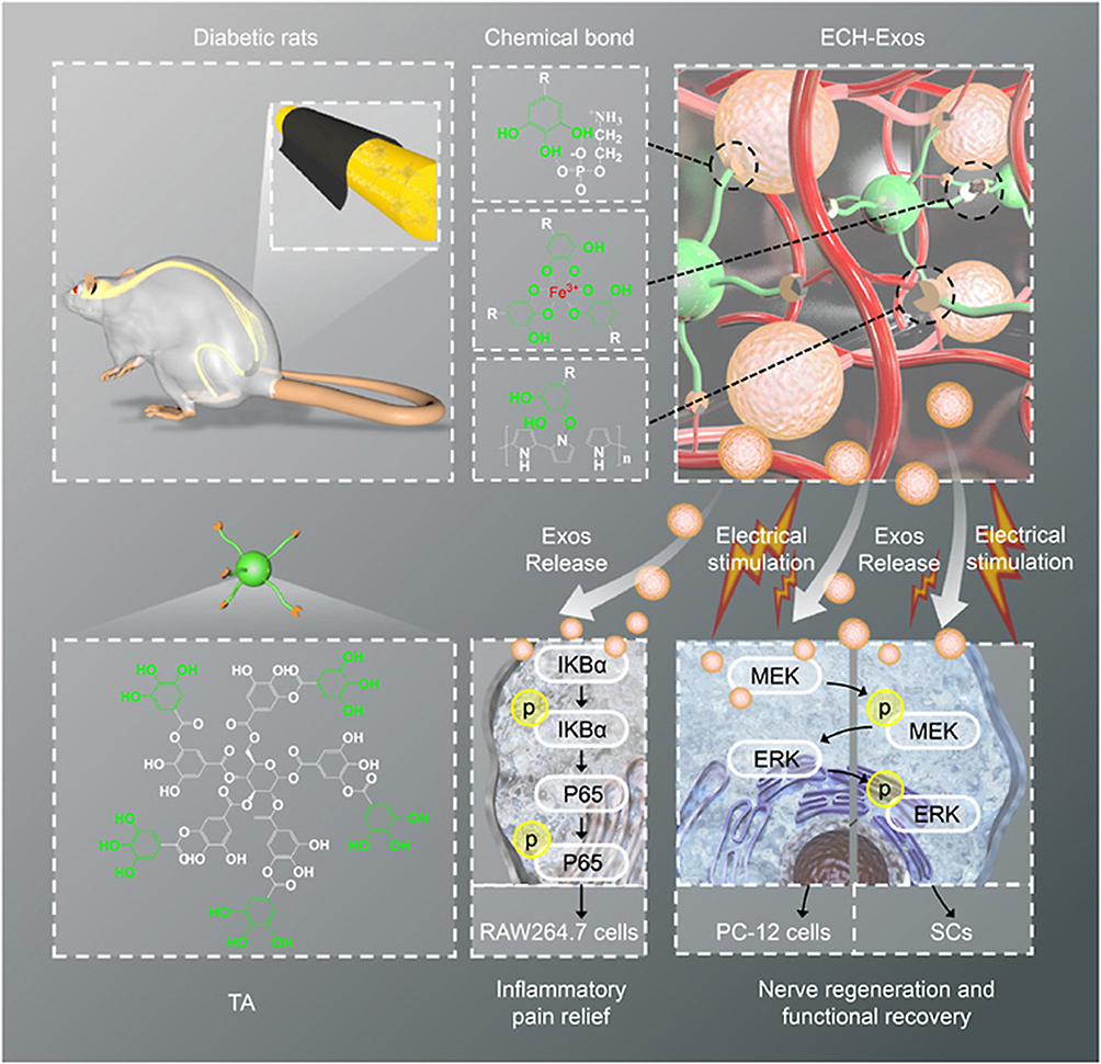 |
Figure 1 Schematic illustration of the molecular composition and interactions of ECH-Exos, highlighting functional moieties, bonding characteristics, and the proposed therapeutic pathway for alleviating neuroinflammation and enhancing neural repair and functional recovery in diabetic peripheral nerve injury. Reproduced under the terms of a Creative Commons Attribution 4.0 International License.36 Copyright 2022, the authors. |
Collectively, organic NPs offer a versatile and clinically translatable platform for macrophage-targeted nerve repair. Taken together, organic nanocarriers enable precise immunomodulation through both exogenous payloads and intrinsic biological interactions, providing a foundation for next-generation nerve repair systems.
Inorganic NPs: Silica- and Magnesium-Based Constructs
Inorganic NPs offer distinct advantages in structural control, surface chemistry, and multifunctionality. Mesoporous silica nanoparticles are especially suited for high-capacity drug loading and surface functionalization with immunomodulatory ligands or peptides. Functionalized mesoporous silica nanoparticles have demonstrated the ability to deliver anti-inflammatory agents to macrophages, promoting a phenotypic shift toward M2 and attenuating local cytokine production at nerve injury sites.37 Despite their versatility, silica-based NPs may pose risks related to long-term tissue accumulation, slow biodegradation, and dose-dependent cytotoxicity, particularly at high concentrations or with improper surface modification. These safety considerations require careful preclinical evaluation.
Magnesium-based nanoparticles uniquely combine immunoregulatory and bioactive ionic functions. Upon degradation, magnesium ions exert direct anti-inflammatory effects by inhibiting NF-κB signaling and enhancing IL-10 expression in macrophages.38 When integrated into nerve conduits or nanofiber matrices, Mg-based NPs not only modulate macrophage responses but also stimulate angiogenesis and neurite outgrowth via the release of pro-regenerative magnesium ion (Mg2+).39 For instance, an injectable bisphosphonate-based hydrogel enabling sustained Mg2+ release was developed to enhance peripheral nerve regeneration, addressing limitations associated with autograft transplantation.39 Mg2+ promoted neurite outgrowth in a dose-dependent manner via activation of the PI3K/Akt pathway and Sema5b signaling. When integrated into polycaprolactone conduits bridging 10 mm nerve gaps, the Mg2+-releasing hydrogel significantly accelerated axonal regeneration, remyelination, and target muscle reinnervation in vivo. Functional assessments confirmed superior recovery compared to Mg2+-free controls. This synergistic platform combining bioactive ion delivery with 3D-engineered scaffolds offers a clinically translatable approach for effective peripheral nerve injury repair (Figure 2). Inorganic NPs thus represent a multifunctional platform capable of delivering immune cues, providing structural support, and actively participating in the biochemical modulation of the nerve microenvironment.
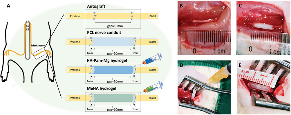 |
Figure 2 Overview of surgical procedures for sciatic nerve repair in SD rats. (A) Experimental grouping and interventions: autograft transplantation, polycaprolactone nerve conduit implantation, and conduit-assisted delivery of either HA–Pam–Mg or MeHA hydrogels. (B) Nerve defect bridging using autologous grafts. (C) Implantation of empty polycaprolactone conduits. (D and E) Hydrogel-assisted nerve repair using HA–Pam–Mg and MeHA systems, respectively, involving initial suture of one nerve stump followed by in situ injection of hydrogel into the conduit lumen. Reproduced under the terms of a Creative Commons Attribution 4.0 International License.39 Copyright 2022, the authors. Abbreviations: S, sural nerve; T, tibial nerve; P, peroneal nerve; HA, hyaluronic acid; Pam, pamidronate; Mg, magnesium; MeHA, methacrylated HA; SEM, scanning electron microscopy. |
Topographical Modulation of Macrophage Behavior
Beyond biochemical composition, the nanoscale topography of biomaterials has emerged as a potent modulator of macrophage phenotype. Aligned nanofiber scaffolds, commonly fabricated via electrospinning, induce macrophage elongation—a morphological feature correlated with M2 polarization and anti-inflammatory gene expression.40 These effects are synergistic with their influence on Schwann cell alignment and axon guidance. Although topographical cues are powerful modulators of macrophage behavior, their fabrication often involves complex manufacturing steps and limited scalability, which may hinder widespread adoption in clinical-grade nerve conduits.
Similarly, micropatterned nerve guidance conduits incorporating nanomaterials such as graphene oxide have been shown to promote M2 differentiation of infiltrating macrophages, while concurrently supporting Schwann cell adhesion and proliferation.41–43 These platforms exemplify the synergy between physical structure and biochemical signaling in immunomodulation.
Functionalization Strategies for Targeted Immunomodulation
Surface functionalization remains a key strategy to enhance NP selectivity for macrophages. Ligands such as mannose or folic acid can be conjugated to NP surfaces to exploit macrophage-expressed receptors, facilitating receptor-mediated endocytosis and intracellular delivery of immunoregulatory payloads.24 Other targeting moieties, including peptides specific for scavenger receptors or aptamers against CCR2, enable more refined discrimination between macrophage subtypes and reduce off-target effects.40,44
Given the dynamic immune and regenerative phases following nerve injury, dual-loaded or sequential-release NPs provide a rational strategy to synchronize therapeutic delivery with biological need. For instance, NPs co-loaded with IL-10 (to dampen M1 activation) and nerve growth factor (NGF) (to stimulate axonal extension) have been shown to orchestrate both immunomodulation and neural repair in animal models.31,45 Multifunctional and temporally responsive NP designs represent the forefront of immunotherapeutic innovation in peripheral nerve engineering, enabling dynamic control over immune and regenerative microenvironments.
Mechanisms of Nano-Immunomodulation in Nerve Regeneration
NPs have rapidly evolved beyond passive delivery vehicles, emerging as dynamic immunomodulatory agents capable of regulating multifaceted regenerative responses after peripheral nerve injury. Through both direct and indirect mechanisms, NPs reprogram immune cells, remodel the lesion microenvironment, and coordinate interactions between macrophages, Schwann cells, and neurons.
Direct Reprogramming of Macrophage Phenotypes
One of the most critical actions of therapeutic NPs is their ability to directly reprogram macrophages from a pro-inflammatory M1 phenotype to a reparative M2 phenotype. This phenotypic switch is essential for resolving inflammation and initiating tissue remodeling. For instance, exosome-inspired NPs loaded with miR-146a-5p have been shown to suppress TRAF6/NF-κB signaling in macrophages, resulting in downregulation of inflammatory cytokines and upregulation of arginase-1 and IL-10, classical M2 markers.23 Similarly, TGF-β locally activate Smad3-dependent signaling pathways in macrophages, promoting macrophage-myofibroblast transition, which plays a dual role in collagen matrix remodeling and wound stabilization during nerve repair.46
Beyond biochemical modulation, piezoelectric NPs, which generate local electric fields in response to mechanical stimulation, have been reported to activate the AMPK pathway in macrophages, further enhancing M2-like polarization and metabolic reprogramming conducive to regeneration.47 These strategies illustrate how nano-enabled platforms can exert cell-intrinsic immunomodulatory effects at the molecular level, dictating macrophage fate decisions in the injury milieu. Thus, NPs offer a means of intracellular immune programming that precisely tunes macrophage activity, enabling a shift from destructive inflammation to constructive regeneration.
Remodeling of the Regenerative Microenvironment
In addition to direct immunocellular interactions, NPs significantly influence the extracellular milieu to foster a regenerative environment. Black phosphorus quantum dots (BPQDs), an ultra-small (<10 nm) and atomically thin semiconductor nanomaterial, have recently emerged as a promising alternative to graphene-based platforms due to their intrinsic biodegradability, photothermal responsiveness, and rich surface chemistry that enables biological functionalization. However, their rapid degradation in aqueous environments and the associated cytotoxicity have limited their biomedical utility, necessitating stabilization strategies to maintain bioactivity while ensuring safety. In a recent study, zero-dimensional black phosphorus quantum dots (BPQDs) modified with antioxidant β-carotene (BPQD@β-carotene) were developed to address the challenges of BP degradation and cytotoxicity.12 BPQD@β-carotene exhibited negligible toxicity and favorable biocompatibility, promoting Schwann cell-mediated neural regrowth, angiogenesis, and immunomodulation. Mechanistically, the stabilized BPQD system activated PI3K/Akt and Ras/ERK1/2 signaling cascades at transcriptomic, proteomic, and metabolomic levels, highlighting its capacity to influence extracellular matrix remodeling and neurovascular niche formation. Embedded within a GelMA/PEGDA scaffold, the nanomaterial facilitated axon remyelination and intraneural vascularization, accelerating functional recovery in rodent and canine models (Figure 3). These findings emphasize the potential of engineered nanomaterials to reshape the extracellular environment and drive clinically translatable tissue regeneration.
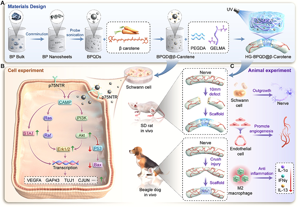 |
Figure 3 Schematic illustration of β-carotene-functionalized BPQDs for promoting Schwann cell-mediated nerve regeneration in peripheral nerve injury. (A) Fabrication of β-carotene-modified black phosphorus quantum dots (BPQD@β-carotene) and their incorporation into a GelMA/PEGDA hydrogel scaffold. (B) BPQD@β-carotene enhances Schwann cell functions, including axonal regeneration, neovascularization, and immunomodulation. (C) In vivo application of BPQD@β-carotene-loaded scaffolds improves functional recovery by supporting axon remyelination and vascular regeneration in rat and beagle dog peripheral nerve injury models. Reproduced with permission.12 American Chemical Society, copyright 2024. |
Moreover, engineering nano-enabled immunomodulation offers a promising strategy for promoting peripheral nerve regeneration. To overcome the limitations posed by wallerian degeneration, a major barrier in nerve repair, a ROS/Ca2+-responsive hydrogel system was developed to deliver engineered extracellular vesicles (E-EV-P@HPCEP) carrying PINK1 mRNA.48 This smart delivery platform enabled targeted release to senescent Schwann cells, indirectly reprogramming the inflammatory-senescent microenvironment post-injury. By activating mitochondrial autophagy and preserving mitochondrial homeostasis, the nanomaterial-mediated system suppressed wallerian degeneration progression and restored tissue repair dynamics (Figure 4). Taken together, through matrix stabilization, angiogenesis, and fibrosis suppression, nano-enabled systems reconstruct the injury microenvironment into a pro-regenerative scaffold for coordinated nerve healing.
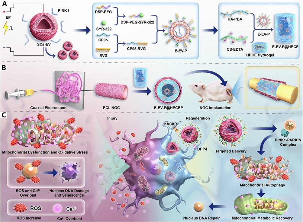 |
Figure 4 Schematic representation of a ROS/Ca2+-responsive hydrogel system enabling targeted delivery of PINK1 mRNA via engineered extracellular vesicles for peripheral nerve repair. (A and B) Preparation of the ROS/Ca2+-responsive dynamic hydrogels. (C) The dynamic hydrogel facilitates spatiotemporally controlled release of dual-targeted EVs to senescent Schwann cells, promoting mitochondrial quality control through PINK1-mediated autophagy activation. Reproduced with permission.48 American Chemical Society, copyright 2024. |
Translational Applications and Biomaterial Integration
The successful clinical translation of nano-immunomodulatory strategies for peripheral nerve regeneration depends on their seamless integration with advanced biomaterials and robust validation in relevant animal models. By embedding NPs within nerve guidance conduits, engineering constructs for in vivo efficacy, and implementing diagnostic tracking platforms, current research is bridging the gap between benchside innovation and bedside application.30,49
NP-Integrated Nerve Guidance Conduits
Among the most promising translational strategies is the incorporation of immunoregulatory NPs into nerve guidance conduits. These scaffolds provide structural support and directional cues for regenerating axons while serving as delivery platforms for immunomodulatory agents.11,50,51 For instance, inspired by the hierarchical architecture of native nerves, a biomimetic nerve guidance conduit was developed by integrating reduced graphene oxide (rGO) and brain-derived neurotrophic factor (BDNF)-loaded GelMA hydrogel onto the anisotropic nanoridges of Morpho butterfly wings.52 This topological and conductive scaffold not only promotes neurite outgrowth and directional alignment of neural stem cells and PC12 cells but also modulates the local immune milieu via conductive cues and sustained neurotrophin release. When rolled into a conduit structure, the scaffold facilitated effective repair of 10 mm sciatic nerve defects in rats, highlighting its therapeutic potential (Figure 5). Advanced constructs have further incorporated Bredigite or similar bioactive ceramic NPs into nerve guidance conduit matrices. These inorganic fillers not only reinforce the mechanical integrity of the conduit but also release osteogenic and angiogenic ions that promote vascularization and macrophage M2 polarization.53,54 The dual action of these composite systems—combining topographical and biochemical cues—illustrates a multifactorial approach to nerve repair. Therefore, NP-loaded nerve guidance conduits represent a clinically translatable platform that unites structural guidance, immune modulation, and biochemical stimulation within a single regenerative system.
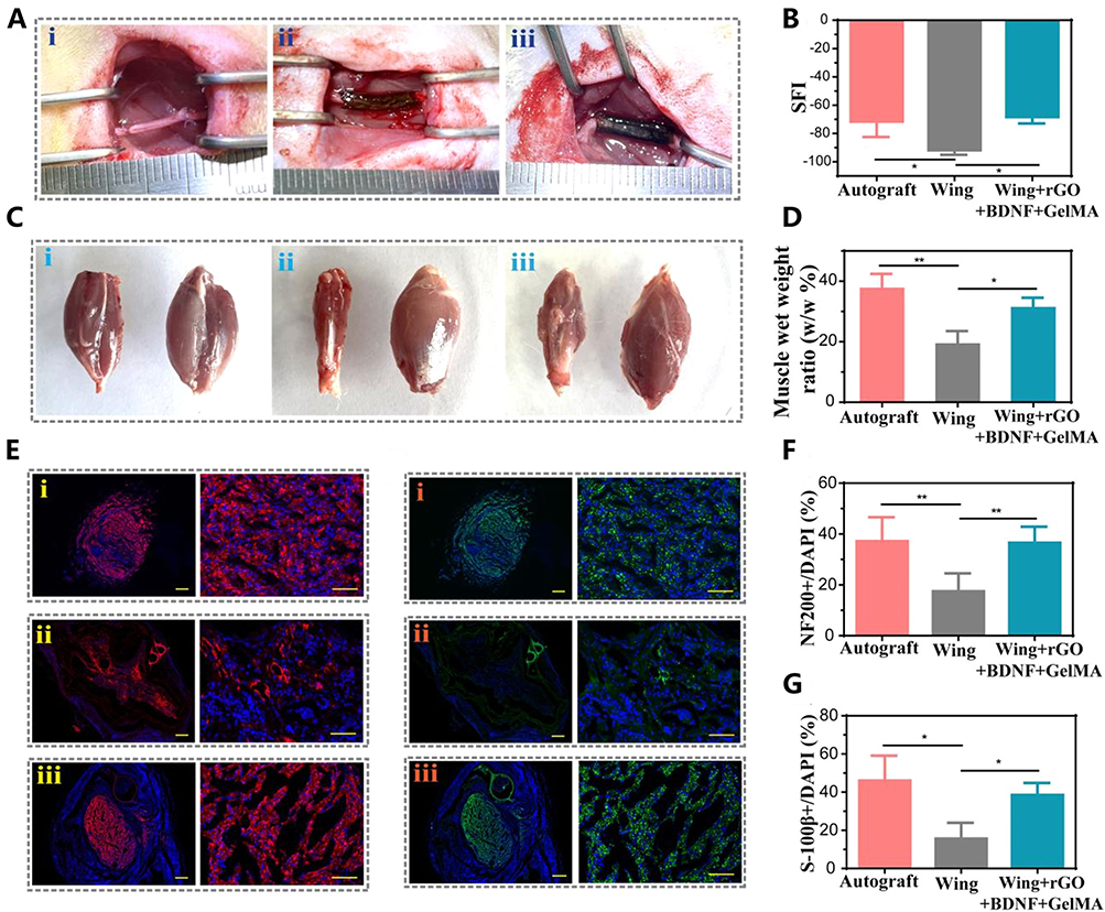 |
Figure 5 Evaluation of sciatic nerve regeneration following nerve guidance conduit implantation. (A) Representative images of surgical implantation in the Autograft (i), Wing-only (ii), and multifunctional Wing+rGO+BDNF+GelMA (iii) groups. (B) Sciatic functional index (SFI) assessment at 8 weeks post-surgery. (C) Gross morphology of target muscles and (D) corresponding muscle wet weight ratios across different groups (i, ii, iii). (E) Immunofluorescence staining for NF200 to assess axonal regeneration in different groups (i, ii, iii). Scale bars: 200 μm (left), 50 μm (right). (F) Immunofluorescence staining for S-100β to assess Schwann cell presence in different groups (i, ii, iii). Scale bars: 200 μm (left), 50 μm (right). (G and H) Quantification of NF200+ (G) and S-100β+ (H) areas normalized to DAPI+ nuclei. *p < 0.05, **p < 0.01. Reproduced with permission.52 American Chemical Society, copyright 2022. |
In vivo Efficacy of NP-Biomaterial Constructs
Extensive preclinical validation of NP-integrated biomaterials has been conducted in rodent models of sciatic nerve injury. These studies report marked improvements in electrophysiological recovery, including increased nerve conduction velocity, compound muscle action potentials, and axon diameter compared to control groups.55–58 Additionally, animals treated with NP-enhanced nerve guidance conduits exhibit reduced muscle atrophy, consistent with effective reinnervation of denervated muscles and restored neuromuscular connectivity.59 Importantly, these nanoengineered systems accelerate the timeline of functional recovery, minimizing chronic deficits that often follow delayed nerve regeneration. Improvements in gait analysis and sensory function further confirm the therapeutic efficacy of these constructs.60,61 The robust in vivo performance of NP-integrated nerve scaffolds provides strong evidence of their regenerative potential and lays the groundwork for eventual clinical application.
Theranostic Integration: Real-Time Monitoring of Immune Responses
Beyond therapeutic function, the convergence of diagnostics and therapy has opened new avenues in precision nerve regeneration. Specifically, ultrasmall superparamagnetic iron oxide (USPIO)-based NPs have been developed to enable non-invasive MRI tracking of macrophage infiltration at injury sites.62 These NPs can be functionalized to target specific macrophage phenotypes, allowing real-time monitoring of immune dynamics during different phases of regeneration.
Such imaging capabilities provide valuable feedback loops for therapy adjustment, enabling clinicians to design NP-based interventions in a patient-specific manner. Moreover, the integration of imaging biomarkers with immunomodulatory platforms enhances our mechanistic understanding of the regeneration process and facilitates personalized treatment planning.27 Theranostic NPs not only expand the functional repertoire of regenerative nanomedicine but also pave the way for precision monitoring and adaptive therapy in peripheral nerve repair.
Challenges and Future Perspectives
The emergence of nanomaterial-based immunomodulation presents a significant shift in peripheral nerve regeneration. However, its clinical application remains constrained by multifaceted challenges including biological specificity, manufacturing scalability, and regulatory oversight. Addressing these bottlenecks while embracing technological innovation is crucial to realizing the full translational potential of nano-immunotherapeutics.
Clinical Barriers to Translation
Despite encouraging results from rodent and small-animal models, the clinical deployment of immunomodulatory NPs faces formidable obstacles. A central concern is in cellular specificity-many current NP systems lack targeting fidelity, raising the risk of off-target effects that may compromise immune homeostasis or induce collateral tissue damage.63 Precise delivery to macrophages or Schwann cells is essential, especially given the narrow therapeutic window between pro-inflammatory clearance and pro-regenerative polarization.
Moreover, the long-term biocompatibility and safety profile of NPs remain inadequately characterized. Questions persist regarding the systemic distribution, degradation kinetics, and potential immunogenicity of NP platforms, especially in large-animal models or humans.64 These issues are magnified in chronic injury or comorbid conditions such as diabetes, where immune dysregulation may alter NP performance.
Scalability and reproducibility also represent major translational bottlenecks. Manufacturing functionally complex NPs at clinical grade and batch-to-batch consistency remains a significant challenge. Regulatory frameworks demand stringent standardization in terms of size, charge, release kinetics, and biological performance.63,65 Collectively, overcoming these barriers will require integrated solutions in targeted design, safety profiling, and scalable manufacturing to advance NP-based immunomodulation from preclinical success to clinical reality.
Emerging Advances in Nano-Immunomodulation
To navigate these challenges, novel engineering strategies are being pursued. A prominent direction is the development of stimuli-responsive NPs that release therapeutics in response to injury-specific cues such as local pH shifts, reactive oxygen species, or protease activity.66,67 These “smart” platforms provide spatiotemporally controlled release, reducing systemic exposure while enhancing on-site immunomodulation.
Personalized nano-immunotherapies are also gaining traction. As immune responses vary with age, sex, metabolic status, and comorbidities, NPs can be designed to reflect patient-specific immune signatures.68 The application of AI-driven modeling and high-throughput screening is expected to accelerate such precision-engineered solutions.
In parallel, multi-targeting nanoplatforms are being designed to address the multifactorial nature of nerve regeneration. These include NPs co-delivering neurotrophic factors, anti-inflammatory agents, and matrix-modulating cues, achieving synergistic effects on both immune and neural compartments.32,50 These emerging strategies redefine the therapeutic scope of NPs, offering the possibility of dynamic, adaptive, and patient-specific modulation of the nerve repair microenvironment.
Perspectives for Clinical Translation
Bridging the translational gap requires a comprehensive and coordinated roadmap. A critical step is the adoption of standardized preclinical models that accurately recapitulate human peripheral nerve injuries, including chronic, delayed, or comorbid conditions. Current reliance on acute injury models in healthy rodents fails to capture the complexity of clinical scenarios.
Equally important is the establishment of harmonized evaluation protocols. Functional recovery metrics should be benchmarked alongside immunophenotyping and histological analysis to ensure reproducibility and comparability across studies. Early engagement with regulatory agencies is indispensable. Proactive dialogue can aid in defining clear criteria for nanoparticle characterization, toxicity screening, and clinical trial design. Such alignment will facilitate smoother navigation through regulatory pathways and accelerate the transition from bench to bedside. By constructing a translational ecosystem grounded in robust models, standardized metrics, and regulatory clarity, the clinical potential of immunomodulatory nanomaterials for peripheral nerve regeneration can be fully realized.
Conclusion
NPs have emerged as precision tools with the potential to revolutionize the treatment of peripheral nerve injury. By enabling spatiotemporal control over immune modulation, these engineered systems offer a means to resolve the immune dysregulation that often hampers nerve regeneration. Through targeted delivery of bioactive agents, tailored interactions with macrophages, and integration with biomaterial scaffolds, NPs can create a pro-regenerative microenvironment that supports both immune balance and neural repair. Importantly, nano-immunomodulation offers therapeutic advantages that could directly improve patient outcomes by overcoming the inherent limitations of current clinical approaches, including donor-site morbidity associated with autografts and the limited regenerative capacity of synthetic conduits. By actively reshaping the immune microenvironment rather than merely serving as passive structural supports, NP-based strategies provide a fundamentally more responsive and adaptive solution for peripheral nerve repair.
Despite remarkable progress at the preclinical level, realizing the full therapeutic potential of nano-immunomodulation in peripheral nerve injury will require sustained, interdisciplinary collaboration. The challenges of refining NP design, ensuring safety, and translating these technologies into clinical practice demand the collective expertise of materials scientists, immunologists, neurobiologists, and clinicians. Looking forward, two priority areas will be particularly critical for advancing the field: (1) enhancing the specificity and durability of macrophage-targeted NP platforms to achieve more precise and sustained immunoregulation, and (2) ensuring long-term biocompatibility, biodegradability, and biosafety to support regulatory approval and clinical translation. Only through such coordinated efforts can we bridge the gap between bench and bedside, bringing advanced nanomedicine solutions to patients with peripheral nerve injuries.
Acknowledgments
We used ChatGPT to enhance the readability and language of our review paper under our supervision, and we have double-checked and edited the content.
Disclosure
The authors report no conflicts of interest in this work.
References
1. Barnes SL, Miller TA, Simon NG. Traumatic peripheral nerve injuries: diagnosis and management. Curr Opin Neurol. 2022;35:718–14. doi:10.1097/WCO.0000000000001116
2. Modrak M, Talukder MAH, Gurgenashvili K, Noble M, Elfar JC. Peripheral nerve injury and myelination: potential therapeutic strategies. J Neurosci Res. 2020;98:780–795. doi:10.1002/jnr.24538
3. Duffy P, McMahon S, Wang X, et al. Synthetic bioresorbable poly-alpha-hydroxyesters as peripheral nerve guidance conduits; a review of material properties, design strategies and their efficacy to date. Biomater Sci. 2019;7:4912–4943. doi:10.1039/c9bm00246d
4. Jha MK, Passero JV, Rawat A, et al. Macrophage monocarboxylate transporter 1 promotes peripheral nerve regeneration after injury in mice. J Clin Invest. 2021;131. doi:10.1172/JCI141964
5. Wu G, Wen X, Kuang R, et al. Roles of macrophages and their interactions with schwann cells after peripheral nerve injury. Cell Mol Neurobiol. 2023;44:11. doi:10.1007/s10571-023-01442-5
6. Cui Y, Wang X, Xu Y, et al. Ropivacaine Promotes Axon Regeneration by Regulating Nav1.8-mediated Macrophage Signaling after Sciatic Nerve Injury in Rats. Anesthesiology. 2023;139:782–800. doi:10.1097/ALN.0000000000004761
7. Liu P, Peng J, Han G-H, et al. Role of macrophages in peripheral nerve injury and repair. Neural Regen Res. 2019;14:1335–1342. doi:10.4103/1673-5374.253510
8. Oshima E, Hayashi Y, Xie Z, et al. M2 macrophage-derived cathepsin S promotes peripheral nerve regeneration via fibroblast-Schwann cell-signaling relay. J Neuroinflammation. 2023;20:258. doi:10.1186/s12974-023-02943-2
9. Chen P, Piao X, Bonaldo P. Role of macrophages in Wallerian degeneration and axonal regeneration after peripheral nerve injury. Acta Neuropathol. 2015;130:605–618. doi:10.1007/s00401-015-1482-4
10. Kwon MJ, Seo Y, Cho H, et al. Nanogel-mediated delivery of oncomodulin secreted from regeneration-associated macrophages promotes sensory axon regeneration in the spinal cord. Theranostics. 2022;12:5856–5876. doi:10.7150/thno.73386
11. Sun X, Bai Y, Zhai H, et al. Devising micro/nano-architectures in multi-channel nerve conduits towards a pro-regenerative matrix for the repair of spinal cord injury. Acta Biomater. 2019;86:194–206. doi:10.1016/j.actbio.2018.12.032
12. Shen J, Sun Y, Liu X, et al. Nerve regeneration potential of antioxidant-modified black phosphorus quantum dots in peripheral nerve injury. ACS Nano. 2024;18(34):23518–23536. doi:10.1021/acsnano.4c07285
13. Xu D, Fu S, Zhang H, et al. Ultrasound-responsive aligned piezoelectric nanofibers derived hydrogel conduits for peripheral nerve regeneration. Adv Mater. 2024;36(28):e2307896. doi:10.1002/adma.202307896
14. Menorca RM, Fussell TS, Elfar JC. Nerve physiology: mechanisms of injury and recovery. Hand Clin. 2013;29:317–330. doi:10.1016/j.hcl.2013.04.002
15. Oladiran O, Shi XQ, Yang M, Fournier S, Zhang J. Inhibition of TLR4 signaling protects mice from sensory and motor dysfunction in an animal model of autoimmune peripheral neuropathy. J Neuroinflammation. 2021;18:77. doi:10.1186/s12974-021-02126-x
16. Hervera A, De Virgiliis F, Palmisano I, et al. Reactive oxygen species regulate axonal regeneration through the release of exosomal NADPH oxidase 2 complexes into injured axons. Nat Cell Biol. 2018;20:307–319. doi:10.1038/s41556-018-0039-x
17. Ishida K, Nagatake T, Saika A, et al. Induction of unique macrophage subset by simultaneous stimulation with LPS and IL-4. Front Immunol. 2023;14:1111729. doi:10.3389/fimmu.2023.1111729
18. Malcangio M. Role of the immune system in neuropathic pain. Scand J Pain. 2019;20:33–37. doi:10.1515/sjpain-2019-0138
19. Min Q, Parkinson DB, Dun XP. Migrating schwann cells direct axon regeneration within the peripheral nerve bridge. Glia. 2021;69:235–254. doi:10.1002/glia.23892
20. Feng R, Muraleedharan Saraswathy V, Mokalled MH, Cavalli V. Self-renewing macrophages in dorsal root ganglia contribute to promote nerve regeneration. Proc Natl Acad Sci U S A. 2023;120:e2215906120. doi:10.1073/pnas.2215906120
21. Zigmond RE, Echevarria FD. Macrophage biology in the peripheral nervous system after injury. Prog Neurobiol. 2019;173:102–121. doi:10.1016/j.pneurobio.2018.12.001
22. Bombeiro AL, Pereira BTN, de Oliveira ALR. Granulocyte-macrophage colony-stimulating factor improves mouse peripheral nerve regeneration following sciatic nerve crush. Eur J Neurosci. 2018;48:2152–2164. doi:10.1111/ejn.14106
23. Sun J, Liao Z, Li Z, et al. Down-regulation miR-146a-5p in schwann cell-derived exosomes induced macrophage M1 polarization by impairing the inhibition on TRAF6/NF-kappaB pathway after peripheral nerve injury. Exp Neurol. 2023;362:114295. doi:10.1016/j.expneurol.2022.114295
24. Govindappa PK, Elfar JC. Erythropoietin promotes M2 macrophage phagocytosis of schwann cells in peripheral nerve injury. Cell Death Dis. 2022;13:245. doi:10.1038/s41419-022-04671-6
25. Xu J, Wen J, Fu L, et al. Macrophage-specific RhoA knockout delays Wallerian degeneration after peripheral nerve injury in mice. J Neuroinflammation. 2021;18:234. doi:10.1186/s12974-021-02292-y
26. Ye Y, Cheng H, Wang Y, et al. Macrophage: a key player in neuropathic pain. Int Rev Immunol. 2024;43:326–339. doi:10.1080/08830185.2024.2344170
27. Dingwall CB, Strickland A, Yum SW, et al. Macrophage depletion blocks congenital SARM1-dependent neuropathy. J Clin Invest. 2022;132. doi:10.1172/JCI159800
28. Lin T, Liu S, Chen S, et al. Hydrogel derived from porcine decellularized nerve tissue as a promising biomaterial for repairing peripheral nerve defects. Acta Biomater. 2018;73:326–338. doi:10.1016/j.actbio.2018.04.001
29. Sun Y, Zhang Y, Guo Y, et al. Electrical aligned polyurethane nerve guidance conduit modulates macrophage polarization and facilitates immunoregulatory peripheral nerve regeneration. J Nanobiotechnology. 2024;22:244. doi:10.1186/s12951-024-02507-3
30. Han S, Gao L, Dou X, et al. Chiral hydrogel nerve conduit boosts peripheral nerve regeneration via regulation of schwann cell reprogramming. ACS Nano. 2024;18:28358–28370. doi:10.1021/acsnano.4c10653
31. Su H, Ye Q, Wang D, et al. A self-assembling peptide-based hydrogel containing NF- κ B inhibitors and NGF for peripheral nerve injury repair. Biofabrication. 2025;17:025031. doi:10.1088/1758-5090/adc340
32. Lin Y, Yu R, Yin G, Chen Z, Lin H. Syringic acid delivered via mPEG-PLGA-PLL nanoparticles enhances peripheral nerve regeneration effect. Nanomedicine. 2020;15:1487–1499. doi:10.2217/nnm-2020-0042
33. Yu J, Lin Y, Wang G, et al. Zein-induced immune response and modulation by size, pore structure and drug-loading: application for sciatic nerve regeneration. Acta Biomater. 2022;140:289–301. doi:10.1016/j.actbio.2021.11.035
34. Fan B, Li C, Szalad A, et al. Mesenchymal stromal cell-derived exosomes ameliorate peripheral neuropathy in a mouse model of diabetes. Diabetologia. 2020;63:431–443. doi:10.1007/s00125-019-05043-0
35. Ren J, Zhu B, Gu G, et al. Schwann cell-derived exosomes containing MFG-E8 modify macrophage/microglial polarization for attenuating inflammation via the SOCS3/STAT3 pathway after spinal cord injury. Cell Death Dis. 2023;14:70. doi:10.1038/s41419-023-05607-4
36. Yang Q, Su S, Liu S, et al. Exosomes-loaded electroconductive nerve dressing for nerve regeneration and pain relief against diabetic peripheral nerve injury. Bioact Mater. 2023;26:194–215. doi:10.1016/j.bioactmat.2023.02.024
37. Kim MS, El-Fiqi A, Kim J-W, et al. Nanotherapeutics of PTEN inhibitor with mesoporous silica nanocarrier effective for axonal outgrowth of adult neurons. ACS Appl Mater Interfaces. 2016;8:18741–18753. doi:10.1021/acsami.6b06889
38. Hosseini A, Sharifzadeh M, Rezayat SM, et al. Benefit of magnesium-25 carrying porphyrin-fullerene nanoparticles in experimental diabetic neuropathy. Int J Nanomed. 2010;5:517–523. doi:10.2147/ijn.s11643
39. Yao Z, Yuan W, Xu J, et al. Magnesium-encapsulated injectable hydrogel and 3D-engineered polycaprolactone conduit facilitate peripheral nerve regeneration. Adv Sci. 2022;9:e2202102. doi:10.1002/advs.202202102
40. Jia Y, Yang W, Zhang K, et al. Nanofiber arrangement regulates peripheral nerve regeneration through differential modulation of macrophage phenotypes. Acta Biomater. 2019;83:291–301. doi:10.1016/j.actbio.2018.10.040
41. Fang Y, Wang C, Liu Z, et al. 3D printed conductive multiscale nerve guidance conduit with hierarchical fibers for peripheral nerve regeneration. Adv Sci. 2023;10:e2205744. doi:10.1002/advs.202205744
42. Zhang D, Yao Y, Duan Y, et al. Surface-anchored graphene oxide nanosheets on cell-scale micropatterned Poly(d, l -lactide- co -caprolactone) conduits promote peripheral nerve regeneration. ACS Appl Mater Interfaces. 2020;12:7915–7930. doi:10.1021/acsami.9b20321
43. Zhang Q, Chen J, Feng Y, et al. Electroactive scaffolds of biodegradable polyurethane/polydopamine-functionalized graphene oxide regulating the inflammatory response and revitalizing the axonal growth cone for peripheral nerve regeneration. J Mater Chem B. 2023;11:6308–6318. doi:10.1039/d3tb00837a
44. Mokarram N, Merchant A, Mukhatyar V, Patel G, Bellamkonda RV. Effect of modulating macrophage phenotype on peripheral nerve repair. Biomaterials. 2012;33:8793–8801. doi:10.1016/j.biomaterials.2012.08.050
45. Day YJ, Liou J-T, Lee C-M, et al. Lack of interleukin-17 leads to a modulated micro-environment and amelioration of mechanical hypersensitivity after peripheral nerve injury in mice. Pain. 2014;155:1293–1302. doi:10.1016/j.pain.2014.04.004
46. Li Y, Cai J, Xu Y, et al. Macrophage-myofibroblast transition contributes to the macrophage elimination and functional regeneration in the late stage of nerve injury. Exp Neurol. 2025;387:115194. doi:10.1016/j.expneurol.2025.115194
47. Wei G, Chen C, Li X, et al. In situ piezoelectricity induces M2 polarization of macrophages to regulate Schwann cells for alleviating neuropathic pain of CCI rats. Biomater Adv. 2025;174:214319. doi:10.1016/j.bioadv.2025.214319
48. Zhao R, Deng X, Tang Y, et al. Mitigating critical peripheral nerve deficit therapy with reactive oxygen Species/Ca 2+ -responsive dynamic hydrogel-mediated mRNA delivery. ACS Nano. 2024;18:16556–16576. doi:10.1021/acsnano.3c13102
49. Song J, Dong J, Yuan Z, et al. Shape-persistent conductive nerve guidance conduits for peripheral nerve regeneration. Adv Healthc Mater. 2024;13:e2401160. doi:10.1002/adhm.202401160
50. Cai Y, Huang Q, Wang P, et al. Conductive hydrogel conduits with growth factor gradients for peripheral nerve repair in diabetics with non-suture tape. Adv Healthc Mater. 2022;11:e2200755. doi:10.1002/adhm.202200755
51. Hu C, Liu B, Huang X, et al. Sea cucumber-inspired microneedle nerve guidance conduit for synergistically inhibiting muscle atrophy and promoting nerve regeneration. ACS Nano. 2024;18:14427–14440. doi:10.1021/acsnano.4c00794
52. Hu Y, Chen Z, Wang H, et al. Conductive nerve guidance conduits based on morpho butterfly wings for peripheral nerve repair. ACS Nano. 2022;16:1868–1879. doi:10.1021/acsnano.1c11627
53. Sun Y, Zhang H, Zhang Y, et al. Li-Mg-Si bioceramics provide a dynamic immuno-modulatory and repair-supportive microenvironment for peripheral nerve regeneration. Bioact Mater. 2023;28:227–242. doi:10.1016/j.bioactmat.2023.05.013
54. Xuan Y, Li L, Yin X, et al. Bredigite-based bioactive nerve guidance conduit for pro-healing macrophage polarization and peripheral nerve regeneration. Adv Healthc Mater. 2024;13:e2302994. doi:10.1002/adhm.202302994
55. Caillaud M, Chantemargue B, Richard L, et al. Local low dose curcumin treatment improves functional recovery and remyelination in a rat model of sciatic nerve crush through inhibition of oxidative stress. Neuropharmacology. 2018;139:98–116. doi:10.1016/j.neuropharm.2018.07.001
56. Soares Dos Santos Cardoso F, Cardoso R, Dos Santos Ramalho B, et al. Inosine accelerates the regeneration and anticipates the functional recovery after sciatic nerve crush injury in mice. Neuroscience. 2019;423:206–215. doi:10.1016/j.neuroscience.2019.09.023
57. Stratton JA, Shah PT, Kumar R, et al. The immunomodulatory properties of adult skin-derived precursor Schwann cells: implications for peripheral nerve injury therapy. Eur J Neurosci. 2016;43:365–375. doi:10.1111/ejn.13006
58. Vallieres N, Barrette B, Wang LX, et al. Betacellulin regulates schwann cell proliferation and myelin formation in the injured mouse peripheral nerve. Glia. 2017;65:657–669. doi:10.1002/glia.23119
59. Dong X, Liu S, Yang Y, et al. Aligned microfiber-induced macrophage polarization to guide schwann-cell-enabled peripheral nerve regeneration. Biomaterials. 2021;272:120767. doi:10.1016/j.biomaterials.2021.120767
60. Schilling BK, Baker JS, Komatsu C, et al. Intramuscular nanofat injection promotes inflammation-induced gastrocnemius regeneration in a syngeneic rat sciatic nerve injury model. Plast Reconstr Surg. 2023;151:947e–958e. doi:10.1097/PRS.0000000000010115
61. Wachs RA, Wellman SM, Porvasnik SL, et al. Apoptosis-decellularized peripheral nerve scaffold allows regeneration across nerve gap. Cells Tissues Organs. 2023;212:512–522. doi:10.1159/000525704
62. Chen MW, Zhang X, Lu L-J, et al. Monitoring of macrophage recruitment enhanced by Toll-like receptor 4 activation with MR imaging in nerve injury. Muscle Nerve. 2018:123–132. doi: 10.1002/mus.26097
63. He L, Tian L, Sun Y, et al. Nano-engineered environment for nerve regeneration: scaffolds, functional molecules and stem cells. Curr Stem Cell Res Ther. 2016;11:605–617. doi:10.2174/1574888x10666151001114735
64. Saracino GA, Cigognini D, Silva D, Caprini A, Gelain F. Nanomaterials design and tests for neural tissue engineering. Chem Soc Rev. 2013;42:225–262. doi:10.1039/c2cs35065c
65. Kong L, Gao X, Qian Y, et al. Biomechanical microenvironment in peripheral nerve regeneration: from pathophysiological understanding to tissue engineering development. Theranostics. 2022;12:4993–5014. doi:10.7150/thno.74571
66. Das S, Sharma M, Saharia D, et al. In vivo studies of silk based gold nano-composite conduits for functional peripheral nerve regeneration. Biomaterials. 2015;62:66–75. doi:10.1016/j.biomaterials.2015.04.047
67. Dong M, Shi B, Liu D, et al. Conductive hydrogel for a photothermal-responsive stretchable artificial nerve and coalescing with a damaged peripheral nerve. ACS Nano. 2020;14:16565–16575. doi:10.1021/acsnano.0c05197
68. Lee J, Choi Y, Song J, et al. Nerve-mimetic adhesive hydrogel electroceuticals: tailoring in situ physically entangled domains in singular polymers. ACS Nano. 2024;18:34949–34961. doi:10.1021/acsnano.4c13097
 © 2026 The Author(s). This work is published and licensed by Dove Medical Press Limited. The
full terms of this license are available at https://www.dovepress.com/terms
and incorporate the Creative Commons Attribution
- Non Commercial (unported, 4.0) License.
By accessing the work you hereby accept the Terms. Non-commercial uses of the work are permitted
without any further permission from Dove Medical Press Limited, provided the work is properly
attributed. For permission for commercial use of this work, please see paragraphs 4.2 and 5 of our Terms.
© 2026 The Author(s). This work is published and licensed by Dove Medical Press Limited. The
full terms of this license are available at https://www.dovepress.com/terms
and incorporate the Creative Commons Attribution
- Non Commercial (unported, 4.0) License.
By accessing the work you hereby accept the Terms. Non-commercial uses of the work are permitted
without any further permission from Dove Medical Press Limited, provided the work is properly
attributed. For permission for commercial use of this work, please see paragraphs 4.2 and 5 of our Terms.