
Back to Journals » Clinical Ophthalmology » Volume 19
Multimodal Imaging Features of Retinal Lesions in Hemophagocytic Lymphohistiocytosis: A Clinical Analysis
Authors Wu W  , Guo H, Yang M , Wang L , Wang Y, Wang K
Received 9 October 2025
Accepted for publication 13 December 2025
Published 21 December 2025 Volume 2025:19 Pages 4875—4884
DOI https://doi.org/10.2147/OPTH.S572896
Checked for plagiarism Yes
Review by Single anonymous peer review
Peer reviewer comments 2
Editor who approved publication: Dr Yousef Fouad
Weizhen Wu, Huiling Guo, Ming Yang, Luping Wang, Yanling Wang,* Kang Wang*
Department of Ophthalmology, Beijing Friendship Hospital, Capital Medical University, Beijing, People’s Republic of China
*These authors contributed equally to this work
Correspondence: Yanling Wang, Email [email protected] Kang Wang, Email [email protected]
Objective: Hemophagocytic lymphohistiocytosis (HLH) associated retinopathy remains poorly characterized in terms of clinical phenotypes and pathogenesis. This retrospective study aimed to systematically define the multimodal imaging features of retinal lesions in patients with HLH and investigate its underlying pathological mechanisms to inform clinical practice.
Methods: A retrospective case series analysis was conducted on 15 HLH patients with retinal lesions. Comprehensive ophthalmic evaluations, including fundoscopy and optical coherence tomography (OCT), were integrated with systemic multimodal imaging data.
Results: Imaging analysis revealed characteristic retinal structural alterations, including retinal hemorrhage (26 eyes, 86.67%), outer retinal atrophy (20 eyes, 66.67%), outer retinal hyperreflective foci (7 eyes, 23.33%), ellipsoid zone disruption (17 eyes, 56.67%) and ellipsoid zone thinning (10 eyes, 33.33%). Multimodal imaging findings suggest a potential association between HLH related retinopathy and a hypoperfused ischemic state of the retina, though this requires further validation through larger scale statistical analysis. Notably, characteristic damage to the outer retinal structures was observed across HLH patients of different etiological subtypes and could manifest at any stage of the disease, including before and after interventions such as chemotherapy and hematopoietic stem cell transplantation.
Discussion: The study data indicate potential involvement of HLH induced systemic cytokine storms, secondary hemoglobin reduction, and hematologic abnormalities such as thrombocytopenia in the observed retinal changes. These interpretations should be understood as hypothesis generating observations within the constraints of descriptive research methodology. Therefore, while cytokine cascade control remains a cornerstone of management, future studies should explore the potential benefit of blood component supplementation as an adjunctive therapy in HLH related retinopathy.
Conclusion: These findings highlight the necessity of establishing early warning indicators for HLH associated retinopathy and conducting multicenter prospective studies to optimize evidence based diagnostic and therapeutic strategies.
Keywords: hemophagocytic lymphohistiocytosis, outer retinal atrophy, retinal hemorrhage
Introduction
Hemophagocytic lymphohistiocytosis (HLH), also known as hemophagocytic syndrome (HPS), is an immune dysregulation syndrome characterized by hyperactivation of the immune system, featuring uncontrolled activation of T lymphocytes, macrophages, and natural killer (NK) cells, along with massive release of inflammatory cytokines, which leads to multi organ dysfunction, failure, and even mortality; however, the precise pathological mechanisms underlying its sustained inflammatory response remain incompletely elucidated. Etiologically, HLH is categorized into familial and secondary forms: familial HLH is primarily caused by hereditary defects in lymphocyte cytotoxicity or mutations in inflammation-related genes, while secondary HLH is commonly triggered by factors such as infections, malignancies, and rheumatic immune diseases.1
Research has identified that HLH can involve retinal lesions, manifesting as retinal hemorrhage, conjunctivitis, keratitis, anterior uveitis, optic papillitis, and choroidal lesions, yet a systematic summarization and analysis of these ocular manifestations are currently lacking.2 Due to the disease’s diverse etiological factors, variability in initial referral departments, and multidisciplinary nature, coupled with atypical ocular involvement symptoms, ophthalmologists face significant diagnostic challenges, increasing the risk of misdiagnosis or underdiagnosis. Early detection of retinal lesions may influence patient treatment outcomes and prognosis.
Our team’s prior retrospective study revealed that retinochoroidal lesions account for up to 49.62% of ocular complications in HLH patients.3 Multimodal imaging enables simultaneous assessment of retinal structure, vascular morphology, and function with superior resolution and dynamic monitoring, significantly enhancing early detection of HLH related retinopathy compared to conventional ophthalmoscopy. This study aims to conduct an indepth exploration of the multimodal imaging features of retinochoroidal structural alterations in HLH patients through retrospective case analysis, with the goal of enhancing clinicians’ recognition of this disease’s characteristics and providing critical evidence for clinical therapeutic decision making and visual function prognosis assessment.
Materials and Methods
This retrospective and observational study analyzed a case series of 15 patients (30 eyes) diagnosed with HLH and exhibiting retinal lesions, who were admitted to our institution between January 2022 and April 2025. The inclusion criterion specifically required evidence of retinal lesions with outer retinal structural alterations. The case series comprised 9 males (60.0%) and 6 females (40.0%), aged 6–52 years (mean 30.2 ± 10.4 years). All cases strictly met the Histiocyte Society’s HLH-2004 diagnostic criteria,4 excluding those with concurrent diabetes, hypertension, acute leukemia, or other systemic diseases potentially confounding retinal pathology assessment.
Treatment protocols adhered to the HLH-1994/2004 guidelines,4 with a backbone regimen of high dose corticosteroids combined with topoisomerase-II inhibitors; supplemental anthracycline antibiotics were administered based on disease severity. Notably, 2 patients (13.3%) had not initiated systematic therapy at baseline ophthalmic examination, while 3 patients (20.0%) had undergone hematopoietic stem cell transplantation(HSCT) before secondary follow up.
Employing a retrospective case series design, the following data were systematically collected:
Ophthalmic examinations: Color fundus photography (Topcon, Japan; Optos, UK), autofluorescence (AF, Optos, UK), spectral domain optical coherence tomography (SD-OCT; Heidelberg Engineering, Germany) (Scan the area centered on the macula with a 6×6mm2 range), OCT angiography (OCTA; Intalight, Germany) (Macula-centered scan, 12×12mm2 field), B-scan ultrasonography (AVISO, France), and fluorescein angiography (FFA, Carl Zeiss Meditec, Germany).
Laboratory parameters: Platelet count and hemoglobin levels. Blinded assessment was conducted (κ=0.85), with all imaging results evaluated by two senior ophthalmologists in physically isolated environments. Standardized deidentified images were used, with patient identifiers, medical histories, and treatment information concealed. No clinical discussions occurred prior to evaluation, and discordant cases were adjudicated by a third party expert.
The study was approved by the ethics committee of the Beijing Friendship Hospital (2025-P2-315-01), and performed under hospital guidance. This retrospective study utilized medical records previously obtained during clinical practice, from which deidentified data were extracted via a clinical big data platform. Given the complete anonymization of the data, rendering individual participants untraceable, and the absence of any involvement of personal privacy or commercial interests, the confidentiality and personal identifiers of all participants were rigorously safeguarded. Based on these ethical compliance considerations, informed consent was waived for this study. The principles of the Declaration of Helsinki were followed.
Statistical Methods: Given the retrospective, observational, and exploratory design with a limited sample size in this case series study, descriptive statistics were employed to characterize the fundus imaging findings across the study cohort.
Results
This study enrolled 15 HLH patients with ocular involvement, with disease duration ranging from 1 to 15 months (mean 4.13 ± 4.01 months). Etiologies included Epstein Barr virus (EBV) infection (8 cases, 53.33%), familial genetic disorders (3 cases, 20.00%), EBV-positive T/ NK-cell lymphoma (2 cases, 13.33%), EBV-positive T-cell lymphoma (1 case, 6.67%), and diffuse large B-cell lymphoma (1 case, 6.67%), indicate insufficient evidence in the current data to support differences between etiological groups.
All patients (30 eyes) underwent complete fundus photography and SD-OCT examinations (100%), while FFA, AF, OCTA, and B-scan ultrasonography were each performed in 2 eyes (6.67%). Eight patients (53.33%) received ≥2 ophthalmic follow-ups, whereas 7 patients (46.67%) completed only baseline examinations.
Ocular manifestations universally included outer retinal structural alterations (15/15 patients) and retinal hemorrhages (15/15 patients), with unilateral or bilateral involvement. Fundus features across 30 eyes comprised (Table 1):
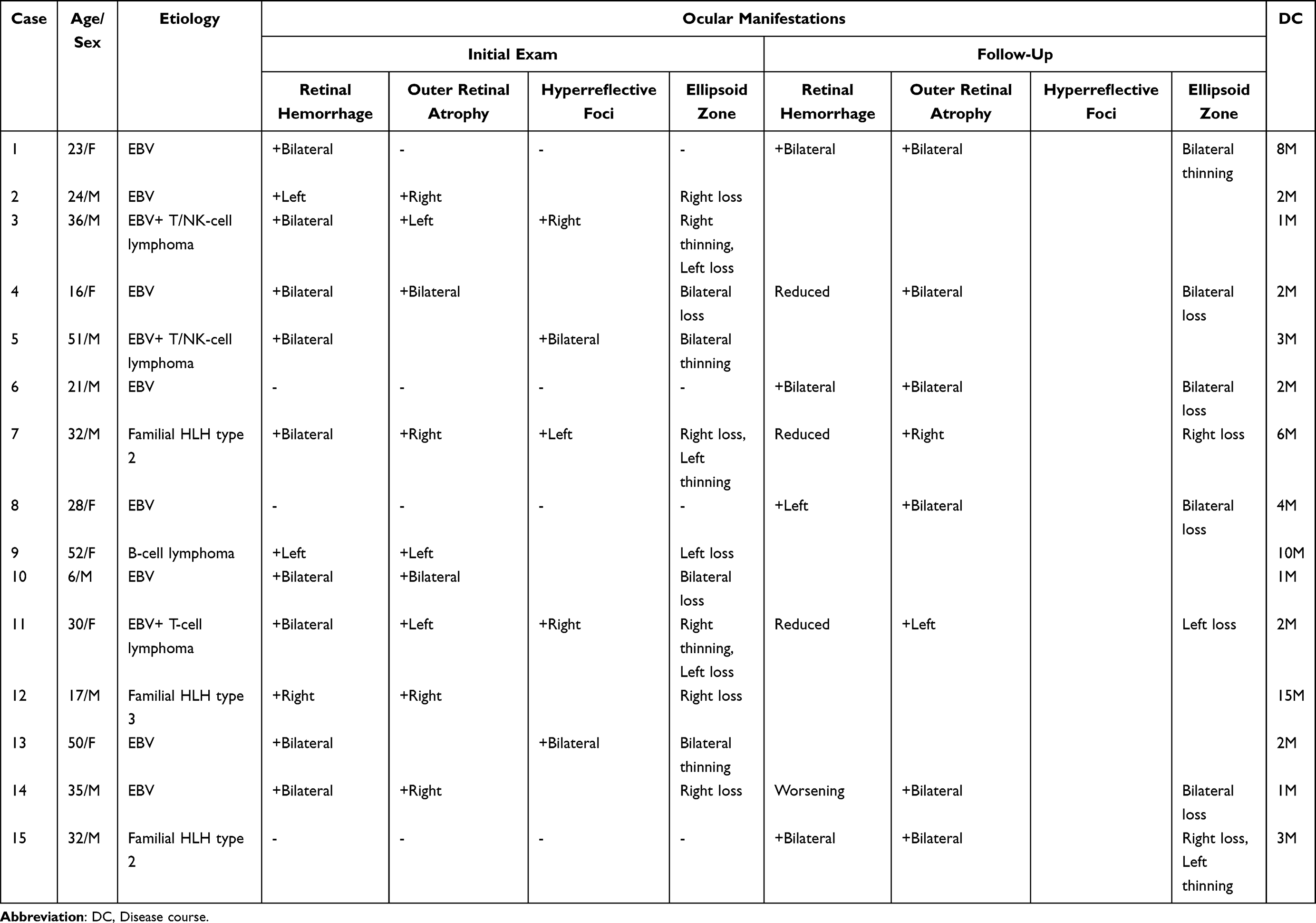 |
Table 1 Clinical Characteristics of the 15 Patients |
Hemorrhage characteristics: Involved all retinal regions (Figure 1). Morphologies included superficial hemorrhages (22 eyes, 73.33%) (Figure 1), Roth spots (6 eyes, 20.00%) (Figure 1), subinternal limiting membrane hemorrhages (Sub-ILM) (3 eyes, 10.00%) (Figure 1), and flame shaped hemorrhages (3 eyes, 10.00%) (Figure 2).
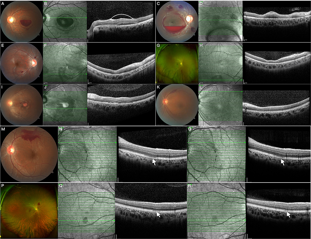 |
Figure 1 (A and B) demonstrate sub-ILM hemorrhage in the macular region with extensive circinate outer retinal atrophy foci in the left eye of Case 3; (C and D) exhibit sub-ILM hemorrhage in the macular region, Roth spot hemorrhage at the vascular arch, and multiple outer retinal atrophy foci in the right eye of Case 10; (E and F) show superficial retinal hemorrhage with multifocal outer retinal atrophy in Case 4; (G and H) reveal isolated intraretinal hemorrhage in the inferior retina accompanied by outer retinal atrophy at the inferior vascular arch in Case 12; (I and J) depict post hemorrhagic hemoglobin absorption status in the macular region with multifocal outer retinal atrophy in the left eye of Case 6; (K and L) display superficial retinal hemorrhage at the superior vascular arch with outer retinal atrophy in Case 9; (M–O) illustrate superficial retinal hemorrhage with outer retinal hyperreflectivity at the superior vascular arch in the left eye of Case 7, showing resolution of hyperreflective lesions on follow up (white arrows); (P–R) demonstrate superficial retinal hemorrhage with hyperreflective outer retinal alterations in the right eye of Case 11, with resolved hyperreflectivity on follow up (white arrows). |
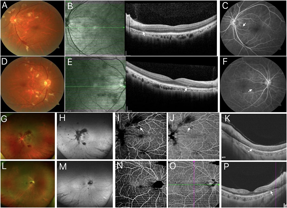 |
Figure 2 Multimodal Imaging of Case 5 and Case 15. (A–F) correspond to Case 5, (A and D) demonstrating bilateral scattered cotton wool spots; (B and E) multifocal outer retinal hyperreflectivity on SD-OCT (white arrows). (C and F) Lesions exhibit early-phase hypofluorescence (white arrows), localized capillary telangiectasia, and microaneurysm formation on FFA. (G–P) represent Case 15, (G and L) showing bilateral flame shaped hemorrhages and left eye Purtscher flecken; (H and M) showing fundus autofluorescence; OCTA reveals outer retinal atrophy superotemporal to the optic disc in the left eye ((K), white arrows) and superficial (I) and deep (J) capillary plexus hypoperfusion (white arrows); and OCTA of the right eye showed retinal atrophy in the temporal outer layer of the optic disc((P), white arrows), with inadequate perfusion of the superficial (N) and deep (O) retinal capillaries found in both the area of the lesion and in the inferotemporal quadrant. |
Outer retinal structural alterations: SD-OCT findings (Figure 1): Outer retinal atrophy (20 eyes, 66.67%) (Figure 1), hyperreflective foci (7 eyes, 23.33%) (Figure 2), ellipsoid zone(EZ) disruption (17 eyes, 56.67%) (Figure 1), EZ thinning (10 eyes, 33.33%) (Figure 1), retinal pigment epithelium (RPE) changes (4 eyes, 13.33%) (Figure 3), and serous detachment (3 eyes, 10.00%) (Figure 3). A significant correlation was observed between ellipsoid zone disruption and outer retinal atrophy (χ2 > 3.84, p<0.05). Near Infrared reflectance (NIR) imaging: Demonstrated well demarcated hyporeflective lesions with darker margins versus centers (Figure 1). Morphologies comprised circular (12 eyes, 40.00%), irregular (17 eyes, 56.67%), and ring shaped (5 eyes, 16.67%) patterns. Multiple lesions per eye involved macular (15 eyes, 50.00%), peripapillary (15 eyes, 50.00%), vascular arcade (8 eyes, 26.67%), and peripheral retinal regions (4 eyes, 13.33%).
 |
Figure 3 Multimodal Fundus Imaging in Case 1 and Case 8. (A–D) represent multimodal imaging of Case 1: (A and B) demonstrate RPE elevation with subneurosensory fluid at initial examination; (C and D) post chemotherapy show resolution of subneurosensory fluid with new onset outer retinal atrophy superotemporal to the optic disc (white arrows). (E–L) correspond to Case 8: (E and I) reveal multiple yellow white punctuate lesions in the posterior pole; (F, G, J and K) exhibit RPE layer abnormalities with subneurosensory fluid on SD-OCT; (H and L) display B-scan ultrasonography showing no significant inflammatory echoes in the vitreous cavity. |
Special case analysis: Two cases (Case 1, 7) had not initiated systemic therapy at baseline examination. Four patients (Case 6, 8, 14, 15) underwent HSCT before secondary follow up (3 months, 3 weeks, 3 weeks, and 2 weeks post HSCT, respectively). All other examinations were conducted during immunosuppressive therapy.
Discussion
HLH is a nonmalignant immune dysregulatory disorder characterized by polyclonal lymphoproliferative hyperplasia of the lymphoid and monocyte macrophage systems with excessive proinflammatory cytokine production, typically inducing pancytopenia, disseminated intravascular coagulation, and cytokine hyper secretion culminating in multiorgan failure; uncontrolled cytotoxic T lymphocyte and natural killer cell activation reciprocally triggers macrophage hyperactivation, precipitating cytokine storm mediated vascular endothelial damage; while etiologically heterogeneous with EBV infection being predominant, this study confirms that outer retinal involvement associated with diverse etiologies of HLH morphologically indistinguishable across causative subgroups and emerging at variable treatment stages is pathophysiologically attributable to HLH itself and unrelated to chemotherapeutic agents or hematopoietic stem cell transplantation sequelae.
In this study, all patients exhibited retinal hemorrhages with diverse manifestations affecting all retinal regions. Our team’s prior retrospective analysis of over 1000 HLH cases revealed that decreased erythrocyte counts, reduced platelet counts, and elevated fibrinogen levels correlated with ocular involvement in HLH patients.3 We therefore hypothesize that retinal hemorrhages stem from hemoglobin and platelet level reductions in HPS patients both being typical diagnostic features of HLH. Reporteds5 incidence of anemic retinopathy reaches 28.3%, with severe anemia (hemoglobin <8 g/dL) and thrombocytopenia (platelet count <50×109/L) constituting high risk factors that demonstrate synergistic effects when coexisting. Rubenstein et al6 documented 44% prevalence of retinopathy among 67 patients with concurrent anemia and thrombocytopenia. These findings strongly suggest that HLH associated severe anemia and thrombocytopenia represent key pathogenic factors in retinochoroidopathy development, though exact mechanisms remain incompletely understood. The established pathological cascade of hypoxia, accelerated blood flow, vascular dilatation, and endothelial damage has been demonstrated to cause retinal hemorrhages and cotton wool spots in anemia.7 Cases 4 and 7 in our series showed marked hemorrhage resolution following platelet and hemoglobin level improvement, supporting our hypothesis that during hemoglobin/ platelet decline in HLH, retinal endothelial integrity becomes compromised by ischemia, vascular wall dilatation and hemodynamic changes, permitting erythrocyte extravasation through endothelial junctions. Sub-ILM hemorrhages as seen in Case 10 (Figure 1) following coughing/ valsalva maneuvers may result from such mechanisms. While minor sub-ILM hemorrhages typically resolve spontaneously, extensive hemorrhages risking macular damage may require ILM laser fenestration or vitrectomy; however, given our proposed pathophysiology, we recommend stabilizing hematological parameters prior to intervention, as isolated ocular treatment proves ineffective without correcting systemic hypoxia according to prior reports.8 While the data suggest a potential pathophysiological link between hemoglobin/platelet levels and retinal lesion, the lack of direct quantitative measurements limits the determination of causality and assessment of predictive validity. Future studies require quantitative validation to confirm the robustness of this mechanism. We further hypothesize that HLH derived cytokines and free radicals contribute to hemorrhage and vascular occlusion, evidenced by studies9 showing perivascular inflammatory infiltration in HLH’s hyperinflammatory state. Hypofibrinogenemia induced disseminated intravascular coagulation(DIC) and massive platelet consumption both precipitate retinal ischemia and vaso occlusive disease.10 Case 15 (Figure 2) developed transplant associated severe anemia with vision loss, exhibiting CRVO like or Purtscher like retinopathy on fundoscopy, with OCTA demonstrating full thickness retinal capillary and choroidal hypoperfusion in outer retinal atrophy zones likely resulting from combined endothelial injury from inflammatory cytokines and hypoxic stress increasing blood viscosity, compounded by DIC from platelet/ fibrinogen depletion. Such pathological changes may affect any retinal/choroidal vascular layer, manifesting as retinochoroidal capillary occlusion or even central retinal artery/ vein occlusion. Simsek et al11 posits that hematologic abnormalities including anemia may disrupt retinal circulation in nonatherosclerotic young patients, causing simultaneous arterial and venous occlusions. Sebrow et al12 reported HLH associated Purtscher like retinopathy cases, attributing these to cytokine mediated DIC where fibrinogen aggregation causes ischemic microemboli findings supporting our hypothesis. Based on these mechanistic analyses, we hypothesize that transfusion therapy targeting hemoglobin/platelet deficits may improve visual outcomes in HLH related retinopathy beyond cytokine storm control. However, due to the limited sample size, this study could not establish statistically significant intervention thresholds for hemoglobin/platelet levels or optimal timing for ophthalmologic screening. The hypothesis remains speculative and requires validation through larger scale prospective studies. Janetos et al documented nearly identical fundus findings to our Case 15 in severe anemia/ thrombocytopenia cases, observing hemorrhage resolution and visual improvement after transfusion therapy during 1 year follow up, emphasizing early hematological correction’s prognostic significance for visual recovery.13
This study identified via SD-OCT that ocular involvement in HLH patients manifests retinal abnormalities ranging from RPE structural alterations to subretinal fluid accumulation and severe serous retinal detachment, resembling central serous chorioretinopathy (CSC). Crucially, Case 1 exhibited subneurosensory retinal fluid at initial ophthalmic examination prior to HLH directed chemotherapy; subsequent follow up after chemotherapy and corticosteroid administration demonstrated significant fluid resolution (Figure 3), thereby differentiating it from typical CSC. This supports the clinical practice that corticosteroid therapy for HLH remains indicated despite CSC like fundus findings. Given the choroid’s hypervascularized nature, it exhibits heightened vulnerability to cytokine mediated inflammatory infiltration in HLH. Postmortem histopathology by Vizcaino et al14 confirmed macrophage/lymphocyte infiltration within the choroid and trabecular meshwork in adult HPS cases. We therefore hypothesize that HLH derived cytokine and free radical storms induce choroidal inflammation, triggering RPE barrier dysfunction and pump failure, which collectively precipitate pigment epithelial detachment (PED) and subretinal fluid accumulation.
This investigation further identified HLH associated outer retinal structural alterations, with SD-OCT unequivocally demonstrating varying degrees of outer retinal atrophy, EZ disruption/ thinning, and outer retinal hyperreflective changes across all 15 cases. NIR imaging revealed hyporeflective lesions with hyperdemarcated margins (Figures 1 and 4), akin to acute macular neuroretinopathy (AMN), though distinctively, HLH related lesions exhibited multifocality beyond the macula, involving multiple retinal regions; NIR detected similar midperipheral lesions, though retrospective analysis limited SD-OCT verification outside the macula, potentially underestimating disease extent. Consistent with Sarraf’s AMN classification,15 all cases manifested SD-OCT features corresponding to Type 2 AMN characterized by outer nuclear layer (ONL) hyperreflectivity progressing to thinning with EZ defects, implicating deep retinal capillary plexus (DCP) occlusion. The outer retina, being a watershed zone supplied by both retinal and choriocapillaris circulations, is inherently vulnerable to ischemic insult.16 Monk et al17 comprehensively analyzed AMN cases with rare etiologies, proposing that beyond dehydration and hypovolemia, leukostasis, increased capillary permeability, endothelial dysfunction with hemorrhagic diathesis, platelet destruction, immune complex deposition induced microarteriolar occlusion, and consumptive coagulopathy may all contribute to focal deep retinal capillary plexus ischemia. Uzun F et al emphasized that these microvascular alterations can occur without visible fundoscopic abnormalities, compromising retinal and choroidal structural integrity and function.18 Consequently, we hypothesize that HLH associated outer retinal injury results from cytokine mediated vascular damage and microvascular thrombosis disrupting the choroid and deep retinal capillary networks, leading to capillary occlusion and outer retinal ischemia. Concurrently, hypoxia from hemoglobin reduction exacerbates endothelial injury, increasing blood viscosity, while diminished platelet and fibrinogen levels promote DIC both mechanisms potentially contributing to choroidal and deep retinal capillary occlusion. In Case 8 (Figure 3), multimodal imaging revealed posterior pole multifocal yellow white punctate lesions, with SD-OCT demonstrating RPE irregular thickening, exudative retinal detachment, and outer retinal atrophy features closely resembling acute posterior multifocal placoid pigment epitheliopathy(APMPPE),19 an entity attributed to primary choriocapillaris ischemia. We therefore hypothesize that Case 8 likely manifests choroidal capillary ischemia through the aforementioned pathological cascade; regrettably, FFA and AF imaging were not performed in this patient to further characterize the vascular deficits. In 2002, Suzuki et al20 reported a case of HLH associated acute posterior multifocal placoid pigment epitheliopathy (APMPPE), whose fundoscopic manifestations were strikingly congruent with our Case 8. Their investigation similarly postulated choriocapillaris ischemia as the underlying pathogenic mechanism. While conventional FFA proved nondiagnostic for deep microvascular occlusion16 (though useful for excluding VKH/APMPPE), OCTA in Cases 14/ 15 objectively demonstrated profound capillary dropout and reduced vessel density in both superficial capillary plexus (SCP) and DCP within atrophic zones, alongside choroidal hypoperfusion. It should be noted that these OCTA based observations cannot be assumed to represent the entire cohort until validated through statistically powered analyses with adequate sample size. Notably, outer retinal hyperreflectivity resolved in Cases 4/ 7/ 11 concurrent with HLH remission and hematologic normalization, whereas progressive outer atrophy occurred in Case 14 (Figure 4) during HLH exacerbation suggesting hyperreflectivity represents an early, potentially reversible ischemic marker, while atrophy signifies irreversible injury. We hypothesize outer retinal structural changes as putative biomarkers for HLH related outer retinal ischemia, warranting prospective validation. Intriguingly, all 5 patients with extensive multifocal outer atrophy had EBV triggered HLH, suggesting viral copathogenicity. The absence of significant vitreous/ retinal inflammation across cases may reflect immunosuppression from chemotherapy/ transplantation or systemic dexamethasone modulation. It is worth noting that chemotherapy induced toxicity may contribute to retinal lesions through oxidative stress or microvascular injury pathways. However, as this study utilized retrospective data lacking systematic records of chemotherapy drug types, doses, or exposure durations, quantification of retinal toxicity was unfeasible. Additionally, the limited sample size precluded subgroup analysis. Consequently, chemotherapy exposure was not included in statistical analyses. We acknowledge this limitation and emphasize the need for future studies to use prospective designs or comprehensive databases to disentangle the contributions of different mechanisms.
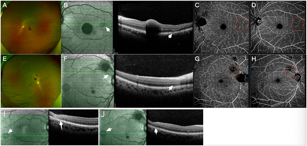 |
Figure 4 Multimodal fundus imaging of Case 14. (A–D) demonstrate subretinal hemorrhage in the macular region of the left eye; SD-OCT reveals localized outer retinal atrophy superotemporal to the macula (white arrow); OCTA shows hypoperfusion and reduced vessel density in both superficial and deep capillary plexuses (red circle). (E–G and H) display flame shaped hemorrhages around the optic disc in the right eye, with outer retinal atrophy (white arrows) in the superotemporal midperipheral retina; OCTA exhibits hypoperfusion in superficial and deep capillary plexuses, with more pronounced alterations in the deep capillary plexus (red circle). I depicts the initial ophthalmic examination, and J the follow up examination, revealing progressive enlargement of the atrophic lesions (white arrow). |
Conclusions
This study employed high resolution multimodal imaging to conduct a retrospective analysis of outer retinal involvement in HLH patients. By synthesizing reported imaging characteristics from existing literature, we systematically characterized the ocular manifestations and investigated underlying pathogenic mechanisms, ultimately enhancing diagnostic precision. Notably, investigations by Li21 and Schafer22 have demonstrated that ocular manifestations particularly acute unilateral vision loss may serve as sentinel features preceding systemic HLH symptoms. Our findings suggest that HLH related ocular involvement should be considered when outer retinal structural abnormalities are detected, accompanied by DCP damage or choroidal capillary ischemia on multimodal imaging, in patients with unexplained persistent fever and cytopenia, to avoid missed or misdiagnosis. However, the retrospective and observational design of our study, along with its limited sample size, may introduce selection bias. Additionally, the inclusion criteria inherently favor patients with retinal lesions, which may itself constitute a selection bias. These findings highlight the urgent necessity to establish early warning indicators for HLH related retinopathy and conduct multicenter prospective studies to strengthen evidence based diagnostic and therapeutic strategies, with future research prioritizing the determination of relationships between hemoglobin/platelet levels and retinal lesion severity, evaluation of temporal associations between transfusion interventions and retinal recovery, assessment of platelet count thresholds’ impact on retinal hemorrhage prevention, and identification of biomarkers predictive of HLH related retinopathy.
Acknowledgments
The authors would like to thank all the participants involved in the study.
Funding
There is no funding to report.
Disclosure
No potential conflict of interest was reported by the authors.
References
1. Henter JI. Hemophagocytic lymphohistiocytosis. N Engl J Med. 2025;392(6):584–598. doi:10.1056/NEJMra2314005
2. Patil A, Mahendradas P, Shetty R. Hemophagocytic lymphohistiocytosis after dengue fever and its ocular features. JAMA Ophthalmol. 2023;141(9):e231069. doi:10.1001/jamaophthalmol.2023.1069
3. Wang L, Suo L, Kou F, et al. Ocular phenotypes in patients with hemophagocytic lymphohistiocytosis: a retrospective analysis in a single center over 7 years. Am J Ophthalmol. 2023;253:119–131. doi:10.1016/j.ajo.2023.05.011
4. Henter JI, Horne A, Aricó M, et al. HLH-2004: diagnostic and therapeutic guidelines for hemophagocytic lymphohistiocytosis. Pediatr Blood Cancer. 2007;48(2):124–131. doi:10.1002/pbc.21039
5. Mansour AM, Lee JW, Yahng SA, et al. Ocular manifestations of idiopathic aplastic anemia: retrospective study and literature review. Clin Ophthalmol. 2014;8:777–787. doi:10.2147/OPTH.S62163
6. Rubenstein RA, Yanoff M, Albert DM. Thrombocytopenia, anemia, and retinal hemorrhage. Am J Ophthalmol. 1968;65(3):435–439. doi:10.1016/0002-9394(68)93098-5
7. Selvan H, Azad SV, Ramesh P, Bhakthaganesh K. Retinopathy in severe aplastic anaemia. BMJ Case Rep. 2019;12(10):e232426. doi:10.1136/bcr-2019-232426
8. Metelitsina TI, Sheth VS, Patel SB, Grassi MA. Peripheral retinopathy associated with aplastic anemia. Retin Cases Brief Rep. 2017;11(2):108–110. doi:10.1097/ICB.0000000000000302
9. Gupta J, Jauhari P, Kumar A, et al. Primary hemophagocytic lymphohistiocytosis with prolonged primary neurologic presentation. Pediatrics. 2023;151(4):e2022057848. doi:10.1542/peds.2022-057848
10. Finn AP, Roehrs P, Grace SF, Vajzovic L. Ischemic retinal vascular disease in an infant with hemophagocytic lymphohistiocytosis. JAMA Ophthalmol. 2017;135(11):1277–1279. doi:10.1001/jamaophthalmol.2017.3258
11. Simsek A, Tekin M, Bilen A, Karadag AS, Bucak IH, Turgut M. Evaluation of choroidal thickness in children with iron deficiency anemia. Invest Ophthalmol Vis Sci. 2016;57(14):5940–5944. doi:10.1167/iovs.15-18713
12. Sebrow DB, Dhrami-Gavazi E, Horowitz JD, Yannuzzi LA. Purtscher retinopathy as a manifestation of hemophagocytic lymphohistiocytosis. Retin Cases Brief Rep. 2017;11(4):335–338. doi:10.1097/ICB.0000000000000360
13. Janetos TM, Bhasin A. Optic disc hemorrhages in severe anemia and thrombocytopenia. J Gen Intern Med. 2023;38(1):234–235. doi:10.1007/s11606-022-07853-2
14. Vizcaino MA, Eberhart CG, Rodriguez FJ. Hemophagocytic lymphohistiocytosis in adults with intraocular involvement: clinicopathologic features of 3 cases. Ocul Oncol Pathol. 2017;4(1):1–11. doi:10.1159/000475551
15. Sarraf D, Rahimy E, Fawzi AA, et al. Paracentral acute middle maculopathy: a new variant of acute macular neuroretinopathy associated with retinal capillary ischemia. JAMA Ophthalmol. 2013;131(10):1275–1287. doi:10.1001/jamaophthalmol.2013.4056
16. Bhavsar KV, Lin S, Rahimy E, et al. Acute macular neuroretinopathy: a comprehensive review of the literature. Surv Ophthalmol. 2016;61(5):538–565. doi:10.1016/j.survophthal.2016.03.003
17. Munk MR, Jampol LM, Cunha Souza E, et al. New associations of classic acute macular neuroretinopathy. Br J Ophthalmol. 2016;100(3):389–394. doi:10.1136/bjophthalmol-2015-306845
18. Bezci Aygün F, Algedik Tokyürek MÖ, Unal Ş, Gümrük F, Kadayifcilar S. Retinal and choroidal circulation impairments in fanconi anemia. Am J Ophthalmol. 2025;272:166–175. doi:10.1016/j.ajo.2025.01.017
19. Jabs DA, Brezin AP, Dick AD; Standardization of Uveitis Nomenclature (SUN) Working Group. Classification criteria for acute posterior multifocal placoid pigment epitheliopathy. Am J Ophthalmol. 2021;228:174–181. doi:10.1016/j.ajo.2021.03.056
20. Suzuki S, Mizota A, Adachi-Usami E. A case of hemophagocytic syndrome with retinal changes resembling acute posterior multifocal placoid pigment epitheliopathy. Retina. 2002;22(2):219–222. doi:10.1097/00006982-200204000-00015
21. Li X, Ma Y, Tang J, Chen T, Ma X. A hemophagocytic lymphohistiocytosis patient that presented with unilateral panuveitis. Ocul Immunol Inflamm. 2017;25(2):275–277. doi:10.3109/09273948.2015.1107593
22. Schafer GL, Jaladat Y, Hawy E. A case of the initial presentation of hemophagocytic lymphohistiocytosis as acute unilateral vision loss. Cureus. 2024;16(11):e73820. doi:10.7759/cureus.73820
 © 2025 The Author(s). This work is published and licensed by Dove Medical Press Limited. The
full terms of this license are available at https://www.dovepress.com/terms
and incorporate the Creative Commons Attribution
- Non Commercial (unported, 4.0) License.
By accessing the work you hereby accept the Terms. Non-commercial uses of the work are permitted
without any further permission from Dove Medical Press Limited, provided the work is properly
attributed. For permission for commercial use of this work, please see paragraphs 4.2 and 5 of our Terms.
© 2025 The Author(s). This work is published and licensed by Dove Medical Press Limited. The
full terms of this license are available at https://www.dovepress.com/terms
and incorporate the Creative Commons Attribution
- Non Commercial (unported, 4.0) License.
By accessing the work you hereby accept the Terms. Non-commercial uses of the work are permitted
without any further permission from Dove Medical Press Limited, provided the work is properly
attributed. For permission for commercial use of this work, please see paragraphs 4.2 and 5 of our Terms.