
Back to Journals » Drug Design, Development and Therapy » Volume 20
Modified Jie-Yu-He-Huan Capsule in Post-Stroke Depression Treatment: Insights from DIA Proteomic and PRM Validation
Authors Wang X, Gao Q, Liu C  , Hu M, Liu Z, Li Z, Zhang H, Wu L, Chen K, Xu K, Geng X, Liu W, Wei S
Received 10 October 2025
Accepted for publication 23 January 2026
Published 4 February 2026 Volume 2026:20 565281
DOI https://doi.org/10.2147/DDDT.S565281
Checked for plagiarism Yes
Review by Single anonymous peer review
Peer reviewer comments 3
Editor who approved publication: Dr Tuo Deng
Xinyu Wang,1– 5,* Qian Gao,6,* Chao Liu,7,8,* Minghui Hu,1,2 Zhiyao Liu,1,2,4 Zifa Li,1– 5 Hao Zhang,1– 4 Lidan Wu,1– 4 Kai Chen,9 Kaiyong Xu,2– 5 Xiwen Geng,1– 5 Wei Liu,1– 4,10 Sheng Wei1– 4
1Institute for Chinese Medicine and Brain Science, Shandong University of Traditional Chinese Medicine, Ji’nan, Shandong, 250355, People’s Republic of China; 2Key Laboratory of Traditional Chinese Medicine Classical Theory, Ministry of Education, Shandong University of Traditional Chinese Medicine, Ji’nan, Shandong, 250355, People’s Republic of China; 3Shandong Key Laboratory of Innovation and Application Research in Basic Theory of Traditional Chinese Medicine, Shandong University of Traditional Chinese Medicine, Ji’nan, Shandong, 250355, People’s Republic of China; 4Shandong Provincial Engineering Research Center for the Prevention and Treatment of Major Brain Diseases with Traditional Chinese Medicine, Shandong University of Traditional Chinese Medicine, Ji’nan, Shandong, 250355, People’s Republic of China; 5Experimental Center, Shandong University of Traditional Chinese Medicine, Ji’nan, Shandong, 250355, People’s Republic of China; 6Department of Basic Medical Sciences, Shijiazhuang Medical College, Shijiazhuang, Hebei, People’s Republic of China; 7Department of Traditional Chinese Medicine, Shandong Provincial Hospital Affiliated to Shandong First Medical University, Ji’nan, Shandong, People’s Republic of China; 8First Clinical Medical College, Shandong University of Traditional Chinese Medicine, Ji’nan, Shandong, People’s Republic of China; 9Shandong Innovation Center of Engineered Bacteriophage Therapeutics, Shandong Academy of Pharmaceutical Sciences, Ji’nan, Shandong, People’s Republic of China; 10Department of Encephalopathy, The second Affiliated Hospital of Shandong University of Traditional Chinese Medicine, Ji’nan, Shandong, People’s Republic of China
*These authors contributed equally to this work
Correspondence: Sheng Wei, Key Laboratory of Traditional Chinese Medicine Classical Theory, Ministry of Education, Shandong University of Traditional Chinese Medicine, Ji’nan, Shandong, People’s Republic of China, Fax +86 531 89628015, Email [email protected] Wei Liu, Department of Encephalopathy, The Second Affiliated Hospital of Shandong University of Traditional Chinese Medicine, Ji’nan, Shandong, People’s Republic of China, Email [email protected]
Background and Purpose: Post-stroke depression (PSD), a prevalent neuropsychiatric complication of stroke, manifests as persistent low mood and depressive symptoms secondary to cerebrovascular injury, contributing to increased morbidity and mortality. Although the modified Jie-Yu-He-Huan (MJYHH) capsule has demonstrated clinical efficacy in alleviating PSD symptoms, its pharmacological targets and mechanisms of action remain unclear.
Methods: Pharmacological screening was performed on healthy C57BL/6 mice, revealing significant antidepressant effects of MJYHH. A PSD rat model was subsequently established through combined middle cerebral artery occlusion (MCAO), social isolation, and chronic mild stress (CMS). Behavioural tests were conducted to evaluate therapeutic outcomes. Data-independent acquisition (DIA) and parallel reaction monitoring (PRM) technologies were employed to investigate the traditional Chinese medicine (TCM) mechanisms.
Results: The PSD model group exhibited characteristic depressive behaviours, which were significantly attenuated by MJYHH treatment. Proteomic analysis identified 16 differentially expressed proteins (DEPs), with subsequent Gene Ontology (GO), KEGG pathway, and protein-protein interaction (PPI) network analyses demonstrating their predominant association with: biosynthetic and metabolic pathways, oxidative stress response, mitochondrial dysfunction.PRM validation and molecular docking studies further implicated three key targets (Rrm2b, Cyp51a1, and Rhot2) as potential mediators of MJYHH’s therapeutic effects.
Conclusion: This integrated investigation combining animal models, omics technologies, and computational approaches elucidates the multi-target mechanism of MJYHH capsule in PSD treatment, providing a scientific foundation for TCM-based intervention strategies.
Keywords: post-stroke depression, modified Jie-Yu-He-Huan capsules, data independent acquisition, parallel reaction monitoring, molecular docking
Introduction
Post-stroke depression (PSD) constitutes a prevalent neuropsychiatric complication following cerebrovascular events, clinically characterised by persistent anhedonia and pathological despair.1 This condition detrimentally influences critical clinical outcomes including functional recovery trajectories, quality of life indices, stroke recurrence rates, and all-cause mortality. Epidemiological evidence suggests approximately 33% of stroke survivors develop clinically significant depressive symptoms during post-acute recovery,2 substantially impeding rehabilitation processes.3 Contemporary research has elucidated PSD’s multifactorial pathogenesis involving neurotransmitter dysregulation, neuroinflammatory cascades, oxidative stress responses, and impaired neuroplasticity. Despite this complexity, therapeutic options remain limited to selective serotonin reuptake inhibitors (SSRIs) such as fluoxetine, which carry risks of adverse effects and dependency.4 This therapeutic gap underscores the imperative for both mechanistic investigations and novel treatment development.
Traditional Chinese medicine (TCM) has demonstrated distinctive advantages in PSD management through its holistic, multi-target approach.5,6 Our previous work established the efficacy of ShuYu and Jie-Yu-He-Huan (JYHH) capsules in rodent models of mood disorders.7,8 The modified JYHH formulation (MJYHH), enhanced with curcumin from Curcuma longa, exhibits neuroprotective properties9 while maintaining the original composition (Paeoniae Radix Alba, Fructus Gardeniae, Albiziae Flos, and Moutan Cortex).7 Although clinically effective for anxiety disorders with favourable safety profiles, MJYHH’s molecular mechanisms in PSD remain incompletely characterised.
Proteomic advancements have revolutionised mechanistic studies in both neurology and TCM research.10,11 Data-independent acquisition (DIA) proteomics offers unparalleled coverage for large-scale protein profiling, while parallel reaction monitoring (PRM) enables high-specificity target validation. This dual approach was successfully employed in cerebral ischemia research,12 establishing its suitability for elucidating complex drug actions.
In this study, we employed behavioural assessments and integrated proteomics (DIA-PRM) to investigate MJYHH’s effects in PSD model rats, focusing on hippocampal protein dynamics. Molecular docking further validated multi-compound/multi-target interactions. Our findings provide novel insights into MJYHH’s pharmacological actions and potential therapeutic targets for PSD.
Materials and Methods
Animals
Sprague-Dawley rats (220 ± 20 g) and C57BL/6J mice (18–22 g, 6–8 weeks old) were obtained from Beijing Vital River Laboratories (Production License: SCXY [jing] 2021–0006). Following a one-week acclimatisation period with ad libitum access to food and water, animals were maintained under controlled conditions (21±1°C) with a reversed 12-hour light/dark cycle (lights on 21:00–09:00). Daily parallel operations were implemented to minimise human interference. All animal experimental procedures adhered to the guidelines for the care and use of laboratory animals as issued by the National Institutes of Health (NIH) and were approved by the Experimental Animal Ethics Committee of Shandong University of Chinese Medicine (SYXK [Lu] 2022–0009). Behavioural testing occurred during the animals’ active phase (10:00–17:00).
Post-Stroke Depression Model Establishment
The PSD model was developed through sequential implementation of middle cerebral artery occlusion (MCAO), social isolation, and 21-day chronic mild stress (CMS), modifying the approach described by Willner et al.13 This protocol was designed to recapitulate the neuropsychological interactions characteristic of PSD.
MCAO was performed using a modified Zea Longa suture method,13 (see Supplementary Figure 1). Briefly, isoflurane-anaesthetised rats were positioned supine, with the ventral neck area shaved and disinfected with povidone-iodine. A 2 cm midline incision exposed the left carotid sheath (Supplementary Figure 1A). Following separation of the sternocleidomastoid muscle, the common carotid artery (CCA) was isolated and two ligatures placed (Supplementary Figure 1B). After proximal permanent ligation and distal temporary occlusion, a 45° arteriotomy was made in the CCA (Supplementary Figure 1C). A nylon filament (diameter 0.26 mm) was advanced 16–19 mm into the internal carotid artery to induce ischaemia (Supplementary Figure 1D). Sham controls underwent identical procedures excluding filament insertion.
Neurological function was evaluated 24 hours post-operation using the Z-Longa scoring system. For this study, we selected model rats demonstrating moderate neurological deficits, corresponding to Z-Longa scores between 1 and 3. The scoring system was implemented as follows: a score of 0 indicated no observable neurological deficits; 1 denoted failure to fully extend the paralysed forelimb; 2 represented circling behaviour toward the paretic side during ambulation; 3 indicated pronounced lateral tilting during movement; 4 signified complete loss of spontaneous locomotion with comatose state; and 5 indicated mortality.
Following assessment, rats scoring between 1–3 were randomly allocated to three experimental groups (PSD, MJJHH and FLX groups) based on baseline measurements of sucrose preference and total distance travelled in open field tests. All animals were individually housed and subjected to chronic mild stress (CMS) for three weeks, with the exception of control sham group animals. Concurrent with CMS exposure, pharmacological interventions were administered throughout the three-week period.
Control sham and PSD group rats received daily oral gavage of 0.5% sodium carboxymethyl cellulose (10 mL/kg/d). The MJYHH group received combined treatment with JYHH (357.427 mg/kg/d) and curcumin (176.918 mg/kg/d) via oral gavage. FLX group animals were administered fluoxetine (2.08 mg/kg/d).14 Behavioural assessments were conducted 1 hour following the final administration.
The CMS protocol incorporated 13 distinct stressors selected to induce depression-like behaviours without causing traumatic distress: strobe light exposure, restraint stress, food deprivation, water deprivation, wet bedding conditions, white noise exposure, circadian rhythm disruption (day-night reversal), empty bottle stimulation, novel object exposure, 45° cage tilting, tail clamping, food stimulation and odour stimulation. These were carefully implemented to provide varied environmental challenges. Each model rat received at least one randomly assigned stressor daily. The complete weekly CMS schedule is detailed in Supplementary Table 1. Behavioural data analysis and evaluation employ a blind method.
Drugs and Reagents
JYHH capsule, a traditional Chinese medicinal formulation, contained standardized extracts of four herbal components: Paeoniae Radix Alba (52.1%), Fructus Gardeniae (27.0%), Albiziae Flos (18.7%), and Moutan Cortex (2.2%). The extraction protocol for these constituents followed our previously published methodology.7 For the present study, a modified JYHH formulation was prepared by supplementing the original capsule composition with curcumin. All herbal components were suspended in distilled water containing 0.5% (w/v) sodium carboxymethyl cellulose to ensure homogeneity. Curcumin (C805205, purity ≥98%) was procured from Shanghai Macklin Biochemical Technology. Fluoxetine hydrochloride (9515AC), employed as a reference compound, was obtained from Eli Lilly and Company.
Safety Evaluation of the MJYHH Capsule
Fifty C57BL/6J mice were randomly assigned to five experimental groups: (1) control group receiving 0.5% sodium carboxymethyl cellulose (0.2 mL/10 g/day); (2) JYHH capsule group administered 510.61 mg/kg/day7 of the standard formulation; (3) curcumin group treated with 252.74 mg/kg/day; (4) MJYHH capsule group receiving both JYHH capsule (510.61 mg/kg/day) and curcumin (252.74 mg/kg/day);15 and (5) fluoxetine group administered 18 mg/kg/day16 as a positive control. All treatments were administered by oral gavage for 30 consecutive days.
Body weight was monitored weekly throughout the experimental period. Behavioural assessments were conducted during the final two days of treatment, with all tests initiated precisely one hour following drug administration to ensure standardised pharmacokinetic conditions.
2,3,5-Triphenyltetrazolium Chloride (TTC) Staining
Following anaesthesia, rat brain tissue was dissected 2 h after MCAO surgery. The excised tissue was placed at −20°C for 20 min to facilitate preparation of 2 mm-thick coronal sections. The brain slices were then incubated in a 2% TTC solution at 37°C for 20 min under light-protected conditions. After staining, the sections were fixed in 4% paraformaldehyde for 5 min, air-dried, and photographed for documentation. All procedures were performed in accordance with the methodology described by Li et al.17
Behaviour Test
Balance Beam Test (BBT)
Motor coordination was assessed using a balance beam apparatus consisting of a horizontal wooden board (2.5 cm wide × 120 cm long), fixed 50 cm above the ground and suspended at each end by a support frame. To mitigate injury risk, a soft cushion was placed beneath the beam.18 Prior to MCAO surgery, all rats underwent training sessions to ensure proficiency in traversing the beam. Animal’s incapable of maintaining balance during pre-surgical trials were excluded from subsequent experiments. At 1 h post-MCAO, rats were positioned at one end of the beam and evaluated using a validated scoring system:
0: Traversed beam steadily with all four limbs;
1: Balanced using three limbs without falling;
2: Demonstrated unstable posture but retained position;
3: One limb slipped but remained on beam >60 s;
4: Unable to walk normally but maintained position >40 s;
5: Fell within 40s of attempted traversal;
6: Immediate inability to stand, resulting in rapid fall.
This protocol was adapted from established methodologies.19
Sucrose Preference Test (SPT)
The SPT was conducted to assess anhedonia, a core symptom of depression.20 Rats were individually housed and acclimatised to two bottles of 1% sucrose solution for 24 h. Subsequently, they were presented with one bottle of pure water and one bottle of 1% sucrose solution for 24 h. After 23 h of water deprivation, rats were given access to both bottles for 1 h, with bottle positions alternated to control for side preferences. Sucrose preference was calculated as:
Sugar preference value = (Sugar consumption/Total liquid consumption) × 100%.
Open Field Test (OFT)
Rats were placed in an open-field arena for 6 min, with behaviour recorded using an infrared camera system (15 frames/s). Locomotor activity was analysed using the XR SuperMaze animal behaviour tracking system.21
Tail Suspension Test (TST)
Depressive-like behaviour was evaluated by suspending mice 60 cm above the ground via adhesive tape placed 1 cm from the tail tip. Immobility time during the final 4 min of the 6-min test was quantified using SMART 3.0 software.22
Forced Swimming Test (FST)
For rats: Animals were placed in a transparent cylindrical tank (30 cm water depth, 23°C) for 10 min. Sessions were recorded using SMART 3.0, with water replaced between trials to minimise olfactory cues.23
For mice: The protocol was adapted as follows: mice were exposed to a 20 cm water column (23°C) for 15 min, dried at 30°C, and retested 24 h later for 5 min. Data were analysed as described for rats.24
Data-Independent Acquisition (DIA) Proteomics
Protein Preparation and Digestion
Rat brain tissues were homogenised in lysis buffer (8 M urea, 50 mM Tris-HCl) supplemented with Roche protease inhibitor cocktail. Following centrifugation (20,000 × g, 4°C, 15 min), the supernatant was reduced with 10 mM dithiothreitol (DTT) at 37°C for 60 min and subsequently alkylated with 20 mM iodoacetamide (IAA) in darkness for 30 min. Protein concentration was determined using the Bradford assay, with sample integrity verified by SDS-PAGE. Proteins were digested with trypsin, and the resulting peptides were desalted using Waters solid-phase extraction cartridges before vacuum drying. Dried peptides were reconstituted in ultrapure water and stored at −20°C.
Nano-LC-MS/MS Quantitative Analysis
Peptides were resuspended in 0.1% formic acid (FA), centrifuged (20,000 × g, 10 min), and loaded onto a self-packed C18 column (100 μm inner diameter, 1.8 μm particles, 35 cm length) using an EASY-nLC™ 1200 system (Thermo Scientific). Separation was achieved at 300 nL/min with the following gradient: 4–27% solvent B (98% acetonitrile, 0.1% FA) over 0–103 min, 27–40% from 103–111 min, 40–90% from 111–113 min, maintained at 90% until 120 min.
Mass spectrometry was performed on an Orbitrap Exploris™ 480 (Thermo Fisher Scientific) with these parameters:
Full MS: 350–1,500 m/z range, 120,000 resolution, 300% normalised AGC target
DIA mode: 53 variable windows (400–1,200 m/z), HCD fragmentation at 32% energy
MS/MS: Orbitrap detection at 30,000 resolution, 200% normalised AGC target.
Protein Identification and Quantification
DIA-NN software processed raw data against UniProt-derived protein sequences using these criteria: trypsin/P digestion (max 2 missed cleavages); fixed carbamidomethylation ©; variable modifications of oxidation (M) and N-terminal acetylation; 20 ppm precursor and 0.05 Da fragment mass tolerances. Results were filtered at 1% false discovery rate (FDR) for downstream analysis.
Parallel Reaction Monitoring (PRM) Validation
Pooled peptide samples were analysed in PRM mode using an Orbitrap Exploris™ 480 with NSI ion source. Candidate differentially expressed proteins (DEPs) were quantified via targeted peptide sequencing, with data processed in Skyline software.
Molecular Docking Studies
Ligand Preparation
Small molecule 3D structures from PubChem were energy-minimised in ChemBio3D Ultra 14.0 (RMS gradient: 0.001) and converted to mol2 format. AutodockTools-1.5.6 assigned charges, rotatable bonds, and hydrogen atoms before saving as PDBQT files.
Protein Preparation
Rrm2b, Cyp51a1, and Rhot2 structures from UniProt were processed in PyMOL 2.3.0 to remove crystallographic waters/ligands. AutodockTools-1.5.6 prepared proteins for docking (hydrogen addition, charge assignment) in PDBQT format.
Docking Parameters
Binding sites were predicted using POCASA 1.1. AutoDock Vina 1.1.2 performed docking with these grid parameters (40 × 40×40 Å, 0.42 Å spacing):
Rrm2b: Centre (3.1655, 27.6395, 77.697)
Cyp51a1: Centre (−14.197, −1.2425, 4.483)
Rhot2: Centre (8.888, 0.841, 3.4265).
Statistical Analysis
Behavioural test data are expressed as mean ± standard error of the mean (SEM). Group comparisons for repeated measures were analysed using two-way analysis of variance (ANOVA), with Bonferroni’s post hoc test applied for pairwise comparisons. All statistical analyses were performed using GraphPad Prism 9.0 software, with significance levels denoted as *p < 0.05, **p < 0.01, ***p < 0.001 and ****p < 0.0001 versus control (Con) groups, or #p < 0.05, ##p < 0.01, ###p < 0.001 and ####p < 0.0001 versus post-stroke depression (PSD) groups. Non-significant differences are indicated by ns.
For proteomic analysis, data processing was conducted in R (version 4.0.0). Raw protein intensities were normalised using the median-centred method. Hierarchical clustering was visualised using the heatmap package, while principal component analysis (PCA) was performed with the metaX package. Differentially expressed proteins were identified through Student’s t-test (p < 0.05) with a fold-change threshold of <0.83 or >1.2. Functional annotation of protein sequences was achieved through hypergeometric enrichment analysis of KEGG pathways and Gene Ontology terms.
Results
Evaluation of Anti-Depressant Efficacy of MJYHH Capsules in Mice
The MJYHH capsules, a novel Chinese herbal formulation comprising Paeoniae Radix Alba, Fructus Gardeniae, Albiziae Flos, Moutan Cortex, and curcumin, were evaluated for efficacy and safety in healthy male C57BL/6J mice. Following 30 days of oral administration, behavioural assessments were conducted using OFT, TST, and FST on days 29–30 (Figure 1A), established paradigms for antidepressant screening.25 No significant differences in body weight were observed across the five experimental groups (control, JYHH capsules, curcumin, MJYHH capsules, and fluoxetine; Supplementary Figure 2A), indicating no adverse metabolic effects of MJYHH treatment. Similarly, OFT results revealed no intergroup variations in total distance travelled, time spent in the central area, or central area entries (Figure 1B and 1C; Supplementary Figure 2B), suggesting unaltered locomotor activity or anxiety-like behaviour. In depression-related behavioural assays, the MJYHH capsules, curcumin, and fluoxetine groups exhibited significantly reduced immobility duration in the TST compared to controls (Figure 1D). A parallel anti-depressant effect was observed in the FST, where MJYHH capsules and fluoxetine, but not curcumin, significantly decreased immobility time (Figure 1E). No group differences were detected in the number of immobility episodes during TST or FST (Supplementary Figure 2C and D). These findings demonstrate that MJYHH capsules are a safe and effective anti-depressant intervention in mice, with efficacy comparable to fluoxetine. The optimised doses identified in this murine model were subsequently applied to rat studies.
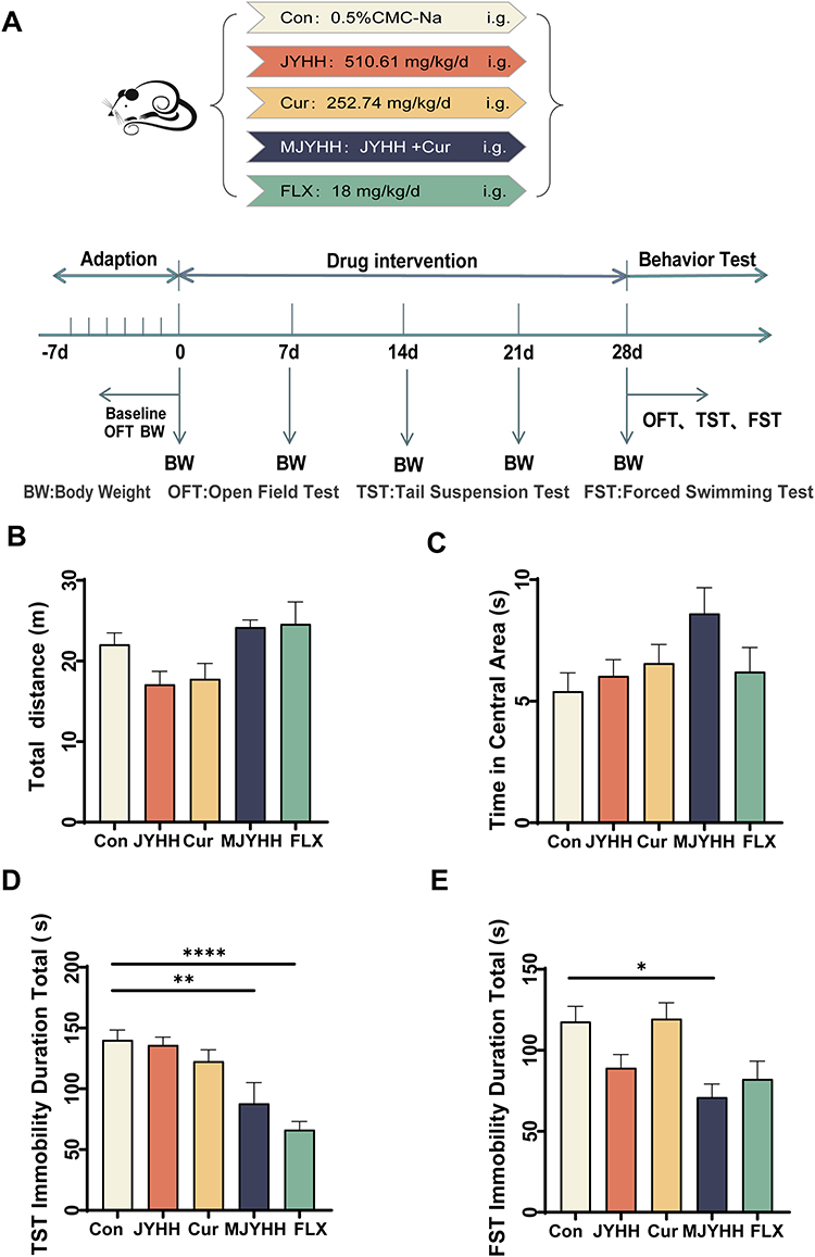 |
Figure 1 Evaluation of MJYHH capsule efficacy in behavioural tests. (A) Schematic representation of the experimental design. (B) Total distance travelled in the open field test (OFT). (C) Time spent in the central area during OFT. (D) Total immobility duration in the tail suspension test (TST). (E) Total immobility duration in the forced swim test (FST). Data are presented as mean ± standard error of the mean (SEM). Statistical significance was determined by two-way ANOVA compared to control groups: *p < 0.05, **p < 0.01, ****p < 0.0001. Abbreviations: i.g., Intragastric Administration; Con, Control Group; JYHH, Jie-Yu-He-Huan capsules group; Cur, Curcumin Group; MJYHH, Modified Jie-Yu-He-Huan capsules group; FLX, Fluoxetine Group. |
MJYHH Capsules Ameliorate Depression-Like Behaviours in PSD Rat Model
To evaluate the therapeutic potential of MJYHH capsules in PSD, we established a rat model through MCAO surgery followed by four weeks of CMS and social isolation. After model validation, PSD rats received either MJYHH capsules or fluoxetine for seven days prior to behavioural assessments, including the BBT, SPT, OFT, and FST (Figure 2A). The successful induction of stroke was confirmed by elevated BB scores (Figure 2B) and TTC staining revealing cerebral infarction (Figure 2C). During CMS exposure, PSD model rats exhibited significantly reduced body weight compared to controls, a trend reversed by both MJYHH and fluoxetine treatment (Figure 2D). In the OFT, while total locomotion distance remained comparable across groups, PSD rats demonstrated marked reductions in centre-area exploration (distance, entries, and duration) relative to sham controls. These deficits were attenuated by MJYHH or fluoxetine administration (Figure 2E–2I). The SPT revealed diminished sucrose preference in PSD rats, indicative of anhedonia, which was significantly restored by both treatments (Figure 2J). Similarly, the FST showed prolonged immobility duration and increased immobility episodes in PSD rats, parameters that were normalised following MJYHH or fluoxetine intervention (Figure 2K and 2L). Collectively, these data demonstrate successful PSD model establishment and validate the efficacy of MJYHH capsules in mitigating depression-like behaviours, with effects comparable to fluoxetine.
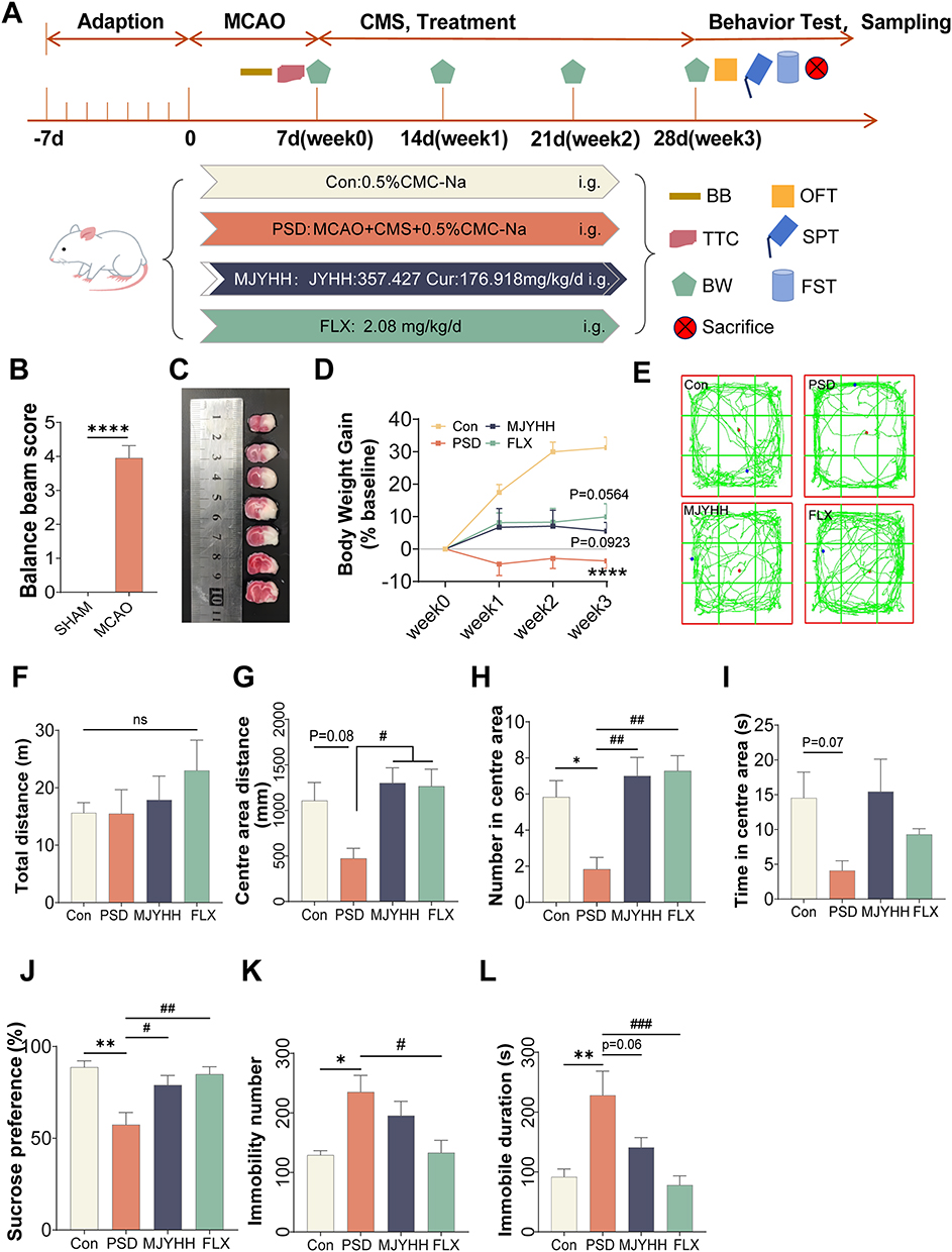 |
Figure 2 Therapeutic effects of MJYHH treatment on post-stroke depression (PSD)-like behaviours. (A) Schematic overview of the experimental design. (B) Balance beam test scores following middle cerebral artery occlusion (MCAO). (C) Representative 2,3,5-triphenyltetrazolium chloride (TTC) staining results. (D) Body weight changes during the experimental period. (E) Representative open field test (OFT) tracking plots. (F) Total distance travelled in the OFT. (G) Distance covered in the central zone during OFT. (H) Number of entries into the central area during OFT. (I) Time spent in the central zone during OFT. (J) Percentage sucrose preference in the sucrose preference test. (K) Immobility duration during the forced swim test (FST). (L) Total number of immobility episodes during FST. Data are presented as mean ± standard error of the mean (SEM). Statistical significance was determined by two-way ANOVA: ns indicates no significant difference; *p < 0.05, **p < 0.01, ****p < 0.0001 versus control (Con) groups; #p < 0.05, ##p < 0.01, ###p < 0.001, versus PSD groups. |
Protein Profiling Reveals Differentially Expressed Proteins (DEPs) in PSD Rats Treated with MJYHH Capsules
To elucidate the molecular mechanisms underlying the therapeutic effects of MJYHH capsules on PSD, we conducted a comprehensive proteomic analysis of hippocampal tissues using DIA technology (Figure 3A). The DIA analysis identified approximately 80,374 peptide segments and 5,178 proteins across all groups (n=5, FDR<0.01, Supplementary Figure 3A–3C). Principal component analysis (PCA) demonstrated strong intra-group clustering with clear inter-group separation, indicating high reproducibility within groups and significant proteomic differences between groups (Figure 3B). Hierarchical clustering analysis further revealed distinct protein expression patterns among the three experimental groups (Figure 3C and 3D). Comparative analysis identified 84 DEPs between control and PSD model rats, and 120 DEPs between MJYHH-treated and PSD model rats (Figure 3E). A Venn diagram illustrated 16 proteins with altered expression common to all three groups (Figure 3F). DEPs were defined based on statistical significance (p < 0.05) and fold-change thresholds (FC < 0.83 or >1.2). These findings provide a molecular framework for understanding how MJYHH capsules ameliorate depression-like behaviours in PSD rats, revealing key protein targets that may underlie their therapeutic efficacy.
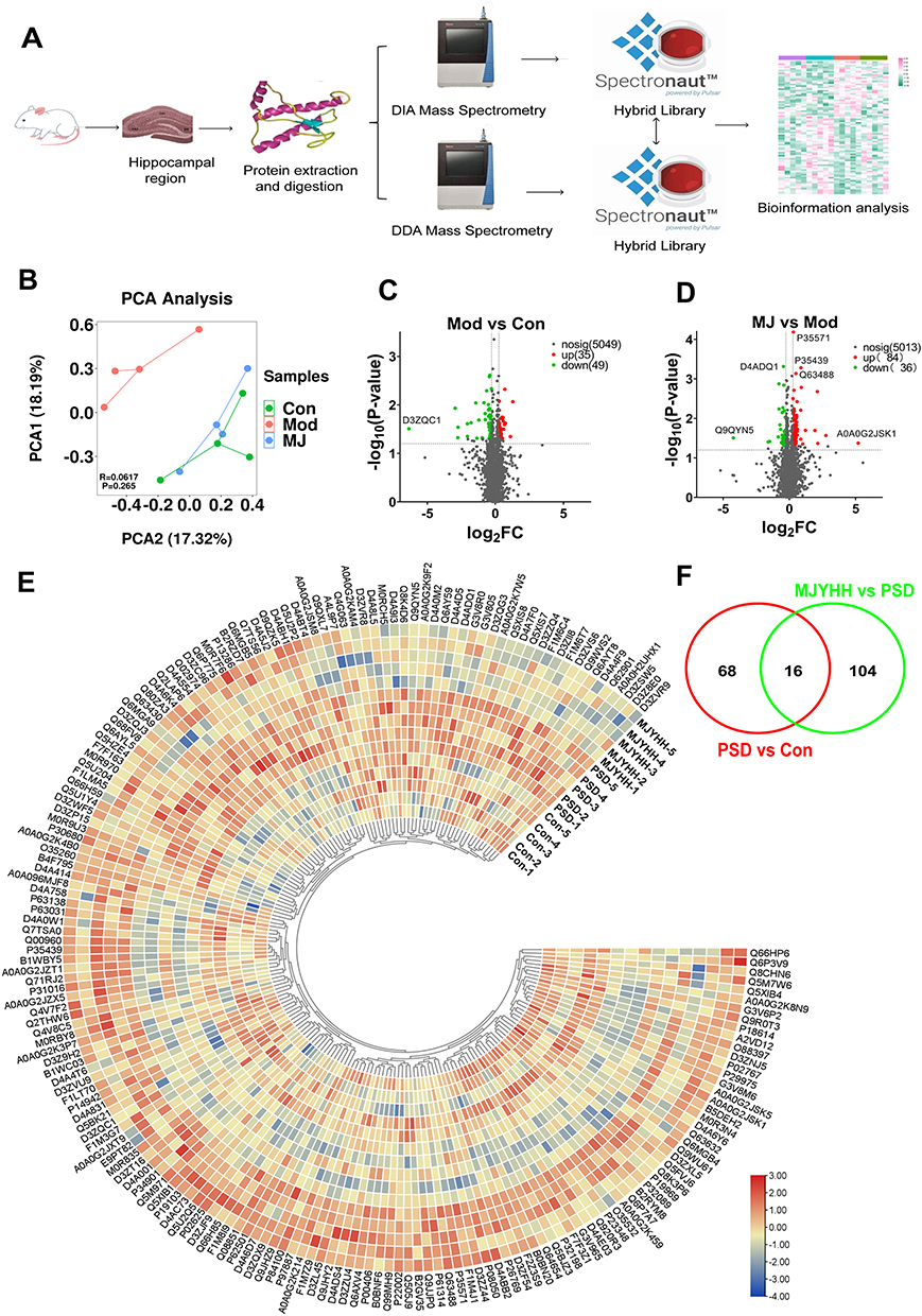 |
Figure 3 Proteomic characterisation of differentially expressed proteins (DEPs) in the hippocampus using data-independent acquisition (DIA) technology. (A) Schematic workflow of DIA mass spectrometry (DIA-MS) for hippocampal protein detection in rat models. (B) Multivariate analysis of protein expression profiles using principal component analysis (PCA). (C and D) Volcano plot analysis identifying statistically significant DEPs (p < 0.05, fold change > 1.5). (E) Hierarchical clustering of DEPs displayed as a heat map. (F) Comparative analysis of DEPs across experimental groups presented as a Venn diagram. |
Bioinformatics Analysis Reveals Key Pathways in PSD Treatment Response
Bioinformatic analysis using the OmicsBean tool26,27 was performed to elucidate the functional implications of DEPs across experimental groups. Comparative analysis between PSD model and control groups identified 84 DEPs with significant functional enrichment (Figure 4A). These proteins were primarily associated with transmembrane transport, oxoacid metabolic processes, and steroid hormone responses in biological processes. Cellular component analysis revealed predominant localisation to cytoplasmic, mitochondrial and endoplasmic reticulum compartments. Molecular function characterisation demonstrated enrichment in ion transmembrane transporter activity, oxidoreductase activity and related transporter functions. Parallel analysis of 120 DEPs between PSD model and MJYHH-treated groups (Figure 4C) showed distinct functional profiles. Biological process enrichment highlighted transmembrane transport, trans-synaptic signalling and organophosphate metabolism. Cellular components were markedly enriched in mitochondrial structures (envelope, membrane and whole organelle) alongside cytoplasmic localisation. Molecular functions mirrored transporter activities observed in the PSD-control comparison, with additional emphasis on inorganic molecular entity transport. Pathway analysis through KEGG revealed distinct mechanistic signatures (Figure 4B and 4D). The PSD-control comparison implicated nine pathways including nicotine/cocaine addiction, long-term potentiation, and lipid metabolism pathways. MJYHH treatment modulated 24 pathways, most notably the Rap1 signalling pathway, chemical carcinogenesis-receptor activation, cAMP signalling, and synaptic plasticity pathways (long-term potentiation, glutamatergic synapse). Metabolic pathways and substance addiction mechanisms remained prominent, with additional involvement of drug metabolism enzymes.
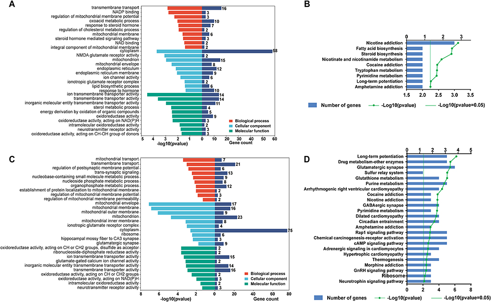 |
Figure 4 Functional enrichment analysis of differentially expressed proteins. (A) Gene Ontology (GO) classification of biological processes, molecular functions and cellular components in the PSD group compared with control (Con). (B) Kyoto Encyclopedia of Genes and Genomes (KEGG) pathway analysis of the PSD versus Con groups. (C) GO term enrichment profile comparing the MJYHH-treated group with the PSD group. (D) KEGG pathway mapping of proteins altered following MJYHH treatment relative to the PSD group. |
Protein-Protein Interaction (PPI) Network Analysis Identifies Key Molecular Targets
PPI analysis of DEPs was performed using STRING and visualised with Cytoscape 3.7.2 (Figure 5). The PPI network between PSD model and control groups comprised 75 nodes and 140 edges, while the network comparing PSD model and MJYHH-treated groups contained 115 nodes and 377 edges. In the visualisation, node colour intensity (orange to blue gradient) and edge thickness were proportional to degree value. Sixteen high-degree DEPs were prioritised by descending degree value: Grin1, Grin2b, Cyp51a1, Nnt, Itgb1, Rhot2, Tspan6, Rrm2b, Pbxip1, Ankzf1, Mpc1, Slc25a25, Slco1a5, LOC498555, RGD1311703, and Mllt11. These proteins represent potential therapeutic targets for MJYHH capsule treatment of PSD.
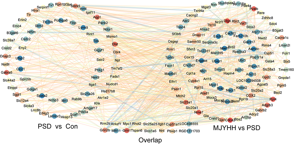 |
Figure 5 Protein-protein interaction (PPI) network analysis of differentially expressed proteins. The network illustrates interactions between proteins identified in the comparison of post-stroke depression (PSD) versus control (Con) groups, as well as modified Jieyuanshen (MJYHH) treatment versus PSD groups. The visualisation demonstrates key molecular interactions and potential hub proteins within these experimental conditions. |
PRM Validates Key Protein Alterations
To validate potential therapeutic targets identified through bioinformatics and PPI analysis, we selected three DEPs (Rrm2b, Cyp51a1, and Rhot2) associated with oxidative stress, biosynthetic metabolism, and mitochondrial function via KEGG analysis. DIA quantification revealed that PSD rats exhibited significantly elevated Rrm2b levels compared to controls (Figure 6A), which were normalised by MJYHH treatment. Conversely, Cyp51a1 and Rhot2 expression was significantly reduced in PSD rats (Figure 6B and 6C), with MJYHH treatment restoring these levels. PRM validation confirmed these findings, demonstrating congruent expression patterns between DIA and PRM methodologies (Figure 6D). These results strongly implicate Rrm2b, Cyp51a1, and Rhot2 as molecular targets mediating the therapeutic effects of MJYHH capsules in PSD.
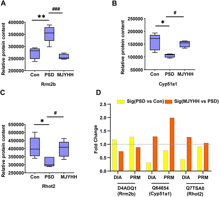 |
Figure 6 Quantitative proteomic analysis of Rrm2b, Cyp51a1, and Rhot2 expression. (A-C) Relative protein abundance of Rrm2b (A), Cyp51a1 (B), and Rhot2 (C) as determined by data-independent acquisition (DIA) mass spectrometry across experimental groups. (D) Parallel reaction monitoring (PRM) validation of hippocampal protein levels in PSD versus control (Con) groups and MJYHH-treated versus PSD groups. Data are presented as mean ± standard error of the mean (SEM). Statistical significance was assessed by two-way ANOVA: *p < 0.05, **p < 0.01, versus Con group; #p < 0.05, ###p < 0.001, versus PSD group. |
Molecular Docking Simulations Reveal Robust Interactions Between MJYHH Components and Therapeutic Targets
To investigate the mechanistic relationship between MJYHH capsule components and therapeutic targets, molecular docking simulations were conducted. The five principal bioactive compounds (paeoniflorin, geniposide, paeonol, quercetin, and curcumin) were docked with three candidate targets: Rrm2b, Cyp51a1, and Rhot2. Binding energies (Table 1) demonstrated significant interactions, with values below −5 kcal/mol indicating spontaneous binding and those below −7 kcal/mol representing strong molecular affinity. Notably, paeonol exhibited binding energies between −5 and −7 kcal/mol with all targets, while other compounds showed even stronger interactions (<-7 kcal/mol).
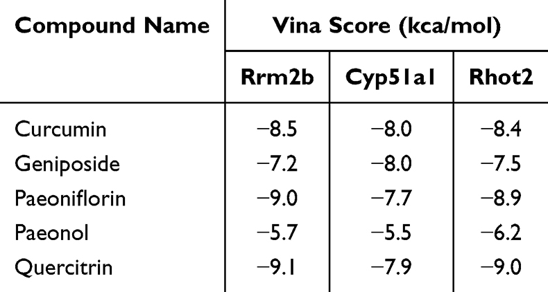 |
Table 1 Molecular Docking Results |
Detailed interaction analyses using PLIP and PyMol revealed specific binding mechanisms (Figure 7–9). For Rrm2b (Figure 7), curcumin formed hydrogen bonds with ARG110, ARG226, PHE183, and SER29, complemented by hydrophobic interactions with GLN113 and GLU21 (Figure 7A). Geniposide established hydrogen bonds at GLU222/ARG292 and hydrophobic contacts with ARG226/LEU230 (Figure 7B). Paeoniflorin engaged five hydrogen bonds (LEU230, SER199, TYR285, ARG292, SER316) and three hydrophobic interactions (Figure 7C), while paeonol bound via ARG110/ARG186 hydrogen bonds and LEU35 hydrophobic interaction (Figure 7D). Quercitrin demonstrated hydrogen bonding with TYR285, SER225, ARG292, and GLY229, alongside ARG226 hydrophobic contact (Figure 7E).
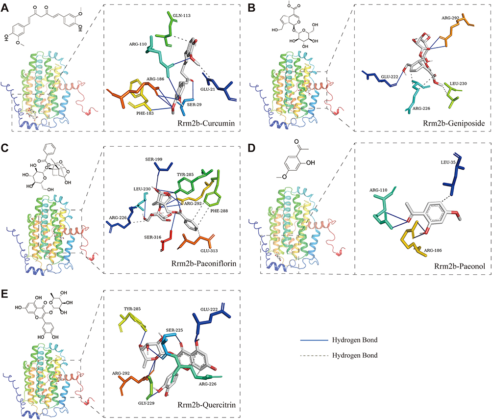 |
Figure 7 Molecular docking analysis of five bioactive compounds with Rrm2b. (A) The binding conformation of curcumin within the active site of Rrm2b. (B) Computational simulation of geniposide interaction with Rrm2b. (C) Structural representation of paeoniflorin bound to Rrm2b. (D) Molecular docking model of paeonol with Rrm2b. (E) Visualisation of quercetin binding to Rrm2b. |
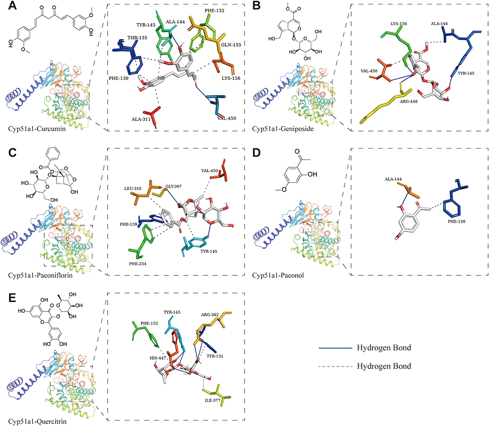 |
Figure 8 Molecular docking analysis of five bioactive compounds with Cyp51a1. (A) The binding conformation of curcumin within the active site of Cyp51a1. (B) Computational simulation of geniposide interaction with Cyp51a1. (C) Structural representation of paeoniflorin bound to Cyp51a1. (D) Molecular docking model of paeonol with Cyp51a1. (E) Visualisation of quercetin binding to Cyp51a1. |
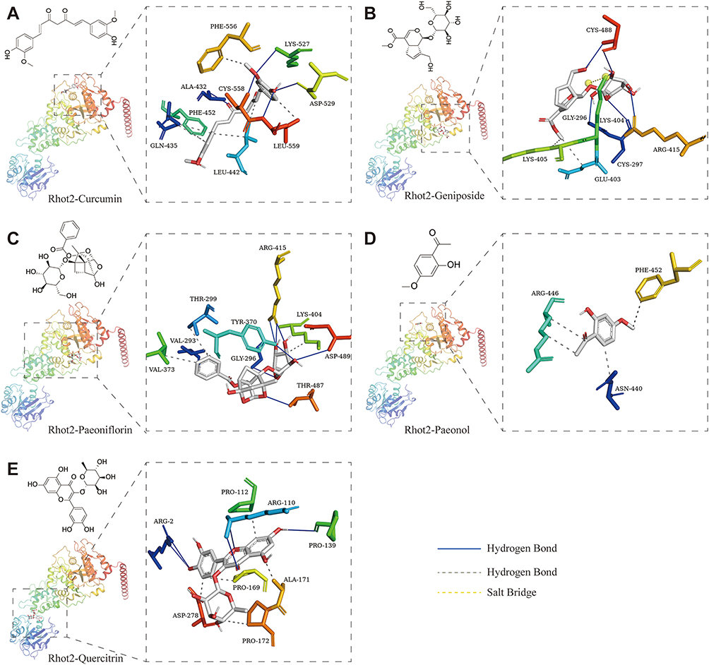 |
Figure 9 Molecular docking analysis of five bioactive compounds with Rhot2. (A) The binding conformation of curcumin within the active site of Rhot2. (B) Computational simulation of geniposide interaction with Rhot2. (C) Structural representation of paeoniflorin bound to Rhot2. (D) Molecular docking model of paeonol with Rhot2. (E) Visualisation of quercetin binding to Rhot2. |
In Rhot2 interactions (Figure 8), curcumin formed hydrogen bonds with CYS558, LYS527, and ASP529, with additional hydrophobic contacts at PHE556, LEU559, and GLN435 (Figure 8A). Geniposide exhibited a salt bridge with LYS404 alongside hydrogen bonds at CYS488/ARG415/CYS297 (Figure 8B). Paeoniflorin displayed four hydrogen bonds (ARG415, ASP489, TYR370, THR487) and multiple hydrophobic interactions (Figure 8C), whereas paeonol relied solely on hydrophobic forces with PHE452/ASN440/ARG446 (Figure 8D). Quercitrin bound via hydrogen bonds to ARG2/ARG110/PRO139 and extensive hydrophobic contacts (Figure 8E).
Cyp51a1 docking (Figure 9) showed curcumin forming hydrogen bonds with ALA144/VAL450 and seven hydrophobic interactions (Figure 9A). Geniposide engaged four hydrogen bonds (VAL450, LYS156, ARG446, TYR145) with complementary hydrophobic contacts (Figure 9B). Paeoniflorin bound through GLY307/TYR145 hydrogen bonds and five hydrophobic interactions (Figure 9C), while paeonol formed a single hydrogen bond with ALA144 and hydrophobic contact at PHE139 (Figure 9D). Quercitrin demonstrated three hydrogen bonds (TYR145, ARG382, HIS447) and hydrophobic interactions with ILE337/PHE152/TYR131 (Figure 9E).
These comprehensive simulations confirm strong binding competence between MJYHH components (curcumin, geniposide, paeoniflorin, paeonol, quercitrin) and the three target proteins, supporting Rrm2b, Cyp51a1, and Rhot2 as plausible therapeutic targets for PSD intervention in the rat model.
Discussion
Our experimental approach involved establishing PSD models in rats through MCAO to simulate stroke, combined with solitary parenting and CMS protocols to accelerate the development of depression-like behaviours. Following 21 days of oral administration of MJYHH capsules, behavioural assessments demonstrated significant improvement in depression-like symptoms in PSD model rats. Proteomic analysis of the hippocampal region revealed three potential molecular targets of MJYHH treatment: Rrm2b, Cyp51a1, and Rhot2, as identified through DIA and PRM techniques. Subsequent GO and KEGG pathway analyses further indicated that these molecular targets are functionally associated with oxidative stress regulation, biosynthetic processes, and mitochondrial dysfunction in PSD. Molecular docking predictions between the major active components of MJYHH capsules and these targets provide additional support for the formulation’s antidepressant effects in stroke-related depression. These findings, combining animal behavioural studies with proteomic bioinformatics analysis, elucidate potential therapeutic targets and molecular mechanisms underlying MJYHH capsule’s efficacy in PSD treatment.
This study offers valuable insights into both the pathogenesis of PSD and the mechanistic basis of traditional Chinese medicine intervention, providing a solid foundation for future research in this field. The integrated experimental approach, incorporating behavioural, proteomic and bioinformatic analyses, strengthens the evidence for MJYHH capsules as a potential therapeutic option for PSD.Current proteomic research in TCM increasingly employs DIA and PRM methodologies due to their complementary analytical strengths. DIA technology offers unbiased, high-throughput proteome coverage, particularly advantageous for comprehensive protein expression profiling in complex biological samples and detection of low-abundance proteins.28,29 Conversely, PRM provides targeted quantification with exceptional sensitivity and selectivity, making it ideal for validating key differential proteins and analysing post-translational modifications.30 The synergistic application of these techniques significantly enhances proteomic data reliability. Recent studies demonstrate the utility of this combined approach in TCM research. Tetrahydroxy stilbene glycoside, an active component derived from Polygonum multiflorum, was shown to ameliorate learning and memory deficits in an Alzheimer’s disease model, with DIA and PRM revealing associated protein alterations and modifications.31 Similarly, proteomic analysis coupled with PRM validation elucidated the therapeutic mechanisms of Yi-shen-hua-shi granules in IgA nephropathy treatment.32 These methodologies have also been successfully applied to characterise the glucolipid metabolic regulatory effects of the Hua Tan Qu Shi recipe.33 In our investigation of MJYHH capsule’s therapeutic mechanisms, we employed this dual-technique strategy. Initial DIA analysis identified 16 differentially expressed proteins among control, PSD and MJYHH-treated groups (Figure 3). Subsequent PRM validation confirmed consistent expression trends for only three of these proteins (Figure 6). This discrepancy may reflect technical limitations of PRM in detecting extremely low-abundance proteins. Nevertheless, the combined DIA-PRM approach provides comprehensive insights into MJYHH’s antidepressant mechanisms and potential therapeutic targets in PSD.
PSD represents a significant neuropsychiatric complication following cerebrovascular events, yet its underlying pathogenesis remains incompletely understood. Current evidence suggests a multifactorial aetiology involving genetic predisposition, lesion localisation, pre-existing medical and psychiatric conditions, alongside psychosocial determinants.34,35 Substantial research has established associations between PSD and neuroinflammatory processes, neuronal injury, and neurotransmitter dysregulation.36–38 The relationship between depressive disorders and inflammatory mechanisms is well-documented.36,39,40 In particular, the central nervous system demonstrates characteristic overactivation of astrocytes and microglia, resulting in excessive secretion of pro-inflammatory cytokines. These inflammatory mediators contribute to oxidative stress imbalance within neural tissues while simultaneously suppressing endogenous antioxidant defences.41 Furthermore, inflammatory processes are known to disrupt the synthesis and release of monoamine neurotransmitters, thereby exacerbating depressive symptomatology.42,43 Contrary to these established mechanisms, our bioinformatic analysis of GO and KEGG pathways revealed mitochondrial function, oxidative stress regulation, and biosynthetic processes as potentially predominant factors in PSD pathogenesis (Figure 4). The therapeutic effects of MJYHH capsules appear particularly relevant to these mitochondrial and oxidative stress pathways in our PSD model. Through subsequent PRM validation and molecular docking studies, we identified Rrm2b, Rhot2, and Cyp51a1 as key molecular targets of MJYHH treatment. Notably, Rrm2b and Rhot2 are both implicated in mitochondrial functional regulation, while Cyp51a1 participates in biosynthetic and metabolic pathways. These findings offer novel insights into the molecular basis of PSD development and may inform future therapeutic strategies. The identification of mitochondrial and biosynthetic pathways as potential intervention targets provides a new framework for understanding PSD pathophysiology and drug development.
Growing evidence demonstrates that TCM exhibits therapeutic potential for PSD through multiple mechanisms, including alleviation of neuronal damage, modulation of oxidative stress and inflammatory responses, and regulation of neurotransmitter release. Recent studies have identified specific molecular pathways involved in these effects. For instance, Di-Huang-Yin-Zi has been shown to activate the P53/SLC7A11 signalling pathway by enhancing P53 ubiquitination and stabilisation, thereby inhibiting ferroptosis in PSD models.44 Similarly, aloe-emodin, a natural compound derived from aloe or rhubarb, exerts neuroprotective effects in PSD treatment primarily through modulation of TRPV4 channels, which reduces pathological water influx into astrocytes following cerebral ischaemia while simultaneously promoting secretion of neurotrophic factors BDNF and NTF3 to ameliorate depressive symptoms.45 Furthermore, Jiawei Kongsheng Zhenzhong Pill has demonstrated efficacy in PSD treatment via activation of the netrin-1/DCC signalling pathway, leading to improvement in both depressive behaviours and neuronal damage.46 Our current investigation further supports the therapeutic potential of TCM in PSD management. The MJYHH capsule treatment significantly attenuated depression-like behaviours in post-stroke rat models. Comprehensive bioinformatics analysis through GO and KEGG pathways revealed a strong association between PSD pathogenesis and oxidative stress responses coupled with mitochondrial dysfunction (Figure 4). Subsequent validation of target molecules particularly highlighted the close relationship between capsule treatment efficacy and mitochondrial functional regulation. These findings collectively suggest that the therapeutic benefits of TCM in PSD may be mediated, at least in part, through modulation of mitochondrial function.
The MJYHH capsules, grounded in traditional Chinese medicine theory, demonstrate therapeutic efficacy through liver regulation, depression alleviation, and mind-calming properties. This formulation combines curcumin with extracts from Paeoniae Radix Alba, Fructus Gardeniae, Albiziae Flos, and Moutan Cortex. Our previous chromatographic analysis identified four primary active components in the JYHH capsules: paeoniflorin, geniposide, quercitrin, and paeonol.7 The current formulation represents an advancement through the addition of curcumin, a compound recognised for its neuroprotective properties mediated through antioxidant and anti-inflammatory mechanisms.47,48 Clinical evidence supports curcumin’s therapeutic potential in emotional disorders, including major depressive disorder, bipolar disorder, schizophrenia, and anxiety disorders.49 Particularly relevant to this study, curcumin has demonstrated efficacy in PSD rodent models by modulating the GAS5/miR-10b/BDNF/Trkβ pathway.50 The inclusion of paeoniflorin, derived from Paeoniae Radix Alba, contributes additional therapeutic benefits through its established antioxidant, anti-inflammatory, antidepressant, and neuroprotective activities.51 A chronic unpredictable mild stress model has shown that paeoniflorin can counteract neuronal apoptosis induced by miR-200a-3p and miR-200b-3p overexpression, thereby protecting primary neurons from corticosterone-induced neurotoxicity.52 Furthermore, paeoniflorin ameliorates depression-like behaviours in PSD models by upregulating BDNF and p-CREB expression in the CA1 region and modulating BDNF, cannabinoid receptors, and CRF in VTA-NAc tissue.53,54 Geniposide, the active component extracted from Fructus Gardeniae, exhibits notable antioxidant, anti-inflammatory, and neuroprotective properties,55,56 with substantial evidence supporting its antidepressant effects.56,57 Quercetin, derived from Albiziae, demonstrates anti-inflammatory activity and has been shown to rapidly improve depression-like behaviours in LPS-induced models by inhibiting PI3K/AKT/NFκB signalling while restoring CREB/BDNF pathways in the hippocampus.58 Paeonol completes this therapeutic profile with its documented neuroprotective effects against neuroinflammation and depressive-like symptoms.59–61 Our molecular docking analysis revealed strong binding affinities (<7 kcal/mol) between four of these components (excluding paeonol) and the three proteomically-identified targets (Figures 7–9, Table 1). These findings collectively demonstrate that the antidepressant effects of MJYHH capsules in PSD rats arise from the synergistic action of multiple bioactive components. The proteomic and molecular docking results further support the multi-component, multi-target mechanism underlying TCM’s therapeutic approach to post-stroke depression.
Several methodological limitations should be acknowledged in this study. The inherent sensitivity constraints of DIA technology may have compromised the detection of proteins with exceptionally low expression levels. Although DIA and PRM provide robust platforms for high-throughput proteomic analysis and targeted verification respectively, their capacity to identify low-abundance proteins remains inferior to enrichment-based methodologies. This technical limitation potentially influenced the comprehensiveness of our proteomic characterisation of PSD, suggesting that future investigations might benefit from incorporating antibody-based enrichment strategies or next-generation mass spectrometry platforms to achieve deeper proteome coverage.
A further consideration arises from the targeted nature of PRM validation, which by design focuses on predefined peptide targets. This approach inevitably excludes potentially relevant but unpredicted molecular species that might contribute to PSD pathophysiology. The predictive modelling of drug-target interactions through machine learning algorithms, while computationally powerful, requires experimental validation to establish biological relevance. These computational predictions would be strengthened by orthogonal evidence from in vitro and in vivo pharmacological studies.
To address these limitations, future research should adopt an integrated multi-omics strategy incorporating metabolomic and transcriptomic datasets. Such an approach would enable the construction of comprehensive molecular networks, providing a more robust framework for mechanistic exploration of PSD. Subsequent experimental validation of identified targets through both in vitro and in vivo models would substantially strengthen the translational relevance of these findings.
The present investigation employed a novel rat model of PSD developed through MCAO combined with social isolation and CMS. Behavioural assessments demonstrated the anti-PSD efficacy of the MJYHH capsules, a traditional Chinese medicine formulation. Subsequent proteomic analysis using DIA technology revealed significant alterations in hippocampal protein expression patterns associated with PSD pathogenesis. The DIA results indicated that the differentially expressed proteins in PSD model rats were predominantly involved in biosynthetic metabolism, oxidative stress responses, and mitochondrial dysfunction. Further validation through PRM and molecular docking analyses identified three key protein targets: Cyp51a1, Rrm2b, and Rhot2. These findings suggest novel molecular mechanisms underlying the therapeutic effects of MJYHH capsules in PSD intervention. This study provides important insights into the molecular pathology of PSD and elucidates potential mechanisms of traditional Chinese medicine treatment. The identification of these protein targets establishes a foundation for future research into PSD pathogenesis and therapeutic development. The integration of behavioural, proteomic, and computational approaches offers a comprehensive framework for understanding the complex neurobiological processes involved in PSD and its treatment.
Conclusion
This comprehensive investigation has systematically demonstrated the therapeutic potential and mechanistic basis of the MJYHH capsule in treating PSD through an integrated multidisciplinary approach. The study employed a well-validated PSD rat model, with behavioural assessments unequivocally confirming the capsule’s efficacy in ameliorating depression-like symptoms. Subsequent proteomic profiling of hippocampal tissue using both DIA and PRM methodologies identified three pivotal molecular targets: RRM2B, CYP51A1, and RHOT2. Bioinformatic analysis through GO and KEGG enrichment revealed that the therapeutic effects are mediated through the modulation of oxidative stress pathways, biosynthetic processes, and mitochondrial function. These findings were further substantiated by molecular docking studies, which demonstrated robust interactions between active MJYHH constituents and the identified target proteins. The present work provides compelling evidence for the multi-target mechanism underlying MJYHH’s anti-PSD effects, while simultaneously offering novel insights into PSD pathogenesis. Importantly, these findings establish a robust scientific foundation for both the clinical translation of this traditional Chinese medicine formulation and the development of novel therapeutics for PSD. The convergence of evidence from in vivo models, multi-omics analyses, and computational approaches significantly advances our understanding of this complex neuropsychiatric condition and its potential treatment modalities.
Abbreviations
MJYHH, Modified Jie-Yu-He-Huan; PSD, Post-Stroke Depression; DIA, Data Independent Acquisition; PRM, Parallel reaction monitoring; DEPs, Differentially expressed proteins; PPI, Protein-protein interaction; TCM, Traditional Chinese medicine; JYHH, Jie-Yu-He-Huan; MCAO, Middle cerebral artery occlusion; CMS, Chronic mild stress; OFT, Open Field Test; SPT, Sucrose Preference Test; BBT, Balance Beam Test; TST, Tail Suspension Test; FST, Forced swimming test; MDD, Major depressive disorder; Con, control groups; FLX, Fluoxetine.
Data Sharing Statement
The data that support the findings of this study are available from the corresponding author, [SW], upon reasonable request.
Ethics Declarations
All animal experiments and protocols complied with international animal experimental ethics and requirements and were approved by the Animal Ethics Committee of Shandong University of Traditional Chinese Medicine (Ethical Number: DDUTCM20251222106; License No. SYXK (LU) 2022 0009).
Author Contributions
All authors have made substantial contributions to this work, encompassing study conception, experimental design, data acquisition, analysis and interpretation. Each contributor participated actively in manuscript preparation through drafting, critical revision and final approval of the submitted version. The authors collectively selected the target journal and accept full responsibility for all aspects of the research presented.
Funding
This study was supported by the National Natural Science Foundation of China (no. 82274383,82305065), High Level Key Disciplines of Traditional Chinese Medicine: Basic Theory of Traditional Chinese Medicine, National Administration of Traditional Chinese Medicine (no. zyyzdxk-2023118), the Special Funding for Taishan Scholars Project (no. tsqn202211137), the Natural Science Foundation of Shandong Province (no. ZR2024QH185, ZR2024QH072, ZR2023QH078, ZR2023QH309), Postdoctoral Fellowship Program (Grade C) of China Postdoctoral Science Foundation under Grant (No.GZC20231507), the China Postdoctoral Science Foundation (no. 2025T181085), the Chinese Medicine and Brain Science Youth Scientific Research Innovation Team, Shandong University of Traditional Chinese Medicine (no. 22202101), Key Project of Scientific Research Fund of Shandong University of Traditional Chinese Medicine (no. KYZK2024Z08), the second batch of Scientific research Fund projects of Shandong University of Traditional Chinese Medicine (no. KYZK2024Q10), the Medical and Health Science and Technology Project of Shandong Province (no. 202302081721).
Disclosure
The authors report no conflicts of interest in this work.
References
1. Gao J, He Y, Shi F, et al. Activation of Sirt6 by icariside II alleviates depressive behaviors in mice with poststroke depression by modulating microbiota-gut-brain axis. J Adv Res. 2025.
2. Gomberg J, Stein LK, Dhamoon MS. Risk of recurrent stroke and mortality among black and white patients with poststroke depression. Stroke. 2024;55(5):1308–22. doi:10.1161/STROKEAHA.123.045743
3. Sarkar A, Sarmah D, Datta A, et al. Post-stroke depression: chaos to exposition. Brain Res. Bull. 2021;168:74–88. doi:10.1016/j.brainresbull.2020.12.012
4. Frank D, Gruenbaum BF, Zlotnik A, Semyonov M, Frenkel A, Boyko M. Pathophysiology and current drug treatments for post-stroke depression: a review. Int J Mol Sci. 2022;23(23).
5. Fan Q, Liu Y, Sheng L, et al. Chaihu-Shugan-San inhibits neuroinflammation in the treatment of post-stroke depression through the JAK/STAT3-GSK3beta/PTEN/Akt pathway. Biomed Pharmacother. 2023;160:114385. doi:10.1016/j.biopha.2023.114385
6. Zhang M, Bai X. Shugan jieyu capsule in post-stroke depression treatment: from molecules to systems. Front Pharmacol. 2022;13:821270. doi:10.3389/fphar.2022.821270
7. Geng X, Wu H, Li Z, et al. Jie-Yu-He-Huan Capsule Ameliorates Anxiety-Like Behaviours in Rats Exposed to Chronic Restraint Stress via the cAMP/PKA/CREB/BDNF Signalling Pathway. Oxid Med Cell Longev. 2021;2021:1703981. doi:10.1155/2021/1703981
8. Geng X, Wang X, Liu K, et al. ShuYu capsule alleviates emotional and physical symptoms of premenstrual dysphoric disorder: impact on ALLO decline and GABA(A) receptor delta subunit in the PAG area. Phytomedicine. 2024;130:155549. doi:10.1016/j.phymed.2024.155549
9. Pei J, Palanisamy CP, Natarajan PM, et al. Curcumin-loaded polymeric nanomaterials as a novel therapeutic strategy for Alzheimer’s disease: a comprehensive review. Ageing Res Rev. 2024;99:102393. doi:10.1016/j.arr.2024.102393
10. Mo J, Liao W, Du J, et al. Buyang huanwu decoction improves synaptic plasticity of ischemic stroke by regulating the cAMP/PKA/CREB pathway. J Ethnopharmacol. 2024;335:118636. doi:10.1016/j.jep.2024.118636
11. Chen H, He Y, Chen S, Qi S, Shen J. Therapeutic targets of oxidative/nitrosative stress and neuroinflammation in ischemic stroke: applications for natural product efficacy with omics and systemic biology. Pharmacol Res. 2020;158:104877. doi:10.1016/j.phrs.2020.104877
12. Wang Z, Sun Y, Bian L, et al. The crosstalk signals of Sodium Tanshinone IIA Sulfonate in rats with cerebral ischemic stroke: insights from proteomics. Biomed. Pharmacother. 2022;151.
13. Longa EZ, Weinstein PR, Carlson S, Cummins R. Reversible middle cerebral artery occlusion without craniectomy in rats. Stroke. 1989;20(1):84–91. doi:10.1161/01.STR.20.1.84
14. Li T, Wang D, Zhao B, Yan Y. Xingnao jieyu decoction ameliorates poststroke depression through the BDNF/ERK/CREB pathway in rats. Evid Based Complement Alternat Med. 2018;2018:5403045. doi:10.1155/2018/5403045
15. Geng X, Zhang H, Hu M, et al. A novel curcumin oil solution can better alleviate the motor activity defects and neuropathological damage of a Parkinson’s disease mouse model. Front Aging Neurosci. 2022;14:984895. doi:10.3389/fnagi.2022.984895
16. David DJ, Samuels BA, Rainer Q, et al. Neurogenesis-dependent and -independent effects of fluoxetine in an animal model of anxiety/depression. Neuron. 2009;62(4):479–493. doi:10.1016/j.neuron.2009.04.017
17. Li W, Shen N, Kong L, et al. STING mediates microglial pyroptosis via interaction with NLRP3 in cerebral ischaemic stroke. Stroke and Vascular Neurol. 2024;9(2):153–164. doi:10.1136/svn-2023-002320
18. Zhang Y, Yang M, Yuan Q, et al. Piperine ameliorates ischemic stroke-induced brain injury in rats by regulating the PI3K/AKT/mTOR pathway. J Ethnopharmacol. 2022;295:115309. doi:10.1016/j.jep.2022.115309
19. Goldstein LB, Davis JN. Beam-walking in rats: studies towards developing an animal model of functional recovery after brain injury. J Neurosci Methods. 1990;31(2):101–107. doi:10.1016/0165-0270(90)90154-8
20. He LW, Zeng L, Tian N, et al. Optimization of food deprivation and sucrose preference test in SD rat model undergoing chronic unpredictable mild stress. Anim. Models Exp. Med. 2020;3(1):69–78. doi:10.1002/ame2.12107
21. Choleris E, Thomas AW, Kavaliers M, Prato FS. A detailed ethological analysis of the mouse open field test: effects of diazepam, chlordiazepoxide and an extremely low frequency pulsed magnetic field. Neurosci Biobehav Rev. 2001;25(3):235–260. doi:10.1016/S0149-7634(01)00011-2
22. Chen Z, Gu J, Lin S, et al. Saffron essential oil ameliorates CUMS-induced depression-like behavior in mice via the MAPK-CREB1-BDNF signaling pathway. J Ethnopharmacol. 2023;300:115719. doi:10.1016/j.jep.2022.115719
23. Casarrubea M, Roy V, Sorbera F, et al. Temporal structure of the rat’s behavior in elevated plus maze test. Behav. Brain Res. 2013;237:290–299. doi:10.1016/j.bbr.2012.09.049
24. Liu S, Xu S, Wang Z, Guo Y, Pan W, Shen Z. Anti-depressant-like effect of sinomenine on chronic unpredictable mild stress-induced depression in a mouse model. Med Sci Monitor. 2018;24:7646–7653. doi:10.12659/MSM.908422
25. Idayu NF, Hidayat MT, Moklas MA, et al. Antidepressant-like effect of mitragynine isolated from Mitragyna speciosa Korth in mice model of depression. Phytomedicine. 2011;18(5):402–407. doi:10.1016/j.phymed.2010.08.011
26. Xie H, Huang H, Tang M, et al. iTRAQ-based quantitative proteomics suggests synaptic mitochondrial dysfunction in the hippocampus of rats susceptible to chronic mild stress. Neurochemical Res. 2018;43(12):2372–2383. doi:10.1007/s11064-018-2664-y
27. Sun N, Sun W, Li S, et al. Proteomics analysis of cellular proteins co-immunoprecipitated with nucleoprotein of influenza a virus (H7N9). Int J Mol Sci. 2015;16(11):25982–25998. doi:10.3390/ijms161125934
28. Lou R, Cao Y, Li S, et al. Benchmarking commonly used software suites and analysis workflows for DIA proteomics and phosphoproteomics. Nat Commun. 2023;14(1):94. doi:10.1038/s41467-022-35740-1
29. Yu F, Teo GC, Kong AT, et al. Analysis of DIA proteomics data using MSFragger-DIA and FragPipe computational platform. Nat Commun. 2023;14(1):4154. doi:10.1038/s41467-023-39869-5
30. Rauniyar N. Parallel reaction monitoring: a targeted experiment performed using high resolution and high mass accuracy mass spectrometry. Int J Mol Sci. 2015;16(12):28566–28581. doi:10.3390/ijms161226120
31. Gao Y, Li J, Hu K, et al. Phosphoproteomic analysis of APP/PS1 mice of Alzheimer’s disease by DIA based mass spectrometry analysis with PRM verification. J Proteomics. 2024;299:105157. doi:10.1016/j.jprot.2024.105157
32. Xu R, Zhang J, Hu X, et al. Yi-shen-hua-shi granules modulate immune and inflammatory damage via the ALG3/PPARgamma/NF-kappaB pathway in the treatment of immunoglobulin a nephropathy. J Ethnopharmacol. 2024;319(Pt 2):117204. doi:10.1016/j.jep.2023.117204
33. Li Y, Ma J, Sun S, et al. DIA-PRM proteomic analysis of phlegm-dampness constitution with glucolipid metabolic disorders by the intervention of hua tan qu shi recipe. Biomed Res Int. 2022;2022:6464431. doi:10.1155/2022/6464431
34. Robinson RG, Jorge RE. Post-stroke depression: a review. Am J Psychiatry. 2016;173(3):221–231. doi:10.1176/appi.ajp.2015.15030363
35. Das J, KR G. Post stroke depression: the sequelae of cerebral stroke. Neurosci Biobehav Rev. 2018;90:104–114. doi:10.1016/j.neubiorev.2018.04.005
36. Lu W, Wen J. Neuroinflammation and post-stroke depression: focus on the microglia and astrocytes. Aging Dis. 2024;16(1):394–407. doi:10.14336/AD.2024.0214-1
37. Jeong S, Chokkalla AK, Davis CK, Vemuganti R. Post-stroke depression: epigenetic and epitranscriptomic modifications and their interplay with gut microbiota. Mol Psychiatry. 2023;28(10):4044–4055. doi:10.1038/s41380-023-02099-8
38. Yin Y, Ju T, Zeng D, et al. “Inflamed” depression: a review of the interactions between depression and inflammation and current anti-inflammatory strategies for depression. Pharmacol Res. 2024;207:107322. doi:10.1016/j.phrs.2024.107322
39. Leonard B, Maes M. Mechanistic explanations how cell-mediated immune activation, inflammation and oxidative and nitrosative stress pathways and their sequels and concomitants play a role in the pathophysiology of unipolar depression. Neurosci Biobehav Rev. 2012;36(2):764–785. doi:10.1016/j.neubiorev.2011.12.005
40. Jiang R, Noble S, Rosenblatt M, et al. The brain structure, inflammatory, and genetic mechanisms mediate the association between physical frailty and depression. Nat Commun. 2024;15(1):4411. doi:10.1038/s41467-024-48827-8
41. Wang JY, Wen LL, Huang YN, Chen YT, Ku MC. Dual effects of antioxidants in neurodegeneration: direct neuroprotection against oxidative stress and indirect protection via suppression of glia-mediated inflammation. Curr Pharm Des. 2006;12(27):3521–3533. doi:10.2174/138161206778343109
42. Oglodek E. Changes in the Serum Levels of Cytokines: IL-1beta, IL-4, IL-8 and IL-10 in depression with and without posttraumatic stress disorder. Brain Sci. 2022;12(3):387. doi:10.3390/brainsci12030387
43. Wang L, Wang R, Liu L, Qiao D, Baldwin DS, Hou R. Effects of SSRIs on peripheral inflammatory markers in patients with major depressive disorder: a systematic review and meta-analysis. Brain Behav Immun. 2019;79:24–38. doi:10.1016/j.bbi.2019.02.021
44. Yang Z, Jiang Y, Xiao Y, et al. Di-Huang-Yin-Zi regulates P53/SLC7A11 signaling pathway to improve the mechanism of post-stroke depression. J Ethnopharmacol. 2024;319(Pt 2):117226. doi:10.1016/j.jep.2023.117226
45. Liu Y, Peng J, Leng Q, Tian Y, Wu X, Tan R. Effects of aloe-emodin on the expression of brain aquaporins and secretion of neurotrophic factors in a rat model of post-stroke depression. Int J Mol Sci. 2023;24(6).
46. Zhao Y, Song A, Liu G, et al. Modulation of netrin-1/DCC signaling pathway by Jiawei Kongsheng Zhenzhong Pill improves synaptic structural plasticity in PSD rats. J Pharmacol Sci. 2025;157(4):242–252. doi:10.1016/j.jphs.2025.02.004
47. Mohan MC, Anjana AS, Hilmi Jaufer TA, Deepti A, Krishnakumar IM, Baby Chakrapani PS. Co-delivery of curcumin-resveratrol-carnosic acid complex promotes neurogenesis and cognitive recovery in a rodent model of repeated mild traumatic brain injury. Biomed Pharmacother. 2025;183:117818. doi:10.1016/j.biopha.2025.117818
48. Lopresti AL. Potential role of curcumin for the treatment of major depressive disorder. CNS Drugs. 2022;36(2):123–141. doi:10.1007/s40263-022-00901-9
49. Mohammadzadeh R, Fathi M, Pourseif MM, et al. Curcumin and nano-curcumin applications in psychiatric disorders. Phytother Res. 2024;38(8):4240–4260. doi:10.1002/ptr.8265
50. C L, L WT, Z LL, L XQ, C M, L Y. Long noncoding RNA GAS5 enhanced by curcumin relieves poststroke depression by targeting miR-10b/BDNF in rats. J Biol Regul Homeost Agents. 2020;34:815–823. doi:10.23812/20-113-A-25
51. Wang XL, Feng ST, Wang YT, Chen NH, Wang ZZ, Zhang Y. Paeoniflorin: a neuroprotective monoterpenoid glycoside with promising anti-depressive properties. Phytomedicine. 2021;90:153669. doi:10.1016/j.phymed.2021.153669
52. Yuan N, Li X, Tang K, et al. Xiaoyaosan inhibits neuronal apoptosis by regulating the miR-200/NR3C1 signaling in the prefrontal cortex of chronically stressed rats. Phytomedicine. 2022;103:154239. doi:10.1016/j.phymed.2022.154239
53. Wang C, Wu C, Yan Z, Cheng X. Ameliorative effect of Xiaoyao-jieyu-san on post-stroke depression and its potential mechanisms. J Nat Med. 2019;73(1):76–84. doi:10.1007/s11418-018-1243-5
54. Hu MZ, Wang AR, Zhao ZY, Chen XY, YB L, Liu B. Antidepressant-like effects of paeoniflorin on post-stroke depression in a rat model. Neurol Res. 2019;41(5):446–455. doi:10.1080/01616412.2019.1576361
55. Wang L, Chen S, Liu S, et al. A comprehensive review of ethnopharmacology, chemical constituents, pharmacological effects, pharmacokinetics, toxicology, and quality control of gardeniae fructus. J Ethnopharmacol. 2024;320:117397. doi:10.1016/j.jep.2023.117397
56. Duan G, Zou T, Wu X, Zhang Y, Liu H, Mei C. Neuroprotective role of geniposide-loaded UMSC nanovesicles in depression via P2ry12 downregulation. Phytomedicine. 2025;140:156581. doi:10.1016/j.phymed.2025.156581
57. Zhao Y, Zhang Q, Yan Y, et al. Antidepressant-like effects of geniposide in chronic unpredictable mild stress-induced mice by regulating the circ_0008405/miR-25-3p/Gata2 and Oip5os1/miR-25-3p/Gata2 networks. Phytother Res. 2023;37(5):1850–1863. doi:10.1002/ptr.7702
58. Sun Y, Zhang H, Wu Z, et al. Quercitrin rapidly alleviated depression-like behaviors in lipopolysaccharide-treated mice: the involvement of pi3k/akt/nf-kappab signaling suppression and CREB/BDNF signaling restoration in the hippocampus. ACS Chem Neurosci. 2021;12(18):3387–3396. doi:10.1021/acschemneuro.1c00371
59. Wei E, Gao A, Mu X, et al. Paeonol ameliorates hippocampal neuronal damage by inhibiting GRM5/GABBR2/beta-arrestin2 and activating the cAMP-PKA signaling pathway in premenstrual irritability rats. Brain Res Bull. 2023;205:110830. doi:10.1016/j.brainresbull.2023.110830
60. Zhang N, Ma Y, Li Y, et al. Paeonol prevents sepsis-associated encephalopathy via regulating the HIF1A pathway in microglia. Int Immunopharmacol. 2024;143(Pt 1):113287. doi:10.1016/j.intimp.2024.113287
61. Kang WC, Lee YS, Park K, et al. Paeonol alleviates postmenopause-induced neuropsychiatric symptoms through the modulation of GPR30 in ovariectomized mice. J Ethnopharmacol. 2024;327:118063. doi:10.1016/j.jep.2024.118063
 © 2026 The Author(s). This work is published and licensed by Dove Medical Press Limited. The
full terms of this license are available at https://www.dovepress.com/terms
and incorporate the Creative Commons Attribution
- Non Commercial (unported, 4.0) License.
By accessing the work you hereby accept the Terms. Non-commercial uses of the work are permitted
without any further permission from Dove Medical Press Limited, provided the work is properly
attributed. For permission for commercial use of this work, please see paragraphs 4.2 and 5 of our Terms.
© 2026 The Author(s). This work is published and licensed by Dove Medical Press Limited. The
full terms of this license are available at https://www.dovepress.com/terms
and incorporate the Creative Commons Attribution
- Non Commercial (unported, 4.0) License.
By accessing the work you hereby accept the Terms. Non-commercial uses of the work are permitted
without any further permission from Dove Medical Press Limited, provided the work is properly
attributed. For permission for commercial use of this work, please see paragraphs 4.2 and 5 of our Terms.