Back to Journals » OncoTargets and Therapy » Volume 12
MicroRNA-221 promotes papillary thyroid carcinoma cells migration and invasion via targeting RECK and regulating epithelial–mesenchymal transition
Authors Wei Z, Gao A, Wang Q, Lou X, Zhao J, Lu Q
Received 10 October 2018
Accepted for publication 6 February 2019
Published 28 March 2019 Volume 2019:12 Pages 2323—2333
DOI https://doi.org/10.2147/OTT.S190364
Checked for plagiarism Yes
Review by Single anonymous peer review
Peer reviewer comments 4
Editor who approved publication: Dr XuYu Yang
Zhao-li Wei,1 Ai-bin Gao,1 Qing Wang,1 Xiu-e Lou,1 Jing Zhao,2 Qing-jun Lu3
1Department of Endocrinology, Binzhou Central Hospital, Binzhou Medical University, Binzhou 251700, Shandong Province, People’s Republic of China; 2Department of Oncology, Provincal Hospital of Shandong University, Jinan 250000, Shandong Province, People’s Republic of China; 3Department of Oncology, Renji Hospital, Shanghai Jiao Tong University School of Medicine, Shanghai 200001, People’s Republic of China
Aim: The aim of this study was to detect the effects and potential mechanisms of microRNA-221 on a series of biological behaviors of papillary thyroid carcinoma (PTC) cells in vitro and in vivo.
Methods: First, we analyzed the relationship between the expression of miR-221 and several clinicopathological features of PTC patients and then detected the expression of the miR-221 in tumor tissues and cell lines. The effects of miR-221 on proliferation and invasion of PTC cells were verified by cell counting kit-8 (CCK-8) assay, wound healing assay and transwell assay. Western blot assay was applied to explore the correlation between miR-221 and RECK expression in PTC K1 cells. Finally, a xenograft model was established to further confirm the tumor-promoting effects of miR-221 in vivo.
Results: Our data indicated that miR-221 was relatively upregulated in metastatic PTC tissues. MiR-221 promoted the proliferation, migration and invasion activities of PTC K1 cells, following variations of epithelial–mesenchymal transition (EMT)-related protein expression. We identified RECK as a direct target of miR-221, revealed its expression to be inversely correlated with miR-221 in PTC samples and showed that its reintroduction reverses miR-221-induced PTC invasiveness. In addition, miR-221 was also verified to promote tumor growth and increase tumor volume and weight in vivo. Taken together, miR-221/RECK axis could be an effective way to regulate biological behaviors of PTC.
Conclusion: MiR-221 may be involved in PTC cell invasion and metastasis by targeting RECK, indicating that the miR-221/RECK pathway could be studied further as a potential new diagnostic or prognostic biomarker for PTC.
Keywords: miR-221, papillary thyroid carcinoma, invasion, RECK
Introduction
Thyroid cancer, the incidence of which has continuously and sharply increased worldwide in the last 10 years, represents the most common malignant tumor in endocrine system diseases.1,2 A large number of epidemiological data indicate that the incidence of thyroid malignancy is affected by hormones, heredity and environmental factors, such as radioactivity, goiter-causing substances, iodine deficiency, etc. In addition, Hashimoto’s thyroiditis may also lead to the occurrence of thyroid cancer. Papillary thyroid carcinoma (PTC), with a higher degree of differentiation and early lymph node metastasis, accounts for approximately 85% of the malignant thyroid tumors.3 By 2019, PTC is predicted to be the 8th most common cancer in women in mainland China.4 Current clinical strategies, such as radical operation, chemotherapy, radiotherapy and other adjuvant therapies, offer curable treatment with 5-year survival rate over 90%. Nevertheless, distant metastasis and recurrence still occurred in several subtypes of PTC, and the 5-year survival rate of advanced PTC is still less than 50%.5 Accordingly, revealing the potential molecular mechanisms is of critical importance for improving the condition of PTC patients, prognosis and the quality of life after operation.
MiRNA is a kind of endogenous small RNA with a length of around 25 nucleotides, which plays various important roles in early development, fat metabolism, cell differentiation and other biological activities.6 Recent studies strengthen the evidence that the aberrant expression of miRNAs is closely related to the occurrence and development of human malignancies. As anti-oncogenes or oncogenes, miRNAs participate in the proliferation, apoptosis and invasion of tumor cells, indicating that they are of great value to study the mechanisms of miRNA in tumor progression.7 Up to now, plenty of miRNAs were reported to be involved in the progression of PTC. For instance, Huang et al8 reported that overexpression of miR-219-5p suppressed PTC cell migration and promoted apoptosis by targeting the estrogen receptor (ER)-α. Zhu et al’s research showed that miR-182 could facilitate cell proliferation and invasion through direct suppression of close homolog of LI (CHL1).9 Overexpression of miR-144-3p was reported to promote the PTC cell proliferation and cell cycle progression by regulating the cell cycle-related proteins such as cyclin D1, cyclin-dependent kinase (CDK) 2 and cell division cycle 25A (CDC25A). Besides, the aberrant expression of miR-144-3p could obviously affect the epithelial–mesenchymal transition (EMT) of PTC via regulating the expression of E-cadherin, N-cadherin and vimentin.10 MiR-146b was shown to be significantly overexpressed in PTC tissues with central compartment lymph node infiltration and associated with high-risk PTC with BRAF mutation. Furthermore, miR-146b expression is an independent risk factor for poor prognosis in PTC together with advanced tumor stage.11 In another study, overexpression of miR-146b-5p was shown to significantly increase the PTC cell proliferation through modulation of the TGF-β signaling pathway.12 Cong et al13 detected the expression of microRNAs in nearly 500 PTC specimens and 60 normal thyroid specimens obtained from The Cancer Genome Atlas database, which indicated that miR-221 was upregulated in PTC tissues compared with normal thyroid tissues. However, very little is currently known about and reported on the function of miR-221. Therefore, it is crucial to investigate the role and relevant mechanisms of miR-221 in PTC. In the present research, we aim to explore various biological behaviors of miR-221 and to detect the possible targets and the regulatory pathway that miR-221 might be involved in.
Materials and methods
Patients studied and tissue collection
Thirty PTC cancer samples and adjacent normal thyroid papillary tissues were collected from the Department of Pathology, Renji Hospital of Shanghai Jiao Tong University between October 2014 and May 2016. The corresponding paracancerous normal tissues from the same PTC patients were obtained 3 cm beyond the boundary of PTC tissues. All tissues were collected and immediately frozen in liquid nitrogen until RNA and protein extraction. Besides, no other treatments were conducted in these patients before surgery and tissue sampling (Table 1). The informed consents were signed by all the patients involved before operation. Written informed consent was obtained from each patient before the surgery. In addition, this work was conducted in accordance with the Declaration of Helsinki and was approved by the ethics committee of the Renji Hospital of Shanghai Jiao Tong University.
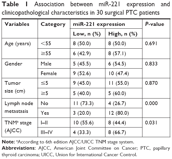 | Table 1 Association between miR-221 expression and clinicopathological characteristics in 30 surgical PTC patients |
Cell lines and cultures
Human PTC cell lines (TPC-1, K1 and BCPAP) and human thyroid follicular epithelial cells (Nthy-ori 3-1) were purchased from Biochemistry and Cell Biology Institute of the Chinese Academy of Sciences (Shanghai, China). Cells were maintained in RPMI 1640 medium supplemented with 10% FBS (Thermo Fisher Scientific, Waltham, MA, USA), 100 U/mL penicillin G and 100 g/mL streptomycin (Sigma-Aldrich Co., St Louis, MO, USA) at 37°C in a humidified 5% CO2 incubator.
RNA preparation and real-time PCR
The total RNA was extracted from the PTC cells with the help of TRIzol reagent (Thermo Fisher Scientific). To reverse transcribe the cDNA, the Omniscript RT (Qiagen NV, Venlo, the Netherlands) was employed using 1 μg of the extracted RNA. The cDNA was then used as a template for quantitative real-time PCR (qRT-PCR) with the help of Taq PCR Master Mix Kit (Qiagen NV) according to the manufacturer’s instructions. The primer sequences used in the present study were as follows: RECK, forward 5′-AGCAACCGAGCCCGTATGT-3′ and reverse 5′-CCGAGTAGGCAGCACACACA-3′; U6, forward 5′-TGGGGTTATACATTGTGAGAGGA-3′ and reverse 5′-GTGTGCTACGGAGTTCAGAGGTT-3′; and β-actin, forward 5′-ATGGATGATGATATCGCCGCGCTC-3′ and reverse 5′-TTTCTCCATGTCGTCCCAGTTGG-3′. β-Actin was used as the internal control. The relative expression levels of each gene were detected and normalized using the 2−ΔΔCt method relative to U6 or β-actin. Experiments were performed in triplicate.
Lentivirus production and cell transfection
RECK overexpression lentivirus was purchased from GeneChem (Shanghai, China), and GV358-EGFP empty vector lentivirus was used as negative controls. K1 cell was infected with the lentivirus supernatants and selected with 3 mg/mL puromycin to produce K1 cell with stable overexpression of RECK (K1 RECK) or its control cell (K1 CON). miR-221 mimics (miR-221) and their corresponding negative control miRNAs (miR-con) were obtained from GenePharma (Shanghai, China).
Cell viability and colony formation assay
Approximately 1×10⁵ K1 cells were seeded in 96-well culture plates and grown in the incubator for 24 h, 48 h, 72 h, 96 h and 120 h, respectively. Before detection, 10 μL of cell counting kit-8 (CCK-8; Beyotime, Shanghai, China) solution was added to each well. Plates were incubated for 1 h in the incubator, and the absorbance was monitored at 490 nm using a microplate reader. Three independent experiments were performed for colony formation assay, and the transfected cells were trypsinized and seeded into 6-well plates (5×10³ cells/well). After 2 weeks, the cells were fixed with 10% formaldehyde for 15 min, stained with 1.0% crystal violet (both from Sigma-Aldrich Co.) for 10 min and then counted and photographed under a light microscope (Olympus Corporation, Tokyo, Japan).
Wound healing assay
K1 cells (5×10⁶ per well) were cultured in 6-well plates for 24 h. Cell layers were subsequently scratched with a standard 200 μL sterile plastic tip, washed 3 times with PBS solution and then cultured in serum-free medium for 24 h and 48 h, respectively. The cells migrated into the gap were observed under a phase microscope qualitatively (Olympus Corporation). The experiments were repeated twice and representative photographs are shown.
Transwell assay
Cell invasion and migration were assessed using 24-well Corning Costar inserts with 8 μm pores (Corning Costar Corp., Cambridge, MA, USA). The upper surface of each insert was coated with Matrigel (BD, Franklin Lakes, NJ, USA; diluted 1: 8) for 6 h in an incubator. A total of 5×10⁵ serum-starved K1 cells were added to upper chambers and incubated at 37°C for different time periods. Non-migrating or non-invading cells were removed with cotton buds from the top chambers. Cells remaining in bottom chambers were fixed with 100% methanol, stained with 0.1% crystal violet in 2% ethanol and quantified visually in 10 random fields using bright-field optics. Experiments were performed in triplicate, and data are reported as mean ± SD of cell numbers.
Western blot analysis
Specimens from each group were homogenized using RIPA buffer (Sigma-Aldrich Co.), and lysates were separated using SDS-PAGE. Proteins were transferred to nitrocellulose membranes and blocked with 5% nonfat milk in PBS (Beyotime) containing 0.05% Tween 20. Western blot was performed using the following antibodies: RECK (ab88249, 1:500; Abcam, Cambridge, UK); Snail (3879S, 1:1,500; Cell Signaling Technology, Danvers, MA, USA); vimentin (5741S, 1:1,500; CST); E-cadherin (14472S, 1:2,000; CST) and β-actin (ab8227, 1:5,000; Abcam); N-cadherin (4061S, 1:1,000; CST) and β-actin (ab8227, 1:5,000; Abcam); anti-rabbit horseradish peroxidase-conjugated secondary antibody (A15021, 1:4,000; Zhongshan Biotech, Guangdong, People’s Republic of China); and anti-mouse horseradish peroxidase-conjugated secondary antibody (A17533, 1:4,000; Zhongshan Biotech).
Dual luciferase reporter assays
The full-length RECK 3′-UTR was amplified from normal human cDNA and cloned into the NotI/XhoI sites downstream of the stop codon of Renilla luciferase in the pGL-control vector (Promega Corporation, Fitchburg, WI, USA). The corresponding mutant constructs were created by mutating the seed regions of the miR-221-binding sites. For the luciferase assay, approximately 5×10³ K1 cells were added into a 6-well plate and incubated at 37°C until the cells were 60%–80% confluent. K1 cells were cotransfected with miR-221 mimic or miR-con and 200 ng plasmid pGL3-RECK WT or pGL3-RECK MUT using Lipofectamine 2000 (Thermo Fisher Scientific) according to the manufacturer’s protocol. Forty-eight hours after transfection, the cells were collected and luciferase activity was detected via the Dual-Luciferase Reporter Assay system (Promega Corporation). The specific activity is expressed as the fold change of the experimental group vs the miR-con group. The tests were repeated independently in triplicate.
In vivo studies
Six-week-old female BALB/c nude mice (20–25 g) were purchased from the Animal Center of Shanghai Jiao Tong University School of Medicine (Shanghai, China) and maintained under specific pathogen-free conditions. K1 cells stably expressing miR-221 or miR-con were suspended in ~100 μL PBS and then injected subcutaneously into the rear flank of mice (3 per groups, 5×10⁶ cells/mouse). Tumor volumes, calculated by the formula V = length × width2/2 (mm3), were measured with a slide caliper every week until the animals were sacrificed. Five weeks after injection, all mice were sacrificed and tumor tissues were resected and weighed. Tumor tissues were used to perform immunohistochemistry (IHC) staining, and total protein of all specimens was extracted to detect the expression of RECK by Western blot. All animal protocols in the present research were approved and supervised by the Institutional Animal Care and Use Committee of Renji Hospital.
Statistical analysis
Values were demonstrated as mean ± SD from 3 independent experiments. Data were analyzed by GraphPad prism 5.01 using Student’s t-test (for comparison between 2 groups) or ANOVA as appropriate. The association between miR-221 expression and clinicopathological variables was assessed by the Chi-square tests. The correlation between RECK and miR-221 expression was evaluated using Spearman’s correlation analysis. The values were considered significant at P<0.05 and P<0.01.
Results
miR-221 expression and clinicopathological features in PTC patients
To reveal the clinical significances of miR-221 in PTC disease, we divided the 30 PTC patients into high miR-221 expression group (n=16) and low miR-221 expression group (n=14) using the mean value (0.421±0.04) of relative expression levels in all 30 PTC specimens as a cutoff. As shown in Table 1, we found that upregulation of miR-221 was associated with TNM stage and lymph node metastasis, indicating that these 2 clinicopathological features are predictors of poor long-term prognosis. Nevertheless, no obvious correlations were found between miR-221 and age, tumor size or gender in the PTC patients.
High expression of miRNA-221 in PTC clinical specimens and cell lines
As shown in Figure 1A, miR-221 expression was significantly lower in normal thyroid tissues compared to PTC specimens with lymph node metastasis, indicating that the metastasis of PTC might be closely related to upregulation of miR-221. To further illustrate the problem, we then explored the expression of miR-221 in several PTC cell lines (TPC-1, K1 and BCPAP) and human thyroid follicular epithelial cells (Nthy-ori 3-1) by qRT-PCR (Figure 1B). We found that the expression level of miR-221 is upregulated in PTC cell lines compared with levels in the human Nthy-ori 3-1 cell line. We therefore generated stable K1 cell line with up/downregulated miR-221 expression using an adenovirus delivery system. miR-221 was demonstrated by PCR analysis (Figure 1C).
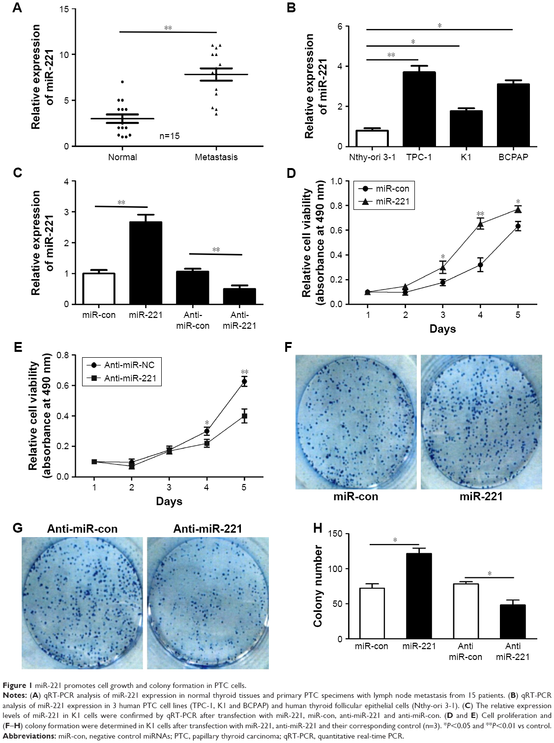 | Figure 1 miR-221 promotes cell growth and colony formation in PTC cells. |
miR-221 strengthens K1 cell growth in vitro
Compared to the control group, K1 cells transfected with miR-221 mimics showed an increased cell proliferation capacity. On the contrary, anti-miR-221-transfected K1 cells grew at a significantly lower rate as compared with the corresponding control group. These findings demonstrated that aberrant expression of miR-221 could regulate K1 cell proliferation in both CCK-8 assays and colony-forming assays (Figure 1D–H).
miR-221 upregulates cell migration and invasion in vitro
To investigate the effect of miR-221 on migration and invasion ability of PTC cells, wound healing assays (Figure 2A and B) and transwell assays (Figure 2C–E) were conducted in K1 cells transfected with miR-221, anti-miR-221 or respective controls. Consistent with the abovementioned clinical data, upregulation of miR-221 in K1 cells obviously enhanced the abilities of cell migration and invasion. In addition, inhibiting miR-221 expression obviously attenuated the abovementioned biological activities of K1 cells in vitro. In conclusion, these data demonstrated that miR-221 could evidently promote migration and invasion of PTC cells in vivo.
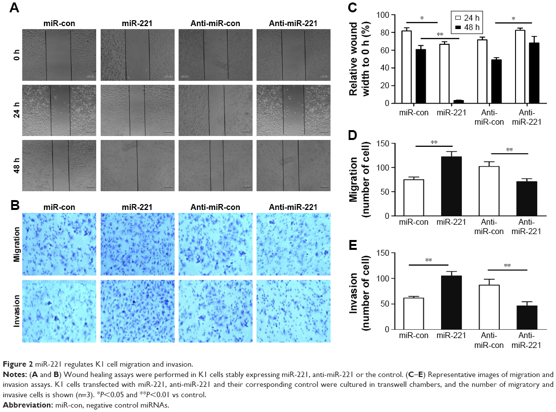 | Figure 2 miR-221 regulates K1 cell migration and invasion. |
miR-221 promotes EMT in K1 cells
Since EMT is closely correlated with tumor metastasis abilities, we then examined several EMT markers in K1 cells transfected with miR-221, anti-miR-221 or their respective control. Consistent with our hypothetical conclusion, qRT-PCR and Western blotting results all showed that the expression of E-cadherin was lower in miR-221 mimic-transfected groups compared with that in miR-con controls, whereas the level of N-cadherin, vimentin and Snail was obviously higher in K1 cells transfected with miR-221 (Figure 3A–C). Activation of the EMT transcription factor Snail and downregulation of the epithelial marker E-cadherin along with the upregulation of mesenchymal markers N-cadherin and vimentin demonstrate the progress of EMT. Therefore, the abovementioned findings indicated that aberrant expression of miR-221 could suppress EMT, which was in accordance with its inhibitory action on migration and invasion of K1 cells.
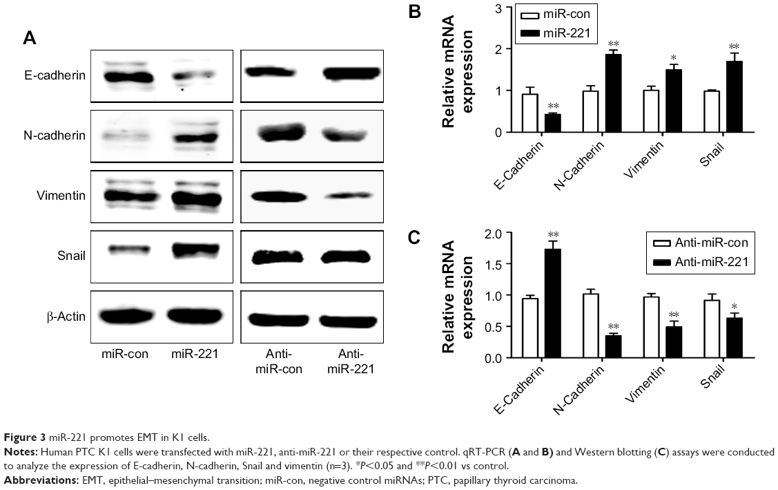 | Figure 3 miR-221 promotes EMT in K1 cells. |
miR-221 upregulates invasion via RECK in human PTC cells
To reveal the mechanism by which miR-221 enhances K1 cell metastasis, we searched for potential target genes of miR-221 using 2 publicly available databases, TargetScan and miRanda. Our analysis of the 3′-UTR sequence of RECK identified a possible binding site for miR-221 (Figure 4A). To test the function of this potential binding site, we inserted wild-type or mutant 3′-UTR sequences immediately downstream of the luciferase reporter gene and coexpressed these with either miR-221 or miR-con in K1 cells. qRT-PCR and Western blotting analyses showed that miR-221 overexpression significantly reduced the levels of RECK mRNA and protein in K1 cells (Figure 4B and C). Furthermore, as shown in Figure 4D, miR-221 overexpression leads to an obvious abatement in relative luciferase activity, whereas activity did not drop at all in the mutant 3′-UTR reporter, indicating that functionality relies on the intact seed sequence. Together, these findings strongly support a direct suppression of RECK by miR-221 by means of mRNA degradation as well as translational repression. We then detected whether RECK took effects in miR-221-mediated PTC progression. After the transfection of miR-221, K1 RECK cells were liable to restore the scratch wounds compared with K1-con cells (Figure 4E). Similar results could be found by transwell assays (Figure 4F and G). These findings indicated that RECK upregulation was able to suppress the promoting metastasis effects of miR-221 in PTC cells and acted as an important mediator in miR-221 regulating invasion and migration of PTC cells. Next, we evaluated whether miR-221 negatively regulated RECK in an EMT-based pathway. Our findings revealed that protein levels of RECK, Snail and vimentin were markedly upregulated in response to miR-221 overexpression in K1-con cell, whereas these changes were not found in K1 RECK cells. Likewise, under the stress of miR-221 overexpression, E-cadherin protein was markedly decreased in K1-con cells but not affected in K1 RECK cells (Figure 4H). These findings suggested that miR-221 had the potential to regulate the expression of EMT markers (Snail, vimentin and E-cadherin) in K1 cells and that these regulations were modulated by RECK. Moreover, RECK overexpression could reverse the effect of miR-221 on these EMT markers in PTC cells.
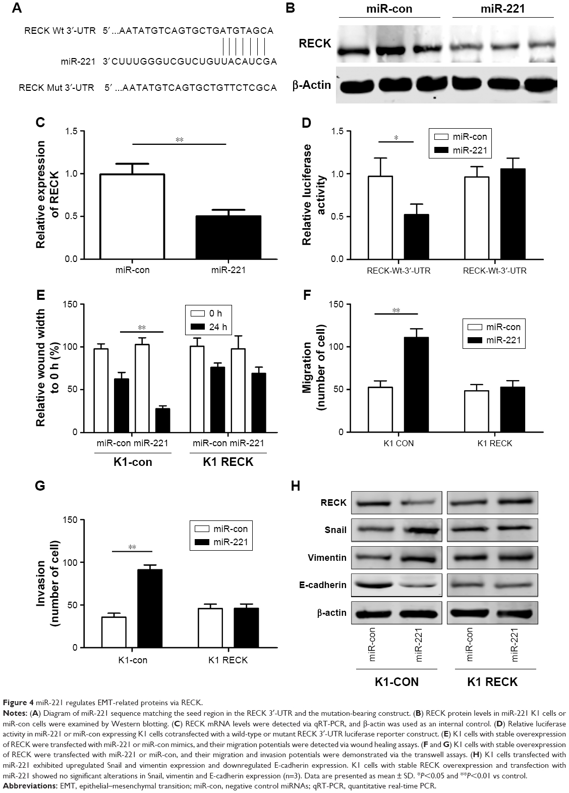 | Figure 4 miR-221 regulates EMT-related proteins via RECK. |
miR-221 enhances growth of K1 cells in vivo
Five weeks later, the mice were executed, and tumor weights were measured. As shown in Figure 5A–C, tumors grew faster in the miR-221 group than in the miR-con group. The average weight of tumors resulting from miR-221 was significantly higher than that of tumors derived from the miR-con group (1,754±138 mg vs 907±87 mg, P<0.01). Furthermore, to determine whether miR-221 downregulated RECK at the protein level in xenografts, we detected RECK expression by Western blot assays. The RECK protein level was decreased in the miR-221 group compared with the miR-con group (Figure 5D). In addition, immunohistochemical staining of Ki-67 antigen indicated that the increased tumor growth in mice was, in part, due to higher proliferation caused by the upregulation of miR-221. The number of Ki-67-antigen-positive cells was higher in the tumor derived from miR-221 cells than that in miR-con cells (Figure 5E and F).
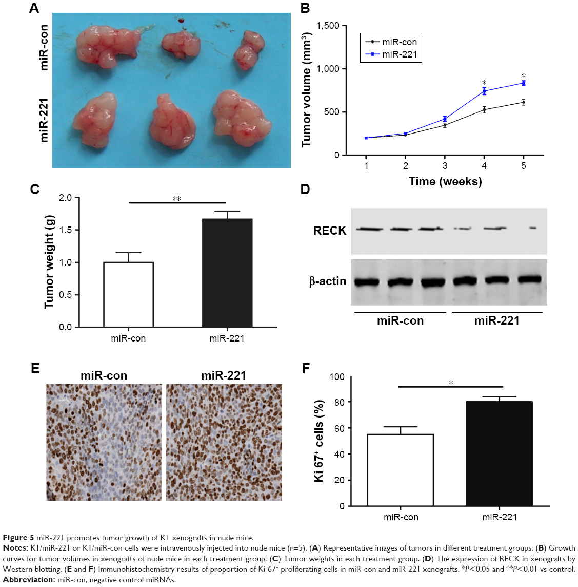 | Figure 5 miR-221 promotes tumor growth of K1 xenografts in nude mice. |
Discussion
MiR-221 belongs to the miR-221/222 cluster, which locates on the X chromosome and shares some identical seed sequences with its homologous miRNA, miR-222.14–17 Previous findings have indicated that miR-221 was obviously upregulated in multiple types of tumor tissues.18–22 For instance, miR-221 was upregulated in ovarian cancer tissues, and patients with increased miR-221 expression levels had a reduced disease-free survival and overall survival compared with those with low miR-221 expression. Transfection of ovarian cancer cells with miR-221 inhibitor induced APAF1 protein expression, suppressed cell proliferation and migration and promoted tumor cell apoptosis.23 Furthermore, Fornari et al24 reported that miR-221 is upregulated in 80% of the liver cancer cells and has an oncogenic function via the inhibition of CDKN1C/p57 and CDKN1B/p27 protein expression. MiR-221 enhanced breast cancer cell growth, migration and invasion, meanwhile propagated the self-renewal of breast cancer stem cells, possibly via targeting the PTEN/Akt pathway.25 MiR-221 played an important role in the control of cancer stem cell homeostasis, and miR-221 was expressed at higher level in the stem cell population of primary and T47D cells compared to differentiated cells.26
The invasion and metastasis processes of tumor cells are composed of multisteps. Degradation of extracellular matrix by matrix metalloproteinase plays an important role in the invasion and metastasis of tumor cells. Some in vivo and in vitro studies have shown that RECK could block the degradation of extracellular matrix by negatively regulating the functions of MMP2 and MMP9 and thus inhibit the invasion and metastasis activities of tumor cells.27 In addition, the expression of RECK protein is reduced or even lost during tumor progression, and so it is very important to restore the expression of RECK protein in tumor cells to inhibit the invasion and metastasis of tumor cells.28 A number of studies have reported the effect of RECK in tumor progression. RECK restrained gastric cancer cell proliferation via inhibiting the NOTCH signaling pathway.29 Collectively, RECK functions as a multirole regulator involved in tumorigenesis. The antitumor activity of RECK was documented in breast cancer by inhibiting erbB signaling, which is associated with chemoresistance.30 RECK was downregulated by miR-96 and ultimately led to proliferation and chemoresistance in esophageal cancer.31 MiR-221-RECK axis was confirmed to be an important path modulating Helicobacter pylori infection-related gastric cancer development.32 Shen et al33 reported that upregulation of miR-590-5p could significantly enhance the cell growth and metastasis and decrease the susceptibility of gastric cancer cells to cisplatin and paclitaxel in vitro. Besides, reintroduction of RECK suppressed the tumor promotion effects of miR-590-5p by inhibiting cell growth and metastasis and increasing drug sensitivity. In the present study, we revealed a novel metastasis mechanism of PTC by which RECK was negatively affected by miR-221 and possessed a negative correlation with E-cadherin protein, enabling thyroid cancer cell metastasis by the regulation of EMT-related proteins.
Conclusion
The primary findings of the present study indicate that miR-221 is obviously upregulated in PTC tissue and cell lines, and its expression level in tumor tissues was significantly correlated with lymph node metastasis or TNM stage. Besides, upregulation of miR-221 in PTC cells enormously enhances cell growth, colony formation, migration and invasion in vitro, as well as promotes xenograft growth in mice. Furthermore, we confirm RECK as an important target gene of miR-221 and find that miR-221 inhibits RECK expression and regulates EMT-related proteins, which contribute to the promotion of PTC cell metastasis. Overall, we elucidate a novel mechanism responsible for thyroid tumorigenesis and indicate that the miR-221/RECK axis may be a useful therapeutic target for PTC.
Disclosure
The authors report no conflicts of interest in this work.
References
Cabanillas ME, McFadden DG, Durante C. Thyroid cancer. Lancet. 2016;388:2783–2795. doi:10.1016/S0140-6736(16)30172-6 | ||
Lim H, Devesa SS, Sosa JA, Check D, Kitahara CM. Trends in thyroid cancer incidence and mortality in the United States, 1974–2013. JAMA. 2017;317:1338–1348. doi:10.1001/jama.2017.2719 | ||
Kim SJ, Myong JP, Jee HG, et al. Combined effect of Hashimoto’s thyroiditis and BRAF(V600E) mutation status on aggressiveness in papillary thyroid cancer. Head Neck. 2016;38:95–101. doi:10.1002/hed.23854 | ||
Wang T, Jiang M, Ren Y, et al. Health-related quality of life of community thyroid cancer survivors in Hangzhou, China. Thyroid. 2018;28:1013–1023. doi:10.1089/thy.2017.0213 | ||
Du L, Wang Y, Sun X, et al. Thyroid cancer: trends in incidence, mortality and clinical-pathological patterns in Zhejiang Province, Southeast China. BMC Cancer. 2018;18:291. doi:10.1186/s12885-018-4242-8 | ||
Rupaimoole R, Slack FJ. MicroRNA therapeutics: towards a new era for the management of cancer and other diseases. Nat Rev Drug Discov. 2017;16:203–222. doi:10.1038/nrd.2016.246 | ||
Santulli G. microRNA: cancer. Adv Exp Med Biol. 2016:2047–2048. | ||
Huang C, Cai Z, Huang M, et al. miR-219-5p modulates cell growth of papillary thyroid carcinoma by targeting estrogen receptor alpha. J Clin Endocrinol Metab. 2015;100:E204–E213. doi:10.1210/jc.2014-2883 | ||
Zhu H, Fang J, Zhang J, et al. miR-182 targets CHL1 and controls tumor growth and invasion in papillary thyroid carcinoma. Biochem Biophys Res Commun. 2014;450:857–862. doi:10.1016/j.bbrc.2014.06.073 | ||
Liu C, Su C, Chen Y, Li G. MiR-144-3p promotes the tumor growth and metastasis of papillary thyroid carcinoma by targeting paired box gene 8. Cancer Cell Int. 2018;18:54. doi:10.1186/s12935-018-0550-y | ||
Chou CK, Chen RF, Chou FF, et al. miR-146b is highly expressed in adult papillary thyroid carcinomas with high risk features including extrathyroidal invasion and the BRAF(V600E) mutation. Thyroid. 2010;20:489–494. doi:10.1089/thy.2009.0027 | ||
Geraldo MV, Yamashita AS, Kimura ET. MicroRNA miR-146b-5p regulates signal transduction of TGF-beta by repressing SMAD4 in thyroid cancer. Oncogene. 2012;31:1910–1922. doi:10.1038/onc.2011.381 | ||
Cong D, He M, Chen S, Liu X, Liu X, Sun H. Expression profiles of pivotal microRNAs and targets in thyroid papillary carcinoma: an analysis of The Cancer Genome Atlas. Onco Targets Ther. 2015;8:2271–2277. doi:10.2147/OTT.S85753 | ||
Song QX, Liu YF, Hu XY, et al. Identification of miRNAs and their target genes in developing soybean seeds by deep sequencing. BMC Plant Biol. 2011;11:5. doi:10.1186/1471-2229-11-68 | ||
Joshi T, Yan Z, Libault M, et al. Prediction of novel miRNAs and associated target genes in Glycine max. BMC Bioinformatics. 2010;11(Suppl 1):S14. doi:10.1186/1471-2105-11-S1-S14 | ||
Aparicio O, Carnero E, Abad X, et al. Adenovirus VA RNA-derived miRNAs target cellular genes involved in cell growth, gene expression and DNA repair. Nucleic Acids Res. 2010;38:750–763. doi:10.1093/nar/gkp1028 | ||
Li Y, Di C, Li W, et al. Oncomirs miRNA-221/222 and tumor suppressors miRNA-199a/195 are crucial miRNAs in liver cancer: a systematic analysis. Dig Dis Sci. 2016;61:2315–2327. doi:10.1007/s10620-016-4156-8 | ||
Ma M, Chen S, Liu Z, et al. miRNA-221 of exosomes originating from bone marrow mesenchymal stem cells promotes oncogenic activity in gastric cancer. Onco Targets Ther. 2017;10:4161–4171. doi:10.2147/OTT.S143315 | ||
Zhao L, Zou D, Wei X, et al. MiRNA-221-3p desensitizes pancreatic cancer cells to 5-fluorouracil by targeting RB1. Tumour Biol. 2016:1–11. doi:10.1007/s13277-016-5445-8 | ||
Liu P, Sun M, Jiang W, Zhao J, Liang C, Zhang H. Identification of targets of miRNA-221 and miRNA-222 in fulvestrant-resistant breast cancer. Oncol Lett. 2016;12:3882–3888. doi:10.3892/ol.2016.5180 | ||
Abak A, Amini S, Estiar MA, Montazeri V, Sakhinia E, Abhari A. Analysis of miRNA-221 expression level in tumors and marginal biopsies from patients with breast cancer (cross-sectional observational study). Clin Lab. 2018;64:169–175. doi:10.7754/Clin.Lab.2017.170821 | ||
Qin J, Luo M. MicroRNA-221 promotes colorectal cancer cell invasion and metastasis by targeting RECK. FEBS Lett. 2014;588:99–104. doi:10.1016/j.febslet.2013.11.014 | ||
Li J, Li Q, Huang H, et al. Overexpression of miRNA-221 promotes cell proliferation by targeting the apoptotic protease activating factor-1 and indicates a poor prognosis in ovarian cancer. Int J Oncol. 2017;50:1087–1096. doi:10.3892/ijo.2017.3898 | ||
Fornari F, Pollutri D, Patrizi C, et al. In hepatocellular carcinoma miR-221 modulates Sorafenib resistance through inhibition of caspase-3-mediated apoptosis. Clin Cancer Res. 2017;23:3953–3965. doi:10.1158/1078-0432.CCR-16-1464 | ||
Li B, Lu Y, Wang H, et al. miR-221/222 enhance the tumorigenicity of human breast cancer stem cells via modulation of PTEN/Akt pathway. Biomed Pharmacother. 2016;79:93–101. doi:10.1016/j.biopha.2016.01.045 | ||
Roscigno G, Quintavalle C, Donnarumma E, et al. MiR-221 promotes stemness of breast cancer cells by targeting DNMT3b. Oncotarget. 2016;7:580–592. doi:10.18632/oncotarget.5979 | ||
Oh J, Takahashi R, Kondo S, et al. The membrane-anchored MMP inhibitor RECK is a key regulator of extracellular matrix integrity and angiogenesis. Cell. 2001;107:789–800. | ||
Chang HC, Cho CY, Hung WC. Downregulation of RECK by promoter methylation correlates with lymph node metastasis in non-small cell lung cancer. Cancer Sci. 2007;98:169–173. doi:10.1111/j.1349-7006.2006.00367.x | ||
Hong KJ, Wu DC, Cheng KH, Chen LT, Hung WC. RECK inhibits stemness gene expression and tumorigenicity of gastric cancer cells by suppressing ADAM-mediated Notch1 activation. J Cell Physiol. 2014;229:191–201. doi:10.1002/jcp.24434 | ||
Hong KJ, Hsu MC, Hung WC. RECK impedes DNA repair by inhibiting the erbB/JAB1/Rad51 signaling axis and enhances chemosensitivity of breast cancer cells. Am J Cancer Res. 2015;5:2422–2430. | ||
Xia H, Chen S, Chen K, Huang H, Ma H. MiR-96 promotes proliferation and chemo- or radioresistance by down-regulating RECK in esophageal cancer. Biomed Pharmacother. 2014;68:951–958. doi:10.1016/j.biopha.2014.10.023 | ||
Liu W, Song N, Yao H, Zhao L, Liu H, Li G. miR-221 and miR-222 simultaneously target RECK and regulate growth and invasion of gastric cancer cells. Med Sci Monitr. 2015;21:2718–2725. doi:10.12659/MSM.894324 | ||
Shen B, Yu S, Zhang Y, et al. miR-590-5p regulates gastric cancer cell growth and chemosensitivity through RECK and the AKT/ERK pathway. Onco Targets Ther. 2016;9:6009–6019. doi:10.2147/OTT.S110923 |
 © 2019 The Author(s). This work is published and licensed by Dove Medical Press Limited. The
full terms of this license are available at https://www.dovepress.com/terms
and incorporate the Creative Commons Attribution
- Non Commercial (unported, 3.0) License.
By accessing the work you hereby accept the Terms. Non-commercial uses of the work are permitted
without any further permission from Dove Medical Press Limited, provided the work is properly
attributed. For permission for commercial use of this work, please see paragraphs 4.2 and 5 of our Terms.
© 2019 The Author(s). This work is published and licensed by Dove Medical Press Limited. The
full terms of this license are available at https://www.dovepress.com/terms
and incorporate the Creative Commons Attribution
- Non Commercial (unported, 3.0) License.
By accessing the work you hereby accept the Terms. Non-commercial uses of the work are permitted
without any further permission from Dove Medical Press Limited, provided the work is properly
attributed. For permission for commercial use of this work, please see paragraphs 4.2 and 5 of our Terms.