Back to Journals » International Journal of Nanomedicine » Volume 17
Microfluidic Methods in Janus Particle Synthesis
Authors Saqib M ![]() , Tran PA
, Tran PA ![]() , Ercan B, Erdem EY
, Ercan B, Erdem EY ![]()
Received 20 April 2022
Accepted for publication 25 August 2022
Published 19 September 2022 Volume 2022:17 Pages 4355—4366
DOI https://doi.org/10.2147/IJN.S371579
Checked for plagiarism Yes
Review by Single anonymous peer review
Peer reviewer comments 2
Editor who approved publication: Dr Yan Shen
Muhammad Saqib,1 Phong A Tran,2 Batur Ercan,3– 5 E Yegan Erdem1,6
1Department of Mechanical Engineering, Bilkent University, Ankara, Turkey; 2Queensland University of Technology (QUT), Brisbane, QLD, 4000, Australia; 3Department of Metallurgical and Materials Engineering, Middle East Technical University, Ankara, Turkey; 4Biomedical Engineering Program, Middle East Technical University, Ankara, Turkey; 5BIOMATEN, Center of Excellence in Biomaterials and Tissue Engineering, Middle East Technical University, Ankara, 06800, Turkey; 6National Nanotechnology Research Center (UNAM), Ankara, Turkey
Correspondence: E Yegan Erdem, Bilkent University, Mechanical Engineering, EA 122, Bilkent, Ankara, 06800, Turkey, Tel +90 312 290 3068, Email [email protected]
Abstract: Janus particles have been at the center of attention over the years due to their asymmetric nature that makes them superior in many ways to conventional monophase particles. Several techniques have been reported for the synthesis of Janus particles; however, microfluidic-based techniques are by far the most popular due to their versatility, rapid prototyping, low reagent consumption and superior control over reaction conditions. In this review, we will go through microfluidic-based Janus particle synthesis techniques and highlight how recent advances have led to complex functionalities being imparted to the Janus particles.
Keywords: anisotropy, droplet-based, asymmetric, nanoparticles, multi-functionality
Introduction
Over the last two decades, scientists have been drawn to the field of nanotechnology in search of novel smart materials with tailored qualities and desired capabilities. The principal aim was to lower the length scale at which matter was handled in order to obtain unprecedented control over material properties. In this context, Janus particles were studied extensively due to their anisotropic nature. The fact that such particles had different physical, chemical and surface properties along different orientations gave rise to exciting prospects in several applications,1–6 including but not limited to cell encapsulation and assembly,7 DNA assays,8 biological multiplexing,9 targeted drug delivery,10–15 noninvasive imaging,16 theranostics,17,18 microlenses,19–21 reflection-mode displays,22 removal of organic and metal pollutants,23,24 and water decontamination.25,26 Since anisotropic particles possess remarkable potential, it was understandable that a large number of methods were reported for their synthesis.27 Janus particle synthesis methods can be broadly classified under three categories, namely masking, phase separation and self-assembly.28–31 Masking method involves immobilizing particles onto a template to block one side, followed by modifying the free side which leads to anisotropy; however, it is a laborious, multi-step process. The phase separation method consists primarily of three steps: dispersion of immiscible polymers in an emulsion, evaporation of the solvent and cross-linking to lock the polymer phases in place. In the self-assembly method, block copolymers are dispersed in a solvent, and assembled through solvent exchange followed by cross-linking of the core. Although phase separation and self-assembly methods are promising techniques, particles’ size distribution is a problem if the particles are synthesized using a batch synthesis route.1,32
Apart from these conventional batch synthesis techniques, several research groups reported synthesis of Janus particles utilizing microfluidic systems to obtain multiple functionalities. In its broadest definition, microfluidics is the study of fluid flow inside channels having less than 1 mm characteristic length. Its main advantage is the capability to use small amounts of sample in detection, analysis, separation and synthesis with higher throughput and sensitivity. At this scale, it is easier to manipulate reagents in a more controllable way with reduced thermal and concentration gradients. It enables each reagent solution to go through an identical process, eventually reducing the variation of size and morphology. In terms of particle synthesis, microfluidic-based devices, due to this high level of control, provide the ability to fabricate monodisperse particles that have a narrow size distribution as compared to particles obtained with batch synthesis techniques.
Flow in a microfluidic system can be continuous or droplet-based. In continuous flow reactors, reagents flow directly inside the channel. On the other hand, in droplet-based microfluidics, reagents move within a channel in the form of nanoliter sized droplets, carried by another immiscible fluid. The droplets move inside the device without getting attached to the device walls due to the presence of a continuous phase between the droplets and the microchannel walls, which reduces channel contamination.
Microfluidic systems have been used for nano- and microparticle synthesis extensively due to their ability to precisely control reaction conditions, such as temperature, concentration, and residence time, which in turn alters nucleation and growth of the particles.33,34 To synthesize Janus particles, two types of materials need to be brought together within the channel. This can be achieved by utilizing a channel design that merges two reagents together either inside a droplet or directly mixing in continuous flow. Several successful microfluidic approaches have been developed. The most popular ones are co-flowing streams, phase separation, double-emulsion, and electrohydrodynamic co-jetting methods.32,35–37 In this review, these techniques will be described as separate subsections, and recent advances in Janus particles synthesis using these methods will be highlighted. Moreover, several other techniques were reported recently; for example, coagulation of nanoparticles inside droplets, controlled merging and crosslinking of droplets and using droplets as particle templates. These techniques do not directly fall under any of the main categories and they will be covered as a separate subsection entitled “miscellaneous techniques”.
Co-Flowing Streams
In a microchannel, fluids with different chemical compositions can flow parallel to each other without mixing. This is due to the viscous forces that suppress diffusion. Fluid flowing in this configuration is named co-flowing streams. For the synthesis of Janus particles, two or more co-flowing streams are separated into droplets by the shear force exerted by another fluid stream and these droplets are later polymerized by UV exposure,38,39 thermal curing or ionic crosslinking.40,41 The co-flowing streams can be miscible or immiscible. Firstly, we will discuss miscible streams. Nisisako et al were the first to demonstrate the synthesis of hemispherical colored droplets and microspheres using this technique.42
 |
Figure 1 An example of a co-flowing microfluidic method to generate Janus droplets. (A) Schematic of the droplet generation process and subsequent polymerization. A mixture of an acrylamide, a cross-linker and a photoinitiator is injected from inlet 1 and 2. The fluids injected from inlet 1 and 2 contain different fluorescent materials. Mineral oil is injected from inlet 3. (B) Chart showing the variety of particles synthesized using this technique. (C) Fluorescent image of the parallel flowing streams showing the maintenance of a clear interface between the two streams. (D) Micrograph of the droplet generation process. Note: Reprinted with permission from Shepherd RF, Conrad JC, Rhodes SK, et al. Microfluidic assembly of homogeneous and janus colloid-filled hydrogel granules. Langmuir. 2006;22(21):8618–8622. Copyright (2006) American Chemical Society.43 |
Shepherd et al contributed to the fundamentals of co-flowing streams technique by synthesizing spherical- and discoidal-shaped Janus hydrogel granules by altering the cross-sectional geometry of the microchannels as shown in Figure 1.43 The maintenance of a clear boundary between the co-flowing streams, and consequently in the generated droplets, depends on the ability to avoid convective and diffusive mixing. These findings were utilized by several other studies on Janus particle synthesis to obtain different kinds of anisotropies such as amphiphilicity,11 optical anisotropy,44,45 and magnetic anisotropy.46,47 Hessberger et al reported synthesis of a liquid crystalline elastomer containing Janus particles using side-by-side capillaries.45 The presence of liquid crystalline elastomer at one half of the Janus particles was shown to enhance the actuating properties of the particles in several applications. Maeda et al used a centrifuge-based microdroplet generator to fabricate Janus particles having magnetic anisotropy.48 Seiffert et al reported the synthesis of Janus microshells (hollow) and microgels using three co-flowing streams. For these microshells, they used a non-polymerizable phase as the middle stream, which was removed later by washing, and for the microgels they used a polymerizable phase having magnetic nanoparticles, which provided the microgels with a magnetic core.49 Xie et al utilized a nanoprecipitation method to synthesize sub-micrometer sized Janus particles capable of carrying payloads with different solubility in their two hemispheres.11 Kamperman et al reported the production of multienzymatic microreactors having magnetic anisotropy using in-air microfluidics, where the co-flowing streams were transported in air instead of a conventional continuous phase fluid and were simultaneously cured by a cross-linking jet showered at the generated droplets.50 They claimed that in-air microfluidics approach increased the production rate by two to three orders of magnitude compared to the conventional microfluidic approach. This work could potentially lead the way to high throughput synthesis of Janus particles, which has been the major challenge in commercialization of droplet-based microfluidic methods to synthesize particles. Their device and the obtained microparticles are shown in Figure 2. In a more recent work, Liu et al demonstrated the synthesis of hydrogel microparticles having magnetic anisotropy via an emulsion-based external gelation method. The set-up consisted of a pair of parallel flowing dispersed phase streams, one containing sodium alginate solution and the other containing a mixture of sodium alginate solution and magnetic nanoparticles, sheared into droplets that were solidified downstream by ionic cross-linking after reacting with calcium ions present in the cross linking agent flowing downstream.51 This recent approach overcomes the shortcomings of the earlier ionic crosslinking method proposed by Zhao et al using a continuous stream of cross-linking agent instead of droplets, and therefore, produces monodisperse uniform shaped spherical particles having a clear interface between the two hemispherical compartments.47
 |
Figure 2 Janus particle generation in air microfluidics. Hydrogel particles with a paramagnetic compartment were synthesized at high throughput compared to channel based microfluidic systems. The size of these particles could be tuned, and the technique allowed monodiperse particle synthesis. Enzymes were encapsulated inside these particles and they were proposed as enzymatic bioreactors. (A) Concept of the device, (B) formation steps of the of the Janus particles, (C and D) real-time images during particle synthesis, (E) synthesized Janus particles under the fluorescent microscope, characterization results of the particles show that (F) the particles are monodisperse and that (G) there is a clear distinction between the two compartments. Note: Reprinted from Kamperman T, Trikalitis VD, Karperien M, et al. Ultrahigh-throughput production of monodisperse and multifunctional janus microparticles using in-air microfluidics. ACS Appl Mater Interfaces. 2018;10(28):23433–23438. Creative Common.50 |
Co-flowing fluids used for the synthesis of Janus particles can also be immiscible. In this case, the structure of the Janus droplet, and in turn, of the particle, is determined by the spreading coefficient. The spreading coefficient is given by 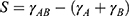 ,52 where
  is the interfacial tension between the two co-flowing phases,
  is the interfacial tension between the co-flowing phase A and continuous phase fluid, and
  is the interfacial tension between the co-flowing phase B and continuous phase fluid.53
 |
Figure 3 Microfluidic device that uses immiscible co-flowing streams to generate Janus and ternary microparticles. (A) Device schematic, where M1 is methacryloxypropyl dimethylsiloxane, M2 is a mixture of pentaerythritol triacrylate, poly(ethylene glycol) diacrylate and acrylic acid, and W is aqueous SDS (sodium dodecyl sulfate) solution. (B) Droplet generation from co-flowing streams, (C–F) optical and fluorescent (inset) microscopy images of the synthesized particles with varying ratios of monomers. (parts of two figures were merged from the original paper). Adapted with permission from Nie Z, Li W, Seo M, et al. Janus and ternary particles generated by microfluidic synthesis: design, synthesis, and self-assembly. J Am Chem Soc. 2006;128(29):9408–9412 . Copyright (2006)American Chemical Society.54 |
The first report on the synthesis of Janus particles using immiscible co-flowing streams was presented by Nie et al where they synthesized Janus and ternary (trisectioned) particles. They also demonstrated the synthesis of Janus particles with varying ratios of two monomer streams.54 Their device and obtained results are shown in Figure 3. Nisisako et al reported the synthesis of truncated Janus particles using one polymerizable and one non-polymerizable fluid as the co-flowing fluids.40 After off-chip UV curing, the non-polymerizable phase was washed and a truncated Janus particle, whose shape was controlled with the interfacial tension, was produced. This technique of having a polymerizable and a non-polymerizable phase has been used in several works. Yu et al utilized this technique to synthesize truncated-, crescent- and hollow-shaped anisotropic colloidal photonic crystal supraparticles for potential use in novel color displays.38 Some other uses of this approach are the two works of Nisasako et al where they fabricated a biconvex microlens using two co-flowing streams and biconcave microlens using three co-flowing streams.20,21 Another interesting example of this technique is the synthesis of self-propelled micromotors for water decontamination, which was achieved by controlled decoration of Ag nanoparticles on the concave surface, TiO2 nanoparticles on the convex surface and magnetic nanoparticles inside the micromotor itself.25 Another interesting study to lay the foundation for other works was reported by Prasad et al where immiscible monomers in a co-flowing system were used to fabricate Janus particles with a dumbbell or a snowman shape depending on the capillary number of each stream. The dumbbell-shaped particles were incorporated with magnetic nanoparticles in one half, followed by a heat treatment to produce concave ceramic hemispheres having different surface textures and magnetic anisotropy.55
 |
Figure 4 (A) Schematic showing the formation of magnetically anisotropic and highly porous polymeric particles due to the two step phase separation between polystyrene and poly(vinyl acetate). (B) Illustration of the two step synthesis of highly porous isotropic magnetic polymer particles. DCM: dichloromethane; PS: polystyrene; PVAc: poly(vinyl acetate), Fe3O4: magnetic nanoparticles. Reprinted with permission from Al Nuumani R, Smoukov SK, Bolognesi G, et al. Highly porous magnetic Janus microparticles with asymmetric surface topology. Langmuir. 2020;36(42):12702–12711. Copyright 2020 American Chemical Society.70 |
Yang et al utilized a side-by-side capillary configuration to synthesize anisotropic magnetic Janus particles where the anisotropy was maintained by having immiscible co-flowing streams.56 Xu et al reported the synthesis of hydrophilic-hydrophobic anisotropic Janus particles using a selective channel surface modification. The surface of the dispersed phase microchannel was modified to make one side of the microchannel hydrophobic and the other side hydrophilic. A mixture of a hydrophilic monomer and a hydrophobic monomer was used as the dispersed phase. Due to the surface modification of the microchannel, the hydrophilic monomer flowed along the hydrophilic part of the microchannel and the hydrophobic monomer flowed along the hydrophobic part of the microchannel. The two parallel streams were sheared into droplets followed by UV exposure to form amphiphilic particles.57 Recently, Haney et al reported the synthesis of hydrophobic-hydrophilic Janus particles as a stabilizing agent in emulsions that could be used as a potential replacement for conventional surfactants.58
Phase Separation
Phase separation provides an excellent pathway for Janus particle synthesis. It relies on the fact that the reaction of monomers to energy absorption depends on their chemistry.59 This method does not require precise control of flow parameters, making it a simpler approach than the co-flowing technique which depends on the precision of the flow parameters to maintain the interface that separates the two halves of the Janus particles. Shah et al reported one of the first studies on the phase separation method where they synthesized Janus microparticles with one side made of a hydrogel and the other side made primarily of aggregated colloidal nanoparticles. They used a two-step phase separation method consisting of heat-induced shrinkage and accumulation of microgels to one hemisphere, and UV polymerization of an acrylamide monomer in the other hemisphere.60 The utilized microgels were cationic, so anionic magnetic beads accumulated in the same hemisphere and rendered the spherical particles magnetically anisotropic. Lone et al reported fabrication of Janus particles with a protruding head to give physical anisotropy using UV-directed phase separation. Firstly, droplets of an aqueous phase, consisting of a thermosensitive and a photosensitive compound, were formed in a flow-focusing geometry using hexadecane as the continuous phase. The UV-directed phase separation was achieved in a two-step procedure where a light-sensitive compound (spironaphthoxazine) present in each droplet pushed the other water-based thermosensitive monomer (Poly(N-isopropylacrylamide)) to the tip of the spherical droplet upon UV exposure as a result of zwitterionic pair effect. This led the way for the photoinitiator from the continuous phase to diffuse into the tip and initiate polymerization and form the anisotropic Janus particle.61 Kim et al reported a novel technique that took advantage of the difference in the rates of phase separation and UV polymerization between two hemispheres to synthesize asymmetric porous Janus particles. They generated droplets of the dispersed phase composed of a nematic liquid crystal, that causes the polarization of light waves, mixed with a polymer. The generated droplets were then collected and exposed to UV radiation. It was observed that the droplet hemisphere directly facing UV radiation swelled (and wrinkled when dried), while the other hemisphere remained smooth making the particle asymmetrically porous.62 Li et al altered the contents of the dispersed phase from an organochloride compound (dichloromethane) to an organic compound (dimethyl carbonate) for phase separation-based Janus particle synthesis. They concluded that by changing the nature of the dispersed phase from an organochloride to an organic fluid, a morphology transition from core-shell microcapsule to Janus could be achieved.63 Another study based on phase separation was reported by Jeong et al where a solution of incompatible monomers was used to form anisotropic particles. The droplet phase was a mixture of three polymers, two of which were incompatible with the third one. As heat was provided, the solvent in the droplets evaporated and the incompatible polymers separated into two compartments giving the particle its Janus characteristic.64 Ekanem et al reported the synthesis of anisotropic particles containing patches on one part of the particles. The extent of patches was found to depend on emulsion formulation, droplet size, and synthesis route. They concluded that smaller droplets resulted in patchy particles and larger droplets contained no patches. The difference in the resulting morphology was attributed to shorter solidification time required for smaller droplets which prevented the complete phase separation of polymers present in each droplet.65
 |
Figure 5 (A) Schematic of a double emulsion microfluidic device and droplets formed with one core. (B) Optical image of double emulsion Janus droplet formation in a PDMS device. (C) Optical image of the collection reservoir where the double emulsion droplets are collected and exposed to UV radiation to initiate polymerization. Reprinted with permission from Chen CH, Shah RK, Abate AR, et al. Janus particles templated from double emulsion droplets generated using microfluidics. Langmuir. 2009;25(8):4320–4323. Copyright (2009) American Chemical Society.72 |
Wang et al synthesized amphiphilic polymer particles having magnetic anisotropy by an off-chip magnetically driven phase separation method.66 Sun et al utilized a simple flow-focusing droplet generator coupled with a downstream serpentine channel to synthesize novel Janus particles having separate lipid (triglyceride type) and polymeric (poly(lactic-co-glycolic acid)) compartments. The morphology of the particles was easily tuned from Janus-patchy to triple, quadruple and core-shell type by varying the amount and type of surfactant incorporated into the dispersed phase.67 Ghosh et al reported the synthesis of snowman-like Janus hydrogel particles by solvent evaporation-based phase separation.68 Xia et al used the solvent loss-based phase separation method to demonstrate how the density of suspended particles could lead to phase separation and shape alterations in Janus particles. They demonstrated that low-density microparticles led to more spherical, and high-density nanoparticles led to flattened particle shapes.69 Utilizing phase separation method based on solvent loss, Ren et al synthesized crescent-shaped Janus particles decorated with manganese oxide nanoparticles on the concave surface and magnetic nanoparticles on the convex surface for water decontamination applications.26 Recently, Al Nuumani et al successfully synthesized anisotropic magnetic Janus particles to be porous on one hemisphere and non-porous on the other one using the process shown in Figure 4.70 This was achieved by a two-step solvent evaporation-based phase separation. In the first step, the phase separation of thermodynamically incompatible monomers caused hemispherical partitioning, and in the second step the phase separation of dichloromethane and heptane led to the formation of pores on one hemisphere. In a more recent work, Lan et al utilized a phase separation technique based on UV exposure coupled with solvent evaporation to synthesize snowman-shaped amphiphilic Janus particles using materials derived from plants that could be potentially used as environment-friendly replacements for microplastics. In contrast to the previous research reports, this work not only presented a novel approach to synthesize amphiphilic Janus particles but also demonstrated a practical solution to address the low throughput drawback of microfluidic platforms by using 10,260 parallel microdroplet generators in a single silicon-and-glass microfluidic device to scale up the synthesis.71
Double Emulsion
In the double emulsion method, droplets of one phase (oil or aqueous) encapsulate the droplets of the other phase to generate encapsulated droplets within a microfluidic channel. This is achieved by selective treatment of the microfluidic channels to form hydrophobic and hydrophilic regions within the same device. Chen et al reported the formation of non-spherical Janus particles using this approach as shown in Figure 5.72 During transportation of droplets inside the channel, the inner droplet was pushed towards the back side of the encapsulating droplet due to hydrodynamic forces and density difference between the two droplets. The double emulsion droplets were then cured with UV exposure to freeze their Janus configuration. Chen et al used this approach to synthesize anisotropic magnetic Janus particles by incorporating magnetic nanoparticles into the inner droplet phase.73 Thiele et al further advanced this methodology by adding a third consecutive droplet generation unit to provide extra microgel spheres which acted as a support to prevent shape change during the UV exposure.74 Using this approach, non-spherical snowman-shaped particles having internally hydrophilic or hydrophobic lobes and an outer hydrophilic shell could be successfully synthesized. These Janus particles were proposed to be a potential replacement for conventional surfactants as an emulsion stabilizer.
 |
Figure 6 Schematic of the microfluidic device used to synthesize dimer like Janus particles. Reprinted with permission from Lu AX, Jiang K, DeVoe DL, et al. Microfluidic assembly of Janus-like dimer capsules. Langmuir. 2013;29(44):13624–13629. Copyright (2013) American Chemical Society.80 |
In the case of double emulsion method, over-reliance on oil-in-water emulsions has been a major hurdle in the ability of the obtained particles to encapsulate biomolecules. For this reason, there is a lot of attention towards utilizing multiple all-aqueous emulsions to synthesize Janus particles.75 In a recent study, Dumas et al demonstrated how water-in-water emulsions could be stabilized by adsorbing chitosan-grafted lipid nanocapsules at the interface between the two aqueous phases. If such a method could be utilized to synthesize Janus particles, not only would it allow encapsulation of biomolecules it would also open new frontiers for drug delivery applications.76
Electrohydrodynamic Co-Jetting
Electrohydrodynamic (EHD) co-jetting is a technique in which co-flowing streams of fluids are pumped in air across an electric field to induce electrified jetting to obtain anisotropic particles. Roh et al were among the first scientists to report the synthesis of Janus particles using this method.77 They studied how changes in the interfacial tension values between the co-flowing fluids caused changes in the particle shapes to obtain peanut, snowman, and spindle-like particle morphologies. Their results showed that these particles exhibited excellent anisotropy and no cross-contamination of payloads occurred between the spherical halves. In another work, Hwang et al reported the synthesis of spherical Janus nanoparticles consisting of two different types of polymer hemispheres for use in controlled release of multiple drugs.78 The synthesized Janus particles demonstrated selective, pH-dependent degradation of each hemispherical compartment. Rahmani et al conducted another study where 100 nm sized Janus particles were obtained by EHD co-jetting by altering process parameters, including polymer composition, polymer concentration and type of surfactant.79 In a recent work, Feng et al reported the synthesis of drug-loaded magnetic Janus particles using EHD co-jetting method. The particles comprised three compartments that possessed different functionalities, namely magnetic nanoparticles for targeting, dyes for fluorescent tracking, and drugs for killing cancer cells. According to their findings, these multicompartmental particles were more efficient in targeting and killing cancer cells than conventional techniques.12
Miscellaneous Techniques
Aside from the techniques discussed above, there are other methodologies for the microfluidic production of anisotropic particles. Lu et al reported the synthesis of dumbbell-shaped anisotropic Janus particles having magnetic properties for micromixing applications. Janus particles were synthesized by the coalescence of two adjacent droplets as shown in Figure 6. One droplet contained chitosan and the other contained magnetic nanoparticles in chitosan.80 As the droplets came into contact, glutaraldehyde was introduced into the microchannel which initiated the cross-linking of chitosan resulting in dumbbell-shaped chitosan particles. Shang et al reported the fabrication of Janus particles composed of iron oxide (Fe3O4) and silicon dioxide (SiO2) in distinct hemispheres using droplets as particle templates. Droplets were cured off-chip in the presence of a magnetic field which resulted in anisotropic Janus particles.81 Using a similar approach, Chen et al reported the production of Fe3O4 and TiO2 Janus particles using branched polymers by off-chip magnetic and gravitational force-based separation, followed by thermal curing.82 Torii et al fabricated anisotropic magnetic Janus particles by an off-chip gelation process initiated by diffusion of ions from the droplet to an agarose gel plate.83 Although effective, off-chip processing is not desirable in microfluidic-based particle synthesis. Varma et al reported the synthesis of anisotropic magnetic Janus particles for protein detection by on-chip magnetic force-based separation, followed by UV polymerization.84 In a more recent work, Wang et al fabricated Janus nanostructures composed of gold nanorods and silver, by a simple microfluidic droplet-based reactor and serpentine mixer.85 They demonstrated the use of these Janus structures as nanosensors for the detection of mercury ions. Such particles could be commercially utilized for the detection of mercury in drinking water to avoid harmful effects of mercury ingestion. Recently, Li et al reported a novel method to synthesize Janus colloidal photonic crystals for potential use on a flexible display. The shortcoming in the earlier conventional methods, for example in the method reported by Yu et al,38 was that the colloidal photonic crystals were formed inside a microfluidic chip and therefore, needed to be transferred onto the grooves on the flexible display by a complex and painstaking process. To address this issue, Li et al designed a system that combined a microfluidic droplet generator with an automatic dispensing printing system to synthesize and print Janus colloidal photonic crystals on a flexible display. The vertical microfluidic droplet generator formed droplets from two sources. These droplets came into proximity of each other and merged to form a Janus structure due to the interfacial tension between the two phases, and were dispensed onto a flexible display. This eliminated the tedious process of transferring the synthesized colloidal structures from a microfluidic device onto a substrate.86
Conclusion
In this review, microfluidic-based synthesis routes for Janus particles have been discussed including co-flowing streams method, phase separation method and double emulsion method among others. While these conventional microfluidic-based synthesis routes enable the fabrication of monodisperse Janus particles having complex functionalities, they exhibit certain shortcomings, such as the need for similar viscosities for dispersed phases in the co-flowing streams, precise control over fluid flow rates for the double-emulsion method, and difficulty in achieving equilibrium in phase separation processes due to constantly changing physical properties of fluids throughout the process. Microfluidic-based techniques provide a superior pathway for the synthesis of Janus particles as compared to conventional batch synthesis techniques due to their ability to synthesize monodisperse particles using a small volume of reagent typically on a single chip. Recent advances in microfluidic-based techniques for Janus particle synthesis provide the ability to fabricate a wide variety of particle shapes using non-hazardous, biogenic materials in the nanometer regime. This is of utmost importance for the potential use of these anisotropic particles in several applications critical to human life, ie removal of contaminants (metal traces, bacteria, microplastics, oil, etc) from water, and detection of bacteria and toxins. On the other hand, for applications requiring a large number of particles, scaling up of production is necessary and is still a challenge for microfluidic-based methods. Furthermore, conventional microfluidic devices are fabricated using specialized equipment which makes their manufacturing expensive. In addition, microfluidic platforms require pumps and external valves to operate, which makes the device prone to leakage. Nevertheless, several parallelization techniques were utilized in recent reports that allowed the production of monodisperse droplets from multiple nozzles at a time, at a generation frequency of several kHz. Moreover, many potential replacements to conventional microfluidics platforms have been introduced, ie, paper-based microfluidics which are easy to manufacture and eliminate the need for pumps and external valves. To conclude, Janus particles have the potential to contribute massively to some of human kinds’ technological challenges, and although microfluidic-based techniques have certain limitations, the recent advances in the field of microfluidics leave us optimistic about the future prospects for the use of these methods for Janus particle synthesis.
Acknowledgments
The authors acknowledge the support provided by the Turkish Scientific and Technological Research Council (TÜBİTAK) 1001 grant (project ID: 219M480).
Disclosure
The authors report no conflicts of interest in this work.
References
1. Safaie N, Ferrier RC. Janus nanoparticle synthesis: overview, recent developments, and applications. J Appl Phys. 2020;127(17):170902. doi:10.1063/5.0003329
2. Perro A, Reculusa S, Ravaine S, et al. Design and synthesis of Janus micro-and nanoparticles. J Mater Chem. 2005;15(35–36):3745–3760. doi:10.1039/b505099e
3. Walther A, Muller AH. Janus particles: synthesis, self-assembly, physical properties, and applications. Chem Rev. 2013;113(7):5194–5261. doi:10.1021/cr300089t
4. Walther A, Müller AH. Janus particles. Soft Matter. 2008;4(4):663–668. doi:10.1039/b718131k
5. Zhang J, Grzybowski BA, Granick S. Janus particle synthesis, assembly, and application. Langmuir. 2017;33(28):6964–6977. doi:10.1021/acs.langmuir.7b01123
6. Jiang S, Chen Q, Tripathy M, et al. Janus particle synthesis and assembly. Adv Mater. 2010;22(10):1060–1071. doi:10.1002/adma.200904094
7. Zhang L, Chen K, Zhang H, et al. Microfluidic templated multicompartment microgels for 3D encapsulation and pairing of single cells. Small. 2018;14(9):1702955. doi:10.1002/smll.201702955
8. Lan J, Chen J, Li N, et al. Microfluidic generation of magnetic-fluorescent Janus microparticles for biomolecular detection. Talanta. 2016;151:126–131. doi:10.1016/j.talanta.2016.01.024
9. Nisisako T, Torii T, Takahashi T, et al. Synthesis of monodisperse bicolored janus particles with electrical anisotropy using a microfluidic Co‐Flow system. Adv Mater. 2006;18(9):1152–1156. doi:10.1002/adma.200502431
10. Khan IU, Serra CA, Anton N, et al. Microfluidic conceived drug loaded Janus particles in side-by-side capillaries device. Int J Pharm. 2014;473(1–2):239–249. doi:10.1016/j.ijpharm.2014.06.035
11. Xie H, She ZG, Wang S, et al. One-step fabrication of polymeric Janus nanoparticles for drug delivery. Langmuir. 2012;28(9):4459–4463. doi:10.1021/la2042185
12. Feng ZQ, Yan K, Li J, et al. Magnetic Janus particles as a multifunctional drug delivery system for paclitaxel in efficient cancer treatment. Mater Sci Eng C. 2019;104:110001. doi:10.1016/j.msec.2019.110001
13. Seo KD, Doh J, Kim DS. One-step microfluidic synthesis of Janus microhydrogels with anisotropic thermo-responsive behavior and organophilic/hydrophilic loading capability. Langmuir. 2013;29(49):15137–15141. doi:10.1021/la403015y
14. Indalkar YR, Gaikwad SS, Ubale AT. Janus particles recent and novel approach in drug delivery: an overview. J Curr Pharma Res. 2013;3(4):1031. doi:10.33786/JCPR.2013.v03i04.010
15. Yang Z, Aarts DA, Chen Y, et al. Janus Particle Synthesis, Self-Assembly and Applications. Royal Society of Chemistry; 2012.
16. Song G, Chen M, Zhang Y, et al. Janus iron oxides@ semiconducting polymer nanoparticle tracer for cell tracking by magnetic particle imaging. Nano Lett. 2018;18(1):182–189. doi:10.1021/acs.nanolett.7b03829
17. Rahiminezhad Z, Tamaddon AM, Borandeh S, et al. Janus nanoparticles: new generation of multifunctional nanocarriers in drug delivery, bioimaging and theranostics. Appl Mater Today. 2020;18:100513. doi:10.1016/j.apmt.2019.100513
18. Zhang Y, Huang K, Lin J, et al. Janus nanoparticles in cancer diagnosis, therapy and theranostics. Biomater Sci. 2019;7(4):1262–1275. doi:10.1039/C8BM01523F
19. Nisisako T, Ando T, Hatsuzawa T. Biconvex polymer microlenses fabricated from microfluidic Janus droplets. J Japan Soc Precis Eng. 2013;79(5):460–466. doi:10.2493/jjspe.79.460
20. Nisisako T, Ando T, Hatsuzawa T. Capillary‐assisted fabrication of biconcave polymeric microlenses from microfluidic ternary emulsion droplets. Small. 2014;10(24):5116–5125. doi:10.1002/smll.201401269
21. Nisisako T, Suzuki H, Hatsuzawa T. Biconvex polymer microlenses with tunable imaging properties designed by Janus droplet microfluidics. Micromachines. 2015;6(10):1435–1444. doi:10.3390/mi6101428
22. Kim SH, Jeon SJ, Jeong WC, et al. Optofluidic synthesis of electroresponsive photonic janus balls with isotropic structural colors. Adv Mater. 2008;20(21):4129–4134.
23. Guix M, Weiz SM, Schmidt OG, et al. Self‐propelled micro/nanoparticle motors. Part Part Syst Charact. 2018;35(2):1700382. doi:10.1002/ppsc.201700382
24. Jurado-Sánchez B, Wang J. Micromotors for environmental applications: a review. Environ Sci. 2018;5(7):1530–1544.
25. Chen A, Ge XH, Chen J, et al. Multi-functional micromotor: microfluidic fabrication and water treatment application. Lab Chip. 2017;17(24):4220–4224. doi:10.1039/C7LC00950J
26. Ren M, Guo W, Guo H, et al. Microfluidic fabrication of bubble-propelled micromotors for wastewater treatment. ACS Appl Mater Interfaces. 2019;11(25):22761–22767. doi:10.1021/acsami.9b05925
27. Marschelke C, Fery A, Synytska A. Janus particles: from concepts to environmentally friendly materials and sustainable applications. Colloid Polym Sci. 2020;298(7):841–865. doi:10.1007/s00396-020-04601-y
28. Poggi E, Gohy JF. Janus particles: from synthesis to application. Colloid Polym Sci. 2017;295(11):2083–2108.
29. Fan X, Yang J, Loh XJ, et al. Polymeric Janus nanoparticles: recent advances in synthetic strategies, materials properties, and applications. Macromol Rapid Commun. 2019;40(5):1800203. doi:10.1002/marc.201800203
30. Du J, O’Reilly RK. Anisotropic particles with patchy, multicompartment and Janus architectures: preparation and application. Chem Soc Rev. 2011;40(5):2402–2416. doi:10.1039/c0cs00216j
31. Lattuada M, Hatton TA. Synthesis, properties and applications of Janus nanoparticles. Nano Today. 2011;6(3):286–308. doi:10.1016/j.nantod.2011.04.008
32. Lone S, Cheong IW. Fabrication of polymeric Janus particles by droplet microfluidics. RSC Adv. 2014;4(26):13322–13333. doi:10.1039/C4RA00158C
33. Erdem EY, Cheng JC, Doyle FM, et al. Multi‐temperature zone, droplet‐based microreactor for increased temperature control in nanoparticle synthesis. Small. 2014;10(6):1076–1080. doi:10.1002/smll.201302379
34. Wahab MA, Erdem EY. Multi-step microfluidic reactor for the synthesis of hybrid nanoparticles. J Micromechan Microengine. 2020;30(8):085006. doi:10.1088/1361-6439/ab8dd2
35. Nisisako T. Recent advances in microfluidic production of Janus droplets and particles. Curr Opin Colloid Interface Sci. 2016;25:1–2. doi:10.1016/j.cocis.2016.05.003
36. Zhao Q, Cui H, Wang Y, Du X. Microfluidic platforms toward rational material fabrication for biomedical applications. Small. 2020;16(9):1903798. doi:10.1002/smll.201903798
37. Yang S, Guo F, Kiraly B, et al. Microfluidic synthesis of multifunctional Janus particles for biomedical applications. Lab Chip. 2012;12(12):2097–2102. doi:10.1039/c2lc90046g
38. Yu Z, Wang CF, Ling L, et al. Triphase microfluidic‐directed self‐assembly: anisotropic colloidal photonic crystal supraparticles and multicolor patterns made easy. Angewandte Chemie. 2012;124(10):2425–2428. doi:10.1002/ange.201107126
39. Yu X, Zhang C, You S, et al. Microfluidic synthesis of multiferroic Janus particles with disk-like compartments. Appl Phys Lett. 2016;108(7):073504. doi:10.1063/1.4942365
40. Nisisako T, Torii T. Formation of biphasic Janus droplets in a microfabricated channel for the synthesis of shape‐controlled polymer microparticles. Adv Mater. 2007;19(11):1489–1493. doi:10.1002/adma.200700272
41. Marquis M, Renard D, Cathala B. Microfluidic generation and selective degradation of biopolymer-based Janus microbeads. Biomacromolecules. 2012;13(4):1197–1203. doi:10.1021/bm300159u
42. Nisisako T, Torii T, Higuchi T. Novel microreactors for functional polymer beads. Chem Eng J. 2004;101(1–3):23–29. doi:10.1016/j.cej.2003.11.019
43. Shepherd RF, Conrad JC, Rhodes SK, et al. Microfluidic assembly of homogeneous and janus colloid-filled hydrogel granules. Langmuir. 2006;22(21):8618–8622.
44. Yin SN, Wang CF, Yu ZY, et al. Versatile bifunctional magnetic‐fluorescent responsive janus supraballs towards the flexible bead display. Adv Mater. 2011;23(26):2915–2919. doi:10.1002/adma.201100203
45. Hessberger T, Braun LB, Henrich F, et al. Co-flow microfluidic synthesis of liquid crystalline actuating Janus particles. J Mater Chem C. 2016;4(37):8778–8786. doi:10.1039/C6TC03378D
46. Yuet KP, Hwang DK, Haghgooie R, et al. Multifunctional superparamagnetic Janus particles. Langmuir. 2010;26(6):4281–4287. doi:10.1021/la903348s
47. Zhao LB, Pan L, Zhang K, et al. Generation of Janus alginate hydrogel particles with magnetic anisotropy for cell encapsulation. Lab Chip. 2009;9(20):2981–2986. doi:10.1039/b907478c
48. Maeda K, Onoe H, Takinoue M, et al. Controlled synthesis of 3D multi‐compartmental particles with centrifuge‐based microdroplet formation from a multi‐barrelled capillary. Adv Mater. 2012;24(10):1340–1346. doi:10.1002/adma.201102560
49. Seiffert S, Romanowsky MB, Weitz DA. Janus microgels produced from functional precursor polymers. Langmuir. 2010;26(18):14842–14847. doi:10.1021/la101868w
50. Kamperman T, Trikalitis VD, Karperien M, et al. Ultrahigh-throughput production of monodisperse and multifunctional janus microparticles using in-air microfluidics. ACS Appl Mater Interfaces. 2018;10(28):23433–23438. doi:10.1021/acsami.8b05227
51. Liu Y, Nisisako T. Microfluidic generation of monodispersed Janus alginate hydrogel microparticles using water-in-oil emulsion reactant. Biomicrofluidics. 2022;16(2):024101. doi:10.1063/5.0077916
52. Geng Y, Ling S, Huang J, et al. Multiphase microfluidics: fundamentals, fabrication, and functions. Small. 2020;16(6):1906357. doi:10.1002/smll.201906357
53. Guzowski J, Korczyk PM, Jakiela S, et al. The structure and stability of multiple micro-droplets. Soft Matter. 2012;8(27):7269–7278. doi:10.1039/c2sm25838b
54. Nie Z, Li W, Seo M, et al. Janus and ternary particles generated by microfluidic synthesis: design, synthesis, and self-assembly. J Am Chem Soc. 2006;128(29):9408–9412. doi:10.1021/ja060882n
55. Prasad N, Perumal J, Choi CH, et al. Generation of monodisperse inorganic–organic janus microspheres in a microfluidic device. Adv Funct Mater. 2009;19(10):1656–1662. doi:10.1002/adfm.200801181
56. Yang YT, Wei J, Li X, et al. A side-by-side capillaries-based microfluidic system for synthesizing size-and morphology-controlled magnetic anisotropy janus beads. Adv Powder Technol. 2015;26(1):156–162. doi:10.1016/j.apt.2014.08.018
57. Xu K, Ge XH, Huang JP, et al. A region-selective modified capillary microfluidic device for fabricating water–oil Janus droplets and hydrophilic–hydrophobic anisotropic microparticles. RSC Adv. 2015;5(58):46981–46988. doi:10.1039/C5RA05690J
58. Haney B, Chen D, Cai LH, et al. Millimeter-size Pickering emulsions stabilized with Janus microparticles. Langmuir. 2019;35(13):4693–4701. doi:10.1021/acs.langmuir.9b00058
59. Pang X, Wan C, Wang M, et al. Strictly biphasic soft and hard Janus structures: synthesis, properties, and applications. Angewandte Chemie Int Edition. 2014;53(22):5524–5538. doi:10.1002/anie.201309352
60. Shah RK, Kim JW, Weitz DA. Janus supraparticles by induced phase separation of nanoparticles in droplets. Adv Mater. 2009;21(19):1949–1953. doi:10.1002/adma.200803115
61. Lone S, Kim SH, Nam SW, et al. Microfluidic synthesis of Janus particles by UV-directed phase separation. Chem Commun. 2011;47(9):2634–2636. doi:10.1039/c0cc04517a
62. Kim J, Joo J, Park SY. Preparation of asymmetric porous Janus particles using microfluidics and directional UV curing. Part Part Syst Charact. 2013;30(11):981–988. doi:10.1002/ppsc.201300194
63. Li W, Dong H, Tang G, et al. Controllable microfluidic fabrication of Janus and microcapsule particles for drug delivery applications. RSC Adv. 2015;5(30):23181–23188. doi:10.1039/C4RA17153E
64. Jeong J, Um E, Park JK, et al. One-step preparation of magnetic Janus particles using controlled phase separation of polymer blends and nanoparticles. RSC Adv. 2013;3(29):11801–11806. doi:10.1039/c3ra41394b
65. Ekanem EE, Zhang Z, Vladisavljević GT. Facile production of biodegradable bipolymer patchy and patchy Janus particles with controlled morphology by microfluidic routes. Langmuir. 2017;33(34):8476–8482. doi:10.1021/acs.langmuir.7b02506
66. Wang X, Feng X, Ma G, et al. Amphiphilic Janus particles generated via a combination of diffusion‐induced phase separation and magnetically driven dewetting and their synergistic self‐assembly. Adv Mater. 2016;28(16):3131–3137. doi:10.1002/adma.201506358
67. Sun XT, Guo R, Wang DN, et al. Microfluidic preparation of polymer-lipid Janus microparticles with staged drug release property. J Colloid Interface Sci. 2019;553:631–638. doi:10.1016/j.jcis.2019.06.069
68. Ghosh S, Schurtenberger P. Microfluidic production of snowman-shaped Janus hydrogel particles. Colloids Surf A. 2019;573:205–210. doi:10.1016/j.colsurfa.2019.04.034
69. Xia M, Go EM, Choi KH, et al. One-step production of highly anisotropic particles via a microfluidic method. J Industr Eng Chem. 2018;64:328–336. doi:10.1016/j.jiec.2018.03.033
70. Al Nuumani R, Smoukov SK, Bolognesi G, et al. Highly porous magnetic Janus microparticles with asymmetric surface topology. Langmuir. 2020;36(42):12702–12711. doi:10.1021/acs.langmuir.0c02315
71. Lan Y, Wu J, Han SH, et al. Scalable synthesis of Janus particles with high naturality. ACS Sustain Chem Eng. 2020;8(48):17680–17686. doi:10.1021/acssuschemeng.0c04929
72. Chen CH, Shah RK, Abate AR, et al. Janus particles templated from double emulsion droplets generated using microfluidics. Langmuir. 2009;25(8):4320–4323. doi:10.1021/la900240y
73. Chen CH, Abate AR, Lee D, et al. Microfluidic assembly of magnetic hydrogel particles with uniformly anisotropic structure. Adv Mater. 2009;21(31):3201–3204. doi:10.1002/adma.200900499
74. Thiele J, Seiffert S. Double emulsions with controlled morphology by microgel scaffolding. Lab Chip. 2011;11(18):3188–3192. doi:10.1039/c1lc20242a
75. Perro A, Coudon N, Chapel JP, Martin N, Béven L, Douliez JP. Building micro-capsules using water-in-water emulsion droplets as templates. J Colloid Interface Sci. 2022;613:681–696. doi:10.1016/j.jcis.2022.01.047
76. Dumas F, Benoit JP, Saulnier P, Roger E. A new method to prepare microparticles based on an Aqueous Two-Phase system (ATPS), without organic solvents. J Colloid Interface Sci. 2021;599:642–649. doi:10.1016/j.jcis.2021.03.141
77. Roh KH, Martin DC, Lahann J. Biphasic Janus particles with nanoscale anisotropy. Nat Mater. 2005;4(10):759–763. doi:10.1038/nmat1486
78. Hwang S, Lahann J. Differentially degradable Janus particles for controlled release applications. Macromol Rapid Commun. 2012;33(14):1178–1183. doi:10.1002/marc.201200054
79. Rahmani S, Villa CH, Dishman AF, et al. Long-circulating Janus nanoparticles made by electrohydrodynamic co-jetting for systemic drug delivery applications. J Drug Target. 2015;23(7–8):750–758. doi:10.3109/1061186X.2015.1076428
80. Lu AX, Jiang K, DeVoe DL, et al. Microfluidic assembly of Janus-like dimer capsules. Langmuir. 2013;29(44):13624–13629. doi:10.1021/la403267j
81. Shang L, Shangguan F, Cheng Y, et al. Microfluidic generation of magnetoresponsive Janus photonic crystal particles. Nanoscale. 2013;5(20):9553–9557. doi:10.1039/c3nr03218c
82. Chen Y, Nurumbetov G, Chen R, et al. Multicompartmental Janus microbeads from branched polymers by single-emulsion droplet microfluidics. Langmuir. 2013;29(41):12657–12662. doi:10.1021/la402417h
83. Aketagawa K, Hirama H, Torii T. Hyper-miniaturisation of monodisperse Janus hydrogel beads with magnetic anisotropy based on coagulation of Fe 3 O 4 nanoparticles. J Mater Sci Chem Eng. 2013;1:1–5. doi:10.4236/msce.2013.12001
84. Varma VB, Wu RG, Wang ZP, et al. Magnetic Janus particles synthesized using droplet micro-magnetofluidic techniques for protein detection. Lab Chip. 2017;17(20):3514–3525. doi:10.1039/C7LC00830A
85. Wang Y, Shang M, Wang Y, et al. Droplet-based microfluidic synthesis of (Au nanorod@ Ag)–polyaniline Janus nanoparticles and their application as a surface-enhanced Raman scattering nanosensor for mercury detection. Analytical Methods. 2019;11(31):3966–3973. doi:10.1039/C9AY01213C
86. Li GX, Qu XW, Hao LW, Li Q, Chen S. A microfluidics‐dispensing‐printing strategy for Janus photonic crystal microspheres towards smart patterned displays. J Polymer Sci. 2022;60:1710–1717. doi:10.1002/pol.20210824
 © 2022 The Author(s). This work is published and licensed by Dove Medical Press Limited. The
full terms of this license are available at https://www.dovepress.com/terms
and incorporate the Creative Commons Attribution
- Non Commercial (unported, 3.0) License.
By accessing the work you hereby accept the Terms. Non-commercial uses of the work are permitted
without any further permission from Dove Medical Press Limited, provided the work is properly
attributed. For permission for commercial use of this work, please see paragraphs 4.2 and 5 of our Terms.
© 2022 The Author(s). This work is published and licensed by Dove Medical Press Limited. The
full terms of this license are available at https://www.dovepress.com/terms
and incorporate the Creative Commons Attribution
- Non Commercial (unported, 3.0) License.
By accessing the work you hereby accept the Terms. Non-commercial uses of the work are permitted
without any further permission from Dove Medical Press Limited, provided the work is properly
attributed. For permission for commercial use of this work, please see paragraphs 4.2 and 5 of our Terms.