Back to Journals » International Journal of Nanomedicine » Volume 20
Metal-Organic Frameworks as Advanced Gene Delivery Vectors: Mechanisms, Functionalization, and Biomedical Applications
Authors Guo Y, Wu H, Mao X, Li Y, Zhu W
Received 15 July 2025
Accepted for publication 9 October 2025
Published 25 October 2025 Volume 2025:20 Pages 12939—12958
DOI https://doi.org/10.2147/IJN.S553945
Checked for plagiarism Yes
Review by Single anonymous peer review
Peer reviewer comments 2
Editor who approved publication: Professor Eng San Thian
Yang Guo,1,2,* Hao Wu,1,* Xinyu Mao,1,2 Yawei Li,1 Wenhe Zhu1
1Research and Experimental Center, Jilin Medical University, Jilin, People’s Republic of China; 2Department of Biochemistry and Molecular Biology, Medical College of Yanbian University, Yanji, Jilin, People’s Republic of China
*These authors contributed equally to this work
Correspondence: Yawei Li, Research and Experimental Center, Jilin Medical University, No. 5 Jilin Street, Fengman District, Jilin, 132013, People’s Republic of China, Email [email protected] Wenhe Zhu, Research and Experimental Center, Jilin Medical University, No. 5 Jilin Street, Fengman District, Jilin, 132013, People’s Republic of China, Email [email protected]
Abstract: Gene therapy has emerged as a transformative therapeutic strategy for addressing genetic disorders, refractory cancers, and infectious diseases; however, its clinical translation is significantly hindered by the lack of efficient, safe, and targeted gene delivery vectors. Conventional viral vectors are limited by immune rejection, narrow packaging capacity, and potential biosafety risks, while early nonviral vectors often suffer from poor targeting ability, low intracellular delivery efficiency, and insufficient protection of genetic cargo. Thus, the development of advanced gene delivery vectors is critical to overcoming these bottlenecks, safeguarding the stability and bioavailability of genetic materials, and unlocking the full therapeutic potential of gene-based therapies. Metal-organic frameworks (MOFs) are a new type of porous nanomaterial with substantial potential for use in gene delivery due to their large specific surface area, tunable pore size, good biocompatibility, and low toxicity. Here, we present a comprehensive review of MOF synthesis strategies, gene delivery mechanisms, and associated progress in biomedical applications. Genes can be effectively loaded onto MOFs through pore encapsulation, surface adsorption, covalent binding, and in situ encapsulation. Subsequently, surface functionalization methods are used to achieve precise delivery. In tumor-targeted therapy, MOFs can specifically recognize cancer cells and release genes in response to the microenvironment, thereby significantly inhibiting tumor growth. In the field of immune regulation, MOF multifunctionality supports the codelivery of genes and immune drugs, synergistically enhancing the antitumor immune response. However, challenges remain in the clinical application of MOFs, including insufficient biostability, low intracellular delivery efficiency, and potential toxicity. These challenges are expected to be addressed in the future through the development of new stable MOF materials, the optimization of surface engineering strategies, and the construction of intelligent responsive systems, yielding more precise and efficient gene therapy development.
Keywords: MOFs, gene therapy, tumor therapy, biomedical applications
Introduction
Gene therapy is an emerging therapeutic technology that primarily treats diseases by transmitting functional genes or regulating gene expression. This technology has shown substantial potential in the treatment of cancer and genetic and infectious diseases.1 Although the concept of gene therapy was proposed almost 50 years ago and thousands of clinical gene therapy approaches have been developed worldwide, few applications have succeeded, and numerous problems remain to be addressed. The main problem is the lack of safe vectors. Viral vectors exhibit a high transfection rate; however, they are associated with immune rejection and limited packaging capacity, limiting their clinical application.2 Nanoplatforms using nonviral vectors have become the most popular approach in research due to their low immunogenicity, high safety, and ease of chemical modification.3
Nano delivery systems use nanomaterials as vectors for the efficient transport of gene therapy drugs to the target area or cells.4 These systems can protect gene therapy drugs from in vivo degradation, making the drugs more stable and targeted, and can improve the cellular drug uptake rate.5 Nano delivery systems can help better target drugs to the intended tissues and cells; furthermore, they can facilitate the release of drugs into cells from the endoplasmic reticulum. Among the various nanocarriers available, MOFs have attracted substantial research attention due to their unique design and porous structure, which underlie a suite of exceptional properties for drug delivery.6,7
Specifically, MOFs stand out among nonviral nanocarriers due to their high loading capacity, pH-responsive degradation in the tumor microenvironment, multifunctionality, excellent biocompatibility, and ease of surface functionalization for targeted delivery.7 Additionally, novel nanoscale platforms such as nanozyme-driven nanomotors (eg, POMotors) have demonstrated significant potential in tumor-targeted therapy through self-propulsion and catalytic activity, further expanding the research scope and application prospects of non-viral carriers.8 MOFs comprise metal ions or clusters combined with organic ligands, resulting in porous, adjustable structures with large surface areas, high porosity, and good chemical stability; thus, they can adsorb a high number of molecules.9 Changing the functional groups on MOFs or loading different reactive substances can alter the pore-surface chemistry of MOFs and enhance their catalytic properties, including sensing and fluorescence effects. Furthermore, MOFs have also been employed in biosensing platforms, such as electrochemical immunosensors for biomarker detection, demonstrating their versatility beyond drug and gene delivery.10 As nanoscale drug carriers, MOFs show substantial potential in oncology, chemotherapy, photothermal therapy, and photodynamic therapy applications.11 Targeted drug delivery with high drug accumulation and increased cell uptake has been observed with MOFs along with controllable drug release, enabling the most effective treatment with the least damage.6
Basic Concepts and Properties of MOFs
Concept of MOFs
MOFs, also known as coordination polymers(CPs), are organic–inorganic porous hybrid polymeric materials synthesized by the self-assembly of organic functional ligands and metal ions/ion clusters through coordination bonds. Mixing a metal ion with an organic ligand yields tens of thousands of MOFs of different pore sizes and structures.12 MOFs differ from inorganic porous and ordinary organic materials as they exhibit the brittle properties of nonmetallic materials combined with the plasticity of organic materials. These complicated topologies of MOFs have generated extensive public interest in applications such as energy storage and conversion, photo and electrocatalysis, and biomedicine.13–15 In recent years, MOFs have received substantial attention in the field of drug delivery due to their large specific surface areas, adjustable pore sizes, ease of functionalization, and good biocompatibility.16 Through careful structural design and modification, MOFs can encapsulate and release genetic drugs more effectively than traditional carriers. Furthermore, MOFs exhibit numerous active sites, enabling functional improvements that can substantially improve the target specificity and transmembrane capability of gene delivery. Recently, researchers have focused on improving the gene transfer capability of MOFs through synthesis and surface modifications, demonstrating promising therapeutic outcomes in vitro and in vivo.17
Main MOF Types and Synthesis Methods
MOFs are a class of ordered porous coordination polymers formed through the self-assembly of metal ions or metal clusters with organic ligands via coordination bonds, resulting in two- or three-dimensional network structures. Various MOF types with different characteristics can be obtained by changing the metal ions and organic ligands, as shown in Figure 1.18 Depending on the metal (or ionic group) selected for MOF production, the ligand type, and the synthesis route, MOFs can be divided as follows. Class I: The UiO series uses a cluster comprising zirconium and oxygen (Zr6O4(OH)4) to form a cage-like structure with dicarboxylic acid ligands. UiO-66 is the smallest ligand and most common structure, exhibiting high heat and chemical resistance due to its size.19 Furthermore, UiO-66 is the most stable MOF material and is extensively used in biomedicine.20 Class II: The MIL series involves a porous network (eg, MIL-101) formed by trivalent metals (eg, Fe, Cr) and carboxylic acid ligands (eg, homobenzotriacetic acid).21,22 Functional modulation is realized through metal substitution, creating a novel method for covalently loading drugs.23 Class III: The ZIF series, such as Zeolitic Imidazolate Framework-8 (ZIF-8), creates structures similar to zeolites using tetrahedral metals (Zn, Co) and imidazolium ligands that combine the high porosity of MOFs with the heat and chemical resistance of zeolites.24 Class IV: IRMOFs are generated with [Zn4O6]+ metal clusters and result in repeating mesh structures with adjustable pore sizes when combined with ligands such as IRMOF-74, enabling precise control over how drugs are loaded and released.25 Class V: The PCN series exhibits a flexible, multistage pore design and different topologies resulting from the combination of metal–organic ligands and synthesis, providing advantages in gas separation and energy storage.26 These five types of materials all exhibit highly specific surface areas, structural designability, and functional modifiability, collectively driving the innovative development of MOFs in fields such as catalysis and biomedicine.
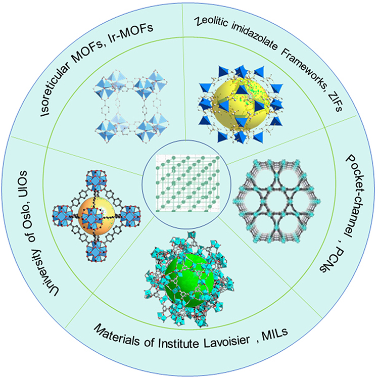 |
Figure 1 Schematic diagram of the five main types of MOFs. |
Unlike conventional methods, which usually employ high temperatures and pressures or solvent environments for the reaction, MOF synthesis methods exploit the structure, shape, and excellent properties of MOFs by precisely regulating the connection between metallic and organic units. Recently, novel synthesis methods using various forms of energy inputs, such as mechanical force, microwaves, ultrasound, and electric fields, have significantly improved the synthesis efficiency, crystal quality, and environmental friendliness of the MOF synthesis process. For example, unique to the MOF synthesis process, hydrothermal/solvent-thermal, ultrasound-assisted, microwave, mechanochemical, microemulsion, and electrochemical methods differ in their thermodynamic states, energy transfer modes, and reaction modulation capabilities.27 The advantages and disadvantages of various synthesis methods are presented in Table 1.
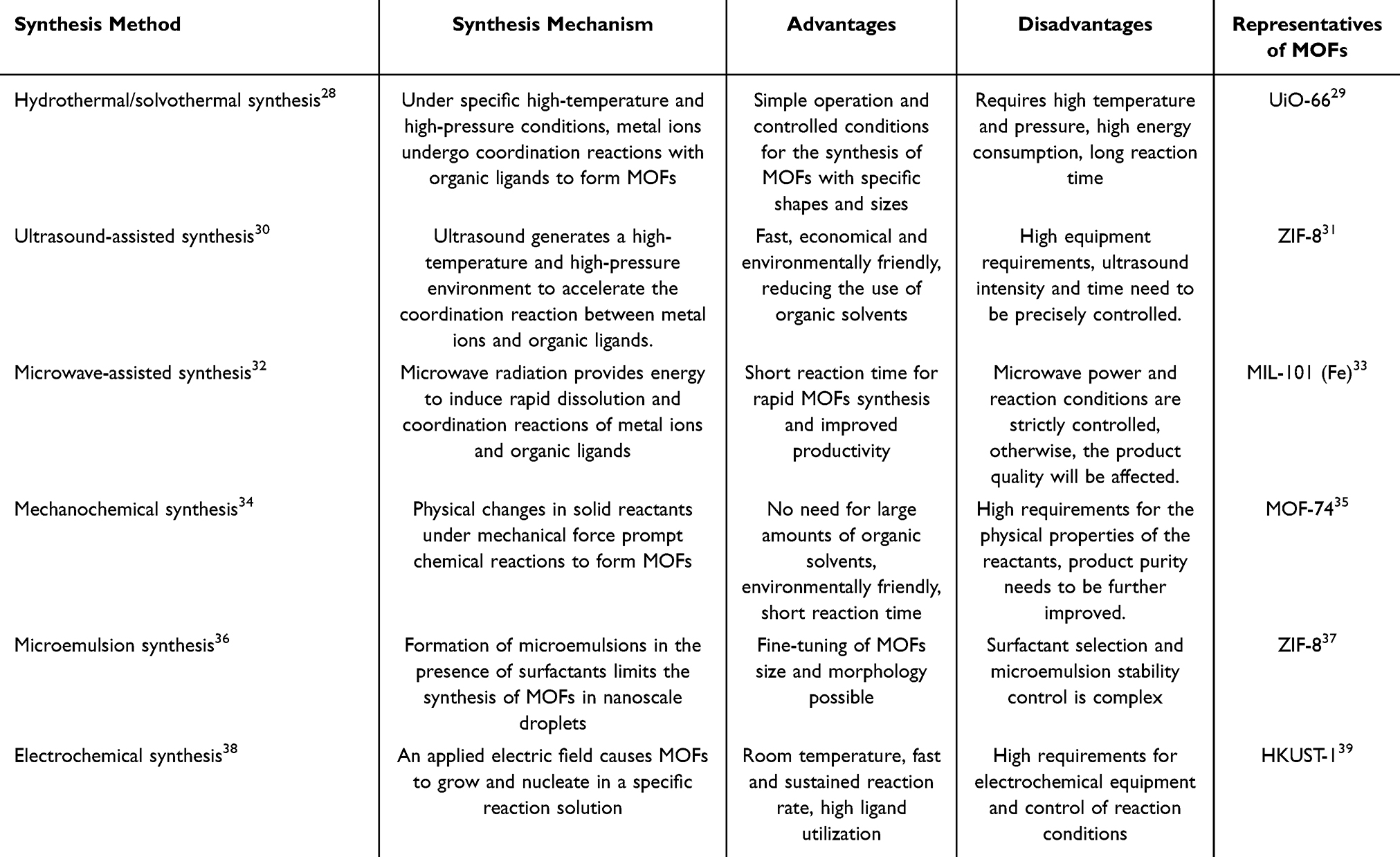 |
Table 1 Mechanisms and Advantages, and Disadvantages of MOFs Synthesis Methods |
Mechanism of Action and Functionalized Design of MOFs for Gene Delivery
MOFs are used for gene transport due to their special structure and properties. MOFs exhibit large specific surface areas and pores, providing sufficient room for numerous genes and enabling successful gene loading.40 Furthermore, the chemical makeup of MOFs is easily changed, and their properties can be adjusted by changing the metal ions or organic ligands according to gene delivery needs.41 In addition, MOF surfaces are easily modified to enhance their interaction with cells and enable the cells to absorb the gene vectors more effectively. Modifying the targeting ligand on the MOF surface enables the targeted delivery of MOFs to specific cells or tissues. This approach could also achieve more precise gene delivery. Moreover, some MOFs exhibit excellent biocompatibility and degradability and are degraded and metabolized slowly inside the body, reducing the potential for toxicity to the organism after gene transfer.42
Advantages of MOFs in Gene Delivery
High Specific Surface Area and Porosity
MOF materials exhibit pores in a fixed arrangement with a high surface area; thus, they can be used as nanoscale reactors or hold various added compositions that increase the overall functionality of the material.43 These special features demonstrate how the shape of MOFs is associated with their performance and enable loading with a substantial quantity of genetic material.44 MOFs can also carry genetic drugs such as siRNAs. The shape of the MOF material helps protect the drug. Biocompatibility and targeting can be improved by surface modification for gene delivery to tumor cells.45 In addition, the higher the porosity of MOFs, the more unsaturated metal sites will be contained within them, increasing the collision and contact rate between organic matter molecules and unsaturated metal sites; thus, increasing the porosity of MOFs can heighten their ability to capture genetic materials.46
Tunable Pore Size
Metal ions and organic ligands can be used to adjust the pore size and surface chemistry of MOFs. This tunability allows the construction of MOFs for different gene-delivery needs. Appropriate adjustments to the specific surface area and pore size of MOFs promote genetic material adsorption. Metals, such as Zn2+, Fe3+, and Zr4+, and organic ligands, such as imidazole and carboxylic acid analogs, can be used to intentionally design the size of the pore and surface charge of the MOFs to match the requirements for different nucleic acid molecule loading.40
MOF Biocompatibility and Low Toxicity
MOFs demonstrate superior biosafety and lower toxicity than traditional viral vectors. MOFs can be designed to be harmless to normal cells through the choice of metal ions and organic ligands. For example, regarding drug delivery systems, MOF nanoparticles in stable complexes with drugs have been evaluated in clinical trials and found to promote in vivo drug release without any obvious allergic reactions or other harmful effects.47
MOF Targeting Capacity
When used as vectors for gene delivery, MOFs exhibit exceptional targeting capacities. MOFs can be modified to identify and bind to target cell surface receptors, improving the accuracy and success rate of targeted gene delivery, thereby reducing off-target damage.48 In oncogene treatment, MOF surfaces have been altered with tumor-specific antibodies to exclusively target tumor cells and improve their efficacy. Tao developed a biologically camouflaged MOF system to improve the efficiency of tumor-targeting siRNA delivery. In their study, MOFs were coated with natural targeting proteins on the tumor cell membrane, helping the MOFs locate and adhere to the tumor cells while maintaining their ability to respond to the tumor environment and ensure siRNA release specifically at the tumor site.47 Similarly, the precise targeting capability of MOFs is not limited to therapeutic applications but extends to diagnostic uses. MOFs functionalized with target-specific antibodies have been successfully employed to capture and detect cancer biomarkers, such as HER2, within electrochemical sensor platforms, demonstrating high accuracy and reliability in targeted biomarker detection.49
Stimulus Responsiveness
MOF nanomaterials have been found to respond to the properties of the tumor microenvironment, demonstrating promise in gene and drug delivery approaches, especially for cancer treatment.50 MOFs can release genetic material in response to certain external stimuli, enabling gene release at the appropriate time and under specific circumstances. This ability improves treatment approaches and enables control over how and when genes are released through the combination of materials, shapes, and elements capable of responding to the surrounding environment. For example, pH-sensitive MOFs can quickly release genetic material into the acidic tumor cell environment, thereby enhancing gene release inside the targeted cells. The speed and method of gene release can be modified by changing the size and shape of MOF pores to meet different treatment needs.51
MOF Versatility
MOFs are efficient gene delivery vehicles for gene therapy and can also be combined with other functions to form multifunctional therapeutic systems. MOFs can be loaded with phototherapy photosensitizers and light-responsive nanostructures to increase tumor ablation. MOF nanosheets are used for photothermal therapy, which uses light energy and near-infrared light to transform and directly destroy the target tumor cells.52,53 MOFs can also be coated with metals such as Mn that increase the MR contrast for MRI.54 In addition, MOFs can be surface-modified with a fluorescent probe, allowing visualization of the drug’s distribution and effects after treatment.55 MOFs can also be used as nanocatalysts to produce ROS in the tumor microenvironment and induce oxidative stress damage in the cancer cells; in combination with gene therapy, this effect could inhibit tumor growth.40,45 The multifunctionality of nanomaterials is further exemplified in catalytic therapy. For instance, single-atom nanozymes (Cu-SAzyme) with atomically dispersed active sites can effectively mimic the antioxidant function of superoxide dismutase (SOD), significantly improving multi-organ damage and survival rates in sepsis models. These findings offer valuable insights for designing smart MOF systems that integrate catalytic activity with drug-delivery capabilities.56
MOF Degradability
MOFs can break down and degrade in vivo after gene delivery, reducing the potential for long-term buildup and associated harm. The controlled breakdown of MOFs is key for their safety and targeting and occurs when bonds break or parts dissolve. For example, MOFs break down into small, safe particles in acidic tumor and lysosome environments, leading to drug release. This effect ensures that the drugs are released where they are needed and prevents harmful buildup.57
MOF materials exhibit good biocompatibility and are the ideal carrier for gene delivery due to their highly specific surface area, precisely adjustable pore structure, and ability to release drugs in response to stimuli.58 Furthermore, MOFs can be designed to achieve targeted delivery through the selection of metals and organic ligands to match specific nucleic acids. The advantages of MOFs are shown in Figure 2. The development of MOFs can address the restrictions associated with traditional vectors, and this approach demonstrates substantial potential in tumor gene therapy and precision therapy.59
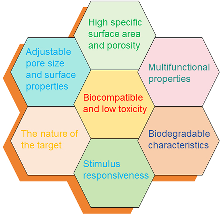 |
Figure 2 Schematic diagram of the advantages of MOFs. |
Gene Delivery Mechanisms
Aperture Encapsulation
Due to the outstanding performance of MOFs, especially their highly specific surface area and high porosity, this approach is highly promising in both medical and material fields.60 These special properties enable the safe and secure loading of MOFs with important genetic materials, such as DNA and RNA, in a process known as pore encapsulation.61 This new encapsulating method enhances the protection of genetic material, allowing it to persist significantly longer than it does with other methods. Researchers can control how the genetic material is inserted by adjusting the MOF size and shape. Controlling the genetic material loaded into MOFs is crucial for gene therapy, targeted drug delivery, and obtaining disease-specific information, enabling the prediction of when the medicine will be released and ensuring it works effectively without causing harm to the patient.62
Surface Mounting
Genetic materials can bind to MOFs via an interaction between the two types of chemical groups on their surface. Genetic materials can adhere to MOFs through electrostatic adsorption or covalent adhesion, helping to keep them securely attached to the MOF surface for gene delivery.63 Electrostatic adsorption is an interaction between charged species. Genetic material is usually negatively charged; thus, it can adhere to MOFs with positively charged surfaces or cationic functional groups via electrostatic interactions. Although this binding mode is relatively weak, it is sufficient to stabilize the attachment of genetic material to the MOF surface and maintain stability during delivery.42
Covalent Binding
Covalent binding is stronger than surface binding because the DNA creates strong bonds with the small molecular groups on the MOF surface, usually necessitating chemical reactions. This approach enables genetic substances to be more strongly attached to the MOF surface during the transportation process, thereby improving the delivery efficiency and stability. Various functional groups, such as amino groups, carboxyl groups, and hydroxyl groups, exist on the surface of MOFs. MOFs can bind with the active groups on DNA or RNA by forming covalent bonds, binding MOFs and genetic material through covalent coupling. Covalent bonds improve the stability of genetic material–MOF binding, providing a “protective shell” for the genetic material, improving its stability, and enabling its release under certain conditions, such as changes in pH values or enzymatic action.62
In Situ Packaging
In-situ Encapsulation (ISE)involves placing the target molecule directly into the MOFs during construction. In this method, the genetic material is mixed with the metal ions and organic ligands during MOF generation.62 ISE relies on the tunability of MOF pores; the metal ions and ligands selected influence the pore size and surface types. Drugs added during the MOF production process are incorporated into the MOF structure by binding to either the metal or organic component.64
Surface Engineering and Functionalization of MOFs
MOFs exhibit considerable advantages in terms of surface functional strategies compared with other nanomaterials. MOFs contain special metals and structures that enable their binding with multiple substances; thus, MOFs can be modified for enhanced cellular uptake and in vivo control. MOF uptake by cells depends mostly on endocytosis pathways (Figure 3). The MOF surface can also be functionalized in many different ways. First, in post-synthesis modification, unoccupied metal sites are modified with molecular coordination. Second, click chemistry can be applied to organic ligands.65 Third, synthetic processes can be used to add regulators as capping agents that are directly incorporated into functional molecules.66 Numerous methods exist for conferring functionality on MOFs, such as the use of surfactants, polyethylene glycol (PEG), substances that enhance uptake, and specific antibodies; thus, MOFs are highly flexible and versatile.67
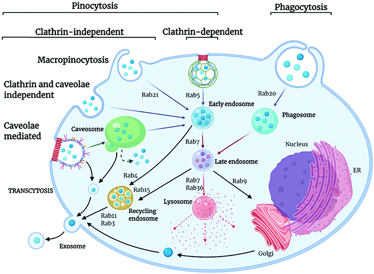 |
Figure 3 The endocytic pathways and roadmap of MOFs.67 Reprinted with permission from.67 |
Surface Modification
PEG Modification
PEG modification is an effective method for altering the surface of materials. When used to modify the surface of MOFs, PEG changes their surface properties, improves their ability to interact with water molecules, and increases their hydrophilicity.68 PEG modification also greatly reduces the in vivo immune response to MOFs. Nonmodified MOFs may be identified and regarded as foreign objects after ingestion and entry into organisms; thus, they will be recognized and eliminated by the immune system through nonspecific recognition after ingestion and the induction of inflammatory and other related immune reactions. PEG is a biocompatible polymer material that can form a “protective shield” on the MOF surface, protecting MOFs from direct contact with the immune system and reducing immunogenicity. Thus, PEG modification improves MOF safety and stability in organisms.69
Chitosan Modifications
Chitosan is a natural polysaccharide with good biocompatibility and biodegradability. Synthetic chitosan-coated MOF materials can serve as effective drug delivery systems. Chitosan coating helps protect the MOFs, prevent MOF breakdown, and reduce drug leakage; thus, this approach enables slow drug release, enhancing its in vivo effects.70 In addition, the combination of chitosan and MOFs results in enhanced antipathogen characteristics, representing a potential novel mechanism for overcoming resistance.
Other Ligand Modification
Changing the MOF surface with amino, carboxyl, and amino acid ligands in a controlled manner can enable the careful adjustment of surface chemical properties. The amino group specifically improves the adsorption performance of acidic gases such as CO2. The carboxyl group can improve the polarity of the pores and their compatibility. Furthermore, the dual nature of amino acids, which have both amino and carboxyl groups, yields better results and enhances the adsorption, catalytic, and separation performance of MOFs.71
Multifunctional
Magnetic Nanoparticle Composite
Combining MOFs with magnetic nanoparticles, such as Fe3O4, enables the construction of magnetic nanoplatforms. This composite material can be used to deliver genes, and a magnetic field can be applied for more precise delivery. The composite material retains the advantages of MOF materials, such as a large specific surface area, adjustable pore size, and good biocompatibility, while also possessing an inherent magnetic response property.72 In gene delivery applications, the addition of Fe3O4 nanoparticles enables the delivery system to be guided by a magnet, allowing the in vivo movement of gene carriers to be controlled by adjusting the intensity or angle of the field. This approach facilitates the delivery of genetic material to a specific location at the desired time, thereby improving gene therapy delivery.73
Drug Codelivery
In addition to gene delivery, MOF materials have substantial potential for combination with other drugs to create new strategies for co-delivering genes and drugs. Researchers have combined anticancer drugs with MOFs to create novel delivery systems that can effectively incorporate anticancer medicine and protect it through the unique structure and other features of MOFs.58 Molecules or groups with targeting functions are introduced to further enhance the accuracy and efficiency of the drug, enabling the above-mentioned composite system to specifically recognize and bind to the surface of tumor cells, providing targeted delivery to the tumor cells. The targeted delivery of drugs via this method reduces damage to nontumor cells, increases the total drug concentration in the tumor tissue, and enhances treatment efficacy.74
Gene Delivery Strategies and Biomedical Applications of MOFs
MOFs are novel nanomaterials with substantial application prospects in gene delivery. MOFs can achieve targeted gene delivery via surface modification or structural design, thereby increasing the accuracy and effectiveness of gene therapy delivery. MOFs can be created as multifunctional vectors that simultaneously load genes and drugs to synergize therapy; furthermore, diagnostic molecules can be included.48 Different genes can also be specially designed and optimized for MOFs to enhance their stability and delivery efficiency (Figure 4). For example, Karimi-Maleh et al successfully synthesized a core–shell Co₃O₄@MOF-74 nanocomposite via a solvothermal method, which exhibited enhanced electrochemical performance and stability, making it highly suitable for applications in biosensing and targeted delivery systems.75 Overall, MOFs exhibit unique advantages and development prospects in gene delivery, presenting substantial opportunities; however, numerous challenges remain in their application.16
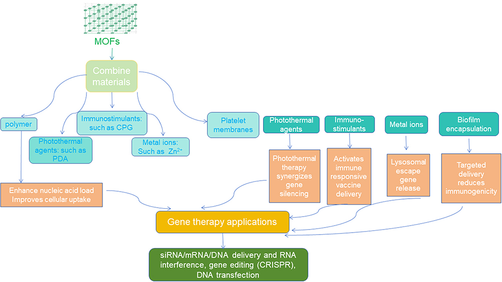 |
Figure 4 Binding of MOFs to other materials and their role in gene delivery. |
Targeted Delivery
Tumor-Targeted Delivery
Tumor therapy with MOFs can be targeted through surface modifications that introduce specific genes, leading to better therapeutic results.76 Saeinasab constructed a specific gene delivery platform for prostate cancer using ZIF-8, a MOF material, to deliver SNHG15 siRNA. The nanoplatform comprised aptamer-PEG-siRNA@ZIF-8 nanoparticles and was constructed via PEGylation modification and EpCAM aptamer binding, remarkably enhancing its stability, cellular uptake, and siRNA targeting capabilities. The experimental results showed that the nanoplatform substantially reduced PC-3 cell proliferation and colony formation in vitro; furthermore, its antitumor activities were found to result from apoptosis induction. In vivo and in vitro experiments demonstrated that the platform could effectively deliver SNHG15 siRNA to inhibit the proliferation of prostate cancer cells and promote apoptosis. In vivo experiments further demonstrated the effectiveness of targeted delivery, with a 78% decrease in tumor volume compared with the non-targeting approach. No obvious systematic toxicity response was observed. Thus, it was demonstrated that ZIF-8 could serve as an effective gene carrier, solving the challenges of instability and the lack of targeted delivery of the siRNA molecule through surface functionalization modification, providing a novel idea for the application of MOFs in tumor gene therapy.77 Similarly, Li designed EGFR-targeted nanoprecursor drugs, termed THE NPs (TPL-HSP conjugates), which release their payload in response to the acidic tumor microenvironment through hydrolyzable ester bonds. These drugs simultaneously inhibit the EGFR/PI3K/AKT pathway, exhibiting a 78% reduction in tumor volume in bladder cancer models, thereby further underscoring the advantages of targeted nanocarriers in solid tumor therapy.78
Mitochondria-Targeted Gene Delivery
Haddad created a system of MOFs modified for improved mitochondrial targeting. Gene therapy for mitochondrial disease can be achieved by modifying target motifs on the MOF surface to enable the specific identification of mitochondria and subsequent delivery of genes. Researchers have developed a customized delivery system that uses a zirconium-based metal–organic framework, known as UIO-66, to enhance the targeting of mitochondria by the anticancer compound Dichloroacetic Acid (DCA). MOFs were synthesized with varying DCA concentration loading and TPP modifications using different experimental methods to enhance the physicochemical properties of the materials. The biological effects of these MOFs were assessed via cytotoxicity assays, super-resolution microscopy, and whole-transcriptome analysis. Experimental results demonstrated that after Triphenylphosphine (TPP) modification, MOFs could target and transport DCA to the perimitochondrial area; a total free drug dose of only 1% was sufficient for the significant induction of mitochondrial morphological fragmentation and activation of apoptosis-related gene expression, which ultimately led to cellular death. These findings could form the experimental basis for mitochondria-targeted gene therapy using MOF material and indicate the feasibility of mitochondria-targeted anticancer therapy.79
Inflammatory Cell-Based Targeted Delivery
MOF materials, as a new type of carrier, can effectively deliver genes, and altering their function via surface modification can overcome biological barriers to targeted delivery, thereby facilitating disease treatment and monitoring. Li developed a method for creating multifunctional nanosystems using MOF material for gene destruction and therapeutic diagnostics. The aforementioned group aimed to address challenges related to nuclease breakdown, targeting, and poor endosomal escape capacity in gene supply systems used for atherosclerosis treatment. They successfully synthesized two iron-based MOF materials, MIL-53 (Fe) and MIL-100 (Fe), and a multifunctional PBMMA-PG polymer bound to the MOFs to improve their stability, targeting capacity, and ability to detach from cells. The modified GAMMA-53 nanoparticles exhibited better gene silencing efficiency and excellent biocompatibility in vitro; furthermore, these nanoparticles demonstrated anti-oxidative properties, reducing the concentration of reactive oxygen species in mouse monocyte macrophage leukemia cells (RAW 264.7). In vivo, GAMMA-53 nanoparticles accurately targeted atherosclerotic plaque sites and greatly improved the detection of these plaques by MRI. Thus, this nanosystem could be used in atherosclerosis diagnostics and has potential applications in atherosclerosis gene therapy.80
Targeted Co-Delivery of siRNA and Cisplatin via RGD-MOF for Synergistic Therapy
Li addressed the challenges of metastasis and chemotherapy resistance in uveal melanoma (UM) by designing a targeted nanodelivery system based on manganese metal-organic frameworks (Mn-MOFs). As shown in Figure 5, the system achieves tumor targeting via integrin αvβ3 through RGD peptides and carries siRNA targeting long non-coding RNA OUM1 and PTPRZ1 alongside cisplatin. The nanoparticles exhibit pH- and reactive oxygen species (ROS)-responsive drug release, enabling synergistic gene silencing, enhanced chemotherapy efficacy, and chemotherapy-dynamic therapy (CDT). Furthermore, by integrating methylene blue (ICG) and Mn²⁺ ions, the system possesses dual-modal fluorescence/MRI imaging capabilities, enabling real-time tracking of tumor drug accumulation and treatment response.81
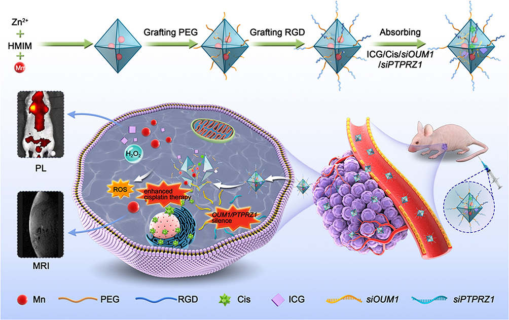 |
Figure 5 Schematic of key steps involved in preparation of porous ICG-COP@MOF-PR (ICG-siOUM1+siPTPRZ1+Cis@MOF-PR) nanostructures and their associated major mechanistic pathways in cancer therapy.81 Reprinted with permission from.81 |
Responsive Release and Adsorption Mechanisms
pH Responsiveness
MOF materials exhibit unique responsive release mechanisms in gene delivery. These materials can respond to different intracellular environments to achieve targeted gene release; pH responsiveness is one of the most frequently investigated mechanisms. The mildly acidic environment of a tumor can cause changes in MOF structure or even degradation, thereby releasing the carried genetic material. This pH-responsive releasing method can make gene delivery more effective and accurate while simultaneously reducing drug release in nontarget areas, thereby protecting normal cells. Cheng designed pH-responsive clusters of metal–organic backbone nanoparticles (Ap-ZIF@SLN#L) to prevent MOF materials from accumulating inefficiently at tumor sites in tumor therapy. Covalently coupling hydrophilic PEG and MMfu to hydrophobic hydroxylated oleylamine (OA), combined with a microfluidics process, further integrated the material into solid lipid nanoshells. These nanoshells were coated with ZIF containing Ap, thus yielding pH-responsive particles. PEG, MMfu, and removal in an acidic tumor environment, along with hydrophobic OA-exposed nanoparticle aggregation, improved tumor cell uptake and accumulation. Ap-ZIF@SLN#L was found to induce the production and release of ROS and Ap and inhibit ZIF autophagy. Cooperation with ZIF promoted tumor cell death and actively infiltrated the tumor microenvironment, inducing the recruitment of M1-type macrophages and CD8+ T cells to the tumor microenvironment These effects substantially inhibited tumor growth and improved the survival rate in mice, and no obvious toxic reactions were observed. These findings provide new ideas and methods for the application of MOFs in tumor therapy. The accumulation of drugs in tumor tissue and their therapeutic effect are the primary areas of focus (Figure 6). Designs focused on pH responsiveness offer valuable references for other applications and may play a significant role in the therapeutic areas of many diseases.82
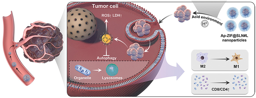 |
Figure 6 Hypothetical mechanistic diagram of the therapeutic effect induced by Ap-ZIF@SLN#L nanoparticles.82 Reprinted with permission from.82 |
In the field of gene delivery, Wang used hydrophilic drugs, nucleic acids, and CRISPR-Cas9 genome editing tools as the basis for creating SMOF NPs with pH-responsive properties. This approach aimed to improve intracellular delivery efficiency. In their study, the researchers prepared pH-responsive SMOF NPs by mixing a water solution containing Zn2+ and the drug with an oil solution, adding a silicon source and an imidazole source to form nanoparticles. The SMOF NPs exhibited a large loading capacity and strong absorption ability. Furthermore, the imidazole groups on the NP surface were protonated in an acidic environment to realize pH-responsive release and endosomal escape by the “proton-sponge effect”. In vitro, the developed MOFs demonstrated higher transfection efficiency and better genome editing capabilities than other commercial reagents. In vivo experiments in mice revealed that SMOF NPs modified with the target ligand ATRA could effectively edit genes in the retinal pigment epithelium. Tests of electrical activity showed that the MOFs are safe, providing an alternative method for delivering genes.83
Sameni investigated the potential of ZIF-8 nanoparticles for gene delivery, with a specific focus on tumor targeting. They found that ZIF-8 could effectively load and protect foreign DNA due to porous structure and pH response characterization, substantially improving gene delivery into sperm cells. The small size of ZIF-8 (~77.8 nm) and its surface charge enabled it to pass through biological membranes; the imidazole ring enabled the particle to escape from endosomes, making it easier for genes to enter the nucleus. The safety of ZIF-8 was also assessed; the particle had no effects on sperm cell survival or embryonic growth, indicating its applicability in vivo.84
Hashemi designed UIO-66@NH2-based MOF nanocarriers and constructed a codelivery system for Epi and Let, overcoming multidrug resistance in breast cancer by loading genes and small-molecule drugs in combination. They prepared drug-loaded nanoparticles via hydrothermal synthesis. In vitro UIO-66-Let/Epi@NH2 significantly increased apoptosis in MCF-7 cells in comparison with the effects of the free drug or the free drug physical mixture. Furthermore, the particles inhibited MCF-7 cell migration and invasion by upregulating the expression of Caspase-3, Caspase-9, and Mfn1 and downregulating the expression of MMP-2 and MMP-9. Furthermore, the particles were less toxic to normal cells (MCF-10A) and could be stably stored at 4°C. Multifunctional carriers such as UIO-66@NH2 can enhance drug targeting and achieve controlled release by utilizing codelivery strategies and exploiting the acidic pH characteristics of the tumor microenvironment.85 This approach could provide new options for overcoming chemotherapy resistance and improving combination therapy.
Li developed a new delivery system using MOF materials to address the issues with complex synthesis, toxic by-products, and low transfection efficiency associated with nonviral gene carriers for delivering plasmid DNAs. They encapsulated pEGFP-C1 into ZIF-8 nanoparticles by bionic mineralization and prepared a ZIF-8/PEI25kD composite vector by coprecipitation. Subsequently, they systematically studied the protection, release, and cell transfection performance of pDNA. The results show that the ZIF-8-PEI 25kD vector significantly enhances the loading ability and protection against enzymatic degradation by improving electrostatic interactions and promoting endosomal release via a pH-responsive release mechanism. Finally, ZIF-8-PEI 25kD achieved a high transfection efficiency in MCF-7 cells, performing comparably to commercial reagents.86
In another study, Salari created a gene delivery system using ZIF-8 MOFs for delivering p53 tumor suppressor genes for cancer treatment. The researchers took full advantage of the structural controllability, high porosity, and pH-responsive degradation of ZIF-8, successfully loading plasmid p53 DNA, which was protected from nucleases and accurately released in the tumor microenvironment via a pH response. The ZIF-8/p53 complex considerably improved the transfection efficiency of the tumor cells in vitro and induced cancer cell apoptosis by restoring p53 protein function in vivo. Further in vivo experiments indicated that this complex effectively and safely targeted and suppressed tumor growth.87 These findings provide novel insights into strategies for developing a multifunctional platform for cancer gene therapy.
Poddar mentioned that siRNA against oncogenes like AR-V7 and CRISPR/Cas9 components against PTEN gene deletions were co-loaded onto ZIF-8 nanoparticles, and prostate-specific membrane antigen (PSMA)-targeting ligands were modified to create a system with targeted delivery capabilities. The platform successfully penetrated the membrane of prostate cancer cells and released the gene-editing tool in response to the acidic environment of the tumor. Furthermore, the platform silenced alternative androgen receptor variants through RNA interference. Together with CRISPR/Cas9, the platform reversed the loss of the PTEN oncogene, inhibited tumor growth, and induced tumor cell apoptosis.88
Near-Infrared Light Responsiveness
MOF vectors can respond to various intra- and extracellular signals during gene delivery, thereby realizing accurate gene release, improving delivery accuracy, and decreasing off-target effects. Notable studies have been conducted on NIR-responsive MOF vectors, revealing that these vectors can achieve spatiotemporally specific gene release under the control of an external light source, providing new strategies for gene therapy. Xu proposed a new microfluidic-assisted MOF biomineralization technique for delivering CRISPR/Cas9 ribonucleoprotein (RNP) to address the problem of low delivery efficiency and hard-to-control delivery resulting from the MOF’s large size, high surface charge, and biosensitivities during gene transfer. By utilizing microfluidics, they biomimetically generated heat-sensitive EuMOFs on photothermally sensitive Prussian blue (PB) templates and effectively encapsulated RNPs as MOFs. PB@RNP-EuMOFs were produced successfully by fine-tuning related microfluidic factors, demonstrating high encapsulation rates, good RNP defensive capabilities, and NIR photostimulation-responsive unloading features. After exposure to NIR light, the nanocarrier released from the endosome faster and reduced GFP expression by 40%. Gene editing activities could be programmed or controlled by changing the light exposure duration, improving editing efficiency compared with the no-light control. This study demonstrates the potential of MOFs as near-infrared light-controlled release vehicles for gene delivery, enhancing the CRISPR/Cas9 RNP delivery system and mitigating off-target effects.89
Application of Gene Delivery Based on MOF Adsorption Mechanism
MOF materials are good carriers for exerting multiple functions. Specific metal and organic ligand incorporation can result in MOFs that effectively load and protect gene-based drugs. Furthermore, the porosity of MOF structures provides sufficient space to co-load small molecules for treating various diseases. Ringaci designed a multifunctional nanocarrier with a core-shell structure to address the challenges of low drug loading capacity and the lack of multidrug codelivery capacity in traditional gene delivery MOFs. The vector contained a magnetic Fe3O4 core covered by a MIL-100 (Fe) network and coated with carboxymethyl dextran (CMD) and PEI on the surface, which enabled the gene to be loaded efficiently onto the vector and small molecules to bind with MIL-100 (Fe) in the pores.90 The vector achieved a transfection efficiency of 68% in HEK293 cells and successfully codelivered Nile blue with plasmids of varying sizes. Fluorescent labeling revealed that both components overlapped significantly in the cells. In vivo experiments demonstrated that this system could effectively express genes and deliver small molecules in mouse lungs. Furthermore, the system exhibited good biocompatibility and safety, indicating its potential as a novel solution and platform for gene therapy and delivery.
Immune Regulation
Gene Delivery and Immune Modulation Synergize to Enhance the Immune Response
MOFs can effectively carry and protect genetic material. Moreover, they can be modified to add targeting molecules or immune factors, enhancing treatment efficacy. MOFs in combination with gene and immunotherapy applications are promising. For instance, Yola developed a bimetallic Ni/Cu–MOF-based immunosensor specifically designed for detecting TNF‑α, achieving an ultra-low detection limit of 2.0 fg/mL, thereby highlighting the significant potential of MOFs in high-sensitivity biomedical diagnostics.91 Feng developed a MOF-based gene delivery method that optimized the therapeutic effect by precisely regulating immune responses in the Tumor Microenvironment (TME) to address the low efficacy of cancer immunotherapy. They explored the potential of MOFs in conjunction with gene delivery and cancer immunotherapy, focusing on the use of MOFs as multifunctional nanocarriers for gene drugs and immunomodulation. pH-responsive, glutathione-sensitive MOFs targeting the TME were effectively developed for the successful loading of siRNAs and immunomodulatory nucleotides. Specific release was achieved through the incorporation of aptamer or targeted ligand modification into the MOFs.92
Chemo-Acquired Combined Immunotherapy Using MOFs
Zhao used a mineralized MOF nanofabrication approach to solve the problem of lymphopresence and immunosuppression of CD8+ T cells in conventional immunotherapy. They used the biomineralization performance of MOFs to deliver lysosomal-targeting drugs to the tumor-infiltrating T cells more accurately. This approach remodeled the acidic environment in lysosomes and reconstituted their metabolism. Moreover, metal ions such as Zn2+ released by MOFs could activate the STING interferon pathway, promote dendritic cell maturation, and facilitate antigen presentation, thereby achieving a synergistic immune effect between innate and adaptive immunity. This method significantly enhanced the T cell-mediated tumor-killing effect through a dual-control mechanism, resulting in tumor–tumor microenvironment remodeling and tumor growth inhibition in a melanoma model. These findings indicate the potential of novel nano-immunomodulation approaches to overcome solid tumors.93 Similarly, Chen demonstrated that oncolytic viruses (eg, T-Vec) are able to release tumor-associated antigens (TAAs) through tumor lysis, which in turn activates dendritic cells (DCs), promoting their maturation and the recruitment of CD8⁺ T cells, as well as reversing the immunosuppressive tumor microenvironment. These findings provide a strong rationale for combining MOFs with immune activators in combinatorial therapeutic strategies.94
MOF-Based Bispecific Antibody Delivery and T-Cell Redirection Therapy
Zhao developed a gene delivery system through MIL-88A metal–organic framework nanoparticles and MC to achieve effective in vivo expression of anti-CD3/anti-EpCAM BiTE. The loading of MC molecules onto MOF nanoparticles by metal-phosphate bonds and electrostatic interaction was demonstrated via X-ray photoelectron spectroscopy (XPS) and dynamic light scattering (DLS) analysis, and the intraperitoneal injection of MOF/MC suspensions into the peritoneum in mice revealed excellent transfection effects with low cytotoxicity. In the intraperitoneal xenograft mouse model constructed with SKOV3 ovarian cells, the researchers indicated that MOFs/MC. BiTE greatly hindered tumor growth and elevated the average survival duration of the mice. Moreover, the MOFs/MC system was found to be a safe and efficacious delivery approach for BiTE immunotherapy of ovarian cancer and has substantial potential clinical applications.95
Novel Gene Delivery
Oral Delivery
In a study, Jiang mentioned that MOFs are applied in oral gene therapy to protect the stability of genes in the stomach, intestines, or other regions of the gastrointestinal tract through a specific structural form. Researchers have carefully investigated MOFs as novel tools for oral gene delivery that address the key issue of low efficiency in regular oral gene therapy. Orally administered genes have difficulties in withstanding the acidic atmosphere of the gastrointestinal tract, resisting enzymatic attack, and traversing the mucous barrier. However, the large specific surface area of MOFs and the ability to customize their porosity and surface function enable structural changes or degradation in acidic environments, initiating drug release. Thus, acid-reactive structures have been constructed to safeguard the gene-based drug with surface modifications enabling targeted delivery and release. The design strategies used when constructing MOFs, including pore size control, biomimicking mineralization, and probiotic metabolite incorporation, illustrate their potential to improve gene-loading efficiency, resist enzymatic degradation, and facilitate cellular uptake. Furthermore, using computational systems to predict the effects of materials, AI can help construct better MOFs to increase the speed of delivery system design and development of personalized treatments.96
Surface-Engineered Delivery
Wu addressed the challenge of poor blood–brain barrier (BBB) penetration, which limits drug delivery for glioblastoma (GBM) treatment, particularly for chemotherapeutic agents such as docetaxel (DTX). They developed a zeolitic imidazolate framework-8 (ZIF-8) based nanosystem by engineering its surface with a 15‑amino acid peptide RVG15 (via RVG15–PEG conjugates), resulting in the construct RVG15–PEG@DTX@ZIF-8. This modification enables specific targeting of nicotinic acetylcholine receptors (nAChRs) on both brain endothelial and GBM cells, facilitating efficient BBB traversal and targeted accumulation within GBM tissue. In orthotopic GBM mouse models, the system significantly inhibited tumor growth, suppressed metastasis, prolonged the survival of tumor-bearing mice, and showed good biocompatibility, providing a promising surface-engineered MOF-based delivery strategy for GBM therapy.97 Zhang aimed to overcome key limitations in osteoarthritis (OA) therapy, including inadequate response to monotherapy, suboptimal therapeutic outcomes due to poor bioavailability and short retention of agents, and the practical constraints of gene delivery. They constructed a surface-engineered, pH-responsive metal–organic framework (MOF) system (MIL-101-NH2@CCM-siRNA, denoted as MCS NPs) for co-delivering the anti-inflammatory drug curcumin (CCM) and HIF-2α siRNA (as illustrated in Figure 7). Using MIL-101-NH2 as the core carrier, CCM was encapsulated via pore adsorption, while siRNA was loaded through surface coordination. The system was further functionalized with hyaluronic acid (HA) to enhance nanoparticle dispersibility, hydrophilicity, and cartilage lubrication. This surface engineering strategy allowed effective protection of siRNA from enzymatic degradation, promoted lysosomal escape, and enabled pH-responsive release of both cargoes in the acidic OA microenvironment. Consequently, the system synergistically silenced HIF-2α expression, inhibited inflammatory responses, and reduced cartilage degeneration. Both in vitro and in vivo studies demonstrated the therapeutic potential of this platform for OA treatment.98
 |
Figure 7 Preparation procedure of MIL-101-NH2@CCM-siRNA nanoparticles and schematic illustration of MIL-101-NH2@CCM-siRNA nanoparticles (premixed with the HA solution) for OA therapy.98 Reprinted with permission from.98 |
Gene Transfer Therapy
Gene therapy methods include gene transfer and editing approaches. Gene transfer refers to the use of vectors to deliver exogenous genes into host cells. The genes expressed then exert a therapeutic effect inside the cell. mRNA transfer is a type of gene transfer that introduces exogenous mRNA into the cell, resulting in therapeutic effects due to the synthesis of therapeutic proteins through the cell’s translation system. MOFs, as novel nanocarriers, offer significant advantages in the field of mRNA delivery. They protect mRNA from being broken down by enzymes, allowing RNA delivery. Lawson created mRNA–PEI@ZIF-8 composite granules in which PEI had been combined with mRNA before being placed inside the ZIF-8 shell layer. PEI is positively charged and can combine with the negatively charged mRNA to form a complex, thereby protecting the mRNA from nuclease degradation and enhancing its binding to the cell membrane. Thus, this approach improves mRNA cell entry. ZIF-8 has good biocompatibility and tunable pore size and pore structure. The composite particles exhibit better stability and less mRNA leakage into biological media. Thus, the RNA is stably transferred in vivo and is slowly released, prolonging the duration of action of RNA in vivo and improving the therapeutic effect. After the composite particle enters the cell, the ZIF-8 layer slowly disintegrates and releases the mRNA–PEI compound. Subsequently, the mRNA is separated and enters the cytoplasm, where it uses the cell’s translation system to produce the corresponding protein. For example, an mRNA that encodes antigenic proteins results in the expression of these proteins after cell delivery, causing an immune system response; ie, when the antigenic protein-encoding RNA enters the cell, the cell makes and exposes the antigenic protein, and the body’s system attacks the cell, producing the desired response.99
Future Prospects
Although MOF systems are promising for gene delivery, numerous challenges remain in their application. First, biostability and degradability are concerns. If the MOFs are not stable in vivo, the gene cargo could be released too early, or the MOFs could break apart and deliver fewer genes. Second, cell absorption rates and toxicity problems cannot be ignored—the metal ion and organic components of MOFs are potentially cytotoxic. Third, gene delivery is challenging. Genetic vectors are highly unstable and can be degraded by in vivo nucleases, the cellular uptake efficiency is low, and intracellular escape is challenging. Furthermore, obstacles in intranuclear transportation complicate the transfection process, resulting in limited delivery. These issues become more challenging when MOF materials are employed as carriers for genetic material. The biocompatibility issue of MOF materials may contradict the needs of cellular uptake and intranuclear transport for gene delivery, affecting the efficiency of gene release. Furthermore, it is difficult to regulate gene expression precisely while maintaining material stability. Further studies investigating the design and optimization of materials are necessary to address the aforementioned challenges and promote the development and application of MOFs in the field of gene therapy.
MOF nanomaterials show substantial potential in gene delivery applications. The development of novel and stable MOF nanomaterials has become a key focus in the research and development of gene delivery systems. By carefully choosing the right metal ions and organic ligands and using new development methods, we can greatly enhance the stability of MOFs in biological settings. Furthermore, incorporating surface changes and targeting MOF delivery systems represents additional improvements. Equipping MOFs with the ability to recognize cells enables them to enter cells more effectively and target the desired sites. Changing the surface and chemistry of MOFs makes them safer in vivo. Disguising MOFs with natural coatings helps prevent immune system attacks. These changes can make MOF delivery systems more useful for gene therapy.
Summary
MOF materials could be very useful for gene therapy due to their special properties. They have a large surface area and numerous pores, which help them transport genetic material. Their pore size and surface can be changed to meet different gene delivery needs. In addition, MOFs are safe and exhibit low toxicity due to their excellent biocompatibility. In addition to their targeting ability, responses to stimuli, multifunctionality, and degradability, MOFs demonstrate substantial advantages compared with other materials for gene delivery. MOFs can be designed to release genes at specific times through surface or structural changes, improving the accuracy and efficiency of gene therapy. MOFs can also act as multifunctional carriers that can release genes, drugs, and detection molecules simultaneously to achieve synergistic therapy. MOFs can be designed to carry different genes (like siRNA, mRNA, and plasmid DNA). These factors improve the stability and efficiency of gene delivery. Overall, MOFs offer unique advantages for gene therapy and provide novel opportunities for progress in this field; however, several challenges remain in their application.
Acknowledgments
This work was supported by the Science and Technology Development Project of Jilin Province (No.20220204038YY).
Disclosure
None of the authors have any conflicts of interest for this work.
References
1. Chancellor D, Barrett D, Nguyen-Jatkoe L, et al. The state of cell and gene therapy in 2023. Mol Ther. 2023;31(12):3376–3388. doi:10.1016/j.ymthe.2023.11.001
2. Shchaslyvyi AY, Antonenko SV, Tesliuk MG, et al. Current state of human gene therapy: approved products and vectors. Pharmaceuticals. 2023;16(10):1416. doi:10.3390/ph16101416
3. Butt MH, Zaman M, Ahmad A, et al. Appraisal for the potential of viral and nonviral vectors in gene therapy: a review. Genes. 2022;13(8):1370. doi:10.3390/genes13081370
4. Ashrafizadeh M, Zarrabi A, Bigham A, et al. (Nano)platforms in breast cancer therapy: drug/gene delivery, advanced nanocarriers and immunotherapy. Med Res Rev. 2023;43(6):2115–2176. doi:10.1002/med.21971
5. Luan XK, Jin X, Li XN, et al. Physiologically stable, epitope-imprinted, and double-gated metal-organic framework drug delivery system for tumor-targeted combination therapy. article. ACS Appl Mater Interfaces. 2025;17(23):34819–34832. doi:10.1021/acsami.5c06175
6. Guo Y, Huang J, Lin M, et al. Nano particle loaded EZH2 inhibitors: Increased efficiency and reduced toxicity for malignant solid tumors. J Transl Int Med. 2025;13(3): 156–169. doi:10.1515/jtim-2025-0020
7. Safarkhani M, Dana N, Taghavimandi F, et al. Exploring metal-organic frameworks in gene delivery: from prostate to lung therapeutics. Article. Appl Mater Today. 2024;41:
8. Tang M, Ni J, Yue Z, et al. Polyoxometalate-nanozyme-integrated nanomotors (POMotors) for self-propulsion-promoted synergistic photothermal-catalytic tumor therapy. Angew Chem Int Ed Engl. 2024;63(6):e202315031. doi:10.1002/anie.202315031
9. Tam CWL, Yam JWP . Harnessing the potential of small extracellular vesicle biomarkers for cancer diagnosis and prognosis with advanced analytical technologies. J Transl Int Med. 2025;13(3): 187–200. doi:10.1515/jtim-2025-0019
10. Yola ML, Atar N. Amperometric galectin-3 immunosensor-based gold nanoparticle-functionalized graphitic carbon nitride nanosheets and core-shell Ti-MOF@COFs composites. Nanoscale. 2020;12(38):19824–19832. doi:10.1039/d0nr05614f
11. Kabiri S, Rahimi R, Mozafari MR, et al. Porphyrin-based MOFs for gene delivery in cancer therapy: recent advances and progress. Review; Early access. Curr Pharm Design. 2025:16. doi:10.2174/0113816128359818250407020852.
12. Chai W, Chen X, Liu J, et al. Recent progress in functional metal-organic frameworks for bio-medical application. Regen Biomater. 2024;11:rbad115. doi:10.1093/rb/rbad115
13. Yanfei L, Ting J, Zhenbao L. Metal-organic frameworks for bioimaging: strategies and challenges. Nanotheranostics. 2022;6(2):143–160. doi:10.7150/ntno.63458
14. Qian Y, Zhang F, Pang H. A review of MOFs and their composites‐based photocatalysts: synthesis and applications. Adv Funct Mater. 2021;31(37):2104231. doi:10.1002/adfm.202104231
15. Liu K-G, Bigdeli F, Panjehpour A, et al. Metal organic framework composites for reduction of CO2. Coord Chem Rev. 2023;493:215257. doi:10.1016/j.ccr.2023.215257
16. Păun C, Motelică L, Ficai D, et al. Metal-organic frameworks: versatile platforms for biomedical innovations. Materials. 2023;16(18). doi:10.3390/ma16186143
17. Khafaga DSR, El-Morsy MT, Faried H, et al. Metal-organic frameworks in drug delivery: engineering versatile platforms for therapeutic applications. RSC Adv. 2024;14(41):30201–30229. doi:10.1039/d4ra04441j
18. Salehipour M, Nikpour S, Rezaei S, et al. Safety of metal–organic framework nanoparticles for biomedical applications: an in vitro toxicity assessment. Inorg Chem Commun. 2023;152:110655. doi:10.1016/j.inoche.2023.110655
19. Dastneshan A, Rahiminezhad S, Mezajin MN, et al. Cefazolin encapsulated UIO-66-NH2 nanoparticles enhance the antibacterial activity and biofilm inhibition against drug-resistant S. aureus: in vitro and in vivo studies. Chem Eng J. 2023;455:140544. doi:10.1016/j.cej.2022.140544
20. Zhang R, Karami AM, Huang Q, et al. UiO-based platforms in biomedicine: advanced nanovehicles for effective treatment. Mater Today Chem. 2025;45:102645. doi:10.1016/j.mtchem.2025.102645
21. Han D, Hao L, Chang M, et al. Facile synthesis of Co-Ni layered double hydroxides nanosheets wrapped on a prism-like metal-organic framework for efficient oxygen evolution reaction. J Colloid Interface Sci. 2023;634:14–21. doi:10.1016/j.jcis.2022.12.026
22. Yang S, Li X, Zeng G, et al. Materials institute lavoisier (MIL) based materials for photocatalytic applications. Coord Chem Rev. 2021;438:213874. doi:10.1016/j.ccr.2021.213874
23. Lin ZQ, Liao DH, Jiang CY, et al. Current status and prospects of MIL-based MOF materials for biomedicine applications. Rsc Medl Chem. 2023;14(10):1914–1933. doi:10.1039/d3md00397c
24. Han Y, Wang F, Zhang J. Design and syntheses of hybrid zeolitic imidazolate frameworks. Coord Chem Rev. 2022;471:214759. doi:10.1016/j.ccr.2022.214759
25. Cai MR, Liang WL, Wang KX, et al. Aperture modulation of isoreticular metal organic frameworks for targeted antitumor drug delivery. ACS Appl Mater Interfaces. 2022;14(32):36366–36378. doi:10.1021/acsami.2c07450
26. Wang D, Yao H, Ye J, et al. Metal-Organic Frameworks (MOFs): classification, Synthesis, Modification, and Biomedical Applications. Small. 2024;20(47):e2404350. doi:10.1002/smll.202404350
27. Zhang Q, Yan S, Yan X, et al. Recent advances in metal-organic frameworks: synthesis, application and toxicity. Sci Total Environ. 2023;902:165944. doi:10.1016/j.scitotenv.2023.165944
28. Huo XW, Yu SW, Xiao SJ, et al. Research progress of metal-organic framework materials in adsorption separation. Cailiao Gongcheng-J Mat Eng. 2021;49(7):10–20. doi:10.11868/j.issn.1001-4381.2020.000559
29. Lucatero E, Bashiri R, Synthesis SMC. Characterization, and evaluation of metal-organic frameworks for oxidative desulfurization: an integrated experiment. Article. J Chem Educ. 2024;101(8):3428–3433. doi:10.1021/acs.jchemed.4c00297
30. Senthil Raja D, Tsai DH. Recent advances in continuous flow synthesis of metal-organic frameworks and their composites. Chem Commun. 2024;60(65):8497–8515. doi:10.1039/d4cc02088j
31. Yi J, Lee G, Park SS. Solvent-induced structural rearrangement in ultrasound-assisted synthesis of metal-organic frameworks. Article; Early Access. Small Methods. 2024;8(12):e2400363. doi:10.1002/smtd.202400363
32. Yang J, Yang YW. Metal–organic frameworks for biomedical applications. Small. 2020;16(10):1906846. doi:10.1002/smll.201906846
33. Dong YN, Hu TD, Pudukudy M, et al. Influence of microwave-assisted synthesis on the structural and textural properties of mesoporous MIL-101(Fe) and NH2-MIL-101(Fe) for enhanced tetracycline adsorption. Mater Chem Phys. 2020;251:
34. Afshariazar F, Morsali A. The unique opportunities of mechanosynthesis in green and scalable fabrication of metal-organic frameworks. J Mater Chem A. 2022;10(29):15332–15369. doi:10.1039/d2ta02699f
35. Beamish-Cook J, Shankland K, Murray CA, et al. Insights into the mechanochemical synthesis of MOF-74. Cryst Growth Des. 2021;21(5):3047–3055. doi:10.1021/acs.cgd.1c00213
36. Vega JF, Poupard MFN, González RM, et al. Effect of the surfactant on the synthesis of metal-organic framework structures. Mater Proceed. 2020;4(1):45. doi:10.3390/IOCN2020-07983
37. Lee J, Park S, Woo S, et al. Soft seed-mediated dimensional control of metal-organic framework nanocrystals through oil-in-water microemulsions. Inorg Chem Front. 2023;10(24):7146–7154. doi:10.1039/d3qi01567j
38. Gatou M-A, Vagena I-A, Lagopati N, et al. Functional MOF-based materials for environmental and biomedical applications: a critical review. Nanomaterials. 2023;13(15):2224. doi:10.3390/nano13152224
39. Kabir M, Rahman M, Islam T, et al. Recent advances of HKUST-1 metal-organic frameworks in the biomedical applications: a comprehensive review. Review Chem Eng J. 2025;513:
40. Li B, Ashrafizadeh M, Jiao T. Biomedical application of metal-organic frameworks (MOFs) in cancer therapy: stimuli-responsive and biomimetic nanocomposites in targeted delivery, phototherapy and diagnosis. Int J Biol Macromol. 2024;260(Pt 2):129391. doi:10.1016/j.ijbiomac.2024.129391
41. Liu B, Jiang M, Zhu D, et al. Metal-organic frameworks functionalized with nucleic acids and amino acids for structure-and function-specific applications: a tutorial review. Chem Eng J. 2022;428:131118. doi:10.1016/j.cej.2021.131118
42. He Y, Li D, Wu L, et al. Metal‐organic frameworks for gene therapy and detection. Adv Funct Mater. 2023;33(12):2212277. doi:10.1002/adfm.202212277
43. Lin RB, Zhang Z, Chen B. Achieving high performance metal-organic framework materials through pore engineering. Acc Chem Res. 2021;54(17):3362–3376. doi:10.1021/acs.accounts.1c00328
44. Wang WJ, Chen D, Li FY, et al. Metal-organic-framework-based materials as platforms for energy applications. Review. Chem. 2024;10(1):86–133. doi:10.1016/j.chempr.2023.09.009
45. Ma Y, Liao J, Cheng H, et al. Advanced gene therapy system for the treatment of solid tumour: a review. Mater Today Bio. 2024;27:101138. doi:10.1016/j.mtbio.2024.101138
46. Sun Y, Zheng L, Yang Y, et al. Metal-organic framework nanocarriers for drug delivery in biomedical applications. Nanomicro Lett. 2020;12(1):103. doi:10.1007/s40820-020-00423-3
47. Tao T, Rehman SU, Xu S, et al. A biomimetic camouflaged metal organic framework for enhanced siRNA delivery in the tumor environment. J Mater Chem B. 2024;12(17):4080–4096. doi:10.1039/d3tb02827e
48. Khulood MT, Jijith US, Naseef PP, et al. Advances in metal-organic framework-based drug delivery systems. Int J Pharm. 2025;673:125380. doi:10.1016/j.ijpharm.2025.125380
49. Yola ML. Sensitive sandwich-type voltammetric immunosensor for breast cancer biomarker HER2 detection based on gold nanoparticles decorated Cu-MOF and Cu2ZnSnS4NPs/Pt/g-C3N4 composite. Mikrochim Acta. 2021;188(3):78. doi:10.1007/s00604-021-04735-y
50. Li B, Yao X, Li J, et al. A tumor microenvironment-activated metal-organic framework-based nanoplatform for amplified oxidative stress-induced enhanced chemotherapy. J Biol Chem. 2023;299(1):102742. doi:10.1016/j.jbc.2022.102742
51. Xie H, Liu X, Huang Z, et al. Nanoscale zeolitic imidazolate framework (ZIF)–8 in cancer theranostics: current challenges and prospects. Cancers. 2022;14(16):3935. doi:10.3390/cancers14163935
52. Chen X, Wang S, Chen Y, et al. Non-invasive activation of intratumoural gene editing for improved adoptive T-cell therapy in solid tumours. Nat Nanotechnol. 2023;18(8):933–944. doi:10.1038/s41565-023-01378-3
53. Huang H, Yuan G, Xu Y, et al. Photoacoustic and magnetic resonance imaging-based gene and photothermal therapy using mesoporous nanoagents. Bioact Mater. 2022;9:157–167. doi:10.1016/j.bioactmat.2021.07.025
54. Qin Q, Yang M, Shi Y, et al. Mn-doped Ti-based MOFs for magnetic resonance imaging-guided synergistic microwave thermal and microwave dynamic therapy of liver cancer. Bioact Mater. 2023;27:72–81. doi:10.1016/j.bioactmat.2023.03.019
55. Li HH, Yang WT, Pan QH. Integration of fluorescent probes into metal-organic frameworks for improved performances. RSC Adv. 2020;10(56):33879–33893. doi:10.1039/d0ra04907g
56. Yang J, Zhang R, Zhao H, et al. Bioinspired copper single-atom nanozyme as a superoxide dismutase-like antioxidant for sepsis treatment. Exploration. 2022;2(4):20210267. doi:10.1002/exp.20210267
57. Chen M, Dong R, Zhang J, et al. Nanoscale metal-organic frameworks that are both fluorescent and hollow for self-indicating drug delivery. ACS Appl Mater Interfaces. 2021;13(16):18554–18562. doi:10.1021/acsami.1c02045
58. Gulati S, Choudhury A, Mohan G, et al. Metal-organic frameworks (MOFs) as effectual diagnostic and therapeutic tools for cancer. J Mater Chem B. 2023;11(29):6782–6801. doi:10.1039/d3tb00706e
59. Ge X, Wong R, Anisa A, et al. Recent development of metal-organic framework nanocomposites for biomedical applications. Biomaterials. 2022;281:121322. doi:10.1016/j.biomaterials.2021.121322
60. Abánades Lázaro I, Chen X, Ding M, et al. Metal–organic frameworks for biological applications. Nat Rev Method Primers. 2024;4(1):42. doi:10.1038/s43586-024-00320-8
61. Tong PH, Zhu L, Zang Y, et al. Metal-organic frameworks (MOFs) as host materials for the enhanced delivery of biomacromolecular therapeutics. Chem Commun. 2021;57(91):12098–12110. doi:10.1039/d1cc05157a
62. Soriano-Giles G, Giles-Mazon EA, Lopez N, et al. Metal organic frameworks (MOFs) as non-viral carriers for DNA and RNA delivery: a review. Rev Inorganic Chem. 2023;43(2):201–219. doi:10.1515/revic-2022-0004
63. Moharramnejad M, Ehsani A, Shahi M, et al. MOF as nanoscale drug delivery devices: synthesis and recent progress in biomedical applications. J Drug Delivery Sci Technol. 2023;81:104285. doi:10.1016/j.jddst.2023.104285
64. Saeb MR, Rabiee N, Mozafari M, et al. Metal–organic frameworks (MOFs) for cancer therapy. Materials. 2021;14(23):7277. doi:10.3390/ma14237277
65. Bednarek C, Schepers U, Thomas F, et al. Bioconjugation in materials science. Adv Funct Mater. 2024;34(20):2303613. doi:10.1002/adfm.202303613
66. Tran VA, Thuan Le V, Doan VD, et al. Utilization of functionalized metal-organic framework nanoparticle as targeted drug delivery system for cancer therapy. Pharmaceutics. 2023;15(3):931. doi:10.3390/pharmaceutics15030931
67. Linnane E, Haddad S, Melle F, et al. The uptake of metal-organic frameworks: a journey into the cell. Chem Soc Rev. 2022;51(14):6065–6086. doi:10.1039/d0cs01414a
68. Lawson HD, Walton SP, Chan C. Metal-organic frameworks for drug delivery: a design perspective. ACS Appl Mater Interfaces. 2021;13(6):7004–7020. doi:10.1021/acsami.1c01089
69. Chen X, Zhuang Y, Rampal N, et al. Formulation of metal-organic framework-based drug carriers by controlled coordination of methoxy peg phosphate: boosting colloidal stability and redispersibility. J Am Chem Soc. 2021;143(34):13557–13572. doi:10.1021/jacs.1c03943
70. Han D, Li Y, Liu X, et al. Rapid bacteria trapping and killing of metal-organic frameworks strengthened photo-responsive hydrogel for rapid tissue repair of bacterial infected wounds. Chem Eng J. 2020;396:125194. doi:10.1016/j.cej.2020.125194
71. Zhao Q, Sun Y, Zhang J, et al. Mixed matrix membranes incorporating amino-functionalized ZIF-8-NH2 in a carboxylic polyimide for molecularly selective gas separation. J Membr Sci. 2024;693:122326. doi:10.1016/j.memsci.2023.122326
72. Liu L, Qi G, Wang M, et al. Construction of intelligent response gene vector based on MOF/Fe3O4/AuNRs for tumor-targeted gene delivery. Int J Biol Macromol. 2024;277(Pt 3):134313. doi:10.1016/j.ijbiomac.2024.134313
73. Abdelhamid HN, Dowaidar M, Hällbrink M, et al. Gene delivery using cell penetrating peptides-zeolitic imidazolate frameworks. Microporous Mesoporous Mater. 2020;300:110173. doi:10.1016/j.micromeso.2020.110173
74. Mohammed MRS, Ahmad V, Ahmad A, et al. Prospective of nanoscale metal organic frameworks NMOFs for cancer therapy. Semi Cancer Biol. 2021;69:129–139. doi:10.1016/j.semcancer.2019.12.015
75. Karimi-Maleh H, Yola ML, Atar N, et al. A novel detection method for organophosphorus insecticide fenamiphos: molecularly imprinted electrochemical sensor based on core-shell Co3O4@MOF-74 nanocomposite. J Colloid Interface Sci. 2021;592:174–185. doi:10.1016/j.jcis.2021.02.066
76. X-f Z, Sun X. Nanomedicines based on nanoscale metal-organic frameworks for cancer immunotherapy. Acta Pharmacol Sin. 2020;41(7):928–935. doi:10.1038/s41401-020-0414-6
77. Saeinasab M, Iranpour S, Hosseini-Giv N, et al. Tumor-targeted delivery of SNHG15 siRNA using a ZIF-8 nanoplatform: towards a more effective prostate cancer therapy. Int J Biol Macromol. 2024;259(Pt 1):129233. doi:10.1016/j.ijbiomac.2024.129233
78. Li G, Song Z, Ru Y, et al. Small-molecule nanoprodrug with high drug loading and EGFR, PI3K/AKT dual-inhibiting properties for bladder cancer treatment. Exploration. 2023;3(5):20220141. doi:10.1002/exp.20220141
79. Haddad S, Lázaro IA, Fantham M, et al. Design of a functionalized metal-organic framework system for enhanced targeted delivery to mitochondria. J Am Chem Soc. 2020;142(14):6661–6674. doi:10.1021/jacs.0c00188
80. Li S, Gao H, Wang H, et al. Tailored polysaccharide entrapping metal-organic framework for RNAi therapeutics and diagnostics in atherosclerosis. Bioact Mater. 2025;43:376–391. doi:10.1016/j.bioactmat.2024.08.041
81. Li Y, Li F, Pan H, et al. Targeted OUM1/PTPRZ1 silencing and synergetic CDT/enhanced chemical therapy toward uveal melanoma based on a dual-modal imaging-guided manganese metal-organic framework nanoparticles. J Nanobiotechnol. 2022;20(1):472. doi:10.1186/s12951-022-01643-y
82. Cheng R, Jiang L, Gao H, et al. A pH-responsive cluster metal-organic framework nanoparticle for enhanced tumor accumulation and antitumor effect. Adv Mater. 2022;34(42):e2203915. doi:10.1002/adma.202203915
83. Wang Y, Shahi PK, Xie R, et al. A pH-responsive silica-metal-organic framework hybrid nanoparticle for the delivery of hydrophilic drugs, nucleic acids, and CRISPR-Cas9 genome-editing machineries. J Control Release. 2020;324:194–203. doi:10.1016/j.jconrel.2020.04.052
84. Sameni M, Moradbeigi P, Hosseini S, et al. ZIF-8 nanoparticle: a valuable tool for improving gene delivery in sperm-mediated gene transfer. Biol Proced Online. 2024;26(1):4. doi:10.1186/s12575-024-00229-2
85. Hashemi A, Hayat-Gheibi SR, Baghbani-Arani F. Co-delivery of epirubicin and letrozole using a metal-organic framework nanoparticle in breast cancer therapy. J Drug Delivery Sci Technol. 2024;95:105515. doi:10.1016/j.jddst.2024.105515
86. Li Y, Zhang K, Liu P, et al. Encapsulation of plasmid DNA by nanoscale metal-organic frameworks for efficient gene transportation and expression. Adv Mater. 2019;31(29):e1901570. doi:10.1002/adma.201901570
87. Salari R, Rastegari B, Hashemi A, Farjadfar A, Masoomi MY. P53 gene therapy with ZIF-8 metal-organic framework: a platform in cancer gene therapy. Acs Omega. 2025;10(11):10891–10902. doi:10.1021/acsomega.4c08739
88. Poddar A, Pyreddy S, Carraro F, et al. ZIF-C for targeted RNA interference and CRISPR/Cas9 based gene editing in prostate cancer. Chem Commun. 2020;56(98):15406–15409. doi:10.1039/d0cc06241c
89. Xu X, Liu C, Wang S, et al. Microfluidic-assisted biomineralization of CRISPR/Cas9 in near-infrared responsive metal-organic frameworks for programmable gene-editing. Nanoscale. 2022;14(42):15832–15844. doi:10.1039/d2nr04095f
90. Ringaci A, Yaremenko AV, Shevchenko KG, et al. Metal-organic frameworks for simultaneous gene and small molecule delivery in vitro and in vivo. Chem Eng J. 2021;418:
91. Yola ML, Atar N. Novel voltammetric tumor necrosis factor-alpha (TNF-α) immunosensor based on gold nanoparticles involved in thiol-functionalized multi-walled carbon nanotubes and bimetallic Ni/Cu-MOFs. Anal Bioanal Chem. 2021;413(9):2481–2492. doi:10.1007/s00216-021-03203-z
92. Feng C, Liang X, Fan R, et al. Metal‐organic frameworks‐based nanomedicines to promote cancer immunotherapy: recent advances and future directions. Medcomm–Biomater Applications. 2024;3(4):e96.
93. Zhao Q, Gong ZJ, Li ZH, et al. Target reprogramming lysosomes of CD8+T cells by a mineralized metal-organic framework for cancer immunotherapy. Adv Mater. 2021;33(17):
94. Chen Y, Tao M, Wu X, et al. Current status and research progress of oncolytic virus. Pharm Sci Adv. 2024;2. doi:10.1002/mba2.96
95. Zhao J, Lu D, Moya S, et al. Bispecific T-cell engager (BiTE) immunotherapy of ovarian cancer based on MIL-88A MOF/MC gene delivery system. Appl Mater Today. 2020;20:100701. doi:10.1016/j.apmt.2020.100701
96. Jiang M, Zhang G, Zeng Q, et al. Weaving the gates of life: pioneering a new era in oral gene delivery with metal-organic frameworks. Chem Eng J. 2024:158522. doi:10.1016/j.cej.2024.158522.
97. Wu H, Liu Y, Chen L, et al. Combined biomimetic MOF-RVG15 nanoformulation efficient over BBB for effective anti-glioblastoma in mice model. Int J Nanomed. 2022;17:6377–6398. doi:10.2147/ijn.S387715
98. Zhang ZJ, Hou YK, Chen MW, et al. A pH-responsive metal-organic framework for the co-delivery of HIF-2α siRNA and curcumin for enhanced therapy of osteoarthritis. J Nanobiotechnol. 2023;21(1):18. doi:10.1186/s12951-022-01758-2
99. Lawson HD, Nguyen HH, Lee KJ, et al. Synthetic strategy for mRNA encapsulation and gene delivery with nanoscale metal-organic frameworks. Article; Early Access. Adv Funct Mater. 2025:22. doi:10.1002/adfm.202504465.
 © 2025 The Author(s). This work is published and licensed by Dove Medical Press Limited. The
full terms of this license are available at https://www.dovepress.com/terms
and incorporate the Creative Commons Attribution
- Non Commercial (unported, 4.0) License.
By accessing the work you hereby accept the Terms. Non-commercial uses of the work are permitted
without any further permission from Dove Medical Press Limited, provided the work is properly
attributed. For permission for commercial use of this work, please see paragraphs 4.2 and 5 of our Terms.
© 2025 The Author(s). This work is published and licensed by Dove Medical Press Limited. The
full terms of this license are available at https://www.dovepress.com/terms
and incorporate the Creative Commons Attribution
- Non Commercial (unported, 4.0) License.
By accessing the work you hereby accept the Terms. Non-commercial uses of the work are permitted
without any further permission from Dove Medical Press Limited, provided the work is properly
attributed. For permission for commercial use of this work, please see paragraphs 4.2 and 5 of our Terms.